- Page 1 and 2:
M E D I C U S ISSN 1409-6366 UDC 61
- Page 3 and 4:
Lezioni I pleksusit brahial tek të
- Page 5 and 6:
Health problems related the greenho
- Page 7 and 8:
Relax Book presentation Personal ne
- Page 9 and 10:
Në praktikën mjekësore, pa marr
- Page 11 and 12:
Besim Aliu, mjek internist Tel. + 3
- Page 13 and 14:
kemi konstatuar rënie të vlerave
- Page 15 and 16:
REZYME: Qëllimi: Të hulumtohet ef
- Page 17 and 18:
në murin e duodenit, që do të th
- Page 19 and 20:
Nga aspekti i shërimit të ulçer
- Page 21 and 22:
PËRFUNDIMET 1. Rezultatet në shë
- Page 23 and 24:
COMPARISSON OF THREE PANTOPRAZOLE-B
- Page 25 and 26:
Konservantët në vaksina Konservan
- Page 27 and 28:
Yellow Fever Hepatiti B Y-F-Vax Ave
- Page 29 and 30:
Absorbanca 0.18 0.16 0.14 0.12 0.1
- Page 31 and 32:
2 Institute of Public Health, Tiran
- Page 33 and 34:
EIQ C 5* DT DT * P S P C * * SY 2 L
- Page 35 and 36:
- Kohëzgjatja e simptomave ishte n
- Page 37 and 38:
6. American Conference of Governmen
- Page 39 and 40:
раст) 1. инсулин 2. хо
- Page 41 and 42:
2.Одредување на хол
- Page 43 and 44:
2.Концентрација на
- Page 45 and 46:
А-Контрола машки (н=
- Page 47 and 48:
2 Psyhiatric hospital of Demir Hisa
- Page 49 and 50:
shëndetit të zhvillojnë programe
- Page 51 and 52:
(BMI midis 25- 29.9), **** Obeze (B
- Page 53 and 54:
Mënyra jo e shëndetshme e të ngr
- Page 55 and 56:
Në termin zakone të të ushqyerit
- Page 57 and 58:
2 Klinika za klini~ka biohemija, Sk
- Page 59 and 60:
Depresy inventiory - BDI) koja e po
- Page 61 and 62:
Grafikon 2a. Raspredelba na ispitan
- Page 63 and 64:
Br. % Br. % Br. % Br. % Nos / / 1 5
- Page 65 and 66:
SYMMARY DEPRESSION IN ABSTINENT HER
- Page 67 and 68:
hipertensionit arterial, si sëmund
- Page 69 and 70:
Gjinia (F) 37/54 20/30 0.94 TA** si
- Page 71 and 72:
Ndikimi i kohëzgjatjes së hiperte
- Page 73 and 74:
1 Internal Clinic, Service of Cardi
- Page 75 and 76:
karboni dhe pluhura), si dhe ndaj n
- Page 77 and 78:
Pasqyra 5 punëtorët e prodhimit t
- Page 79 and 80:
Figura 2 Pasqyra 9 Prevalenca e SPO
- Page 81 and 82:
është konstatuar në punëtorët
- Page 83 and 84:
6. Sessa T, Vecchione C, Mole R, Ja
- Page 85 and 86:
Петар Аврамовски 1 ,
- Page 87 and 88:
заболувањата. Испи
- Page 89 and 90:
Слика 1 - Колор Допл
- Page 91 and 92:
2. Görg C, Riera-Knorrenschild J,
- Page 93 and 94:
Групата која беше т
- Page 95 and 96:
Испитани се 40 пацие
- Page 97 and 98:
DPI 0,4 0,35 0,3 0,25 0,2 0,15 0,1
- Page 99 and 100:
ЗАКЛУЧОЦИ 1. Вредно
- Page 101 and 102:
In our study were estimate together
- Page 103 and 104:
поставува прашањет
- Page 105 and 106:
Табела 1. Максималн
- Page 107 and 108:
1. Континуирано се н
- Page 109 and 110:
Irina Panovska-Stavridis Univerzite
- Page 111 and 112:
Cel na na{ata studija e da vovede m
- Page 113 and 114:
PAS +/- 10 56 M 90 11 135 21 M1 POX
- Page 115 and 116:
Sigurna potvrda za postavuvawe na d
- Page 117 and 118:
PUNIM PROFESIONAL / PROFESIONAL PAP
- Page 119 and 120:
psikologjike. Stresi ndryshon nga n
- Page 121 and 122:
Në total numri i personave të pë
- Page 123 and 124:
Ne u përpoqëm të kuptonim me cil
- Page 125 and 126:
REFERENCAT Gjendja emocionale e fë
- Page 127 and 128:
Mерита Ismajli-Marku, lekar, n
- Page 129 and 130:
Da se napravi analiza na naj~estite
- Page 131 and 132:
Grafikon 2: Definitivni dijagnozi p
- Page 133 and 134:
distalna simetri~na nevropatija (11
- Page 135 and 136:
patients (15,34%). Radicular lessio
- Page 137 and 138:
Adolescentot ne sozreva ~ekorej}i d
- Page 139 and 140:
postoi nekoj problem,a od niv izdoe
- Page 141 and 142:
kriza e promenata na sostavot na se
- Page 143 and 144:
families which have certain problem
- Page 145 and 146:
� покривање на тро
- Page 147 and 148:
ЛИЧНИ ТРОШОЦИ (2005 П
- Page 149 and 150:
Целта и суштината н
- Page 151 and 152:
задължителното оси
- Page 153 and 154:
group 1. Adverse effects from the d
- Page 155 and 156:
(P=0.01) and expulsion time (P=0.00
- Page 157 and 158:
DISCUSSION Spontaneous passage of u
- Page 159 and 160:
15. Dellabella M, Milanese G, Muzzo
- Page 161 and 162:
were International Prostate Score S
- Page 163 and 164:
Study group Control group Diastolic
- Page 165 and 166:
9. Roehrborn CG, Siami P, Barkin J
- Page 167 and 168:
prediktor me ndikim të madh në ko
- Page 169 and 170:
Për paraqitjen e ndikimit të komo
- Page 171 and 172:
Pas testimit të bërë, nuk ësht
- Page 173 and 174:
sinjifikancë të rëndësishme sta
- Page 175 and 176:
Amputation is not only a loss of a
- Page 177 and 178:
influence of different demographic
- Page 179 and 180:
informed their doctor for use of al
- Page 181 and 182:
16. Moore, J., Phipps, K., Marcer,
- Page 183 and 184:
mostra të cilat u morrën në 5 sa
- Page 185 and 186:
12 dysheme kontaminim 13 parvaz dri
- Page 187 and 188:
8. pllake muri neg 9. dysheme konta
- Page 189 and 190: Inspektimet e mbikqyrjes të person
- Page 191 and 192: FAKTORËT PREDISPOZUES TË PNEUMONI
- Page 193 and 194: variable të shpërndarë normalish
- Page 195 and 196: përdorur kritere specifike për t
- Page 197 and 198: Key words : mechanical ventilation,
- Page 199 and 200: Sëmundjet kronike janë konfirmuar
- Page 201 and 202: DIURETIKËT ANTIDIABETIKËT KARDIOT
- Page 203 and 204: Nga tabela 3 vijmë në përfundim
- Page 205 and 206: Frakturat osteoporotike mbesin prob
- Page 207 and 208: 15. Gurwitz JH, Soumerai SB, Avorn
- Page 209 and 210: pove}eto dejnosti, a vo mnogu slu~a
- Page 211 and 212: Od vkupniot broj na slepi lica koj
- Page 213 and 214: Tabela 2. sledat onie na vozrast od
- Page 215 and 216: 10 Etiolo{kiot faktor se razgleduva
- Page 217 and 218: Od navedite rezultati, se zabele`uv
- Page 219 and 220: SUMMARY THE CAUSES OF BLINDNESS IN
- Page 221 and 222: ezi i supeve, për shkak të vësht
- Page 223 and 224: Në tab. 2 janë prezentuar faktor
- Page 225 and 226: Sipas autorëve polak (2,3) inciden
- Page 227 and 228: Shtatzania e shumëfishtë, pesha e
- Page 229 and 230: Marija Saboli}-Asenova 1 , Vesna Sa
- Page 231 and 232: Figura 1. Prisatni visokovoltirani
- Page 233 and 234: emenosta. Razli~ni se mislewata i z
- Page 235 and 236: Vigabatrin (VGB). The diagnosis was
- Page 237 and 238: konsekuenca dëmtuese në funkcioni
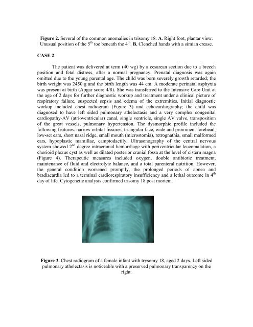
- Page 239: the common features in both cases a
- Page 243 and 244: (7, 9). Congenital heart disease is
- Page 245 and 246: Универзитетска Кли
- Page 247 and 248: Nën lëkurë është shtresa e ind
- Page 249 and 250: Gjatë operacionit Në përfundim t
- Page 251 and 252: Defekti i krijuar pas heqjes së tu
- Page 253 and 254: 3. Borah GL, Hidalgo DA, Wey PD: Re
- Page 255 and 256: RESUME Background: Marfan's Syndrom
- Page 257 and 258: Figure 3 Here ... Figure 3 a- Right
- Page 259 and 260: 4. Hindle NW, Crawford JS: Dislocat
- Page 261 and 262: Antibiotikët shkaktojnë problem n
- Page 263 and 264: Ketokonazole alkaloid® (Alkaloid),
- Page 265 and 266: Levonelle® (Schering), tableta 0,7
- Page 267 and 268: REZYME: Bioteknologjia ka aplikim t
- Page 269 and 270: • ADN-biblioteka Figura 1 Qëllim
- Page 271 and 272: • Fibroza kistike është çrregu
- Page 273 and 274: PËRFUNDIMI Figura 2 Bioteknologjia
- Page 275 and 276: Tel.++38970 207 350, ++38976 425 47
- Page 277 and 278: Во 70 до 90% од испита
- Page 279 and 280: психијатар, се до н
- Page 281 and 282: this integration of forensic psychi
- Page 283 and 284: Etika mjekësore cakton standarde m
- Page 285 and 286: Dr. sci. Agron M. Rexhepi DORACAK A
- Page 287 and 288: Përgatitja teknike e shkëlqyeshme
- Page 289 and 290: pranishmit i përshëndeti dhe u d
- Page 291 and 292:
� Florence LUSTMAN, inspector gen
- Page 293 and 294:
TAKIME MJEKËSORE / MEDICAL MEETING
- Page 295 and 296:
evista mjeksore ―Praxis Medicus
- Page 297 and 298:
19) Prof. Dr. Bexhet Canhasi, profe
- Page 299 and 300:
NGA AKTIVITETET E SHMSHM
- Page 301 and 302:
Dr. Omer Xhemali, kardiokirurg, Gje
- Page 303 and 304:
- titullin e punimit; - emrin dhe m
- Page 305 and 306:
preskoknat vo ovaj broj ili ke nema
- Page 307 and 308:
Do të thonin ata që flasin anglis
- Page 309 and 310:
Se slozuvam da bidete uste postrogi
- Page 311 and 312:
Hysen Ukmata 1928 - 2008 Hysen Ukma
- Page 313 and 314:
Ass. dr sci Adem Haziri, Fakulteti
- Page 315 and 316:
përshkruhet qartë, kurse metodat
- Page 317:
3. Ju keni të drejtë të pranoni