

1 Úvod:
1 Úvod:
1 Úvod:

You also want an ePaper? Increase the reach of your titles
YUMPU automatically turns print PDFs into web optimized ePapers that Google loves.
ightness knob" actually changes the size of the spot on the sample; changing it from<br />
a wide dispersed spot to a pinpoint beam.<br />
3. The beam is restricted by the condenser aperture (usually user selectable), knocking<br />
out high angle electrons (those far from the optic axis, the dotted line down the center)<br />
4. The beam strikes the specimen and parts of it are transmitted<br />
5. This transmitted portion is focused by the objective lens into an image<br />
6. Optional Objective and Selected Area metal apertures can restrict the beam; the<br />
Objective aperture enhancing contrast by blocking out high-angle diffracted electrons,<br />
the Selected Area aperture enabling the user to examine the periodic diffraction of<br />
electrons by ordered arrangements of atoms in the sample<br />
7. The image is passed down the column through the intermediate and projector lenses,<br />
being enlarged all the way<br />
8. The image strikes the phosphor image screen and light is generated, allowing the user<br />
to see the image. The darker areas of the image represent those areas of the sample<br />
that fewer electrons were transmitted through (they are thicker or denser). The lighter<br />
areas of the image represent those areas of the sample that more electrons were<br />
transmitted through (they are thinner or less dense)<br />
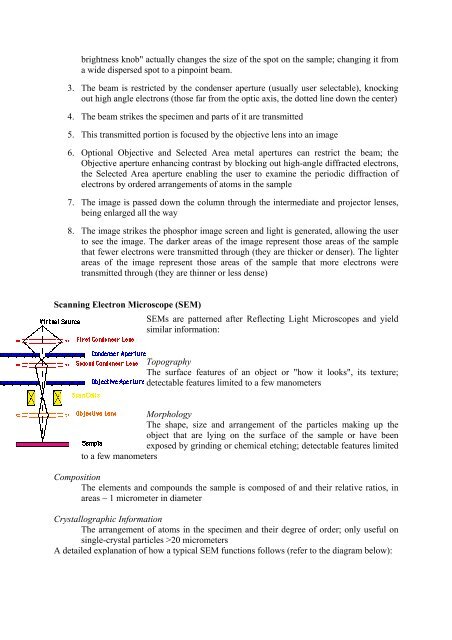
Scanning Electron Microscope (SEM)<br />
SEMs are patterned after Reflecting Light Microscopes and yield<br />
similar information:<br />
Topography<br />
The surface features of an object or "how it looks", its texture;<br />
detectable features limited to a few manometers<br />
Morphology<br />
The shape, size and arrangement of the particles making up the<br />
object that are lying on the surface of the sample or have been<br />
exposed by grinding or chemical etching; detectable features limited<br />
to a few manometers<br />
Composition<br />
The elements and compounds the sample is composed of and their relative ratios, in<br />
areas ~ 1 micrometer in diameter<br />
Crystallographic Information<br />
The arrangement of atoms in the specimen and their degree of order; only useful on<br />
single-crystal particles >20 micrometers<br />
A detailed explanation of how a typical SEM functions follows (refer to the diagram below):


