Acta Ped Vol 42 N 3 - Sociedade Portuguesa de Pediatria
Acta Ped Vol 42 N 3 - Sociedade Portuguesa de Pediatria
Acta Ped Vol 42 N 3 - Sociedade Portuguesa de Pediatria

You also want an ePaper? Increase the reach of your titles
YUMPU automatically turns print PDFs into web optimized ePapers that Google loves.
<strong>Acta</strong> <strong>Ped</strong>iatr Port 2011:<strong>42</strong>(3):108-10<br />
Oliveira PH et al. – Intussusception in an HIV-infected child<br />
Physical examination revealed a well-nourished and afebrile<br />
patient. He was prostrated. Vital signs were stable. The<br />
abdomen was disten<strong>de</strong>d and bowel sounds were diminished.<br />
There was a ten<strong>de</strong>r mass in the left upper and lower quadrants.<br />
Initial abdominal plain film <strong>de</strong>monstrated scarcity of air in the<br />
colon, and two air-fluid levels, consistent with partial small<br />
bowel obstruction. Abdominal ultrasonography revealed intestinal<br />
intussusception with the leading edge of the intussusceptum<br />
at the level of the left colon. A classic “target” sign was<br />
observed on transverse section. In the centre there was an<br />
echogenic mass that suggested a pathologic lead point such as<br />
a Meckel’s diverticulum or a polyp. Child’s age, clinical context<br />
and ultrasonographic findings preclu<strong>de</strong>d hydrostatic reduction.<br />
He was submitted to laparotomy and a double ileo-ileal and<br />
ileo-cecocolic intussusception was found. Manual reduction<br />
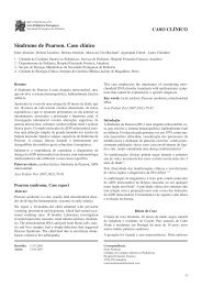
was easily performed. The lead points were two ileal doughnut-shaped<br />
intra-luminal masses at 2,25 meters and 1 meter<br />
respectively from ileocecal valve. The former had central<br />
<strong>de</strong>pression with doubtful viability (Figura, a) and the latter<br />
had central perforation (Figura, b). A double segmental resection<br />
of the ileon encompassing each mass was performed, followed<br />
by double mechanical anastomoses.<br />
thoracic scan performed at thirteenth postoperative day,<br />
<strong>de</strong>tected additional masses at the medium right lung, head and<br />
uncinate process of the pancreas and both kidneys and thoracic,<br />
mesenteric and iliac lymph no<strong>de</strong>s. The patient<br />
respon<strong>de</strong>d well to antiretroviral therapy and chemotherapy<br />
and evolution was favourable. The patient completed the<br />
chemotherapy treatment about an year and a half ago and has<br />
not evi<strong>de</strong>nce of recurrence till this moment.<br />
Discussion<br />
Intussusception is a frequent cause of bowel obstruction in<br />
young children and the greatest inci<strong>de</strong>nce occurs in infants<br />
between ages 5 and 9 months 4 .<br />
Double intussusception is an extremely rare variant of intussusception,<br />
which is almost impossible to diagnose preoperatively.<br />
The diagnosis is usually ma<strong>de</strong> during laparotomy and<br />
manual reduction is usually easily performed 5 .<br />
Pathologic lead points occur in 4-8% of intussusceptions but<br />
are more commonly found in ol<strong>de</strong>r children 6 . More than a half<br />
of the few reported cases of double intussusception in the<br />
child had an un<strong>de</strong>rlying pathologic lead point 7 .<br />
Non-Hodgkin’s lymphomas are responsible for 17% of noni<strong>de</strong>opathic<br />
intussusception 8 . They usually occur after the age<br />
of 3, are more frequent in boys and there’s often a 8-day history<br />
of symptoms with worsening patient status 8,9 .<br />
Intussusception triggered by a lymphoma is unlikely to be<br />
completely reduced by enema, and surgery for manual reduction<br />
is always required 3 . Following reduction, in the majority<br />
of cases, the diagnosis of lymphoma can be accomplished<br />
from peripheral samples such as peritoneal fluid, pleural effusion,<br />
mesenteric lymph no<strong>de</strong>s, bone marrow aspirate or by<br />
tumor biopsy 2,9 . However, in single and localized disease, segmentar<br />
bowel resection should be consi<strong>de</strong>red only if it enables<br />
complete tumor removal, in or<strong>de</strong>r both to confirm the diagnosis<br />
and to reduce the intensity of chemotherapy 2,3,10 .<br />
Complicated cases such as irreducible intussusception or with<br />
perforation or necrosis must be managed with segmentar<br />
resection 9 .<br />
The diagnosis of lymphoma should be systematically evoked<br />
in children over the age of 3, especially if clinical or ultrasonographic<br />
findings are not typical 2,9 .<br />
According to literature, intussusception leads to early <strong>de</strong>tection<br />
of intestinal Burkitt’s lymphoma and prognosis is<br />
favourable 3 .<br />
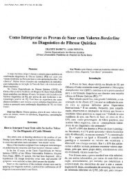
Figure – a. Ileal mass that acted as intussusception leading point, with<br />
central <strong>de</strong>pression; b- Ileal mass that acted as leading point, with central<br />
perforation.<br />
The postoperative course was uneventful, and the patient was<br />
discharged home on sixth postoperative day. Pathology studies<br />
revealed an atypical Burkitt’s lymphoma. Abdominal and<br />
References<br />
1. Farrier J, Dinerman C, Hoyt D, Coimbra R. Intestinal lymphoma<br />
causing intussusception in HIV + patient: a rare presentation. Cur<br />
Surg 2004; 61: 386-8.<br />
2. Delarue A, Bergeron C, Mechinaud-Lacroix F, Coze C, Raphael M,<br />
Patte C. <strong>Ped</strong>iatric non-Hodgkin's lymphoma: primary surgical management<br />
of patients presenting with abdominal symptoms.<br />
Recommendations of the Lymphoma Committee of the French Society<br />
to Combat <strong>Ped</strong>iatric Cancers (SFCE). J Chir 2008;145(5):454-8.<br />
109