Mixoma Cardíaco, revisão biblio - Faculdade de Ciências da Saúde
Mixoma Cardíaco, revisão biblio - Faculdade de Ciências da Saúde
Mixoma Cardíaco, revisão biblio - Faculdade de Ciências da Saúde

Create successful ePaper yourself
Turn your PDF publications into a flip-book with our unique Google optimized e-Paper software.
Tese <strong>de</strong> Mestrado, <strong>Mixoma</strong> <strong>Cardíaco</strong>, <strong>revisão</strong> <strong>biblio</strong>gráfica e análise <strong>de</strong> um caso clínico<br />
O mixoma cardíaco localiza-se na aurícula esquer<strong>da</strong> em cerca <strong>de</strong> 60% a<br />
85% dos casos (ARANA-ARRI et al, 2007; MATSUSHITA, 2007; MENG et al;<br />
GREBENC, 2000), na aurícula direita em cerca <strong>de</strong> 10% e nos ventrículos em<br />
cerca <strong>de</strong> 5% dos casos (ARANA-ARRI et al, 2007, YU et al, 2007). O<br />
envolvimento do anel mitral, folhetos <strong>da</strong> válvula mitral (ALEXANDER et al,<br />
1998), veia cava inferior (ALEXANDER et al, 1998; DEVIG, 1980), bem como<br />
<strong>da</strong>s duas aurículas é menos frequente (REYNEN, 1995). No entanto os<br />
resultados existentes na literatura sobre esta matéria são divergentes.<br />
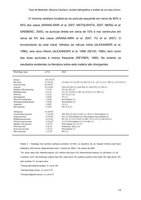
Histology type n (%) Site a<br />
Benign<br />
Myxoma<br />
Non-myxoma<br />
Lipoma<br />
Papillary fibroelastoma<br />
Rhabdomioma<br />
Fibroma<br />
Hemangioma<br />
Myocitic hamartoma<br />
Hemangioendothelioma<br />
Angioma<br />
Myxolipoma<br />
Malignant<br />
Unclassified sarcoma<br />
Angiosarcoma<br />
Rhabdomiosarcoma<br />
Fibrous histiocytoma<br />
Spindle cell sarcoma<br />
Fibrosarcoma<br />
Synovial sarcoma<br />
Myxosarcoma<br />
Tabela 2 – Histologia dos tumores cardíacos primários (n=149), na casuística <strong>de</strong> um hospital Chinês on<strong>de</strong> foram<br />
analisados 149 tumores, diagnosticados entre 1 Janeiro <strong>de</strong> 1999 e 1 <strong>de</strong> Janeiro <strong>de</strong> 2000.<br />
a AV, aortic valve; IAS, interatrial septum; IVC, inferior vena cava; IVS, interventricular septum; LA, left atrium; LV, lef<br />
t ventricle; LVOT, left ventricular outflow tract; MV, mitral valve; PS, posterior surface of the heart; RA, right atrium; RV,<br />
right ventricle; TV, tricuspid valve.<br />
118 (79,2) b<br />
75 (63,6) c<br />
43 (36,4) c<br />
22 (18,6) c<br />
7 (5,9) c<br />
6 (5,1) c<br />
3 (2,5) c<br />
1 (0,8) c<br />
1 (0,8) c<br />
1 (0,8) c<br />
1 (0,8) c<br />
1 (0,8) c<br />
31 (20,8) b<br />
7 (22,6) d<br />
6 (19,4) d<br />
6 (19,4) d<br />
5 (16,1) d<br />
3 (9,7) d<br />
2 (6,5) d<br />
1 (3,2) d<br />
1 (3,2) d<br />
b Among total patients studied, i.e. out of 149<br />
c Among benign tumors, i.e. out of 118<br />
d Among malignant tumors, i.e. out of 31<br />
LA (64), LV (3), RV (3), MV (2), LA+ AV (1), LA+ MV (1), LA+IVS (1)<br />
IAS (18), RA (1), IAS+RA (1), IAS+IVC (1), PS (1)<br />
AV (6), RV (1)<br />
LV (1), LVOT (1), RV (2), IVS (1), LV+MV (1)<br />
AV (1), LV (2)<br />
TV (1)<br />
Pericardium (1)<br />
Pericardium (1)<br />
LV (1)<br />
MV+LA (1)<br />
MV+RA (2), RA (2), RV (1), IVS (1), Pericardium (1)<br />
RA (3), Pericardium (2), Pericardium+myocardium (1)<br />
RV (2), LV (1), LVOT (1) MV+AV+LA (1), Left IAS (1)<br />
LA (3), Pericardium (1), LA+MV (1)<br />
Pericardium (2), LA (1)<br />
Pulmonary artery (2)<br />
Pericardium (1)<br />
MV (1)<br />
14