20x-1280x - Lidl Service Website
20x-1280x - Lidl Service Website
20x-1280x - Lidl Service Website

You also want an ePaper? Increase the reach of your titles
YUMPU automatically turns print PDFs into web optimized ePapers that Google loves.
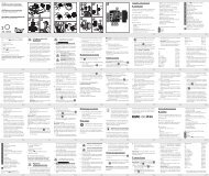
All parts (Fig. 1-6):<br />
B 5x WF Eypiece<br />
C 16x WF Eyepiece<br />
D Barlow lens<br />
E PC-Ocular<br />
F Eyepiece holder<br />
G Microscope head<br />
H Objective-revolver<br />
I Microscope stage<br />
J Focus wheel<br />
1) LED lighting (transmitted<br />
light)<br />
1! Electricity supply<br />
1@ Microscope base<br />
1# Photomizer SE software<br />
1$ Wall connector<br />
1% 10 Slides, 10 Covering glasses<br />
and 5 preparations in a<br />
plastic box<br />
1. General/Location:<br />
1^ matted lens<br />
1& Condenser lens<br />
1* Dimmer<br />
1( Colour filter disc<br />
2) LED lighting (direct light)<br />
2! Direct light / transmitted<br />
light switch<br />
2@ a) Microscopy instruments;<br />
b) pipette; c) pincers<br />
2# Prawn breeding plant<br />
2$ MicroCut<br />
2% Specimens: a) yeast; b) Gum<br />
media (specimen inclusion<br />
medium); c) sea salt;<br />
d) Prawn eggs<br />
2^ Carrying case<br />
Locking screw<br />
Mechanical plate<br />
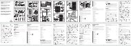
First you must make sure that your microscope is on a stable and solid<br />
surface.<br />
An electricity supply is required for observation with the electric illuminator.<br />
2. Electric LED lighting with dimmer<br />
Check before use whether the light switch (Fig. 1,21) is set to “off”.<br />
The microscope has two lighting units. Lighting can be of three types.<br />
Set the switch (Fig 1, 21) to „II“ to light the specimen from above<br />
(reflected light) or „I“ to light it from below (direct light). Use setting „III“<br />
to have the specimen simultaneously subjected to direct and transmitted<br />
light. The transmitted light unit (Fig 1, 10) is used for transparent<br />
specimens (those on glass slides). To view solid non-transparent<br />
specimens use the direct light unit (Fig 1, 20). Use of both forms of<br />
lighting simultaneously is only recommended for semi-transparent<br />
specimens. This operating mode is not recommended for direct light<br />
specimens on slides as it may cause reflection on the slide.<br />
To operate the mains power pack supplied (Fig. 6, 14) is first<br />
connected to the microscope and a mains power socket (220-230V).<br />
Use the switch (Fig 1, 21) to select the desired lighting mode and set<br />
the dimmer to the desired brightness (Fig. 1, 18).<br />
As your device has continuously controllable lighting (dimmer) optimal<br />
illumination of the object to be viewed is guaranteed.<br />
3. Colour filter disc<br />
The colour filter under the microscope table (Fig. 1, 19) aids in viewing<br />
very bright and transparent objects. Just select the right colour for the<br />
specimen in question. The components of colourless/transparent<br />
objects (e.g. starch particles, single-cell specimens) can thus be better<br />
recognised.<br />
4. Interchangeable illumination lenses<br />
The illumination of your microscope contains two lenses (Fig. 6,<br />
16+17). These are – according to the object being viewed – to be attached<br />
to the LED illumination for best results. The ground glass lens<br />
Fig. 6, 16) is already mounted on the lighting unit. To change the lenses,<br />
simply screw them on and off. Just turn the upper lighting unit<br />
(Fig. 1, 17).<br />
An overview of purposes:<br />
Matted lens (Fig. 6, 16) to be used for views with the PC eyepiece<br />
Viewing of extremely small items with eyepiece (Fig. 6, 1+2) and<br />
Barlow lens (Fig. 6, 3)<br />
Condenser lens (Fig. 6, 17) to be used for viewing of standard items<br />
with eyepieces (Fig. 6, 1+2) and Barlow lens (Fig. 6, 3)<br />
- 4 -<br />
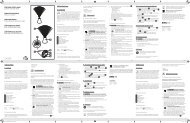
5. Microscope setup<br />
The microscope’s eyepiece (Fig 1, 6) will now be prepared for the first<br />
observation.<br />
First, loosen the screw (Fig. 1, X) and rotate the eyepiece into a convenient<br />
position.<br />
Begin every observation with the lowest magnification.<br />
Place the microscope’s table (Fig. 1, 8) with the focus knob (Fig 1, 9)<br />
into the lowest position and rotate the objective revolver (Fig. 1, 7) until<br />
it locks on the lowest magnification (4x)<br />
Tip:<br />
First, drive the Microscope’s table (Fig. 1, 8) in its lowest<br />
position before changing the objective in order to prevent<br />
damages.<br />
Insert the 5x eyepiece (Fig. 2, 1) in the Barlow lens (Fig. 2, 3).<br />
Take care, that the Barlow lens is inserted completely in the monocular<br />
head (Fig. 2, 5).<br />
6. Observation<br />
After you have set up the microscope with the corresponding illumination,<br />
the following principles are important:<br />
Begin each observation with a simple observation at lowest magnification,<br />
so that the centre and position of the object to be viewed is in<br />
focus.<br />
The higher the magnification the more light is required for good picture<br />
quality.<br />
Place a permanent slide culture (Fig. 3, 15) directly under the<br />
microscope lens on the plate (Fig. 3, 8) and clamp it on the cross-table<br />
(Fig. 3, Y). To do so push the lever (Fig. 3, C) aside. The specimen to<br />
be examined must be directly over the lighting. If not turn the two<br />
knurled screws on the mechanical plate.<br />
Tip:<br />
On the mechanical plate (Fig 3, Y) there are two knurled screws<br />
(Figs 3 A+B). They are used to precisely position the specimen<br />
laterally (Fig 3, A) and vertically (Fig 3, B).<br />
Look through the eyepiece (Fig. 1, 1/2) and turn carefully the focusing<br />
wheel (Fig. 1, 9)) until you can see a sharp picture.<br />
Now you can get a higher magnification, while you pull out slowly the<br />
Barlow lens (Fig. 4, 3) of the monoculare barrel (Fig. 4, 5). With nearly<br />
entirely pulled out Barlow lens the magnification is raised to 2x.<br />
For still higher magnification you can put the 16x eyepiece (Fig. 6, 2)<br />
into the objective revolver and set on higher position (10x / 40x).<br />
Important Hint:<br />
Depending on the preparation higher magnifications do not<br />
always lead to better pictures.<br />
Please notice:<br />
With changing magnification (eyepiece or objective lens<br />
changes, pulling out of the Barlow lens) the sharpness of the<br />
image must be newly defined by turning the focusing wheel (6).<br />
Please be very careful when doing this. When you move the<br />
mechanical plate upwards to fast the objective lens and the<br />
slide can touch and become damaged.<br />
7. Viewed Object – condition and preparation<br />
7.1 Condition<br />
With the Barlow lens nearly fully extended magnification can be doubled.<br />
Both transparent and non-transparent specimens can be examined<br />
with this microscope, which is a direct as well as transmitted light<br />
model. If opaque specimens are examined - such as small animals,<br />
plant parts, tissue, stone and so on - the light is reflected from the<br />
specimen through the lens and eyepiece, where it is magnified, to the<br />
eye (reflected light principle, switch position I). If opaque specimens<br />
are examined the light from below goes through the specimen, lens<br />
and eyepiece to the eye and is magnified en route (direct light principle,<br />
switch position II). Many small organisms of the water, plant parts<br />
and finest animal components have now from nature these transparent<br />
characteristic, other ones must be accordingly prepared. Is it that