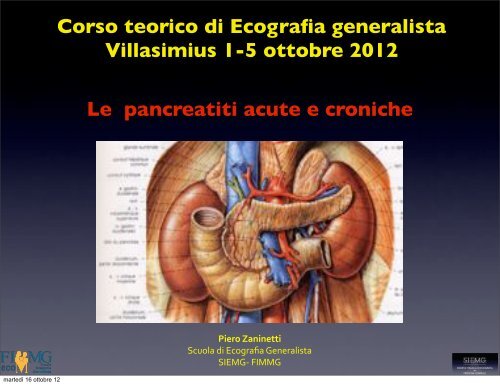
Le pancreatiti acute e croniche Corso teorico di Ecografia ... - siemg
Le pancreatiti acute e croniche Corso teorico di Ecografia ... - siemg
Le pancreatiti acute e croniche Corso teorico di Ecografia ... - siemg

You also want an ePaper? Increase the reach of your titles
YUMPU automatically turns print PDFs into web optimized ePapers that Google loves.
<strong>Corso</strong> <strong>teorico</strong> <strong>di</strong> <strong>Ecografia</strong> generalistaVillasimius 1-5 ottobre 2012<strong>Le</strong> <strong>pancreatiti</strong> <strong>acute</strong> e <strong>croniche</strong>martedì 16 ottobre 12Piero&Zaninetti&Scuola'<strong>di</strong>'<strong>Ecografia</strong>'GeneralistaSIEMG5'FIMMG
Fattori eziologici• Calcolosi biliare• Abuso alcoolico70-80 %• Infezioni• ipertrigliceridemia• Ipercalcemia• Tumori pancreatici e ampollari• Cause jatrogene(chirurgiaaddominale e ERCP)martedì 16 ottobre 12
Pancreatite acuta• Classificazione <strong>di</strong> Atlanta ( 1992)• Pancreatite lieve(80%): edemaeventualmente associato a piccoli focolai <strong>di</strong>necrosi parenchimale• Pancreatite severa(20 %): necrosiparenchimale,necrosi del tessuto a<strong>di</strong>posoperipancreatico,focolai emorragici intra eperi ghiandolari,essudazioneintraperitonealemartedì 16 ottobre 12
upture can occur and may be catastrophic. SplenicCriteri <strong>di</strong> Balthazar ( 1990 )Table 2:GradeABCDEBalthazar gra<strong>di</strong>ng systemDescriptionNormal-appearing pancreasFocal or <strong>di</strong>ffuse enlargement of thepancreasPancreatic gland abnormalitiesaccompanied by mild peripancreaticinflammatory changesFluid collection in a single locationTwo or more fluid collections nearthe pancreas or gas in or adjacentto the pancreasmartedì 16 ottobre 12
accuracy in the depiction of necrotising <strong>pancreatiti</strong>s. 26Table 1Ranson and APACHE II criteria.Criteri <strong>di</strong> Ranson e Apache IIRanson CriteriaAPACHE IIAge >55 yearsAge >55 yearsWBC >16,000/mLWBC 14,900/mLGlucose >200 mg/dLRectal temperature 38.4 CLDH >350 IU/mLMAP 109 mmHgAST >250 IU/LHR 109 bpmHct decrease >10 RR 24BUN increase >5 mg/dL pH 7.49Calcium 6 L Creatinine 1.4 mg/100 mLHct 45.9%GCSChronic health pointsAbbreviations: WBC: white blood cell count; LDH: lactate dehydrogenase;AST: aspartate transaminase; Hct: haematocrit; BUN: blood urea nitrogen;martedì 16 ottobre PO 2 12 : arterial partial pressure of oxygen; MAP: mean arterial pressure; HR:
CT Severity IndexBalthazar Stage PointsA 0B 1C 2D 3E 4% Necrosis Points0 030 230-50 4>50 6Severity IndexPOINTS SEVERITY0 – 3 Low4 – 6 Me<strong>di</strong>um7 - 10 Highmartedì 16 ottobre 12
of the pancreatic microcirculation. It is usually well establishedby 96 h after onset of clinical symptoms, but generallynot before 48 h. 26 The relative amounts of solid and liquidIn<strong>di</strong>ce <strong>di</strong> gravità CT mo<strong>di</strong>ficatocomponents in a region of necrosis are dependent on the time( Mortele AJR 2004 )inhomogeneousand solid necroticTable 4Mo<strong>di</strong>fied CT severity index [Mortele AJR 2004].Prognostic in<strong>di</strong>catorPancreatic InflammationNormal pancreas 0Intrinsic pancreatic abnormalities with2inflammatory changes in the peri-pancreatic fatPancreatic or peri-pancreatic fluid collection4or peri-pancreatic fat necrosisPancreatic necrosisNone 030% 4Extra-pancreatic complicationsOne or more of pleural effusion, ascites,2vascular complications (venous thrombosis, arterialhaemorrhage or pseudoaneurysm formation),parenchymal complications (infarction,haemorrhageor sub-capsular fluid collection) or GI involvement(inflammation, perforation or intramural fluid collection)ScoreWhere mild <strong>acute</strong> <strong>pancreatiti</strong>s is 0e2 points, moderate is 4e6 points andsevere is 7e10 points.Figure 3 Pancreatidemonstrating anpancreatic head (wabdominal pain. A rattenuation of 17Hparenchyma in the tdefined as 30% pamartedì 16 ottobre 12
Diagnosi• Dolore addominale• Esami laboratoristici ( elevazioneamilasi e lipasi > 3 volte il limitesup della norma)• <strong>Ecografia</strong>• Tomografia computerizzatamartedì 16 ottobre 12
<strong>Ecografia</strong>• Alterazioni a carico del pancreas• Patologia litiasica biliare• Valutazione parenchima epatico( etilista)• Diagnosi e monitoraggiocomplicanzemartedì 16 ottobre 12
Pancreas: organo <strong>di</strong> <strong>di</strong>fficile valutazione ecografica• Visibilità globale <strong>di</strong>fficile nel 15-20 % dei casi• La <strong>di</strong>fficoltà <strong>di</strong> visualizzazione aumenta nella sequenza testacorpo-coda• La regione periampollare è <strong>di</strong> solito la più facile da visualizzare• Esame operatore <strong>di</strong>pendentemartedì 16 ottobre 12
Pancreas: organo <strong>di</strong> <strong>di</strong>fficile valutazione ecografica• Visibilità globale <strong>di</strong>fficile nel 15-20 % dei casi• La <strong>di</strong>fficoltà <strong>di</strong> visualizzazione aumenta nella sequenza testacorpo-coda• La regione periampollare è <strong>di</strong> solito la più facile da visualizzare• Esame operatore <strong>di</strong>pendentemartedì 16 ottobre 12
Limiti dell’esame ecografico• Difficoltosa visualizzazione delpancreas( meteorismo ,obesità )• Nel 40 % delle forme lievi l’esameecografico è normale• Difficoltosa <strong>di</strong>fferenziazione dellearee edematose da quellenecrotichemartedì 16 ottobre 12
martedì 16 ottobre 12Pancreatite lieve
Alterazioni ecografiche• Variazioni <strong>di</strong> volume ( aumento<strong>di</strong>ffuso o focale, a volte riduzione )martedì 16 ottobre 12
Alterazioni ecografiche• Variazioni <strong>di</strong> volume ( aumento<strong>di</strong>ffuso o focale, a volte riduzione )martedì 16 ottobre 12
Alterazioni ecografiche• Alterazioni della ecogenicità• ipoecogenicità <strong>di</strong>ffusa• ipoecogenicità segmentaria• <strong>di</strong>somogeneitàmartedì 16 ottobre 12
Alterazioni ecografiche• Alterazioni della ecogenicità• ipoecogenicità <strong>di</strong>ffusa• ipoecogenicità segmentaria• <strong>di</strong>somogeneitàmartedì 16 ottobre 12
!Pancreatite focale !• Area ipoecogena <strong>di</strong>somogenea"""• Può deformare il profilo ghiandolare""• Può comprimere le strutture circostanti ( Vie biliari ) ""• Carcinoma <strong>di</strong> solito ipovascolarizzato e ipoperfuso, pancreatitefocale più vascolarizzata per linfiltrato flogistico""• Spesso necessario esame bioptico "martedì 16 ottobre 12
!Pancreatite focale !• Area ipoecogena <strong>di</strong>somogenea"""• Può deformare il profilo ghiandolare""• Può comprimere le strutture circostanti ( Vie biliari ) ""• Carcinoma <strong>di</strong> solito ipovascolarizzato e ipoperfuso, pancreatitefocale più vascolarizzata per linfiltrato flogistico""• Spesso necessario esame bioptico "martedì 16 ottobre 12
martedì 16 ottobre 12Adenocarcinoma duttale!
martedì 16 ottobre 12Pancreatite severa
Alterazioni ecografiche• Aree <strong>di</strong> necrosi: zone ipoecogene che sialternano ad aree <strong>di</strong> ecostruttura normale• Aree ecoprive a contenuto fluido percolliquazione necrotica• Contorni ghiandolari irregolari emaldelineatimartedì 16 ottobre 12
Alterazioni ecografiche• Aree <strong>di</strong> necrosi: zone ipoecogene che sialternano ad aree <strong>di</strong> ecostruttura normale• Aree ecoprive a contenuto fluido percolliquazione necrotica• Contorni ghiandolari irregolari emaldelineatimartedì 16 ottobre 12
Complicanze della pancreatiteacuta•The original 1992 Atlanta Classification: local complicationsAcute peripancreatic fluid collection Occur early in the course of <strong>acute</strong> <strong>pancreatiti</strong>s; are located in or near the pancreas; and alwayslack a wall of granulation or fibrous tissuePancreatic necrosisA <strong>di</strong>ffuse or focal area(s) of nonviable pancreatic parenchyma,which is typically associated with peripancreatic fat necrosisAcute pseudocystA collection of pancreatic juice enclosed by a wall of fibrous or granulation tissue that arises as aconsequence of <strong>acute</strong> <strong>pancreatiti</strong>s or pancreatic trauma, or chronic <strong>pancreatiti</strong>sPancreatic abscessA circumscribed intra-abdominal collection of pus, usually in proximity to the pancreas, containing littleor no pancreatic necrosis, which arises as a consequence of <strong>acute</strong> <strong>pancreatiti</strong>s or pancreatic traumamartedì 16 ottobre 12
Table 3Revised Atlanta Classification: CT criteria for local pancreatic complicationsLocal ComplicationMorphologic CT CriteriaAcute peripancreatic fluid collection Typically less than 4 weeks old after onset of symptomsOccurs in interstitial edematous <strong>pancreatiti</strong>sHomogeneous collection with fluid densityConfined by normal peripancreatic fascial planesNo fully definable wall encapsulating the collectionAdjacent to pancreas (no intrapancreatic extension)PseudocystTypically older than 4 weeks after onset of symptomsOccurs in interstitial edematous <strong>pancreatiti</strong>sHomogeneous collection (round or oval) with fluid densityWell-defined wall (ie, completely encapsulated)Absence of nonliquid componentAdjacent to pancreas (no intrapancreatic extension)Acute necrotic collectionTypically less than 4 weeks old after onset of symptomsOccurs in necrotizing <strong>pancreatiti</strong>sHeterogeneous collection with liquid and nonliquid density andvarying degrees of loculationNo fully definable wall encapsulating the collectionLocated intrapancreatic and/or extrapancreaticWalled-off necrosisTypically older than 4 weeks after onset of symptomsOccurs in necrotizing <strong>pancreatiti</strong>sHeterogeneous collection with liquid and nonliquid density andvarying degrees of loculationsWell-defined wall (ie, completely encapsulated)Located intrapancreatic and/or extrapancreaticPost-necrosectomy Pseudocyst Occurs after necrosectomy for necrotizing <strong>pancreatiti</strong>sHomogeneous collection (round or oval) with fluid densityWell-defined wall (ie, completely encapsulated)Absence of nonliquid componentLocated intrapancreatic and/or extrapancreaticmartedì 16 ottobre 12
Raccolta acuta fluida pancreatica/peripancreatica a insorgenza precoce• Area transonica ,con contorni nondelineati ,che si adatta alla cavità che lacontiene• Non presenta componenti solide al suointerno ed è costituita da succopancreatico ricco <strong>di</strong> enzimimartedì 16 ottobre 12
Raccolta fluida pancreatica /peripancreatica post necrotica• Si sviluppa come sequela della necrosipancreatica• Contenuto <strong>di</strong>somogeneo ,con detriti <strong>di</strong>tessuto a<strong>di</strong>poso e <strong>di</strong> parenchimapancreatico in sospensione• Spesso conseguente a necrosi coinvolgenteil Wirsung e perciò in comunicazione conessomartedì 16 ottobre 12
Localizzazione• Intrapancreatica• Retrocavità degli epiploon• Spazio pararenale anteriore• Me<strong>di</strong>astinica ( attraverso il forame<strong>di</strong>aframmatico)• Nei foglietti mesocolici e mesenterici• Docce parieto colichemartedì 16 ottobre 12
martedì 16 ottobre 12Retrocavità degli epiploon
Retrocavità degli epiploonFigure 1d. Drawing of the anatomy of the greater and lesser omenta (a) and axial (b), coronal (c),and sagittal (d) <strong>di</strong>agrams of the upper abdomen.Yoo E et al. Ra<strong>di</strong>ographics 2007;27:707-720©2007 by Ra<strong>di</strong>ological Society of North Americamartedì 16 ottobre 12
Retrocavità degli epiploonFigure 1d. Drawing of the anatomy of the greater and lesser omenta (a) and axial (b), coronal (c),and sagittal (d) <strong>di</strong>agrams of the upper abdomen.Yoo E et al. Ra<strong>di</strong>ographics 2007;27:707-720©2007 by Ra<strong>di</strong>ological Society of North Americamartedì 16 ottobre 12
Retrocavità degli epiploonFigure 1c. Drawing of the anatomy of the greater and lesser omenta (a) and axial (b), coronal (c),and sagittal (d) <strong>di</strong>agrams of the upper abdomen.Yoo E et al. Ra<strong>di</strong>ographics 2007;27:707-720©2007 by Ra<strong>di</strong>ological Society of North Americamartedì 16 ottobre 12
Retrocavità degli epiploonFigure 1c. Drawing of the anatomy of the greater and lesser omenta (a) and axial (b), coronal (c),and sagittal (d) <strong>di</strong>agrams of the upper abdomen.Yoo E et al. Ra<strong>di</strong>ographics 2007;27:707-720©2007 by Ra<strong>di</strong>ological Society of North Americamartedì 16 ottobre 12
Rapporti del pancreas con TM eSBMFigure 5. Lateral illustration of the relationship of the pancreas (P) to the transverse mesocolon(TM) and small bowel mesentery (SBM).Vikram R et al. Ra<strong>di</strong>ographics 2009;29:e34©2009 by Ra<strong>di</strong>ological Society of North Americamartedì 16 ottobre 12
martedì 16 ottobre 12Raccolta fluida peripancreatica
Raccolta nella retrocavitàepiploonmartedì 16 ottobre 12
Pseudocisti pancreatica• Formazione a contenuto fluido ,anecogenache persiste > <strong>di</strong> 4 settimane• Presenza <strong>di</strong> parete propria infiammatoria• Contorni definiti e arrotondati• Residui necrotici, sepimentazioni,calcificazioni• Effetto massa correlato alle <strong>di</strong>mensionimartedì 16 ottobre 12
Pseudocisti pancreatica• regressione spontanea (50 % dei casi)• complicazione (30 % dei casi )• stabilità ( 20 % dei casi )martedì 16 ottobre 12
Pseudocisti pancreatica:complicanze• Estensione agli organi a<strong>di</strong>acenti• Emorragia• Peritonite• Ostruzione del duodeno• Ostruzione biliare• Infezionemartedì 16 ottobre 12
Emorragia• Area ecogena <strong>di</strong>somogenea più e menoestesa ,generalmente in sedeperipancreatica ,che tende a colliquarsi<strong>di</strong>ventando ecopriva• Necessaria la conferma con TC che rileva ladensità tipica del sanguemartedì 16 ottobre 12
Ascesso o necrosi infetta• Evoluzione <strong>di</strong> una raccolta fluida• Comparsa <strong>di</strong> echi interni fluttuanti o declivi• Spots iperecogeni espressione dellapresenza <strong>di</strong> aria• Quadro clinico ( febbre,dolore,leucocitosi)martedì 16 ottobre 12
Complicanze vascolari• Trombosi venosa• Pseudoaneurismamartedì 16 ottobre 12
Trombosi venosa• Danno infiammatorio intimale ocompressione estrinseca• Vena splenica (è la più frequente 10-40%dei casi )• Vena mesenterica superiore• Vena portamartedì 16 ottobre 12
Pseudoaneurismi• erosione delle pareti <strong>di</strong> un vaso• formazione anecogena pulsante• Conferma <strong>di</strong>agnostica con ecocolorDopplermartedì 16 ottobre 12
Pancreatite acuta : 6 settimane dopol’esor<strong>di</strong>omartedì 16 ottobre 12
Pancreas <strong>di</strong> <strong>di</strong>mensioni aumentateipoecogeno con ecostruttura<strong>di</strong>somogeneamartedì 16 ottobre 12
martedì 16 ottobre 12Margini irregolari
martedì 16 ottobre 12area ipo-anecogena nella parte anteriore della testa
martedì 16 ottobre 12raccolta fluida nella retrocavità degli epiploon
area rotondeggiante anecogena conspots ecogeni , priva <strong>di</strong> parete propriamartedì 16 ottobre 12
martedì 16 ottobre 12area <strong>di</strong>somogenea ipo anecogena della coda
accolta fluida posteriore al pancreasvena splenica compressamartedì 16 ottobre 12
Pancreatite cronica• Alcool• Iperlipidemia• Ipertiroi<strong>di</strong>smo• Fibrosi cistica• Cause ere<strong>di</strong>tarie e i<strong>di</strong>opatichemartedì 16 ottobre 12
Imaging ecografico• Alterazioni delle <strong>di</strong>mensioni ghiandolari• Alterazioni della ecogenicità• Alterazioni della ecostruttura• Calcificazioni parenchimali• Alterazioni <strong>di</strong> calibro del dotto <strong>di</strong> Wirsung• Calcolosi intraduttale• Variazioni del calibro della SMVmartedì 16 ottobre 12
Alterazioni delle <strong>di</strong>mensionighiandolari• Presenti nel 50% dei casi, soprattutto neglista<strong>di</strong> avanzati <strong>di</strong> malattia• Atrofia <strong>di</strong>ffusa• Alterazioni focalimartedì 16 ottobre 12
Alterazioni della ecogenicità• Solitamente aumentata per fibrosi einfiltrazione a<strong>di</strong>posa• Reperto non specifico( presente in anzianie obesi )martedì 16 ottobre 12
Alterazioni della ecostruttura• Nel 40% dei casi ( precoci ) è normale• Nel 60 % dei casi è <strong>di</strong>somogenea( coesistenza <strong>di</strong> foci <strong>di</strong> fibrosi iperecogeni efoci <strong>di</strong> flogosi ipoecogeni )martedì 16 ottobre 12
Calcificazioni parenchimali• Costituiscono il criterio <strong>di</strong>agnostico piùimportante• Depositi <strong>di</strong> calcio carbonato su una matriceproteica ( plugs) o in aree <strong>di</strong> necrosiinterstiziale• Spots iperecogeni con o senza conod’ombra posterioremartedì 16 ottobre 12
Calcificazioni parenchimali• Costituiscono il criterio <strong>di</strong>agnostico piùimportante• Depositi <strong>di</strong> calcio carbonato su una matriceproteica ( plugs) o in aree <strong>di</strong> necrosiinterstiziale• Spots iperecogeni con o senza conod’ombra posterioremartedì 16 ottobre 12
Calcificazioni parenchimali• Costituiscono il criterio <strong>di</strong>agnostico piùimportante• Depositi <strong>di</strong> calcio carbonato su una matriceproteica ( plugs) o in aree <strong>di</strong> necrosiinterstiziale• Spots iperecogeni con o senza conod’ombra posterioremartedì 16 ottobre 12
Alterazioni <strong>di</strong> calibro del dotto <strong>di</strong>Wirsung• Calibro normale < 3 mm• Irregolarità del profilo con alternanza <strong>di</strong>tratti ectasici• Nelle neoplasie spesso <strong>di</strong>latazione uniformecon profili regolari• Segno specifico ,ma non sensibilemartedì 16 ottobre 12
Alterazioni <strong>di</strong> calibro del dotto <strong>di</strong>Wirsung• Ultrasonography of the PancreasGiulia A. Zamboni, MD*, Maria Chiara Ambrosetti, MD, Mirko D’Onofrio,MD, Roberto Pozzi Mucelli, MDRa<strong>di</strong>ol Clin N Am 50 (2012) 395–406• “la <strong>di</strong>latazione ( > 3 mm) è presente nel 52,3%dei pazienti affetti da pancreatite cronica “• da Bolon<strong>di</strong> L, Priori P, Gullo L, et al.Relationship betweenmorphological changes detected by ultra- sonography and pancreaticexocrine function in chronic <strong>pancreatiti</strong>s.Pancreas 1987;2(2):222–9.martedì 16 ottobre 12
Calcolosi intraduttale• Aggregati proteici con depositi <strong>di</strong> calciocarbonato• Mobilità con<strong>di</strong>zionata dalle <strong>di</strong>mensioni• Patognomonici <strong>di</strong> pancreatite cronicamartedì 16 ottobre 12
Calcolosi intraduttale• Ultrasonography of the PancreasGiulia A. Zamboni, MD*, Maria Chiara Ambrosetti, MD, Mirko D’Onofrio,MD, Roberto Pozzi Mucelli, MDRa<strong>di</strong>ol Clin N Am 50 (2012) 395–406“the presence of calcifications is the most important and pathognomonic <strong>di</strong>agnosticcriterion for chronic <strong>pancreatiti</strong>s”Homma T, Harada H, Koizumi M. Diagnostic criteria for chronic <strong>pancreatiti</strong>sby the Japan Pancreas Society. Pancreas 1997;15(1):14–5.martedì 16 ottobre 12
Pancreatite cronica: un nuovosegno ecografico• Impairment of Changes in the Diameter of thePancreatic Portion of the Superior MesentericVeinAn Ultrasonographic Sign of Chronic Pancreatitis or FibrosisHiroshi Kitamura, MD, Kazuhiko Nomura, MD, Masayuki Arai, MD, Masakazu Kobayashi, MD, Hideharu Miyabayashi, MD, Kiyoshi Furuta,MD, Shoichiro Koike, MD, Kan Nakagawa, MD• J Ultrasound Med 2009; 28:1229–1234 • 0278-4297/09martedì 16 ottobre 12
Pancreatite cronica: un nuovosegno ecografico• La pressione nella porta e nei suoi rami è influenzata dallapressione intra addominale e intratoracica: tali pressioniaumentano durante la inspirazione profonda• Il <strong>di</strong>ametro della SMV ,misurato in scansione sagittaleaumenta significativamente durante la inspirazione profonda• Una minore elasticità del parenchima pancreatico perpancreatite cronica o fibrosi ,riduce in modo significativo lavariazione <strong>di</strong> calibro del tratto pancreatico della SMVmartedì 16 ottobre 12
_online.qxp:Layout 1 8/13/09 1:40 PM Page 1232Variazione del <strong>di</strong>ametro deltratto pancreatico della SMVSuperior Mesenteric Vein Diameter in Chronic PancreatitisAFigure 2. Images from a patient with a normal pancreas. A longitu<strong>di</strong>nal image of the pancreatic portion of the SMV in the expiration phase duringnormal breathing (A) shows its minimal <strong>di</strong>ameter. The <strong>di</strong>ameter of the SMV lumen is markedly enlarged on deep inspiration (B). The number 1 thatlabels the cursor is automatically numbered for numeric order of measurements by a preinstalled application.Bmartedì 16 ottobre 12istence of hyperechoic and hypoechoic foci andmost important <strong>di</strong>agnostic criterion for CP is the
in up to 40% of cases, and this is expected, especiallyin the early stages of the <strong>di</strong>sease. 10,16–18_online.qxp:Layout 1 8/13/09 1:40 PM Page 1232Accor<strong>di</strong>ng to the Japan Pancreas Society, 8 thetial necrosis. On ultrasonography, these appearas hyperechoic foci with posterior sha<strong>di</strong>ng; however,they may be hardly detectable if the calcifi-Variazione del <strong>di</strong>ametro deligure 3. Images from a patient with CP. There is no change in the <strong>di</strong>ameter of the SMV lumen between the expiratory (A) and deep inspiratory (Bhases. Superior The number Mesenteric 1 that labels Vein the Diameter cursor is automatically in Chronic numbered Pancreatitis for numeric order of measurements by a preinstalled application.tratto pancreatico della SMVBAFigure 2. Images from a patient with a normal pancreas. A longitu<strong>di</strong>nal image of the pancreatic portion of the SMV in the expiration phase duringnormal breathing (A) shows its minimal <strong>di</strong>ameter. The <strong>di</strong>ameter of the SMV lumen is markedly enlarged on deep inspiration (B). The number 1 thatlabels the cursor is automatically numbered for numeric order of measurements by a preinstalled application.232 J Ultrasound Med 2009; 28:1229–1234Bmartedì 16 ottobre 12istence of hyperechoic and hypoechoic foci andmost important <strong>di</strong>agnostic criterion for CP is the
in up to 40% of cases, and this is expected, especiallyin the early stages of the <strong>di</strong>sease. 10,16–18_online.qxp:Layout 1 8/13/09 1:40 PM Page 1232Accor<strong>di</strong>ng to the Japan Pancreas Society, 8 thespecific sign of CP. The pancreatic echo texture isinhomogeneous and coarse because of the coex-tial necrosis. On ultrasonography, these appearas hyperechoic foci with posterior sha<strong>di</strong>ng; however,they may be hardly detectable if the calcifi-Variazione del <strong>di</strong>ametro deligure 3. Images from a patient with CP. There is no change in the <strong>di</strong>ameter of the SMV lumen between the expiratory (A) and deep inspiratory (Bhases. Superior The number Mesenteric 1 that labels Vein the Diameter cursor is automatically in Chronic numbered Pancreatitis for numeric order of measurements by a preinstalled application.tratto pancreatico della SMVTable 2. Changes in the DiameterBof the Pancreatic Portion of theSMV in the Normal and CP GroupsMinimal Maximal CaliberGroup Diameter, mm Diameter, mm Change, %Normal (n = 12) 6.4 ± 1.9 11.1 ± 2.7 79.5 ± 43.8CP (n = 12) 8.0 ± 2.0 8.1 ± 2.0 1.4 ± 7.3Values are mean ± SD.1231AFigure 2. Images from a patient with a normal pancreas. A longitu<strong>di</strong>nal image of the pancreatic portion of the SMV in the expiration phase duringnormal breathing (A) shows its minimal <strong>di</strong>ameter. The <strong>di</strong>ameter of the SMV lumen is markedly enlarged on deep inspiration (B). The number 1 thatlabels the cursor is automatically numbered for numeric order of measurements by a preinstalled application.232 J Ultrasound Med 2009; 28:1229–1234Bmartedì 16 ottobre 12istence of hyperechoic and hypoechoic foci andmost important <strong>di</strong>agnostic criterion for CP is the
martedì 16 ottobre 12Pancreatite cronica
Pancreatite cronica focale o“mass forming “• 20 % dei casi• Massa ipoecogena a volte contenentecalcificazioni• Coinvolge tipicamente la testa pancreatica• Di <strong>di</strong>fficile <strong>di</strong>stinzione ecografica rispetto alcarcinoma• CEUS e biopsiamartedì 16 ottobre 12
CEUS• Nelle fasi precoci impregnazione simile alparenchima circostante• Wash-out simile a quello della ghiandolapancreatica• Impregnazione correlata alla intensità delprocesso infiammatoriomartedì 16 ottobre 12
Pancreatite cronica autoimmune• Meccanismo patogenetico autoimmune• In associazione a colangite sclerosante ecolite ulcerosa• Infiltrato linfocitario periduttale• Evoluzione in fibrosimartedì 16 ottobre 12
Pancreatite cronica autoimmune• Pancreas <strong>di</strong>ffusamente aumentato <strong>di</strong> volume• Aspetto a salsiccia• Dotto <strong>di</strong> Wirsung compresso o filiforme• Assenza <strong>di</strong> flogosi peripancreatica• Assenza <strong>di</strong> calcificazioni• Ecogenicità marcatamente ridottamartedì 16 ottobre 12
martedì 16 ottobre 12Conclusioni
<strong>Ecografia</strong> nella pancreatite acuta• Per confermare la <strong>di</strong>agnosi clinica• Per una <strong>di</strong>agnosi <strong>di</strong> eziologia•• Per il follow up•Per la <strong>di</strong>mostrazione <strong>di</strong> complicanzeConsente una valutazione morfologica <strong>di</strong>massima , non essendo in grado <strong>di</strong> identificare<strong>di</strong>rettamente la necrosi parenchimalemartedì 16 ottobre 12
<strong>Ecografia</strong> nella pancreatitecronica• Meto<strong>di</strong>ca <strong>di</strong> prima istanza, economica,rapida,non invasiva• Tecnica <strong>di</strong> scelta per il follow up• Forti limiti nella DD fra pancreatite cronicafocale e carcinomamartedì 16 ottobre 12
martedì 16 ottobre 12grazie per la attenzione