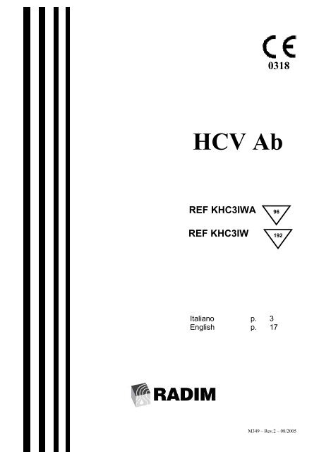
0318 HCV Ab REF KHC3IWA REF KHC3IW - Radim S.p.A.
0318 HCV Ab REF KHC3IWA REF KHC3IW - Radim S.p.A.
0318 HCV Ab REF KHC3IWA REF KHC3IW - Radim S.p.A.

Create successful ePaper yourself
Turn your PDF publications into a flip-book with our unique Google optimized e-Paper software.
<strong>0318</strong><br />
<strong>HCV</strong> <strong>Ab</strong><br />
<strong>REF</strong> <strong><strong>KHC3IW</strong>A</strong><br />
<strong>REF</strong> <strong>KHC3IW</strong><br />
96<br />
192<br />
Italiano p. 3<br />
English p. 17<br />
M349 – Rev.2 – 08/2005
REAGENTI DEL KIT - KIT REAGENTS (96 Test)<br />
Reag. Quant. Stato fisico,<br />
Physical state<br />
MICROPLATE 96 Pronti per l'uso, Ready for use<br />
CONTROL(- 1 x 4.0 mL Pronto per l'uso, Ready for use<br />
CONTROL (+ 1 x 2.0 mL Pronto per l'uso, Ready for use<br />
CAL 2 Liofilo, Lyoph.<br />
WASHBUF (20X 1 x 50 mL Conc.<br />
CONJ 1 x 16 mL Pronto per l'uso, Ready for use<br />
SUBS(TMB 1 x 16 mL Pronto per l'uso, Ready for use<br />
DILAS 3 x 3.0 mL Pronto per l'uso, Ready for use<br />
DILSPE 1 x 50 mL Pronto per l'uso, Ready for use<br />
H2SO4 (O.3 M 1 x 16 mL Pronto per l'uso, Ready for use<br />
REAGENTI DEL KIT - KIT REAGENTS (192 Test)<br />
Reag. Quant. Stato fisico,<br />
Physical state<br />
MICROPLATE 192 Pronti per l'uso, Ready for use<br />
CONTROL ( - 2 x 4.0 mL Pronto per l'uso, Ready for use<br />
CONTROL ( + 1 x 4.0 mL Pronto per l'uso, Ready for use<br />
CAL 4 Liofilo, Lyoph.<br />
WASHBUF(20X 2 x 50 mL Conc.<br />
CONJ 2 x 16 mL Pronto per l'uso, Ready for use<br />
SUBS(TMB 2 x 16 mL Pronto per l'uso, Ready for use<br />
DILAS 5 x 4.0 mL Pronto per l'uso, Ready for use<br />
DILSPE 1 x 100 mL Pronto per l'uso, Ready for use<br />
H2SO4 (O.3 M 2 x 16 mL Pronto per l'uso, Ready for use<br />
<strong><strong>KHC3IW</strong>A</strong> - <strong>KHC3IW</strong> – <strong>HCV</strong> <strong>Ab</strong><br />
M349 – Rev. 2 – 08/2005 – Pag. 2/32
DOSAGGIO IMMUNOENZIMATICO DI TERZA GENERAZIONE PER LA<br />
DETERMINAZIONE DEGLI ANTICORPI ANTI VIRUS DELL’EPATITE “C” IN<br />
PLASMA E SIERO UMANO.<br />
PER USO DIAGNOSTICO IN VITRO<br />
“Il kit può essere usato per la ricerca degli anticorpi nelle unità di sangue ed il follow-up di pazienti<br />
affetti da <strong>HCV</strong>.”<br />
INTRODUZIONE<br />
L’Organizzazione Mondiale della Sanità (OMS) definisce l’infezione da Epatite C come segue:<br />
“L’Epatite C è un’infezione virale del fegato che è stata attribuita a epatiti “non A, non B” parentalmente trasmesse<br />
fino all’identificazione dell’agente eziologico nel 1989. La scoperta e la caratterizzazione del virus dell’epatite C<br />
(<strong>HCV</strong>) porta alla comprensione del suo primario ruolo nelle epatiti post-trasfusione e la sua tendenza ad indurre<br />
infezioni persistenti.<br />
L’<strong>HCV</strong> è una delle maggiori cause di epatiti acute e malattie croniche del fegato, inclusi cirrosi e cancro al fegato.<br />
Globalmente si stima che 170 milioni di persone siano cronicamente infette e da 3 a 4 milioni di persone siano<br />
nuovamente infettate ogni anno. L’<strong>HCV</strong> è trasmesso principalmente per contatto diretto con sangue umano. Le<br />
maggiori cause di <strong>HCV</strong> nel mondo sono l’uso di unità di sangue non controllato per le trasfusioni e il riutilizzo di<br />
aghi e siringhe non adeguatamente sterilizzati. Nessun vaccino è al momento disponibile per prevenire l’epatite<br />
C ed il trattamento delle epatiti C croniche è troppo costoso perché i popoli dei paesi in via di sviluppo possano<br />
permetterselo. Allora, da una prospettiva globale, il più grande impatto sui malati di epatite C sarà ottenuto<br />
focalizzando gli sforzi sulla riduzione dei rischi della trasmissione di <strong>HCV</strong> da esposizioni nosocomiali (trasfusioni<br />
di sangue, iniezioni non sicure) e comportamenti ad alto rischio ( iniezione di droghe).<br />
Il virus dell’ epatite C (<strong>HCV</strong>) è uno dei virus (A, B, C, D, e E), che sono responsabili della maggioranza dei casi di<br />
epatite virale. E’ un virus ad RNA avvolto della famiglia Flaviviridae che mostra di avere uno stretto spettro<br />
d’ospite. Gli uomini e gli scimpanzé sono le sole specie suscettibili all’infezione ed entrambi sviluppano le stesse<br />
malattie. Una caratteristica importante del virus è la relativa mutabilità del genoma, che è probabilmente legata<br />
all’alta tendenza (80%) nell’indurre infezioni croniche. L’<strong>HCV</strong> è riunito in diversi genotipi distinti che possono<br />
essere importanti nella determinazione della gravità della malattia e nella risposta al trattamento.<br />
Il periodo di incubazione dell’infezione da <strong>HCV</strong>, prima dell’inizio dei sintomi clinici, va da 15 a 150 giorni. Nelle<br />
infezioni acute i sintomi più comuni sono affaticamento e itterizia; comunque la maggior parte dei casi (tra il 60% e<br />
il 70%), anche quelli che sviluppano l’infezione cronica, sono a-sintomatici. Circa l’80% dei nuovi pazienti infettati<br />
sviluppa un’infezione cronica. Cirrosi si sviluppano all’incirca dal 10 % al 20% dei pazienti che hanno un’infezione<br />
cronica, mentre cancro al fegato si manifesta tra l’1% e il 5% delle persone che hanno un’infezione cronica per un<br />
periodo da 20 a 30 anni. La maggior parte di pazienti che soffrono di cancro al fegato ma che non presentano<br />
infezione da epatite B mostrano infezione da <strong>HCV</strong>. Il meccanismo per cui l’infezione da <strong>HCV</strong> porta al cancro al<br />
fegato non è ancora ben chiaro. L’epatite C accresce la gravità delle malattie del fegato quando coesiste con<br />
altre condizioni epatiche. In particolare, le malattie del fegato progrediscono più rapidamente in persone con<br />
malattie epatiche provocate da abuso di alcol e con infezione da <strong>HCV</strong>. L’ <strong>HCV</strong> è trasmesso principalmente per<br />
contatto diretto con sangue infetto. La trasmissione attraverso trasfusioni di sangue non esaminato per l’<strong>HCV</strong>, il<br />
riutilizzo di aghi, siringhe e altre attrezzature mediche non adeguatamente sterilizzate, o attraverso lo scambio di<br />
aghi tra drogati, è molto ben documentata. La trasmissione per via sessuale o perinatale può anche avvenire ma<br />
meno frequentemente. Altri modi di trasmissione legati a pratiche comportamentali, sociali, culturali (body<br />
piercing, circoncisioni e tatuaggi) possono avvenire se vengono utilizzati strumenti non adeguatamente sterilizzati.<br />
L’ <strong>HCV</strong> non è trasmesso attraverso starnuti, abbracci, tosse, cibo o acqua, condividendo posate o bicchieri o per<br />
contatto casuale.<br />
Sia nei paesi sviluppati che quelli in via di sviluppo gruppi ad alto rischio includono gli utilizzatori di droghe<br />
iniettabili, i riceventi di sangue non esaminato, emofilici, dializzati e persone con numerosi partners sessuali che<br />
praticano rapporti non protetti. Nei paesi sviluppati si stima che il 90% delle persone con infezione da <strong>HCV</strong><br />
cronica siano principalmente utilizzatori di droghe iniettabili e quelli con una storia di trasfusioni di sangue non<br />
esaminato o emoderivati. Nella maggior parte dei paesi in via di sviluppo, dove sangue e emoderivati non<br />
esaminati sono ancora usati, il principale mezzo di trasmissione dell’infezione sono strumenti per le iniezioni non<br />
sterilizzati e trasfusioni di sangue non controllato. Inoltre le persone che praticano riti sacrificali e circoncisioni<br />
sono a rischio se usano o riusano ferri non sterilizzati.<br />
<strong><strong>KHC3IW</strong>A</strong> - <strong>KHC3IW</strong> – <strong>HCV</strong> <strong>Ab</strong><br />
M349 – Rev. 2 – 08/2005 – Pag. 3/32
L’OMS stima che circa 170 milioni di persone, il 3% della popolazione mondiale, sono infettate da <strong>HCV</strong> e sono a<br />
rischio di sviluppare cirrosi e/o cancro al fegato. La prevalenza dell’infezione <strong>HCV</strong> in Africa, Medio Oriente, Sud<br />
Est asiatico e Pacifico occidentale è alta se comparata con Nord America ed Europa.<br />
I test diagnostici per <strong>HCV</strong> sono usati per prevenire l’infezione attraverso lo screening dei donatori di sangue e<br />
plasma, per stabilire la diagnosi clinica e prendere adeguate decisioni riguardo la cura di un paziente. I test<br />
diagnostici oggi disponibili sono basati su dosaggi immunoenzimatici (EIA) per la rilevazione di specifici anticorpi<br />
<strong>HCV</strong>. Il sistema EIA può rilevare più del 95% di pazienti cronicamente infetti, ma solo dal 50% al 70% delle<br />
infezioni acute. Un saggio immunoblot ricombinante (RIBA) che identifica gli anticorpi che reagiscono con antigeni<br />
individuali <strong>HCV</strong> è spesso usato come test supplementare per la conferma di un risultato EIA positivo. Test per<br />
<strong>HCV</strong> basati sull’amplificazione dell’ RNA (es. PCR, saggio a DNA legato) è anche stato usato per la conferma del<br />
risultato serologico e per l’assegnazione di un’efficace terapia antivirale. Un risultato positivo indica la presenza di<br />
infezione attiva e la possibilità di diffusione dell’infezione e/o lo sviluppo di malattie croniche del fegato.<br />
Farmaci antivirali come l’interferone assunto da solo o in combinazione con ribavirina, possono essere usati per il<br />
trattamento di persone con epatite C cronica, ma il costo del trattamento è molto alto. Il trattamento con<br />
interferone da solo è efficace in circa il 10%-20% dei pazienti. L’interferone combinato con ribavirina è efficace nel<br />
30%-50% dei pazienti. La ribavirina non sembra essere efficace quando usata da sola.<br />
Non c’è nessun vaccino efficace contro l’<strong>HCV</strong>. La ricerca procede ma l’alta mutabilità del genoma dell’<strong>HCV</strong><br />
complica lo sviluppo di un vaccino. La mancanza di conoscenze di qualche risposta immuno-protettiva seguente<br />
l’infezione da <strong>HCV</strong> impedisce anche la ricerca del vaccino. Nemmeno si sa se il sistema immunitario è in grado di<br />
eliminare il virus.<br />
Qualche studio comunque ha mostrato la presenza di anticorpi neutralizzanti il virus nei pazienti affetti da <strong>HCV</strong>. In<br />
assenza di un vaccino devono essere prese tutte le precauzioni per prevenire l’infezione, incluse (a) screening e<br />
test di sangue e organi; (b) disattivazione del virus in plasmi e prodotti derivati; (c) accrescimento e mantenimento<br />
delle pratiche di controllo dell’infezione nei protocolli d’attenzione sanitaria, come l’appropriata sterilizzazione di<br />
strumenti medici e dentali; (d) la promozione di cambiamenti nei comportamenti tra la gente comune e gli<br />
operatori sanitari per ridurre l’uso eccessivo di iniezioni e la pratica di iniezioni sicure; (e) la riduzione del rischio<br />
per persone che fanno uso di droga e pratiche sessuali ad alto rischio.<br />
Il genoma codifica per i componenti strutturali, una proteina nucleocapsidica e glicoproteine dell’envelope, e i<br />
costituenti funzionali coinvolti nella replicazione del virus e nel processamento delle sue proteine.<br />
La regione codificante nucleocapsidica sembra essere la più conservativa tra i differenti genotipi isolati nel<br />
mondo.<br />
PRINCIPIO DEL TEST<br />
Le micropiastre sono coattate con antigeni <strong>HCV</strong>-specifici derivanti dalle regioni “core” e “ns” codificanti per i<br />
conservativi e i determinanti antigenici immunodominanti (peptide core, peptidi ricombinanti NS3, NS4 e NS5).<br />
La fase solida è prima trattata con il campione diluito e gli anticorpi <strong>HCV</strong> sono catturati, se presenti, dagli antigeni.<br />
Dopo aver allontanato tramite lavaggio tutti gli altri componenti del campione,nella seconda incubazione gli<br />
anticorpi anti <strong>HCV</strong> catturati sono rilevati tramite l’aggiunta di specifici anticorpi policlonali anti IgG&M, marcati con<br />
perossidasi (HRP).<br />
L’enzima catturato sulla fase solida , reagendo con la miscela cromogeno/substrato, genera un segnale ottico che<br />
è proporzionale alla quantità di anticorpi anti <strong>HCV</strong> presenti nel campione.<br />
Un valore di cut-off permette di trasformare le densità ottiche rilevate in risultati positivi e negativi dovuti alla<br />
presenza di anticorpi anti-<strong>HCV</strong>.<br />
COMPONENTI<br />
Il kit contiene reagenti sufficienti per eseguire i seguenti dosaggi da 96 test (codice <strong><strong>KHC3IW</strong>A</strong>) e 192 test<br />
(codice <strong>KHC3IW</strong>).<br />
1. Micropiastra MICROPLATE<br />
Piastra da 12 strips di 8 pozzetti scomponibili attivate con peptide Core, peptidi ricombinanti NS3, NS4 e NS5. Le<br />
piastre sono sigillate in buste con essiccante.<br />
Kit da 96 test: 1 micropiastra<br />
Kit da 192 test: 2 micropiastre<br />
<strong><strong>KHC3IW</strong>A</strong> - <strong>KHC3IW</strong> – <strong>HCV</strong> <strong>Ab</strong><br />
M349 – Rev. 2 – 08/2005 – Pag. 4/32
2. Controllo Negativo CONTROL(-<br />
Controllo pronto all’uso. Contiene l’1% di proteine da siero di capra, tampone Na-citrato 10 mM a pH 6.0 +/-0.1,<br />
0.5% Tween 20, 0.09% Na-azide e 0.1% Kathon GC come conservanti.<br />
Il controllo negativo è codificato con colore verde oliva.<br />
Kit da 96 test: 1 x 4.0 mL/flacone<br />
Kit da 192 test: 2 x 4.0 mL/flacone<br />
3. Controllo Positivo CONTROL(+<br />
Controllo pronto all’uso. Contiene l’1% di proteine da siero di capra, anticorpi umani positivi all’<strong>HCV</strong>, tampone Nacitrato<br />
10 mM a pH 6.0 +/-0.1, 0.5% Tween 20, 0.09% Na-azide e 0.1% Kathon GC come conservanti.<br />
Il controllo positivo è codificato con colore verde scuro.<br />
Kit da 96 test: 1 x 2 mL/flacone<br />
Kit da 192 test: 1 x 4.0 mL/flacone<br />
4. Calibratore CAL<br />
Calibratore liofilo. Deve essere disciolto con il volume di acqua grado EIA riportato sull’etichetta. Contiene<br />
proteine da siero bovino fetale, anticorpi umani per <strong>HCV</strong> il cui contenuto è calibrato su NIBSC Working Standard<br />
codice 99/588/003-WI, 10mM di tampone Na-citrato a pH 6.0+/-0.1, 0.3 mg/ml di gentamicina solfato e 0.1% di<br />
Kathon GC come conservanti.<br />
Nota: il volume necessario per sciogliere il contenuto della fiala può variare da lotto a lotto.<br />
Si prega di usare il corretto volume riportato sull’etichetta.<br />
Kit da 96 test: 2 flaconi<br />
Kit da 192 test: 4 flaconi<br />
5. Soluzione di lavaggio concentrata WASHBUF(20X<br />
Soluzione concentrata 20X. Una volta diluita, la soluzione di lavaggio contiene tampone fosfato 10mM a pH 7.0<br />
+/-0.2 e 0.05% Tween 20 and 0.05% Kathon GC<br />
Kit da 96 test: 1 x 50 mL/flacone<br />
Kit da 192 test: 2 x 50 mL/flacone<br />
6. Coniugato Enzimatico CONJ<br />
Reagente pronto all’uso e codificato di colore rosso. Contiene perossidasi di rafano coniugata ad anticorpi<br />
policlonali di capra contro IgG e IgM umane, 5% BSA, tampone Tris 10mM a pH 6.8+/-0.1, 0.1% Kathon GC and<br />
0.02% gentamicina solfato come conservanti.<br />
Kit da 96 test: 1 x 16 mL/flacone<br />
Kit da 192 test: 2 x 16 mL/flacone<br />
7. Cromogeno/Substrato SUBS (TMB<br />
Componente pronto all’uso. Contiene tampone citrato-fosfato 50 mM a pH 3.5-3.8, dimetil-solfossido al 4%,<br />
0.03% di tetrametil-benzidina (TMB) e 0.02% di perossido d’idrogeno (H2O2).<br />
Nota: Deve essere conservato protetto dalla luce in quanto sensibile alla forte illuminazione.<br />
Kit da 96 test: 1 x 16 mL/flacone<br />
Kit da 192 test: 2 x 16 mL/flacone<br />
8. Diluente del Saggio DILAS<br />
Soluzione tamponata di Tris 10mM a pH 8.0 +/-0.1 contenente 0.1% di Kathon GC per il pretrattamento dei<br />
campioni e dei controlli in piastra. Blocca le interferenze.<br />
Nota: Usa tutto il contenuto di una fiala prima di aprirne una seconda. Il reagente è sensibile<br />
all’ossidazione.<br />
Kit da 96 test: 3 x 3.0 mL/flacone<br />
Kit da 192 test: 5 x 4.0 mL/flacone<br />
9. Acido Solforico H2SO4 (O.3 M<br />
Contiene una soluzione 0.3 M di H2SO4.<br />
Attenzione: Irritante (Xi R36/38; S2/26/30)<br />
Kit da 96 test: 1 x 16 mL/flacone<br />
Kit da 192 test: 2 x 16 mL/flacone<br />
10. Diluente Campione DILSPE<br />
Contiene 1% di proteine di siero di capra, tampone Na-citrato 10mM a pH 6.0 +/-0.1, 0.5% di Tween 20, 0.09% di<br />
Na-azide e 0.1% di Kathon GC come conservanti. Usare per diluire i campioni.<br />
Nota: Il diluente cambia colore da verde oliva a verde scuro-blu in presenza del campione.<br />
<strong><strong>KHC3IW</strong>A</strong> - <strong>KHC3IW</strong> – <strong>HCV</strong> <strong>Ab</strong><br />
M349 – Rev. 2 – 08/2005 – Pag. 5/32
Kit da 96 test: 1 x 50 mL/flacone<br />
Kit da 192 test: 1 x 100mL/flacone<br />
11. Copripiastra<br />
Kit da 96 test: n° 2<br />
Kit da 192 test: n° 4<br />
12. Istruzioni per l’Uso n° 1<br />
MATERIALI RICHIESTI MA NON FORNITI<br />
Dosaggio Manuale<br />
1. Micropipette calibrate (200Sl e 10Sl) e puntali usa e getta.<br />
2. Acqua di grado EIA (distillata o deionizzata, trattata con carbone per rimuovere agenti ossidanti usati come<br />
disinfettanti).<br />
3. Timer con intervallo di tempo di 60 min o più.<br />
4. Fogli di carta assorbente.<br />
5. Incubatore termostatico calibrato per micropiastre ELISA in grado di fornire una temperatura di +37°C.<br />
6. Lettore ELISA per micropiastre, calibrato, con filtro 450nm e possibilmente 620-630nm per la determinazione<br />
del bianco.<br />
7. Lavatore calibrato di micropiastre ELISA.<br />
8. Vortex o strumenti similari per miscelare.<br />
Dosaggio Automatico<br />
− Il dispositivo può essere utilizzato con strumentazione automatica di kit ELISA su micropiastra.<br />
− Si garantisce l'applicabilità su strumentazione RADIM e/o SEAC.<br />
− Qualora si utilizzi strumentazione automatica di altri fornitori, è responsabilità dell'utilizzatore assicurarsi che il<br />
kit sia stato opportunamente validato.<br />
AVVERTENZE E PRECAUZIONI<br />
1. Il kit deve essere usato solo da personale tecnico specializzato e correttamente addestrato, sotto la<br />
supervisione del medico responsabile del laboratorio.<br />
2. Quando il kit è usato per lo screening di unità di sangue e componenti del sangue deve essere usato in un<br />
laboratorio certificato e qualificato dall’autorità nazionale in quel campo (Ministero della Sanità o simili) per<br />
eseguire questo tipo di analisi.<br />
3. Tutto il personale coinvolto nell’esecuzione del saggio deve indossare abiti protettivi da laboratorio, guanti in<br />
lattice senza talco e occhiali. L’uso di ogni dispositivo appuntito (aghi) o tagliente (lame) dovrebbe essere evitato.<br />
Tutto il personale coinvolto dovrebbe essere addestrato sulle procedure di sicurezza personale, come<br />
raccomandato dal Centro per il Controllo delle Malattie di Atlanta, US, e riportato nella pubblicazione dell’Istituto<br />
Nazionale di Sanità americano: “BioSicurezza nei Laboratori Microbiologici e Biomedici”, ed. 1984. Fare<br />
comunque riferimento alla legislazione nazionale in vigore al momento.<br />
4. Tutto il personale coinvolto nel maneggiare i campioni dovrebbe essere vaccinato per HBV e HAV, per i quali<br />
sono disponibili vaccini sicuri ed efficaci.<br />
5. L’ambiente di laboratorio dovrebbe essere controllato così da evitare contaminazioni da polvere e agenti<br />
microbiologici nell’aria, quando si aprono le fiale e la micropiastra del kit e quando viene eseguito il test.<br />
Proteggere il cromogeno/substrato dalla luce forte ed evitare vibrazioni del banco di lavoro una volta iniziato il<br />
test.<br />
6. Dal ricevimento, conservare il kit a 2- 8°C in un frigorifero o camera fredda a temperatura controllata.<br />
7. Non scambiare i componenti tra differenti lotti del kit. E’ raccomandato non scambiare i componenti di due kit<br />
dello stesso lotto.<br />
8. Controllare che i reagenti siano limpidi e non contengano grosse particelle o aggregati. Se ciò accade<br />
allertare il supervisore del laboratorio per iniziare le necessarie procedure per la sostituzione del kit.<br />
9. Evitare contaminazioni incrociate tra i campioni (sieri o plasmi) usando puntali monouso e cambiandoli dopo<br />
ogni campione.<br />
10. Evitare contaminazioni incrociate tra i reagenti del kit usando puntali monouso e cambiandoli per l’uso di ogni<br />
componente.<br />
11. Non usare il kit dopo la data di scadenza impressa sulla confezione esterna e sull’ etichetta di ogni singola<br />
fiala all’interno.<br />
12. Trattare tutti i campioni come potenzialmente infetti. Tutti i sieri umani dovrebbero essere maneggiati secondo<br />
il Livello 2 di BioSicurezza, come raccomandato dal Centro per il Controllo delle Malattie, Atlanta, US, insieme<br />
<strong><strong>KHC3IW</strong>A</strong> - <strong>KHC3IW</strong> – <strong>HCV</strong> <strong>Ab</strong><br />
M349 – Rev. 2 – 08/2005 – Pag. 6/32
con quanto riportato nella pubblicazione dell’Istituto di Sanità americano: “BioSicurezza nei laboratori<br />
Microbiologici e Biomedicali”, ed. 1984. Fare comunque riferimento alla legislazione nazionale in vigore al<br />
momento.<br />
13. L’uso di contenitori di plastica monouso è raccomandato per la preparazione dei componenti liquidi o per I<br />
componenti trasferiti nelle postazioni automatizzate, questo per evitare contaminazioni incrociate.<br />
14. I prodotti di scarto durante l’uso del kit devono essere eliminati secondo le direttive nazionali e le leggi<br />
riguardanti i rifiuti di sostanze chimiche e biologiche di laboratorio. In particolare, gli scarichi liquidi generati dalla<br />
procedura di lavaggio, da avanzi dei controlli e dai campioni devono essere trattati come materiali potenzialmente<br />
infetti e inattivati prima di essere eliminati. Si suggerisce di inattivarli mediante trattamento con una soluzione di<br />
ipoclorito di sodio al 10% per 16-18 ore o disattivazione al calore in autoclave a 121°C per 20 minuti.<br />
15. Rovesciamenti accidentali dei campioni durante le operazioni devono essere assorbiti con fogli di carta<br />
imbevuti di ipoclorito di sodio e poi sciacquati con acqua. I fogli di carta vanno poi gettati nell’apposito contenitore<br />
dei rifiuti per materiali biologici.<br />
16. L’acido solforico è irritante. In caso di rovesciamento lavare la superficie con abbondante acqua.<br />
17. Altri materiali di scarto generati dall’uso del kit (ad esempio: i puntali usati per controlli e campioni,<br />
micropiastre usate) dovrebbero essere maneggiati come potenzialmente infetti e riposti in accordo alle direttive<br />
nazionali e alle leggi concernenti lo smaltimento dei rifiuti di laboratorio.<br />
CAMPIONI: PREPARAZIONE E RACCOMANDAZIONI<br />
1. Il sangue è prelevato asetticamente mediante prelievo in vena e i campioni di plasma o siero sono preparati<br />
usando le tecniche standard di preparazione dei campioni per analisi cliniche di laboratorio. Nessuna influenza è<br />
stata osservata nella preparazione del campione con citrato, EDTA o eparina.<br />
2. Evitare ogni aggiunta di conservanti ai campioni, specialmente sodio azide, che può influenzare l’attività<br />
enzimatica del coniugato, generando risultati falsi negativi.<br />
3. I campioni devono essere chiaramente identificati con codici o nomi per evitare confusione nell’interpretazione<br />
dei risultati. Quando il kit è usato per lo screening di unità di sangue è fortemente raccomandato di etichettare<br />
con codici a barre e leggere elettronicamente.<br />
4. Campioni emolizzati (rossi) e visibilmente iperlipemici (lattiginosi) devono essere scartati perché potrebbero<br />
generare risultati falsi. I campioni contenenti residui di fibrina o grosse particelle o filamenti e strutture microbiche<br />
dovrebbero essere scartati perché potrebbero dare origine a risultati falsi.<br />
5. Sieri e plasmi possono essere conservati a +2°…+8°C fino a 5 giorni dopo il prelievo. Per conservazioni più<br />
lunghe i campioni possono essere congelati a –20°C per diversi mesi. Qualsiasi campione congelato non può<br />
essere congelato e scongelato più di una volta, perché questo genera particelle che possono influenzare il<br />
risultato del test.<br />
6. Se sono presenti delle particelle centrifugare a 2000 rpm per 20 minuti o filtrare con filtri a 0.2-0.8um per pulire<br />
il campione da testare.<br />
PREPARAZIONE DEI COMPONENTI E AVVERTENZE<br />
Studi condotti su un kit aperto non hanno mostrato alcuna rilevante perdita di attività fino a 6 riutilizzi dello stesso<br />
materiale in 6 mesi.<br />
1. Micropiastra:<br />
Permettere che la micropiastra raggiunga la temperatura ambiente (almeno 1h) prima di aprire la busta.<br />
Controllare che l’essiccante non sia diventato verde scuro, indicando un difetto di produzione. In questo caso<br />
chiamare il servizio clienti.<br />
Le strips non utilizzate devono essere riposte nell’apposita busta, in presenza dell’essiccante fornito, sigillate<br />
fermamente e conservate a +2°-8°C.<br />
Dopo la prima apertura le strips residue sono stabili fino a quando l’indicatore di umidità presente all’interno della<br />
busta dell’essiccante vira da giallo a verde.<br />
2. Controllo Negativo:<br />
Pronto all’uso. Miscelare su vortex prima dell’uso.<br />
3. Controllo Positivo::<br />
Pronto all’uso. Miscelare su vortex prima dell’uso. Maneggiare questo componente come potenzialmente infettivo,<br />
anche se l’<strong>HCV</strong>, eventualmente presente nel controllo, è stato chimicamente inattivato.<br />
4. Calibratore:<br />
Sciogliere attentamente il contenuto liofilo della fiala con il volume di acqua di grado EIA riportato sulla sua<br />
etichetta.<br />
Miscelare su vortex prima dell’uso.<br />
<strong><strong>KHC3IW</strong>A</strong> - <strong>KHC3IW</strong> – <strong>HCV</strong> <strong>Ab</strong><br />
M349 – Rev. 2 – 08/2005 – Pag. 7/32
Maneggiare questo componente come potenzialmente infettivo anche se l’<strong>HCV</strong>, eventualmente presente nel<br />
controllo, è stato chimicamente inattivato.<br />
Nota: Una volta sciolto, il calibratore non è stabile. Conservare in aliquote a –20°C.<br />
5. Soluzione di lavaggio concentrata:<br />
L’intero contenuto della soluzione concentrata 20X deve essere diluito con acqua bidistillata fino a 1000 ml e<br />
miscelata delicatamente prima dell’uso.<br />
Poiché nel flacone potrebbero essere presenti dei cristalli, quando si prepara la soluzione prestare particolare<br />
attenzione nel far sciogliere completamente tutto il contenuto.<br />
Nella preparazione evitare di generare schiuma perché la presenza di bolle può diminuire l’efficacia del lavaggio.<br />
Nota: Una volta diluita, la soluzione di lavaggio è stabile per una settimana a +2°…+8°C.<br />
6. Coniugato Enzimatico:<br />
Pronto all’uso. Miscelare su vortex prima dell’uso.<br />
Prestare attenzione per non contaminare il liquido con ossidanti chimici, polveri o microbi presenti nell’aria.<br />
Se questo componente deve essere trasferito usare solo contenitori di plastica possibilmente sterili.<br />
7. Cromogeno/Substrato:<br />
Pronto all’uso. Miscelare su vortex prima dell’uso.<br />
Prestare attenzione a non contaminare il liquido con ossidanti chimici, polveri o microbi presenti nell’aria.<br />
Non esporre a forte illuminazione, non metterlo a contatto con agenti ossidanti e superfici metalliche.<br />
Se questo componente deve essere trasferito usare solo contenitori di plastica possibilmente sterili.<br />
8. Diluente del Saggio:<br />
Pronto all’uso. Miscelare su vortex prima dell’uso.<br />
9. Acido Solforico:<br />
Pronto all’uso. Miscelare su vortex prima dell’uso.<br />
Attenzione: Irritante (Xi R36/38; S2/26/30)<br />
Legenda: R 36/38 = Irritante per gli occhi e la pelle.<br />
S 2/26/30 = In caso di contatto con gli occhi, sciacquare immediatamente con acqua e consultare il medico.<br />
10. Diluente del Campione:<br />
Pronto all’uso. Miscelare su vortex prima dell’uso.<br />
STRUMENTAZIONE IN COMBINAZIONE CON IL KIT<br />
1. Le micropipette devono essere calibrate per rilasciare il corretto volume richiesto dal saggio e devono<br />
essere sottoposte a regolare decontaminazione (alcool denaturato, candeggina al 10%, soluzione<br />
disinfettante ospedaliera) di quelle parti che potrebbero accidentalmente entrare in contatto con il<br />
campione. Esse dovrebbero essere regolarmente controllate per mostrare una precisione dell’1% e una<br />
correttezza di +/-2%. La decontaminazione dei componenti residui o fuoriusciti del kit deve eseguita con<br />
regolarità.<br />
2. L’incubatore ELISA dovrebbe essere tarato a 37°C (tolleranza di +/-0.5°C) e regolarmente controllato per<br />
assicurare il mantenimento della temperatura corretta. Sia incubatori a secco che bagnomaria sono<br />
utilizzabili per le incubazioni, se gli strumenti sono convalidati per l’incubazione di test ELISA.<br />
3. Il lavatore ELISA è estremamente importante per la totale riuscita del saggio. Il lavatore deve essere<br />
convalidato con attenzione e correttamente ottimizzato usando apparati di controllo e kit di riferimento,<br />
prima di utilizzarlo per gli esami di routine. Di solito sono sufficienti 4-5 cicli di lavaggio (aspirazione +<br />
dispensazione = 1 ciclo) da 350 ul per assicurare che il saggio dia il risultato aspettato. Si suggerisce un<br />
intervallo di “soaking” di 20-30 secondi tra i cicli. Per stabilire correttamente il loro numero è<br />
raccomandato di eseguire un test di prova con i controlli del kit e campioni di riferimento ben caratterizzati<br />
come positivi o negativi, e controllare la corrispondenza ai valori riportati sotto, nella sezione “Controllo<br />
di Qualità interno”. La regolare calibrazione del volume erogato e la manutenzione del lavatore<br />
(decontaminazione e pulizia degli aghi) deve essere compiuta secondo le indicazioni del produttore.<br />
4. I tempi di incubazione hanno una tolleranza di +/-5%.<br />
5. Il lettore di micropiastre ELISA deve essere dotato di un filtro di lettura di 450nm e idealmente di un<br />
secondo filtro (620-630nm) per le operazioni del bianco. Le sue prestazioni standard dovrebbero essere<br />
(a) ampiezza di banda ≤ 10nm; (b) intervallo di assorbimento da 0 a 2.0; (c) linearità 2.0; (d) ripetibilità<br />
1%. Il bianco è determinato secondo le istruzioni contenute nella sezione “Procedura del Saggio”. Il<br />
sistema ottico del lettore deve essere calibrato regolarmente per assicurare la corretta misurazione della<br />
densità ottica. La manutenzione dovrebbe essere fatta regolarmente secondo le istruzioni del produttore.<br />
<strong><strong>KHC3IW</strong>A</strong> - <strong>KHC3IW</strong> – <strong>HCV</strong> <strong>Ab</strong><br />
M349 – Rev. 2 – 08/2005 – Pag. 8/32
6. Quando si usa una stazione di lavoro automatizzata per kit ELISA tutti i passi critici (dispensazione,<br />
incubazione, lavaggio, lettura, elaborazione dei dati) devono essere attentamente controllati, calibrati e<br />
regolarmente sistemati per ottenere corrispondenza con i valori riportati nelle sezione “Controllo di<br />
Qualità interno”. Il protocollo del saggio deve essere installato nel sistema operativo dell’unità e<br />
convalidato come per il lavatore e il lettore. In più, la parte della stazione che manipola i componenti<br />
liquidi (dispensazione e lavaggio) deve essere convalidata e correttamente impostata. Particolare<br />
attenzione va mostrata nell’evitare il trascinamento (carry-over) da parte degli aghi usati per la<br />
dispensazione e il lavaggio. Questo deve essere studiato e controllato per minimizzare la possibilità di<br />
contaminazione da pozzetti adiacenti. L’uso di stazioni di lavoro automatizzate ELISA è raccomandata<br />
per lo screening di sangue quando il numero dei campioni da testare è maggiore di 20-30 per corsa della<br />
macchina.<br />
7. Quando si utilizzano strumenti per automazione,nel caso in cui il contenitore per i flaconi dello strumento<br />
non si adatti ai flaconi del kit, trasferire la soluzione in essi contenuta in flaconi idonei allo strumento ed<br />
etichettare gli stessi con la stessa etichetta staccatala flacone originale. Questa operazione è importante<br />
al fine di evitare scambi del contenuto dei flaconi durante il trasferimento. Quando il test è terminato, porre<br />
i contenitori secondari opportunamente etichettati e saldamente tappati a 2..8°C.<br />
8. Il servizio clienti offre assistenza agli utilizzatori per l’impostazione ed il controllo degli strumenti usati in<br />
combinazione con il kit, per assicurare concordanza con le richieste descritte.<br />
CONTROLLI E OPERAZIONI PRE SAGGIO<br />
1. Controllare la data di scadenza del kit, stampata sull’etichetta esterna della scatola. Non usare se scaduto.<br />
2. Controllare che i componenti liquidi non siano contaminati da particelle o aggregati visibili a occhio nudo.<br />
Controllare che il Cromogeno/Substrato sia incolore o blu pallido aspirando un piccolo volume dello stesso<br />
con una pipetta di plastica sterile trasparente. Controllare che nessuna rottura della confezione sia avvenuta<br />
nel trasporto e nessuna fuoriuscita di liquido sia presente all’interno della scatola. Controllare che la busta di<br />
alluminio, contenente la micropiastra, non sia bucata o danneggiata.<br />
3. Diluire tutto il contenuto della soluzione di lavaggio concentrata 20X come descritto sopra.<br />
4. Sciogliere il calibratore come descritto sopra.<br />
5. Permettere a tutti I componenti del kit di raggiungere la temperatura ambiente (circa 1h) e poi miscelare<br />
come descritto.<br />
6. Impostare l’incubatore ELISA a 37°C e preparare il lavatore ELISA avvinandolo con la soluzione di lavaggio<br />
diluita, secondo le istruzioni del produttore. Impostare il corretto numero di cicli di lavaggio, come indicato nel<br />
punto 3 della sezione “Strumentazione in combinazione con il Kit”.<br />
7. Controllare che il lettore ELISA sia acceso da almeno 20 minuti prima della lettura.<br />
8. Se si utilizza una stazione di lavoro automatizzata, accenderla, controllare le impostazioni e assicurarsi di<br />
usare il protocollo corretto.<br />
9. Controllare che le micropipette siano impostate al volume richiesto.<br />
10. Controllare che tutti gli strumenti siano disponibili e pronti all’uso.<br />
11. In caso di problemi non procedere oltre con il test e informare il supervisore.<br />
PROCEDURA<br />
Il saggio deve essere eseguito secondo quanto riportato sotto, prestando attenzione nell’osservare gli stessi<br />
tempi d’incubazione per tutti i campioni da testare.<br />
Saggio Automatizzato:<br />
In caso il test sia condotto automaticamente con un sistema ELISA, suggeriamo di fare aspirare allo strumento<br />
prima 200 Sl di Diluente del Campione e poi 10 Sl di campione.<br />
Il campione diluito viene poi dispensato direttamente nel pozzetto della micropiastra. Prima che il successivo<br />
campione sia aspirato, gli aghi devono essere ben lavati per evitare reazioni incrociate tra i campioni.<br />
Non diluire controlli e calibratore che sono già pronti all’uso.<br />
Dispensare 200 Sl di controlli e calibratore nelle appropriate celle della micropiastra.<br />
Nota Importante: Controllare visivamente che i campioni siano stati diluiti e dispensati nei pozzetti appropriati.<br />
Occorre verificare che il colore dei campioni dispensati viri al verde-blu scuro mentre il colore del controllo<br />
negativo resta verde oliva.<br />
Per le successive operazioni seguire le istruzioni operative riportate sotto per il Saggio Manuale.<br />
E’ fortemente raccomandato controllare che l’intervallo di tempo tra la dispensazione del primo e dell’ultimo<br />
campione venga controllata dallo strumento e presa in considerazione per ritardare dello stesso intervallo le<br />
operazioni di lavaggio.<br />
<strong><strong>KHC3IW</strong>A</strong> - <strong>KHC3IW</strong> – <strong>HCV</strong> <strong>Ab</strong><br />
M349 – Rev. 2 – 08/2005 – Pag. 9/32
Saggio Manuale:<br />
1. Inserire il corretto numero di strips o di pozzetti nell’apposito sostegno. Lasciare il pozzetto A1 vuoto per le<br />
operazioni di azzeramento.<br />
2. Dispensare 200 Sl di controllo negativo in triplo, 200 Sl di Calibratore in doppio e 200 Sl di controllo positivo<br />
in singolo nelle celle appropriate. Non diluire Controlli e Calibratore essendo già prediluiti e pronti all’uso!<br />
3. Aggiungere 200 Sl di Diluente del Campione (DILSPE) a tutti i pozzetti dei campioni; poi dispensare 10 Sl di<br />
campione in ogni pozzetto propriamente identificato. Agitare delicatamente la piastra, evitando fuoriuscite e<br />
rischi di contaminazione dei pozzetti adiacenti, per disciogliere completamente il campione nel suo diluente.<br />
Nota importante: Controllare che il colore del Diluente del Campione, dopo l’aggiunta del campione, viri da<br />
verde chiaro a verde-blu scuro, indicando che il campione è stato aggiunto.<br />
4. Dispensare 50 Sl di Diluente del Saggio (DILAS) in tutte le celle dei controlli, del calibratore e dei campioni.<br />
Controllare che il colore dei campioni viri a blu scuro.<br />
5. Incubare la micropiastra per 45 min a +37°C.<br />
Nota Importante: Le strips devono essere coperte con l’apposito foglio adesivo fornito solo quando il test è<br />
eseguito manualmente. Non coprire la strips quando si usa uno strumento ELISA automatizzato.<br />
6. Lavare la micropiastra con un lavatore automatico, effettuando 4-5 cicli di lavaggio, rilasciando e aspirando<br />
350 Sl/pozzetto di soluzione di lavaggio diluita (vedi paragrafo 3 della sezione “Strumentazione in<br />
combinazione con il kit).<br />
7. Pipettare 100 Sl di Coniugato Enzimatico in tutte le celle tranne quella del bianco, e coprire con il foglio<br />
adesivo. Controllare che questo componente di colore rosso sia stato dispensato in tutti i pozzetti, eccetto A1.<br />
Nota Importante: Attenzione a non urtare la superficie della plastica interna della cella con il puntale pieno di<br />
coniugato. Possono avvenire contaminazioni.<br />
8. Incubare la micropiastra per 45 min a +37°C.<br />
9. Lavare le celle come descritto al punto 6.<br />
10. Pipettare 100 Sl di miscela Cromogeno/Substrato in ogni pozzetto e anche in A1. Incubare la micropiastra a<br />
temperatura ambiente (18°-24°C) per 15 minuti al riparo dalla luce diretta.<br />
Nota Importante: Non esporre a forte illuminazione diretta. Può determinare fondi alti.<br />
11. Pipettare 100 Sl di Acido Solforico in tutti i pozzetti usando la stessa sequenza di dispensazione adottata nel<br />
punto 10 per arrestare la reazione enzimatica. L’aggiunta di acido farà virare il controllo positivo e i campioni<br />
positivi da blu a giallo.<br />
12. Misurare l’intensità di colore della soluzione in ogni pozzetto, (vedi paragrafo 5 della sezione<br />
“Strumentazione in combinazione con il kit), con un filtro di lettura a 450 nm e possibilmente con un filtro a<br />
620-630 nm per le operazioni del bianco in posizione A1.<br />
Note Importanti:<br />
1. Se il secondo filtro non è disponibile assicurarsi che non siano presenti impronte di dita sul fondo della<br />
micropiastra prima della lettura a 450nm. Tali impronte potrebbero generare falsi positivi.<br />
2. La lettura deve essere eseguita subito dopo l’aggiunta di Acido Solforico e comunque mai più di 20 minuti<br />
dopo tale aggiunta. Potrebbe avvenire un’auto-ossidazione del cromogeno che porterebbe ad un risultato di<br />
fondo alto.<br />
“Qualora si utilizzasse nel procedimento operativo uno strumento automatico per micropiastre RADIM e/o<br />
SEAC, far riferimento al relativo manuale. In caso di utilizzo della suddetta strumentazione la lettura<br />
spettrofotometrica viene eseguita automaticamente a 3 lunghezze d’onda: 450, 405 e 620 nm, permettendo<br />
l’ampliamento del range di lettura.”<br />
SCHEMA DI DOSAGGIO<br />
Metodo Operazioni<br />
Controlli & Calibratore<br />
Diluente Campioni (DILSPE)<br />
Campione<br />
Diluente del Saggio (DILAS)<br />
200 Sl<br />
200Sl<br />
10Sl<br />
50 Sl<br />
<strong><strong>KHC3IW</strong>A</strong> - <strong>KHC3IW</strong> – <strong>HCV</strong> <strong>Ab</strong><br />
M349 – Rev. 2 – 08/2005 – Pag. 10/32
1^ incubazione 45 min<br />
Temperatura +37°C<br />
Lavaggio 4-5 cicli<br />
350Sl/pozz.<br />
Coniugato Enzimatico 100 Sl<br />
2^ incubazione 45 min<br />
Temperatura +37°C<br />
Lavaggio 4-5 cicli<br />
350Sl/pozz.<br />
TMB/H2O2<br />
100 Sl<br />
3^ incubazione 15 min<br />
Temperatura t.a. (18-24°C)<br />
Acido Solforico 100 Sl<br />
Lettura OD 450nm<br />
Un esempio di schema di dispensazione è riportato sotto:<br />
Micropiastra<br />
1 2 3 4 5 6 7 8 9 10 11 12<br />
A BLK S2<br />
B NC S3<br />
C NC S4<br />
D NC S5<br />
E CAL S6<br />
F CAL S7<br />
G PC S8<br />
H S1 S9<br />
Legenda: BLK = Bianco NC = Controllo Negativo<br />
CAL = Calibratore PC = Controllo Positivo S = Campione<br />
CONTROLLO DI QUALITA’ INTERNO<br />
Un controllo è eseguito, sui controlli e il calibratore ogni volta che il kit è usato, per verificare che i loro valori di<br />
OD450nm siano quelli attesi e riportati nella tabella sotto.<br />
Controllo Requisiti<br />
Bianco < 0.100 OD450nm valore<br />
Controllo Negativo (NC) < 0.050 valore medio OD450nm dopo sottrazione del bianco<br />
Calibratore S/Co > 1.1<br />
Controllo Positivo > 1.000 OD450nm val.<br />
Se i risultati del test corrispondono ai requisiti sopra stabiliti, procedere alla sezione successiva.<br />
Se così non fosse, non procedere ulteriormente e operare come segue:<br />
Problema Controllare<br />
Bianco<br />
1. che la soluzione Cromogeno/Substrato non si sia contaminata durante il saggio<br />
> 0.100 OD450nm<br />
Controllo Negativo 1. che la procedura di lavaggio e le impostazioni del lavatore siano convalidate<br />
(NC)<br />
secondo gli studi di pre-qualificazione;<br />
> 0.050 OD450nm dopo 2. che sia stata usata la corretta soluzione di lavaggio e che il lavatore sia stato<br />
sottrazione del bianco avvinato prima dell’uso;<br />
3. che nessun errore sia stato commesso nella procedura del saggio (dispensazione<br />
del controllo positivo invece del negativo);<br />
4. che non sia avvenuta nessuna contaminazione del controllo negativo o delle suoi<br />
pozzetti a causa di schizzi dei campioni positivi o del coniugato enzimatico;<br />
5. che le micropipette non siano contaminate con campioni positivi o coniugato<br />
enzimatico;<br />
6. che gli aghi del lavatore non siano bloccati o parzialmente ostruiti.<br />
<strong><strong>KHC3IW</strong>A</strong> - <strong>KHC3IW</strong> – <strong>HCV</strong> <strong>Ab</strong><br />
M349 – Rev. 2 – 08/2005 – Pag. 11/32
Calibratore<br />
S/Co < 1.1<br />
Controllo Positivo<br />
< 1.000 OD450nm<br />
1. che le procedure siano state correttamente eseguite;<br />
2. che nessun errore sia stato eseguito nella sua dispensazione (es.: dispensazione<br />
del controllo positivo invece del calibratore)<br />
3. che la procedura di lavaggio e le impostazioni del lavatore siano convalidate<br />
secondo gli studi di pre-qualificazione;<br />
4.che non sia avvenuta nessuna contaminazione esterna del calibratore.<br />
1. che le procedure siano state correttamente eseguite;<br />
2. che nessun errore sia stato commesso nella distribuzione dei controlli<br />
(dispensazione del controllo negativo al posto del positivo). In questo caso il<br />
controllo negativo mostrerà una OD450nm> 0.150.<br />
3. che la procedura di lavaggio e le impostazioni del lavatore siano convalidate<br />
secondo gli studi di pre-qualificazione;<br />
4. che non sia avvenuta nessuna contaminazione esterna del controllo positivo<br />
Dovessero esserci ulteriori problemi dopo i controlli riportati in tabella, fare riferimento al supervisore per altri<br />
provvedimenti.<br />
CALCOLO DEL CUT-OFF<br />
I risultati del test sono calcolati sulla media di un valore di cut-off determinato con la seguente formula:<br />
Cut-Off = NC OD450nm medio + 0.350<br />
Il valore trovato per il test è usato per l’interpretazione dei risultati come descritto nel paragrafo successivo.<br />
Nota Importante:Quando il calcolo dei risultati è fatto dal sistema automatizzato ELISA assicurarsi che sia<br />
utilizzata la formula corretta per calcolare il cut-off ed ottenere la corretta interpretazione dei risultati.<br />
INTERPRETAZIONE DEI RISULTATI<br />
I risultati del test sono interpretati come il rapporto tra l’OD450nm del campione e il valore del Cut-Off (S/Co) in<br />
accordo alla seguente tabella:<br />
S/Co Interpretazione<br />
< 0.9 Negativo<br />
0.9 - 1.1 Equivoco<br />
> 1.1 Positivo<br />
Un risultato negativo indica che il paziente non è stato infettato da <strong>HCV</strong> o che l’unità di sangue può essere<br />
trasfusa.<br />
Quei pazienti che mostrano un risultato equivoco devono essere testati nuovamente attraverso l’esame di un<br />
secondo campione prelevato 1-2 settimane dopo. L’unità di sangue non deve essere trasfusa.<br />
Un risultato positivo indica la presenza dell’infezione da <strong>HCV</strong>, quindi il paziente deve essere trattato di<br />
conseguenza e l’unità di sangue deve essere scartata.<br />
Note Importanti:<br />
1. L’interpretazione dei risultati dovrebbe essere fatta sotto la supervisione del responsabile del laboratorio per<br />
ridurre il rischio di errori di giudizio.<br />
2. Ogni risultato positivo dovrebbe essere confermato da un metodo alternativo in grado di rilevare gli anticorpi<br />
IgG e IgM (test di conferma) prima di formulare una diagnosi di epatite virale.<br />
3. Come provato nello studio di performance clinico, il kit è in grado di determinare una seroconversione in<br />
anticorpi anti <strong>HCV</strong> core prima di alcuni prodotti commerciali presenti sul mercato. Non classificare quindi il<br />
risultato come falso positivo ed esaminare ulteriormente il campione con un test di conferma.<br />
4. Dal momento che il saggio è in grado di determinare anche anticorpi di classe IgM, potrebbero essere rilevate<br />
discrepanze con altri prodotti commerciali per la determinazione di anticorpi anti-<strong>HCV</strong> mancanti di coniugato<br />
anti-IgM. La reale positività del campione per gli anticorpi <strong>HCV</strong> dovrebbe essere poi confermata esaminando<br />
anche la reattività IgM, importante per la diagnosi dell’infezione da <strong>HCV</strong>.<br />
5. Quando i risultati sono trasmessi dal laboratorio ad un centro informatico, prestare attenzione per non<br />
trasferire dati errati.<br />
6. La diagnosi di epatite virale deve essere fatta e riferita al paziente solo da personale medico qualificato.<br />
Un esempio di calcolo è riportato sotto:<br />
I seguenti dati non devono essere usati al posto dei numeri reali ottenuti dall’utilizzatore.<br />
<strong><strong>KHC3IW</strong>A</strong> - <strong>KHC3IW</strong> – <strong>HCV</strong> <strong>Ab</strong><br />
M349 – Rev. 2 – 08/2005 – Pag. 12/32
Controllo Negativo: 0.019 – 0.020 – 0.021 OD450nm<br />
Valore medio: 0.020 OD450nm<br />
Inferiore a 0.050 – Accettato<br />
Controllo Positivo: 2.189 OD450nm<br />
Maggiore di 1.000: Accettato<br />
Cut-Off = 0.020+0.350 = 0.370<br />
Calibratore: 0.550 - 0.530 OD450nm<br />
Valore medio: 0.540 OD450nm S/Co = 1.4<br />
S/Co maggiore di 1.1 – Accettato<br />
Campione 1: 0.070 OD450nm<br />
Campione 2: 1.690 OD450nm<br />
Campione 1 S/Co < 0.9 = negativo<br />
Campione 2 S/Co > 1.1 = positivo<br />
PRESTAZIONI<br />
La valutazione delle prestazioni è stata condotta in accordo a quanto riportato in Common Technical<br />
Specifications o CTS (art. 5, Capitolo 3 di IVD Direttiva 98/79/EC).<br />
1. LIMITE DI RILEVAZIONE<br />
Il limite di rilevazione del saggio è stato calcolato per mezzo del British Working Standard per anti-<strong>HCV</strong>, NIBSC<br />
cod. 99/588-003-WI. La tabella sotto riporta i valori medi di OD450nm di questo standard diluito in plasma<br />
negativo e poi esaminato.<br />
Diluizione Lot # 1 Lot # 2<br />
Fattore S/Co S/Co<br />
1 X 2.0 2.0<br />
2 X 1.1 1.2<br />
4 X 0.7 0.8<br />
8 X 0.5 0.5<br />
Plasma Negativo 0.3 0.3<br />
Inoltre, il campione codificato Accurun 1 –Series 3000- fornito da Boston Biomedica Inc., USA, è stato valutato in<br />
toto mostrando i seguenti risultati:<br />
<strong>KHC3IW</strong> Accurun 1 S/Co<br />
Lot ID Series<br />
1201 3000 1.5<br />
0602 3000 1.5<br />
1202 3000 1.9<br />
Anche 7 campioni, testati positivi per <strong>HCV</strong> con Ortho <strong>HCV</strong> 3.0 SAVe, codice 930820, lot. # EXE065-1, sono stati<br />
diluiti in plasma negativo agli anticorpi anti-<strong>HCV</strong> per generare diluizioni limitanti e poi testati ancora su <strong>KHC3IW</strong>,<br />
lot. # 1202, e Ortho.<br />
La seguente tabella riporta i dati ottenuti.<br />
Campione Diluizioni <strong>KHC3IW</strong> Ortho 3.0<br />
n°<br />
limite S/Co S/Co<br />
1 256 X 1.9 1.3<br />
2 256 X 1.9 0.7<br />
3 256 X 2.4 1.0<br />
4 128 X 2.5 3.2<br />
5 85 X 3.3 1.4<br />
6 128 X 2.2 0.8<br />
7 135 X 3.2 2.2<br />
<strong><strong>KHC3IW</strong>A</strong> - <strong>KHC3IW</strong> – <strong>HCV</strong> <strong>Ab</strong><br />
M349 – Rev. 2 – 08/2005 – Pag. 13/32
2. SPECIFICITA’ E SENSIBILITA’ DIAGNOSTICHE<br />
La valutazione delle prestazioni del dispositivo è stata eseguita in un trial esterno condotto su più di 5000<br />
campioni.<br />
2.1 Specificità Diagnostica:<br />
E’ definita come la probabilità del saggio di ottenere un risultato negativo in assenza dell’analita specifico. Sono<br />
stati esaminati in totale più di 5000 campioni provenienti da donatori abituali e non.<br />
La specificità diagnostica è stata accertata utilizzando come riferimento un kit approvato dalla US FDA.<br />
5043 donatori sono stati testati fornendo una specificità del 99.5%.<br />
210 pazienti ospedalizzati sono stati testati per <strong>HCV</strong>; è stata trovata una specificità diagnostica del 99.5%.<br />
Inoltre, la specificità diagnostica è stata accertata testando 162 campioni potenzialmente interferenti (altre<br />
malattie infettive, anticorpi positivi E. coli, pazienti affetti da malattie epatiche non virali, pazienti in dialisi, donne in<br />
gravidanza, emolizzati, lipemici, etc.). E’ stato accertato un valore di specificità del 100%.<br />
Non è stata osservata nessuna falsa reattività dovuta al metodo di preparazione dei campioni. Sia plasmi, derivati<br />
con differenti tecniche standard di preparazione (citrato, EDTA, eparina), che sieri sono stati usati per determinare<br />
i valori di specificità.<br />
Sono stati testati campioni congelati per verificare interferenze dovute alla raccolta e conservazione.<br />
Nessuna interferenza è stata osservata.<br />
2.2 Sensibilità Diagnostica<br />
E’ definita come la probabilità del saggio di ottenere un risultato positivo in presenza di un analita specifico.<br />
La sensibilità diagnostica è stata accertata esternamente su un numero totale di 359 campioni; è stata trovata una<br />
sensibilità diagnostica del 100%. Internamente sono stati testati più di 50 campioni positivi, fornendo ancora un<br />
valore di sensibilità diagnostica del 100%.<br />
Sono stati testati campioni positivi con infezioni provocate da genotipi differenti di <strong>HCV</strong>.<br />
Inoltre, sono stati studiati la maggior parte dei pannelli di seroconversione disponibili del Boston Biomedica Inc..<br />
Alcuni risultati sono riportati sotto.<br />
Campione<br />
ID<br />
<strong>KHC3IW</strong><br />
risultati<br />
S/Co<br />
BBI WWHV 301<br />
Ortho<br />
<strong>HCV</strong> 3<br />
S/Co<br />
Campione<br />
ID<br />
<strong>KHC3IW</strong><br />
results<br />
S/Co<br />
Ortho<br />
<strong>HCV</strong> 3<br />
S/Co<br />
01 11.3 > 5.0 11 3.2 > 5.0<br />
02 11.3 > 5.0 12 11.3 > 5.0<br />
03 11.3 > 5.0 13 11.3 > 5.0<br />
04 10.7 > 5.0 14 11.2 > 5.0<br />
05 0.1 0.0 15 8.0 > 5.0<br />
06 11.2 > 5.0 16 6.3 > 5.0<br />
07 4.2 > 5.0 17 9.0 > 5.0<br />
08 0.2 0.1 18 10.6 > 5.0<br />
09 8.5 > 5.0 19 1.6 > 5.0<br />
10 11.3 > 5.0 20 10.7 > 5.0<br />
<strong><strong>KHC3IW</strong>A</strong> - <strong>KHC3IW</strong> – <strong>HCV</strong> <strong>Ab</strong><br />
M349 – Rev. 2 – 08/2005 – Pag. 14/32
BBI PHV 920 BBI PHV 908<br />
Campione <strong>KHC3IW</strong> Ortho SAVe<br />
ID S/Co S/Co<br />
01 0.1 0.0<br />
02 0.1 0.0<br />
03 0.2 0.0<br />
04 0.7 0.4<br />
05 2.0 2.1<br />
06 4.4 2.9<br />
07 4.7 > 4.8<br />
08 4.9 > 4.8<br />
09 5.7 > 4.8<br />
10 6.2 > 4.8<br />
BBI PHV 905<br />
Campione <strong>KHC3IW</strong> Ortho 3.0<br />
ID S/Co S/Co<br />
01 0.2 0.0<br />
02 0.2 0.0<br />
03 0.2 0.0<br />
04 0.4 0.5<br />
05 0.5 0.7<br />
06 1.4 1.6<br />
07 3.7 3.2<br />
08 5.2 3.7<br />
09 6.0 4.5<br />
Infine il prodotto è stato testato sul pannello EFS Ac <strong>HCV</strong>, lot n° 01/08.03.22C/01/A, fornito dall’ Etablissement<br />
Francais Du Sang (EFS), France, con i seguenti risultati:<br />
EFS Panel Ac <strong>HCV</strong><br />
Lot # 1 Lot # 2 Lot # 2 Risultati<br />
Campione S/Co S/Co S/Co attesi<br />
<strong>HCV</strong> 1 2.2 2.4 2.6 Positivo<br />
<strong>HCV</strong> 2 1.6 2.0 2.1 Positivo<br />
<strong>HCV</strong> 3 1.5 1.7 1.6 Positivo<br />
<strong>HCV</strong> 4 5.2 6.5 5.5 Positivo<br />
<strong>HCV</strong> 5 1.6 1.8 1.6 Positivo<br />
<strong>HCV</strong> 6 0.4 0.4 0.4 Negativo<br />
Campione <strong>KHC3IW</strong> Ortho 3.0<br />
ID S/Co S/Co<br />
01 0.2 0.0<br />
02 0.2 0.0<br />
03 0.2 0.0<br />
04 0.4 0.5<br />
05 0.5 0.7<br />
06 1.4 1.6<br />
07 3.7 3.2<br />
08 5.2 3.7<br />
09 6.0 4.5<br />
10 7.2 4.8<br />
11 7.9 4.9<br />
12 8.1 5.1<br />
13 8.2 5.1<br />
3. PRECISIONE:<br />
E’ stata calcolata su due campioni, uno negativo e uno basso positivo, esaminati in 16 replicati in tre dosaggi<br />
separati.<br />
I risultati sono riportati come segue:<br />
<strong><strong>KHC3IW</strong>A</strong> - <strong>KHC3IW</strong> – <strong>HCV</strong> <strong>Ab</strong><br />
M349 – Rev. 2 – 08/2005 – Pag. 15/32
Lotto # 1202<br />
Campione negativo (N = 16)<br />
Valori medi 1° esame 2° esame 3° esame Media dei<br />
valori<br />
OD 450nm 0.094 0.099 0.096 0.096<br />
Std.Deviation 0.008 0.007 0.008 0.007<br />
CV % 8.7 6.6 7.9 7.7<br />
Cal # 2 – 7K (N = 16)<br />
Valori medi 1° esame 2° esame 3° esame Media dei<br />
valori<br />
OD 450nm 0.396 0.403 0.418 0.406<br />
Std.Deviation 0.023 0.029 0.027 0.026<br />
CV % 5.9 7.1 6.4 6.5<br />
S/Co 1.1 1.1 1.2 1.1<br />
Lotto # 0602<br />
Campione Negativo (N = 16)<br />
Valori medi 1° esame 2° esame 3° esame Media dei<br />
valori<br />
OD 450nm 0.097 0.096 0.094 0.096<br />
Std.Deviation 0.009 0.010 0.008 0.009<br />
CV % 8.9 10.1 8.4 9.1<br />
Cal # 2 – 7K (N = 16)<br />
Valori medi 1° esame 2° esame 3° esame Media dei<br />
valori<br />
OD 450nm 0.400 0.395 0.393 0.396<br />
Std.Deviation 0.021 0.025 0.026 0.024<br />
CV % 5.4 6.2 6.6 6.1<br />
S/Co 1.2 1.2 1.1 1.2<br />
Lotto # 0602/2<br />
Campione Negativo (N = 16)<br />
Valori medi 1° esame 2° esame 3° esame Media dei<br />
valori<br />
OD 450nm 0.087 0.091 0.088 0.089<br />
Std.Deviation 0.009 0.007 0.008 0.008<br />
CV % 10.0 8.2 8.6 8.9<br />
Cal # 2 – 7K (N = 16)<br />
Valori medi 1° esame 2° esame 3° esame Media dei<br />
valori<br />
OD 450nm 0.386 0.390 0.391 0.389<br />
Std.Deviation 0.023 0.021 0.023 0.022<br />
CV % 6.0 5.3 5.8 5.7<br />
S/Co 1.1 1.2 1.2 1.2<br />
La variabilità mostrata nelle tabelle sopra non ha determinato una misclassificazione dei campioni.<br />
LIMITAZIONI<br />
Risultati falsi positivi, testati nuovamente, e non confermati in RIBA o altri metodi simili di conferma, sono stati<br />
stimati come minori dello 0.1% della popolazione normale.<br />
I campioni congelati contenenti particelle di fibrina o aggregati dopo scongelamento hanno mostrato di generare<br />
falsi positivi.<br />
LEGENDA SIMBOLI: vedi p. 30<br />
<strong><strong>KHC3IW</strong>A</strong> - <strong>KHC3IW</strong> – <strong>HCV</strong> <strong>Ab</strong><br />
M349 – Rev. 2 – 08/2005 – Pag. 16/32
THIRD GENERATION ENZYME INMMUNOASSAY FOR THE DETERMINATION OF ANTI<br />
HEPATITIS “C” VIRUS ANTIBODY IN HUMAN SERUM AND PLASMA<br />
For “in vitro” diagnostic use only.<br />
“The kit may be used for the screening of blood units and the follow-up of <strong>HCV</strong>-infected patients”<br />
INTRODUCTION<br />
The World Health Organization (WHO) define Hepatitis C infection as follows:<br />
“Hepatitis C is a viral infection of the liver which had been referred to as parenterally transmitted "non A, non B<br />
hepatitis" until identification of the causative agent in 1989. The discovery and characterization of the hepatitis C<br />
virus (<strong>HCV</strong>) led to the understanding of its primary role in post-transfusion hepatitis and its tendency to induce<br />
persistent infection.<br />
<strong>HCV</strong> is a major cause of acute hepatitis and chronic liver disease, including cirrhosis and liver cancer. Globally, an<br />
estimated 170 million persons are chronically infected with <strong>HCV</strong> and 3 to 4 million persons are newly infected<br />
each year. <strong>HCV</strong> is spread primarily by direct contact with human blood. The major causes of <strong>HCV</strong> infection<br />
worldwide are use of unscreened blood transfusions, and re-use of needles and syringes that have not been<br />
adequately sterilized. No vaccine is currently available to prevent hepatitis C and treatment for chronic hepatitis<br />
C is too costly for most persons in developing countries to afford. Thus, from a global perspective, the greatest<br />
impact on hepatitis C disease burden will likely be achieved by focusing efforts on reducing the risk of <strong>HCV</strong><br />
transmission from nosocomial exposures (e.g. blood transfusions, unsafe injection practices) and high-risk<br />
behaviours (e.g. injection drug use).<br />
Hepatitis C virus (<strong>HCV</strong>) is one of the viruses (A, B, C, D, and E), which together account for the vast majority of<br />
cases of viral hepatitis. It is an enveloped RNA virus in the flaviviridae family which appears to have a narrow host<br />
range. Humans and chimpanzees are the only known species susceptible to infection, with both species<br />
developing similar disease. An important feature of the virus is the relative mutability of its genome, which in turn<br />
is probably related to the high propensity (80%) of inducing chronic infection. <strong>HCV</strong> is clustered into several distinct<br />
genotypes which may be important in determining the severity of the disease and the response to treatment.<br />
The incubation period of <strong>HCV</strong> infection before the onset of clinical symptoms ranges from 15 to 150 days. In acute<br />
infections, the most common symptoms are fatigue and jaundice; however, the majority of cases (between 60%<br />
and 70%), even those that develop chronic infection, are a symptomatic. <strong>Ab</strong>out 80% of newly infected patients<br />
progress to develop chronic infection. Cirrhosis develops in about 10% to 20% of persons with chronic infection,<br />
and liver cancer develops in 1% to 5% of persons with chronic infection over a period of 20 to 30 years. Most<br />
patients suffering from liver cancer who do not have hepatitis B virus infection have evidence of <strong>HCV</strong> infection.<br />
The mechanisms by which <strong>HCV</strong> infection leads to liver cancer are still unclear. Hepatitis C also exacerbates the<br />
severity of underlying liver disease when it coexists with other hepatic conditions. In particular, liver disease<br />
progresses more rapidly among persons with alcoholic liver disease and <strong>HCV</strong> infection. <strong>HCV</strong> is spread primarily<br />
by direct contact with human blood. Transmission through blood transfusions that are not screened for <strong>HCV</strong><br />
infection, through the reuse of inadequately sterilized needles, syringes or other medical equipment, or through<br />
needle-sharing among drug-users, is well documented. Sexual and perinatal transmission may also occur,<br />
although less frequently. Other modes of transmission such as social, cultural, and behavioural practices using<br />
percutaneous procedures (e.g. ear and body piercing, circumcision, tattooing) can occur if inadequately sterilized<br />
equipment is used. <strong>HCV</strong> is not spread by sneezing, hugging, coughing, food or water, sharing eating utensils, or<br />
casual contact.<br />
In both developed and developing countries, high risk groups include injecting drug users, recipients of<br />
unscreened blood, haemophiliacs, dialysis patients and persons with multiple sex partners who engage in<br />
unprotected sex. In developed countries, it is estimated that 90% of persons with chronic <strong>HCV</strong> infection are<br />
current and former injecting drug users and those with a history of transfusion of unscreened blood or blood<br />
products. In many developing countries, where unscreened blood and blood products are still being used, the<br />
major means of transmission are unsterilized injection equipment and unscreened blood transfusions. In addition,<br />
people who use traditional scarification and circumcision practices are at risk if they use or re-use unsterilized<br />
tools.<br />
WHO estimates that about 170 million people, 3% of the world’s population, are infected with <strong>HCV</strong> and are at risk<br />
of developing liver cirrhosis and/or liver cancer. The prevalence of <strong>HCV</strong> infection in some countries in Africa, the<br />
Eastern Mediterranean, South-East Asia and the Western Pacific (when prevalence data are available) is high<br />
compared to some countries in North America and Europe.<br />
<strong><strong>KHC3IW</strong>A</strong> - <strong>KHC3IW</strong> – <strong>HCV</strong> <strong>Ab</strong><br />
M349 – Rev. 2 – 08/2005 – Pag. 17/32
Diagnostic tests for <strong>HCV</strong> are used to prevent infection through screening of donor blood and plasma, to establish<br />
the clinical diagnosis and to make better decisions regarding medical management of a patient. Diagnostic tests<br />
commercially available today are based on Enzyme immunosorbent assays (EIA) for the detection of <strong>HCV</strong> specific<br />
antibodies. EIAs can detect more than 95% of chronically infected patients but can detect only 50% to 70% of<br />
acute infections. A recombinant immunoblot assay (RIBA) that identifies antibodies which react with individual<br />
<strong>HCV</strong> antigens is often used as a supplemental test for confirmation of a positive EIA result. Testing for <strong>HCV</strong><br />
circulating by amplification tests RNA (e.g. polymerase chain reaction or PCR, branched DNA assay) is also being<br />
utilized for confirmation of serological results as well as for assessing the effectiveness of antiviral therapy. A<br />
positive result indicates the presence of active infection and a potential for spread of the infection and or/the<br />
development of chronic liver disease.<br />
Antiviral drugs such as interferon taken alone or in combination with ribavirin, can be used for the treatment of<br />
persons with chronic hepatitis C, but the cost of treatment is very high. Treatment with interferon alone is effective<br />
in about 10% to 20% of patients. Interferon combined with ribavirin is effective in about 30% to 50% of patients.<br />
Ribavirin does not appear to be effective when used alone.<br />
There is no vaccine against <strong>HCV</strong>. Research is in progress but the high mutability of the <strong>HCV</strong> genome complicates<br />
vaccine development. Lack of knowledge of any protective immune response following <strong>HCV</strong> infection also<br />
impedes vaccine research. It is not known whether the immune system is able to eliminate the virus.<br />
Some studies, however, have shown the presence of virus neutralizing antibodies in patients with <strong>HCV</strong> infection.<br />
In the absence of a vaccine, all precautions to prevent infection must be taken including (a) screening and testing<br />
of blood and organ donors; (b) Virus inactivation of plasma derived products; (c) implementation and maintenance<br />
of infection control practices in health care settings, including appropriate sterilization of medical and dental<br />
equipment; (d) promotion of behaviour change among the general public and health care workers to reduce<br />
overuse of injections and to use safe injection practices; and (e) Risk reduction counselling for persons with highrisk<br />
drug and sexual practices. “<br />
The genome encodes for structural components, a nucleocapsid protein and two envelope glycoproteins, and<br />
functional constituents involved in the virus replication and protein processing.<br />
The nucleocapsid-encoding region seems to be the most conservative among the isolates obtained all over the<br />
world.<br />
PRINCIPLE OF THE TEST<br />
Microplates are coated with <strong>HCV</strong>-specific antigens derived from “core” and “ns” regions encoding for conservative<br />
and immunodominant antigenic determinants (Core peptide, recombinant NS3, NS4 and NS5 peptides).<br />
The solid phase is first treated with the diluted sample and <strong>HCV</strong> <strong>Ab</strong> are captured, if present, by the antigens.<br />
After washing out all the other components of the sample, in the 2 nd incubation bound <strong>HCV</strong> antibodies, IgG and<br />
IgM as well, are detected by the addition of polyclonal specific anti hIgG&M antibodies, labelled with peroxidase<br />
(HRP).<br />
The enzyme captured on the solid phase, acting on the substrate/chromogen mixture, generates an optical signal<br />
that is proportional to the amount of anti <strong>HCV</strong> antibodies present in the sample. A cut-off value let optical densities<br />
be interpreted into <strong>HCV</strong> antibody negative and positive results.<br />
COMPONENTS<br />
The Kit contains reagents necessary for carrying out 96 tests (code <strong><strong>KHC3IW</strong>A</strong>) or for 192 test (code <strong>KHC3IW</strong>).<br />
1. Microplate MICROPLATE<br />
12 strips of 8 microwells coated with Core peptide, recombinant NS3, NS4 and NS5 peptides. Plates are sealed<br />
into a bag with desiccant.<br />
Kit 96 test: 1 microplate<br />
Kit 192 test: 2 microplates<br />
2. Negative Control CONTROL(-<br />
Ready to use control. It contains 1% goat serum proteins, 10 mM Na-citrate buffer pH 6.0 +/-0.1, 0.5% Tween 20,<br />
0.09% Na-azide and 0.1% Kathon GC as preservatives. The negative control is olive green colour coded.<br />
Kit 96 test: 1 x 4.0 mL/vial<br />
Kit 192 test: 2 x 4.0 mL/vial<br />
<strong><strong>KHC3IW</strong>A</strong> - <strong>KHC3IW</strong> – <strong>HCV</strong> <strong>Ab</strong><br />
M349 – Rev. 2 – 08/2005 – Pag. 18/32
3. Positive Control CONTROL (+<br />
Ready to use control. It contains 1% goat serum proteins, human antibodies positive to <strong>HCV</strong>, 10 mM Na-citrate<br />
buffer pH 6.0 +/-0.1, 0.5% Tween 20, 0.09% Na-azide and 0.1% Kathon GC as preservatives. The Positive<br />
Control is dark green colour coded.<br />
Kit 96 test: 1 x 2 mL/vial<br />
Kit 192 test: 1 x 4 mL/vial<br />
4. Calibrator CAL<br />
Lyophilized calibrator. To be dissolved with the volume of EIA grade water reported on the label. It contains foetal<br />
bovine serum proteins, human antibodies to <strong>HCV</strong> whose content is calibrated on the NIBSC Working Standard<br />
code 99/588-003-WI, 10 mM Na-citrate buffer pH 6.0 +/-0.1, 0.3 mg/ml gentamicine sulphate and 0.1% Kathon<br />
GC as preservatives.<br />
Note: The volume necessary to dissolve the content of the vial may vary from lot to lot. Please use the<br />
right volume reported on the label .<br />
Kit 96 test: 2 vials<br />
Kit 192 test: 4 vials<br />
5. Wash buffer concentrate WASHBUF (20X<br />
20x concentrated solution. Once diluted, the wash solution contains 10 mM phosphate buffer pH 7.0+/-0.2, 0.05%<br />
Tween 20 and 0.05% Kathon GC.<br />
Kit 96 test: 1 x 50 mL/vial<br />
Kit 192 test: 2 x 50 mL/vial<br />
6. Enzyme Conjugate CONJ<br />
Ready to use and red colour coded reagent. It contains Horseradish Peroxidase conjugated goat polyclonal<br />
antibodies to human IgG and IgM, 5% BSA, 10 mM Tris buffer pH 6.8+/-0.1, 0.1% Kathon GC and 0.02%<br />
gentamicine sulphate as preservatives.<br />
Kit 96 test: 1 x 16 mL/vial<br />
Kit 192 test: 2 x 16 mL/vial<br />
7. Chromogen/Substrate SUBS TMB<br />
Ready-to-use component. It contains 50 mM citrate-phosphate buffer pH 3.5-3.8, 4% dimethylsulphoxide, 0.03%<br />
tetra-methyl-benzidine or TMB and 0.02% hydrogen peroxide or H2O2.<br />
Note: To be stored protected from light as sensitive to strong illumination.<br />
Kit 96 test: 1 x 16 mL/vial<br />
Kit 192 test: 2 x 16 mL/vial<br />
8. Assay Diluent DILAS<br />
10 mM tris buffered solution pH 8.0 +/-0.1 containing 0.1% Kathon GC for the pre-treatment of samples and<br />
controls in the plate, blocking interference.<br />
Note: Use all the content of one vial before opening a second one. The reagent is sensitive to oxidation.<br />
Kit 96 test: 3 x 3 mL/vial<br />
Kit 192 test: 5 x 4 mL/vial<br />
9. Sulphuric Acid H2SO4 (O.3 M<br />
It contains 0.3 M H2SO4 solution.<br />
Attention: Irritant (Xi R36/38; S2/26/30)<br />
Kit 96 test: 1 x 16 mL/vial<br />
Kit 192 test: 2 x 16 mL/vial<br />
10. Sample Diluent: DILSPE<br />
It contains 1% goat serum proteins, 10 mM Na-citrate buffer pH 6.0 +/-0.1, 0.5% Tween 20, 0.09% Na-azide and<br />
0.1% Kathon GC as preservatives. To be used to dilute the sample.<br />
Note: The diluent changes colour from olive green to dark bluish green in the presence of sample.<br />
Kit 96 test: 1 x 50 mL/vial<br />
Kit 192 test: 1 x 100 mL/vial<br />
11. Plate sealing foils:<br />
Kit 96 test: n° 2<br />
Kit 192 test: n° 4<br />
12. Package insert n° 1<br />
<strong><strong>KHC3IW</strong>A</strong> - <strong>KHC3IW</strong> – <strong>HCV</strong> <strong>Ab</strong><br />
M349 – Rev. 2 – 08/2005 – Pag. 19/32
MATERIALS REQUIRED BUT NOT PROVIDED<br />
Manual Test<br />
1. Calibrated Micropipettes (200ul and 10ul) and disposable plastic tips.<br />
2. EIA grade water (bidistilled or deionised, charcoal treated to remove oxidizing chemicals used as<br />
disinfectants).<br />
3. Timer with 60 minute range or higher.<br />
4. <strong>Ab</strong>sorbent paper tissues.<br />
5. Calibrated ELISA microplate thermostatic incubator capable to provide a temperature of +37°C.<br />
6. Calibrated ELISA microwell reader with 450nm (reading) and possibly with 620-630nm (blanking) filters.<br />
7. Calibrated ELISA microplate washer.<br />
8. Vortex or similar mixing tools.<br />
Automatic Test<br />
− This test can be used with automatic instrument for ELISA kits on microplate.<br />
− We guarantee its applications on RADIM and/or SEAC automatic instruments.<br />
− While using a non RADIM or SEAC automatic instrument for microplate, it is under end user responsibility, to<br />
make sure that it was appropriately tested for ELISA kits.<br />
WARNINGS AND PRECAUTIONS<br />
1. The kit has to be used by skilled and properly trained technical personnel only, under the supervision of a<br />
medical doctor responsible of the laboratory.<br />
2. When the kit is used for the screening of blood units and blood components, it has to be used in a laboratory<br />
certified and qualified by the national authority in that field (Ministry of Health or similar entity) to carry out this type<br />
of analysis.<br />
3. All the personnel involved in performing the assay have to wear protective laboratory clothes, talc-free gloves<br />
and glasses. The use of any sharp (needles) or cutting (blades) devices should be avoided. All the personnel<br />
involved should be trained in biosafety procedures, as recommended by the Center for Disease Control, Atlanta,<br />
U.S. and reported in the National Institute of Health’s publication: “Biosafety in Microbiological and Biomedical<br />
Laboratories”, ed. 1984.<br />
4. All the personnel involved in sample handling should be vaccinated for HBV and HAV, for which vaccines are<br />
available, safe and effective.<br />
5. The laboratory environment should be controlled so as to avoid contaminants such as dust or air-born<br />
microbial agents, when opening kit vials and microplates and when performing the test. Protect the<br />
Chromogen/Substrate from strong light and avoid vibration of the bench surface where the test is undertaken.<br />
6. Upon receipt, store the kit at 2..8°C into a temperature controlled refrigerator or cold room.<br />
7. Do not interchange components between different lots of the kits. It is recommended that components<br />
between two kits of the same lot should not be interchanged.<br />
8. Check that the reagents are clear and do not contain visible heavy particles or aggregates. If not, advise the<br />
laboratory supervisor to initiate the necessary procedures for kit replacement.<br />
9. Avoid cross-contamination between serum/plasma samples by using disposable tips and changing them after<br />
each sample.<br />
10. Avoid cross-contamination between kit reagents by using disposable tips and changing them between the use<br />
of each one.<br />
11. Do not use the kit after the expiration date stated on the external container and internal (vials) labels.<br />
12. Treat all specimens as potentially infective. All human serum specimens should be handled at Biosafety Level<br />
2, as recommended by the Center for Disease Control, Atlanta, U.S. in compliance with what reported in the<br />
Institutes of Health’s publication: “Biosafety in Microbiological and Biomedical Laboratories”, ed. 1984.<br />
13. The use of disposable plastic-ware is recommended in the preparation of the liquid components or in<br />
transferring components into automated workstations, in order to avoid cross contamination.<br />
14. Waste produced during the use of the kit has to be discarded in compliance with national directives and laws<br />
concerning laboratory waste of chemical and biological substances. In particular, liquid waste generated from the<br />
washing procedure, from residuals of controls and from samples has to be treated as potentially infective material<br />
and inactivated before waste. Suggested procedures of inactivation are treatment with a 10% final concentration<br />
of household bleach for 16-18 hrs or heat inactivation by autoclave at 121°C for 20 min..<br />
15. Accidental spills from samples and operations have to be adsorbed with paper tissues soaked with household<br />
bleach and then with water. Tissues should then be discarded in proper containers designated for<br />
laboratory/hospital waste.<br />
16. The Sulphuric Acid is an irritant. In case of spills, wash the surface with plenty of water<br />
17. Other waste materials generated from the use of the kit (example: tips used for samples and controls, used<br />
microplates) should be handled as potentially infective and disposed according to national directives and laws<br />
concerning laboratory wastes.<br />
<strong><strong>KHC3IW</strong>A</strong> - <strong>KHC3IW</strong> – <strong>HCV</strong> <strong>Ab</strong><br />
M349 – Rev. 2 – 08/2005 – Pag. 20/32
SPECIMEN: PREPARATION AND RECOMMANDATIONS<br />
1. Blood is drawn aseptically by venepuncture and plasma or serum is prepared using standard techniques<br />
of preparation of samples for clinical laboratory analysis. No influence has been observed in the<br />
preparation of the sample with citrate, EDTA and heparin.<br />
2. Avoid any addition of preservatives to samples; especially sodium azide as this chemical would affect the<br />
enzymatic activity of the conjugate, generating false negative results.<br />
3. Samples have to be clearly identified with codes or names in order to avoid misinterpretation of results.<br />
When the kit is used for the screening of blood units, bar code labeling and electronic reading is strongly<br />
recommended.<br />
4. Haemolysed (red) and visibly hyperlipemic (“milky”) samples have to be discarded as they could generate<br />
false results. Samples containing residues of fibrin or heavy particles or microbial filaments and bodies<br />
should be discarded as they could give rise to false results.<br />
5. Sera and plasma can be stored at +2°..8°C for up to five days after collection. For longer storage periods,<br />
samples can be stored frozen at –20°C for several months. Any frozen samples should not be<br />
frozen/thawed more than once as this may generate particles that could affect the test result.<br />
6. If particles are present, centrifuge at 2.000 rpm for 20 min or filter using 0.2-0.8u filters to clean up the<br />
sample for testing.<br />
PREPARATION OF COMPONENTS AND WARNINGS<br />
A study conducted on an opened kit has not pointed out any relevant loss of activity up to 6 re-use of the device<br />
and up to 6 months.<br />
1. Microplates:<br />
Allow the microplate to reach room temperature (about 1 hr) before opening the container. Check that the<br />
desiccant is not turned to dark green, indicating a defect of manufacturing.<br />
In this case call the customer service.<br />
Unused strips have to be placed back into the aluminium pouch, in presence of desiccant supplied, firmly zipped<br />
and stored at +2°..8°C.<br />
When opened the first time, residual strips are stable till the indicator of humidity inside the desiccant bag turns<br />
from yellow to green.<br />
2. Negative Control:<br />
Ready to use. Mix well on vortex before use.<br />
3. Positive Control:<br />
Ready to use. Mix well on vortex before use. Handle this component as potentially infective, even if <strong>HCV</strong>,<br />
eventually present in the control, has been chemically inactivated.<br />
4. Calibrator:<br />
Dissolve carefully the content of the lyophilised vial with the volume of EIA grade water reported on its label.<br />
Mix well on vortex before use.<br />
Handle this component as potentially infective, even if <strong>HCV</strong>, eventually present in the control, has been<br />
chemically inactivated.<br />
Note: When dissolved the Calibrator is not stable. Store in aliquots at –20°C.<br />
5. Wash buffer concentrate:<br />
The whole content of the 20x concentrated solution has to be diluted with bidistilled water up to 1000 ml and<br />
mixed gently end-over-end before use.<br />
Once diluted, the wash solution is stable for 1 week at +2..8° C.<br />
In the preparation avoid foaming as the presence of bubbles could give origin to a bad washing efficiency.<br />
Note: Once diluted, the wash solution is stable for 1 week at +2..8° C.<br />
6. Enzyme conjugate:<br />
Ready to use. Mix well on vortex before use.<br />
Be careful not to contaminate the liquid with oxidizing chemicals, air-driven dust or microbes.<br />
If this component has to be transferred use only plastic, possibly sterile disposable containers.<br />
7. Chromogen/Substrate:<br />
Ready to use. Mix well on vortex before use.<br />
Be careful not to contaminate the liquid with oxidizing chemicals, air-driven dust or microbes.<br />
Do not expose to strong illumination, oxidizing agents and metallic surfaces.<br />
If this component has to be transferred use only plastic, possible sterile disposable container.<br />
<strong><strong>KHC3IW</strong>A</strong> - <strong>KHC3IW</strong> – <strong>HCV</strong> <strong>Ab</strong><br />
M349 – Rev. 2 – 08/2005 – Pag. 21/32
8. Assay Diluent:<br />
Ready to use. Mix well on vortex before use.<br />
9. Sulphuric Acid:<br />
Ready to use. Mix well on vortex before use.<br />
Attention: Irritant (Xi R36/38; S2/26/30)<br />
Legenda: R 36/38 = Irritating to eyes and skin.<br />
S 2/26/30 = In case of contact with eyes, rinse immediately with plenty of water and seek medical advice.<br />
10. Sample Diluent:<br />
Ready to use. Mix well on vortex before use.<br />
INSTRUMENTS AND TOOLS USED IN COMBINATION WITH THE KIT<br />
1. Micropipettes have to be calibrated to deliver the correct volume required by the assay and must be submitted<br />
to regular decontamination (70% ethanol, 10% solution of bleach, hospital grade disinfectants) of those parts<br />
that could accidentally come in contact with the sample or the components of the kit. They should also be<br />
regularly maintained in order to show a precision of 1% and a trueness of +2%. Decontamination of spills or<br />
residues of kit components should also be carried out regularly<br />
2. The ELISA incubator has to be set at +37°C (tolerance of +1°C) and regularly checked to ensure the correct<br />
temperature is maintained. Both dry incubators and water baths are suitable for the incubations, provided that<br />
the instrument is validated for the incubation of ELISA tests.<br />
3. The ELISA washer is extremely important to the overall performances of the assay. The washer must be<br />
carefully validated and correctly optimized using the kit controls/calibrator and reference panels, before using<br />
the kit for routine laboratory tests. Usually 4-5 washing cycles (aspiration + dispensation of 350ul/well of<br />
washing solution = 1 cycle) are sufficient to ensure that the assay performs as expected. A soaking time of<br />
20-30 seconds between cycles is suggested. In order to set correctly their number, it is recommended to run<br />
an assay with the kit controls/calibrator and well-characterized negative and positive reference samples, and<br />
check to match the values reported below in the section “Internal Quality Control”. Regular calibration of the<br />
volumes delivered and maintenance (decontamination and cleaning of needles) of the washer has to be<br />
carried out according to the instructions of the manufacturer.<br />
4. Incubation times have a tolerance of +5%.<br />
5. The ELISA microplate reader has to be equipped with a reading filter of 450nm and ideally with a second filter<br />
(620-630nm) for blanking purposes. Its standard performances should be (a) bandwidth < 10 nm; (b)<br />
absorbance range from 0 to > 2.0; (c) linearity to > 2.0; repeatability > 1%. Blanking is carried out on the well<br />
identified in the section “Assay Procedure”. The optical system of the reader has to be calibrated regularly to<br />
ensure that the correct optical density is measured. It should be regularly maintained according to the<br />
manufacturer ‘s instructions.<br />
6. When using an ELISA automated workstation, all critical steps (dispensation, incubation, washing, reading,<br />
shaking, data handling) have to be carefully set, calibrated, controlled and regularly serviced in order to<br />
match the values reported in the section “Internal Quality Control”. The assay protocol has to be installed in<br />
the operating system of the unit and validated as for the washer and the reader. In addition, the liquid<br />
handling part of the station (dispensation and washing) has to be validated and correctly set. Particular<br />
attention must be paid to avoid carry over by the needles used for dispensing samples and for washing. This<br />
must be studied and controlled to minimize the possibility of contamination of adjacent wells due to strongly<br />
reactive samples, leading to false positive results. The use of ELISA automated work stations is<br />
recommended for blood screening and when the number of samples to be tested exceed 20-30 units per run.<br />
7. When using automatic devices, in case the vial holder of the instrument does not fit with the vials supplied in<br />
the kit, transfer the solution into appropriate containers and label them with the same label peeled out from<br />
the original vial. This operation is important in order to avoid mismatching contents of vials, when transferring<br />
them. When the test is over, return the secondary labeled containers to 2..8°C, firmly capped.<br />
8. The customer service offers support to the user in the setting and checking of instruments used in<br />
combination with the kit, in order to assure full compliance with the requirements described.<br />
PRE ASSAY CONTROLS AND OPERATIONS<br />
1. Check the expiration date of the kit printed on the external label of the kit box. Do not use if expired.<br />
2. Check that the liquid components are not contaminated by naked-eye visible particles or aggregates.<br />
Check that the Chromogen/Substrate is colorless or pale blue by aspirating a small volume of it with a<br />
sterile transparent plastic pipette. Check that no breakage occurred in transportation and no spillage of<br />
liquid is present inside the box. Check that the aluminum pouch, containing the microplate, is not punctured<br />
or damaged.<br />
3. Dilute all the content of the 20x concentrated Wash Solution as described above.<br />
4. Dissolve the Calibrator as described above.<br />
<strong><strong>KHC3IW</strong>A</strong> - <strong>KHC3IW</strong> – <strong>HCV</strong> <strong>Ab</strong><br />
M349 – Rev. 2 – 08/2005 – Pag. 22/32
5. Allow all the other components to reach room temperature (about 1 hr) and then mix as described.<br />
6. Set the ELISA incubator at +37°C and prepare the ELISA washer by priming with the diluted washing<br />
solution, with the right number of washing cycles as found in section “Instruments and tools used in<br />
combination with the kit”, paragraph 3.<br />
7. Check that the ELISA reader has been turned on at least 20 minutes before reading.<br />
8. If using an automated workstation, turn it on, check settings and be sure to use the right assay protocol.<br />
9. Check that the micropipettes are set to the required volume.<br />
10. Check that all the other equipment is available and ready to use.<br />
11. In case of problems, do not proceed further with the test and advise the supervisor.<br />
ASSAY PROCEDURE<br />
The assay has to be carried out according to what reported below, taking care to maintain the same incubation<br />
time for all the samples in testing.<br />
Automated assay:<br />
In case the test is carried out automatically with an ELISA system, we suggest to make the instrument aspirate<br />
200 ul Sample Diluent and then 10 ul sample.<br />
Sample diluted is then carefully dispensed directly into the appropriate sample well of the microplate. Before the<br />
next sample is aspirated, needles have to be duly washed to avoid any cross-contamination among samples.<br />
Do not dilute controls/calibrator as they are ready to use.<br />
Dispense 200 ul controls/calibrator in the appropriate control/calibration wells.<br />
Important Note: Visually monitor that samples have been diluted and dispensed into appropriate wells. This is<br />
simply achieved by checking that the colour of dispensed samples has turned to dark bluish-green while the<br />
colour of the negative control has remained olive green.<br />
For the next operations follow the operative instructions reported below for the Manual Assay.<br />
It is strongly recommended to check that the time lap between the dispensation of the first and the last sample will<br />
be calculated by the instrument and taken into consideration by delaying the first washing operation accordingly.<br />
Manual assay:<br />
1. Place the required number of Microwells in the microwell holder. Leave the 1 st well empty for the operation of<br />
blanking.<br />
2. Dispense 200 ul of Negative Control in triplicate, 200 ul Calibrator in duplicate and 200 ul Positive Control in<br />
single in proper wells. Do not dilute Controls and Calibrator as they are pre-diluted, ready to use !<br />
3. Add 200 ul of Sample Diluent (DILSPE) to all the sample wells; then dispense 10 ul sample in each properly<br />
identified well. Mix gently the plate, avoiding overflowing and contaminating adjacent wells, in order to fully<br />
disperse the sample into its diluent.<br />
Important note: Check that the colour of the Sample Diluent, upon addition of the sample, changes from light<br />
green to dark bluish green, monitoring that the sample has been really added.<br />
4. Dispense 50 ul Assay Diluent (DILAS) into all the controls/calibrator and sample wells. Check that the color of<br />
samples has turned to dark blue.<br />
5. Incubate the microplate for 45 min at +37°C.<br />
Important note: Strips have to be sealed with the adhesive sealing foil, supplied, only when the test is carried<br />
out manually. Do not cover strips when using ELISA automatic instruments.<br />
6. Wash the microplate 4-5 times with an automatic washer by delivering and aspirating 350ul/well of diluted<br />
washing solution (see section “Instruments and tools used in combination with the kit”, paragraph 3).<br />
7. Pipette 100Sl Enzyme Conjugate into each well, except the 1 st blanking well, and cover with the sealer. Check<br />
that this red coloured component has been dispensed in all the wells, except A1.<br />
Important note: Be careful not to touch the plastic inner surface of the well with the tip filled with the Enzyme<br />
Conjugate. Contamination might occur.<br />
8. Incubate the microplate for 45 min at +37°C.<br />
9. Wash microwells as in step 6.<br />
10. Pipette 100Sl Chromogen/Substrate mixture into each well, the blank well included. Then incubate the<br />
microplate, protected from light, at room temperature (18-24°C) for 15 minutes.<br />
Important note: Do not expose to strong direct illumination. High background might be generated.<br />
<strong><strong>KHC3IW</strong>A</strong> - <strong>KHC3IW</strong> – <strong>HCV</strong> <strong>Ab</strong><br />
M349 – Rev. 2 – 08/2005 – Pag. 23/32
11. Pipette 100Sl Sulphuric Acid into all the wells using the same pipetting sequence as in step 10 to stop the<br />
enzymatic reaction. Addition of acid will turn the positive control and positive samples from blue to yellow.<br />
12. Measure the colour intensity of the solution in each well, (see section “Instruments and tools used in<br />
combination with the kit”, paragraph 5) at 450nm filter (reading) and possibly at 620-630nm (background<br />
subtraction), blanking the instrument on A1.<br />
Important notes:<br />
1. If the second filter is not available ensure that no finger prints are present on the bottom of the microwell<br />
before reading at 450nm. Finger prints could generate false positive results on reading.<br />
2. Reading has to be carried out just after the addition of the Stop Solution and anyway not any longer than 20<br />
minutes after its addition. Some self oxidation of the chromogen can occur leading to high background.<br />
While using for the procedure a RADIM and/or SEAC automatic instrument for microplates, refer to its<br />
relative manual. By using the above instrumentation the spectrophotometric reading will be performed<br />
automatically at 3 different wavelengths: 450, 405 and 620 nm, thereby allowing a wider curve range.<br />
ASSAY SCHEME:<br />
Method Operations<br />
Controls & Calibrator<br />
Sample Diluent (DILSPE)<br />
Samples<br />
Assay Diluent (DILAS)<br />
An example of dispensation scheme is reported below:<br />
200 Sl<br />
200Sl<br />
10Sl<br />
50 Sl<br />
1 st incubation 45 min<br />
Temperature +37°C<br />
Wash step 4-5 cycles<br />
350Sl/well<br />
Enzyme conjugate 100 Sl<br />
2 nd incubation 45 min<br />
Temperature +37°C<br />
Wash step 4-5 cycles<br />
TMB/H2O2<br />
350Sl/well<br />
100 Sl<br />
3 rd incubation 15 min<br />
Temperature r.t. (18-24°C)<br />
Sulphuric Acid 100 Sl<br />
Reading OD 450nm<br />
Microplate<br />
1 2 3 4 5 6 7 8 9 10 11 12<br />
A BLK S2<br />
B NC S3<br />
C NC S4<br />
D NC S5<br />
E CAL S6<br />
F CAL S7<br />
G PC S8<br />
H S1 S9<br />
Legenda: BLK = Blank NC = Negative Control<br />
CAL = Calibrator PC = Positive Control S = Sample<br />
INTERNAL QUALITY CONTROL<br />
A check is carried out on the controls and the calibrator any time the kit is used in order to verify whether their<br />
OD450nm values are as expected and reported in the table below.<br />
<strong><strong>KHC3IW</strong>A</strong> - <strong>KHC3IW</strong> – <strong>HCV</strong> <strong>Ab</strong><br />
M349 – Rev. 2 – 08/2005 – Pag. 24/32
Check Requirements<br />
Blank well < 0.100 OD450nm value<br />
Negative Control (NC) < 0.050 mean OD450nm value after blanking<br />
Calibrator S/Co > 1.1<br />
Positive Control > 1.000 OD450nm value<br />
If the results of the test match the requirements stated above, proceed to the next section.<br />
If they do not, do not proceed any further and operate as follows:<br />
Problem Check<br />
Blank well<br />
1. that the Chromogen/Sustrate solution has not got contaminated during the<br />
> 0.100 OD450nm assay<br />
Negative Control (NC) 1. that the washing procedure and the washer settings are as validated in the<br />
> 0.050 OD450nm after pre qualification study;<br />
blanking<br />
2. that the proper washing solution has been used and the washer has been<br />
primed with it before use;<br />
3. that no mistake has been done in the assay procedure (dispensation of<br />
positive control instead of negative control);<br />
4. that no contamination of the negative control or of their wells has occurred<br />
due to positive samples or to the enzyme conjugate spill;<br />
5. that micropipettes haven’t got contaminated with positive samples or with the<br />
enzyme conjugate<br />
6. that the washer needles are not blocked or partially obstructed.<br />
Calibrator<br />
1. that the procedure has been correctly executed;<br />
2. that no mistake has been done in its distribution (ex.: dispensation of<br />
S/Co < 1.1<br />
negative control instead of calibrator).<br />
3. that the washing procedure and the washer settings are as validated in the<br />
pre qualification study;<br />
4. that no external contamination of the calibrator has occurred.<br />
Positive Control<br />
1. that the procedure has been correctly executed;<br />
< 1.000 OD450nm 2. that no mistake has been done in the distribution of controls (dispensation of<br />
negative control instead of positive control. In this case, the negative control will<br />
have an OD450nm value > 0.150, too.<br />
3. that the washing procedure and the washer settings are as validated in the<br />
pre qualification study;<br />
4. that no external contamination of the positive control has occurred.<br />
Should these problems happen, after checking, report any residual problem to the supervisor for further actions.<br />
CALCULATION OF THE CUT-OFF<br />
The tests results are calculated by means of a cut-off value determined with the following formula:<br />
Cut-Off = NC mean OD450nm + 0.350<br />
The value found for the test is used for the interpretation of results as described in the next paragraph.<br />
Important note: When the calculation of results is done by the operative system of an ELISA automated work<br />
station be sure that the proper formulation is used to calculate the cut-off value and generate the right<br />
interpretations of results.<br />
INTERPRETATION OF RESULTS<br />
Test results are interpreted as ratio of the sample OD450nm and the Cut-Off value (or S/Co) according to the<br />
following table:<br />
S/Co Interpretation<br />
< 0.9 Negative<br />
0.9 - 1.1 Equivocal<br />
> 1.1 Positive<br />
<strong><strong>KHC3IW</strong>A</strong> - <strong>KHC3IW</strong> – <strong>HCV</strong> <strong>Ab</strong><br />
M349 – Rev. 2 – 08/2005 – Pag. 25/32
A negative result indicates that the patient has not been infected by <strong>HCV</strong> or that the blood unit may be transfused.<br />
Any patient showing an equivocal result should be tested again on a second sample taken 1-2 weeks later from<br />
the patient and examined. The blood unit should not be transfused.<br />
A positive result is indicative of <strong>HCV</strong> infection and therefore the patient should be treated accordingly or the blood<br />
unit should be discarded.<br />
Important notes:<br />
1. Interpretation of results should be done under the supervision of the responsible of the laboratory to reduce<br />
the risk of judgement errors and misinterpretations.<br />
2. Any positive result should be confirmed by an alternative method capable to detect IgG and IgM antibodies<br />
(confirmation test) before a diagnosis of viral hepatitis is formulated.<br />
3. As proved in the Performance Evaluation of the product, the assay is able to detect seroconversion to anti<br />
<strong>HCV</strong> core antibodies earlier than some other commercial kits. Therefore a positive result, not confirmed with<br />
these commercial kits, does not have to be ruled out as a false positive result ! The sample has to be anyway<br />
submitted to a confirmation test<br />
4. As long as the assay is able to detect also IgM antibodies some discrepant results with other commercial<br />
products for the detection of anti <strong>HCV</strong> antibodies - lacking anti hIgM conjugate in the formulation of the<br />
enzyme conjugate and therefore missing IgM reactivity - may be present. The real positivity of the sample for<br />
antibodies to <strong>HCV</strong> should be then confirmed by examining also IgM reactivity, important for the diagnosis of<br />
<strong>HCV</strong> infection.<br />
5. When test results are transmitted from the laboratory to an informatics centre, attention has to be done to<br />
avoid erroneous data transfer.<br />
6. Diagnosis of viral hepatitis infection has to be done and released to the patient only by a qualified medical<br />
doctor.<br />
An example of calculation is reported below:<br />
The following data must not be used instead or real figures obtained by the user.<br />
Negative Control: 0.019 – 0.020 – 0.021 OD450nm<br />
Mean Value: 0.020 OD450nm<br />
Lower than 0.050: Accepted<br />
Positive Control: 2.189 OD450nm<br />
Higher than 1.000: Accepted<br />
Cut-Off = 0.020+0.350 = 0.370<br />
Calibrator: 0.550 - 0.530 OD450nm<br />
Mean value: 0.540 OD450nm S/Co = 1.4<br />
S/Co higher than 1.1: Accepted<br />
Sample 1: 0.070 OD450nm<br />
Sample 2: 1.690 OD450nm<br />
Sample 1 S/Co < 0.9 = negative<br />
Sample 2 S/Co > 1.1 = positive<br />
PERFORMANCES<br />
Evaluation of Performances has been conducted in accordance to what reported in the Common Technical<br />
Specifications or CTS (art. 5, Chapter 3 of IVD Directive 98/79/EC).<br />
1. LIMIT OF DETECTION<br />
The limit of detection of the assay has been calculated by means of the British Working Standard for anti-<strong>HCV</strong>,<br />
NIBSC code 99/588-003-WI. The table below reports the mean OD450nm values of this standard when diluted in<br />
negative plasma and then examined.<br />
Dilution Lot # 1 Lot # 2<br />
Factor S/Co S/Co<br />
1 X 2.0 2.0<br />
2 X 1.1 1.2<br />
4 X 0.7 0.8<br />
8 X 0.5 0.5<br />
Negative plasma 0.3 0.3<br />
<strong><strong>KHC3IW</strong>A</strong> - <strong>KHC3IW</strong> – <strong>HCV</strong> <strong>Ab</strong><br />
M349 – Rev. 2 – 08/2005 – Pag. 26/32
In addition the sample coded Accurun 1 – series 3000 - supplied by Boston Biomedica Inc., USA, has been<br />
evaluated “in toto” showing the results below:<br />
<strong>KHC3IW</strong> Accurun 1 S/Co<br />
Lot ID Series<br />
1201 3000 1.5<br />
0602 3000 1.5<br />
1202 3000 1.9<br />
In addition, n° 7 samples, tested positive for <strong>HCV</strong> <strong>Ab</strong> with Ortho <strong>HCV</strong> 3.0 SAVe, code 930820, lot. # EXE065-1,<br />
were diluted in <strong>HCV</strong> <strong>Ab</strong> negative plasma in order to generate limiting dilutions and then tested again on <strong>KHC3IW</strong>,<br />
lot. # 1202, and Ortho.<br />
The following table reports the data obtained.<br />
Sample Limit <strong>KHC3IW</strong> Ortho 3.0<br />
n° Dilution S/Co S/Co<br />
1 256 X 1.9 1.3<br />
2 256 X 1.9 0.7<br />
3 256 X 2.4 1.0<br />
4 128 X 2.5 3.2<br />
5 85 X 3.3 1.4<br />
6 128 X 2.2 0.8<br />
7 135 X 3.2 2.2<br />
2. DIAGNOSTIC SPECIFICITY AND SENSITIVITY<br />
The Performance Evaluation of the device was carried out in a trial conducted on more than total 5000 samples.<br />
2.1 Diagnostic specificity:<br />
It is defined as the probability of the assay of scoring negative in the absence of specific analyte. A total of more<br />
5000 unselected donors, including 1 st time donors, were examined.<br />
The diagnostic specificity was assessed against a kit US FDA approved.<br />
5043 blood donors were tested providing a specificity of 99.5%.<br />
210 hospitalized patients were tested for <strong>HCV</strong> <strong>Ab</strong>; a diagnostic specificity of 99.5% was found.<br />
Moreover, diagnostic specificity was assessed by testing 162 potentially interfering specimens (other infectious<br />
diseases, E.coli antibody positive, patients affected by non viral hepatic diseases, dialysis patients, pregnant<br />
women, hemolized, lipemic, etc.). A value of specificity of 100% was assessed.<br />
No false reactivity due to the method of specimen preparation has been observed. Both plasma, derived with<br />
different standard techniques of preparation (citrate, EDTA and heparin), and sera have been used to determine<br />
the value of specificity.<br />
Frozen specimens have been tested, as well, to check for interferences due to collection and storage.<br />
No interference was observed.<br />
2.2 Diagnostic Sensitivity<br />
It defined as the probability of the assay of scoring positive in the presence of specific analyte.<br />
The diagnostic sensitivity has been assessed externally on a total number of 359 specimens; a diagnostic<br />
sensitivity of 100% was found. Internally more than other 50 positive samples were tested, providing a value of<br />
diagnostic sensitivity of again 100%.<br />
Positive samples from infections carried out by different genotypes of <strong>HCV</strong> were tested as well.<br />
Furthermore, most of seroconversion panels available from Boston Biomedica Inc., have been studied.<br />
Results are reported below for some of them.<br />
<strong><strong>KHC3IW</strong>A</strong> - <strong>KHC3IW</strong> – <strong>HCV</strong> <strong>Ab</strong><br />
M349 – Rev. 2 – 08/2005 – Pag. 27/32
Sample<br />
ID<br />
<strong>KHC3IW</strong><br />
results<br />
S/Co<br />
BBI WWHV 301<br />
Ortho<br />
<strong>HCV</strong> 3<br />
S/Co<br />
Sample<br />
ID<br />
<strong>KHC3IW</strong><br />
results<br />
S/Co<br />
Ortho<br />
<strong>HCV</strong> 3<br />
S/Co<br />
01 11.3 > 5.0 11 3.2 > 5.0<br />
02 11.3 > 5.0 12 11.3 > 5.0<br />
03 11.3 > 5.0 13 11.3 > 5.0<br />
04 10.7 > 5.0 14 11.2 > 5.0<br />
05 0.1 0.0 15 8.0 > 5.0<br />
06 11.2 > 5.0 16 6.3 > 5.0<br />
07 4.2 > 5.0 17 9.0 > 5.0<br />
08 0.2 0.1 18 10.6 > 5.0<br />
09 8.5 > 5.0 19 1.6 > 5.0<br />
10 11.3 > 5.0 20 10.7 > 5.0<br />
BBI PHV 920 BBI PHV 908<br />
Sample <strong>KHC3IW</strong> Ortho SAVe<br />
ID S/Co S/Co<br />
01 0.1 0.0<br />
02 0.1 0.0<br />
03 0.2 0.0<br />
04 0.7 0.4<br />
05 2.0 2.1<br />
06 4.4 2.9<br />
07 4.7 > 4.8<br />
08 4.9 > 4.8<br />
09 5.7 > 4.8<br />
10 6.2 > 4.8<br />
BBI PHV 905<br />
Sample CVAB.CE Ortho 3.0<br />
ID S/Co S/Co<br />
01 0.2 0.0<br />
02 0.2 0.0<br />
03 0.2 0.0<br />
04 0.4 0.5<br />
05 0.5 0.7<br />
06 1.4 1.6<br />
07 3.7 3.2<br />
08 5.2 3.7<br />
09 6.0 4.5<br />
Finally the Product has been tested on the panel EFS Ac <strong>HCV</strong>, lot n° 01/08.03.22C/01/A, supplied by the<br />
Etablissement Francais Du Sang (EFS), France, with the following results:<br />
EFS Panel Ac <strong>HCV</strong><br />
Lot # 1 Lot # 2 Lot # 2 Results<br />
Sample S/Co S/Co S/Co expected<br />
<strong>HCV</strong> 1 2.2 2.4 2.6 positive<br />
<strong>HCV</strong> 2 1.6 2.0 2.1 positive<br />
<strong>HCV</strong> 3 1.5 1.7 1.6 positive<br />
<strong>HCV</strong> 4 5.2 6.5 5.5 positive<br />
<strong>HCV</strong> 5 1.6 1.8 1.6 positive<br />
<strong>HCV</strong> 6 0.4 0.4 0.4 negative<br />
Sample CVAB.CE Ortho 3.0<br />
ID S/Co S/Co<br />
01 0.2 0.0<br />
02 0.2 0.0<br />
03 0.2 0.0<br />
04 0.4 0.5<br />
05 0.5 0.7<br />
06 1.4 1.6<br />
07 3.7 3.2<br />
08 5.2 3.7<br />
09 6.0 4.5<br />
10 7.2 4.8<br />
11 7.9 4.9<br />
12 8.1 5.1<br />
13 8.2 5.1<br />
<strong><strong>KHC3IW</strong>A</strong> - <strong>KHC3IW</strong> – <strong>HCV</strong> <strong>Ab</strong><br />
M349 – Rev. 2 – 08/2005 – Pag. 28/32
3. PRECISION:<br />
It has been calculated on two samples, one negative and one low positive, examined in 16 replicates in three<br />
separate runs.<br />
Results are reported as follows:<br />
Lot # 1202<br />
Negative Sample (N = 16)<br />
Mean values 1st run 2nd run 3 rd run Average value<br />
OD 450nm 0.094 0.099 0.096 0.096<br />
Std.Deviation 0.008 0.007 0.008 0.007<br />
CV % 8.7 6.6 7.9 7.7<br />
Cal # 2 – 7K (N = 16)<br />
Mean values 1st run 2nd run 3 rd run Average value<br />
OD 450nm 0.396 0.403 0.418 0.406<br />
Std.Deviation 0.023 0.029 0.027 0.026<br />
CV % 5.9 7.1 6.4 6.5<br />
S/Co 1.1 1.1 1.2 1.1<br />
Lot # 0602<br />
Negative Sample (N = 16)<br />
Mean values 1st run 2nd run 3 rd run Average<br />
OD 450nm 0.097 0.096 0.094 0.096<br />
Std.Deviation 0.009 0.010 0.008 0.009<br />
CV % 8.9 10.1 8.4 9.1<br />
Cal # 2 – 7K (N = 16)<br />
Mean values 1st run 2nd run 3 rd run Average value<br />
OD 450nm 0.400 0.395 0.393 0.396<br />
Std.Deviation 0.021 0.025 0.026 0.024<br />
CV % 5.4 6.2 6.6 6.1<br />
S/Co 1.2 1.2 1.1 1.2<br />
Lot # 0602/2<br />
Negative Sample (N = 16)<br />
Mean values 1st run 2nd run 3 rd run Average<br />
OD 450nm 0.087 0.091 0.088 0.089<br />
Std.Deviation 0.009 0.007 0.008 0.008<br />
CV % 10.0 8.2 8.6 8.9<br />
Cal # 2 – 7K (N = 16)<br />
Mean values 1st run 2nd run 3 rd run Average<br />
OD 450nm 0.386 0.390 0.391 0.389<br />
Std.Deviation 0.023 0.021 0.023 0.022<br />
CV % 6.0 5.3 5.8 5.7<br />
S/Co 1.1 1.2 1.2 1.2<br />
The variability shown in the tables above did not result in sample misclassification.<br />
LIMITATIONS<br />
Repeatable false positive results were assessed as less than 0.1% of the normal population.<br />
Frozen samples containing fibrin particles or aggregates after thawing have been observed to generate some<br />
false results.<br />
SIMBOLS LEGEND: see p. 30<br />
<strong><strong>KHC3IW</strong>A</strong> - <strong>KHC3IW</strong> – <strong>HCV</strong> <strong>Ab</strong><br />
M349 – Rev. 2 – 08/2005 – Pag. 29/32
SIMBOLI, SYMBOLS, SYMBOLES, SÍMBOLOS, SÍMBOLOS, SYMBOLE, QRSTUVW, SYMBOLIT,<br />
SYMBOLER<br />
<strong>REF</strong> codice di riferimento o di ordine /<br />
reference or order code / Référence ou<br />
numéro de commande / referencia o<br />
número de pedido / referência ou número<br />
da encomenda / Referenz oder<br />
Bestellnummer / abcdaef ghijeklif m<br />
gnhnoopqrnf / Refarans veye siparis<br />
numarst / referenuní nebo objednací uíslo<br />
LOT Lotto / lot / Lot / lote / lote / charge /<br />
gnhlrcn / parti / šarxe<br />
Data di scadenza / expiry date / date<br />
d’expiration / Fecha de caducidad / Data<br />
de vencimento / Verfallsdatum /<br />
ySphiSzkrn qm{zf / Son kullanma targhi /<br />
datum expirace<br />
IVD Per uso diagnostico in-vitro / For in-vitro<br />
diagnostic use / Pour diagnostic in-vitro /<br />
Para uso diagnóstico In-vitro / aplicação<br />
do diagnóstico In-vitro / Für den<br />
Gebrauch in der IN-VITRO-DIAGNOSTIK<br />
/ odn in vitro cdnokbÄldam ÅhmÄz / in –vitro<br />
diagnostik kullantm / pro pouxití in-vitro<br />
Marcatura CE secondo le direttive IVD<br />
98/79/CE / CE marking according to IVD<br />
guidelines 98/79/EC / marquage CE<br />
conforme aux directives IVD 98/7/EC /<br />
marcado CE según directiva de IVD<br />
98/79/CE / marcação-CE segundo a<br />
directriz-IVD 98/79/CE / CE-Markierung<br />
bei Erfüllung der IVD Richtlinie 98/79/EG /<br />
ÇzSnkÄz CE ÉÑÄpd aidkildamf iczornf<br />
IVD 98/79/EC / 98/79/EC IVD tüzüÖüne<br />
göre CE isareti / CE oznauení dle IVD<br />
98/79/EU<br />
Conservare a 2-8°C / keep at 2-8°C /<br />
conserver à 2-8°C / Conservar a 2-8°C /<br />
conservar a 2-8°C / Lagerung bei 2-8°C /<br />
áàqn{z Äliâf 2-8°C / 2-8°C da saklaytntz<br />
/ skladovat päi 2-8°C<br />
Fabbricante / Manufacturer / Fabriquant /<br />
produzido por / Fabricante / produkt der /<br />
anlnÄapâÑãplnd nge / taraftndan<br />
üretilmistir / výrobce<br />
Rischio biologico / Biohazard / Risque<br />
Biologique / Riesgo Biológico / Risco<br />
Biológico / Bdiqiodaef arkcâkif /<br />
EN 980 – EDMA<br />
96<br />
192<br />
Consultare la metodica operativa / consult<br />
instructions for use / consulter le mode<br />
opératoire / consultar las instrucciones de<br />
uso / consultar as instruções de uso /<br />
Schauen Sie die Arbeitsanleitung an /<br />
ÄâµÉiâqpâlprlp ldf iczorpf ÅhmÄzf /<br />
kullantmda basvurulacak bilgiler / Sledujte<br />
návod k pouxití<br />
Sufficiente per 96 test / sufficient for 96<br />
tests / suffisant pour 96 déterminations /<br />
suficiente para 96 determinaciones /<br />
Componentes para 96 testes / genügend<br />
für 96 Tests / pgnhapr odn 96 lpÄl / 96 test<br />
için yeterli / dostauující pro 96 testê<br />
Sufficiente per 192 test / sufficient for 192<br />
tests / suffisant pour 192 déterminations /<br />
suficiente para 192 determinaciones /<br />
Componentes para 192 testes /<br />
genügend für 192 Tests / pgnhapr odn 192<br />
lpÄl / 192 test için yeterli / dostauující pro<br />
192 testê<br />
RDATE Data di Riferimento / Reference date /<br />
Date de référence / Fecha de referencia /<br />
Data de refêrencia / Referenzdatum /<br />
zµphiµzkrn ÉnëµikeµzÄzf / Referans<br />
Targhi / referenuní datum<br />
RCNS Ricostituire con / reconstitute with /<br />
reconstituer avec / reconstituir con /<br />
reconstituir com / rekonstituiren mit /<br />
nknÄâÄlÑlnd µp / ile kartsttrma /<br />
rekonstituovat s<br />
H2O Acqua distillata o deionizzata / deionized<br />
or distilled water / eau deionisée ou<br />
distillée / agua destilada o desionizada /<br />
água destilada ou deionizada /<br />
Deionisiertes oder Destilliertes Wasser /<br />
ngdikdĵíki -ngpÄlnoµíki kphe /<br />
deiyonize veya distile su / deionizovaná<br />
nebo destilovaná voda<br />
<strong><strong>KHC3IW</strong>A</strong> - <strong>KHC3IW</strong> – <strong>HCV</strong> <strong>Ab</strong><br />
M349 – Rev. 2 – 08/2005 – Pag. 30/32
BIBLIOGRAFIA - <strong>REF</strong>ERENCES<br />
1. CDC. Public Health Service inter-agency guidelines for screening donors of blood, plasma, organs, tissues, and<br />
semen for evidence of hepatitis B and hepatitis C. MMWR 1991;40(No. RR-4):1-17.<br />
2. Alter MJ. Epidemiology of hepatitis C. Hepatology 1997;26:62S-5S.<br />
3. McQuillan GM, Alter MJ, Moyer LA, Lambert SB, Margolis HS. A population based serologic study of hepatitis C<br />
virus infection in the United States. In Rizzetto M, Purcell RH, Gerin JL, Verme G, eds. Viral Hepatitis and Liver<br />
Disease, Edizioni Minerva Medica, Turin, 1997, 267-70.<br />
4. Dufour MC. Chronic liver disease and cirrhosis. In Everhart JE, ed. Digestive diseases in the United States:<br />
epidemiology and impact. US Department of Health and Human Services, Public Health Service, National Institutes<br />
of Health, National Institute of Diabetes and Digestive and Kidney Diseases. Washington, DC: US Government<br />
Printing Office, 1994; NIH publication no. 94-1447, 615-45.<br />
5. Alter MJ, Hadler SC, Judson FN, et al. Risk factors for acute non-A, non-B hepatitis in the United States and<br />
association with hepatitis C virus infection. JAMA 1990;264:2231-35.<br />
6. Alter HJ, Holland PV, Purcell RH, et al. Posttransfusion hepatitis after exclusion of commercial and hepatitis-B<br />
antigen-positive donors. Ann Intern Med 1972;77:691-9.<br />
7. Alter HJ, Purcell RH, Holland PV, Feinstone SM, Morrow AG, Moritsugu Y. Clinical and serological analysis of<br />
transfusion-associated hepatitis. Lancet 1975;2:838-41.<br />
8. Seeff LB, Wright EC, Zimmerman HJ, McCollum RW, VA Cooperative Studies Group. VA cooperative study of posttransfusion<br />
hepatitis and responsible risk factors. Am J Med Sci 1975;270:355-62.<br />
9. Feinstone SM, Kapikian AZ, Purcell RH, Alter HJ, Holland PV. Transfusion-associated hepatitis not due to viral<br />
hepatitis type A or B. N Engl J Med 1975;292:767-70.<br />
10. Choo QL, Kuo G, Weiner AJ, Overby LR, Bradley DW. Isolation of a cDNA clone derived from a blood-borne non-A,<br />
non-B viral hepatitis genome. Science 1989;244:359-62.<br />
11. Kuo G, Choo QL, Alter HJ, et al. An assay for circulating antibodies to a major etiologic virus of human non-A, non-B<br />
hepatitis. Science 1989;244:362-4.<br />
12. Alter HJ, Purcell RH, Shih JW, et al. Detection of antibody to hepatitis C virus in prospectively followed transfusion<br />
recipients with acute and chronic non-A, non-B hepatitis. N Engl J Med 1989;321:1494-1500.<br />
13. Aach RD, Stevens CE, Hollinger FB, et al. Hepatitis C virus infection in post-transfusion hepatitis. An analysis with<br />
first- and second-generation assays. N Engl J Med 1991;325:1325-9.<br />
14. Alter MJ, Margolis HS, Krawczynski K, Judson, FN, Mares A, Alexander WJ, et al. The natural history of communityacquired<br />
hepatitis C in the United States. N Engl J Med 1992;327:1899-1905.<br />
15. Alter, MJ. Epidemiology of hepatitis C in the west. Semin Liver Dis 1995;15:5-14.<br />
16. Donahue JG, Nelson KE, Muåoz A, et al. Antibody to hepatitis C virus among cardiac surgery patients, homosexual<br />
men, and intravenous drug users in Baltimore, Maryland. Am J Epidemiol 1991;134:1206-11.<br />
17. Zeldis JB, Jain S, Kuramoto IK, et al. Seroepidemiology of viral infections among intravenous drug users in northern<br />
California. West J Med 1992;156:30-5.<br />
18. Fingerhood MI, Jasinski DR, Sullivan JT. Prevalence of hepatitis C in a chemically dependent population. Arch Intern<br />
Med 1993;153:2025-30.<br />
19. Garfein RS, Vlahov D, Galai N, Doherty, MC, Nelson, KE. Viral infections in short-term injection drug users: the<br />
prevalence of the hepatitis C, hepatitis B, human immunodeficiency, and human T-lymphotropic viruses. Am J Pub<br />
Health 1996;86:655-61.<br />
20. Brettler DB, Alter HJ, Deinstag JL, Forsberg AD, Levine PH. Prevalence of hepatitis C virus antibody in a cohort of<br />
hemophilia patients. Blood 1990;76:254-6.<br />
21. Troisi CL, Hollinger FB, Hoots WK, et al. A multicenter study of viral hepatitis in a United States hemophilic<br />
population. Blood 1993;81:412-8.<br />
22. Kumar A, Kulkarni R, Murray DL, et al. Serologic markers of viral hepatitis A, B, C, and D in patients with hemophilia.<br />
J Med Virology 1993;41:205-9.<br />
<strong><strong>KHC3IW</strong>A</strong> - <strong>KHC3IW</strong> – <strong>HCV</strong> <strong>Ab</strong><br />
M349 – Rev. 2 – 08/2005 – Pag. 31/32
23. Tokars JI, Miller ER, Alter MJ, Arduino MJ. National surveillance of dialysis associated diseases in the United States,<br />
1995. ASAIO Journal 1998;44:98-107.<br />
24. Osmond DH, Charlebois E, Sheppard HW, et al. Comparison of risk factors for hepatitis C and hepatitis B virus<br />
infection in homosexual men. J Infect Dis 1993;167:66-71.<br />
25. Weinstock HS, Bolan G, Reingold AL, Polish LB: Hepatitis C virus infection among patients attending a clinic for<br />
sexually transmitted diseases. JAMA 1993;269:392-4.<br />
26. Thomas DL, Cannon RO, Shapiro CN, Hook EW III, Alter MJ. Hepatitis C, hepatitis B, and human immunodeficiency<br />
virus infections among non-intravenous drug-using patients attending clinics for sexually transmitted diseases. J<br />
Infect Dis 1994;169:990-5.<br />
27. Buchbinder SP, Katz MH, Hessol NA, Liu J, O'Malley PM, Alter, MJ. Hepatitis C virus infection in sexually active<br />
homosexual men. J Infect 1994;29:263-9.<br />
28. Thomas DL, Zenilman JM, Alter HJ, et al. Sexual transmission of hepatitis C virus among patients attending sexually<br />
transmitted diseases clinics in Baltimore--an analysis of 309 sex partnerships. J Infect Dis 1995;171:768-75.<br />
29. Thomas DL, Factor SH, Kelen GD, Washington AS, Taylor E Jr, Quinn TC. Viral hepatitis in health care personnel at<br />
The Johns Hopkins Hospital. Arch Intern Med 1993;153:1705-12.<br />
30. Cooper BW, Krusell A, Tilton RC, Goodwin R, Levitz RE. Seroprevalence of antibodies to hepatitis C virus in highrisk<br />
hospital personnel. Infect Control Hosp Epidemiol 1992;13:82-5.<br />
RADIM S.p.A. - Via del Mare, 125 - 00040 Pomezia (Roma) Italia<br />
Tel.: +39 06 91.249.1 - Fax: +39 06 91.249.443<br />
National Order Entry: +39 06 91.249.702<br />
Export Department: +39 06 91.249.701<br />
Customer Care: +39 06 91.249.700<br />
info@radim.it - www.radim.it