Misura delle grandezze dosimetriche in tomografia computerizzata ...
Misura delle grandezze dosimetriche in tomografia computerizzata ...
Misura delle grandezze dosimetriche in tomografia computerizzata ...

You also want an ePaper? Increase the reach of your titles
YUMPU automatically turns print PDFs into web optimized ePapers that Google loves.
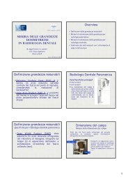
L<strong>in</strong>ee Guida <strong>in</strong> TC<br />
<strong>Misura</strong> <strong>delle</strong> <strong>grandezze</strong><br />
<strong>dosimetriche</strong> <strong>in</strong> <strong>tomografia</strong><br />
<strong>computerizzata</strong><br />
Dr. G. Zatelli, , Dr. S. Mazzocchi, USL10 Firenze; Dr. A.<br />
Ciccarone, AO Meyer Firenze<br />
In 1999 the European Commission published<br />
the European Guidel<strong>in</strong>es on Quality Criteria for<br />
Computed Tomography<br />
I valori di dose di riferimento non sono da <strong>in</strong>tendersi<br />
ad uso locale su un paziente s<strong>in</strong>golo, bensì come la<br />
media <strong>delle</strong> dosi osservate <strong>in</strong> un gruppo<br />
rappresentativo di pazienti.<br />
Tipici livelli di dose <strong>in</strong> eccesso di quella di riferimento<br />
dovranno essere opportunamente giustificati o ridotti.<br />
In generale le dosi al paziente dovranno sempre<br />
essere ridotte ai livelli più bassi che sono<br />
ragionevolmente praticabili e al contempo consistenti<br />
con gli obiettivi cl<strong>in</strong>ici dell’esame.<br />
esame.<br />
TC e <strong>in</strong>dice di dose<br />
TC e <strong>in</strong>dice di dose<br />
La dose <strong>in</strong> TC dipende da<br />
Spessore nom<strong>in</strong>ale dello strato<br />
Avanzamento del lett<strong>in</strong>o per unità di strato<br />
acquisito <strong>in</strong> 360°<br />
Parametri di esposizione<br />
Mezzo di contrasto che aggiunge ulteriori<br />
scansioni<br />
<br />
Computed Tomography Dose Index (CTDI)<br />
Indice di dose <strong>in</strong> TC<br />
CTDI convenzionale<br />
Computed<br />
Tomography Dose<br />
Index (CTDI)<br />
T = spessore nom<strong>in</strong>ale di<br />
strato<br />
N = numero di immag<strong>in</strong>i <strong>in</strong><br />
360°<br />
D(z) ) = valore della dose<br />
assorbita <strong>in</strong> aria lungo il<br />
profilo z<br />
+∞<br />
CTDI = 1<br />
D<br />
N × T<br />
∫<br />
−∞<br />
( z) ⋅ dz<br />
E<br />
B<br />
A A C<br />
D<br />
Z axis<br />
Different CTDIs:<br />
Def<strong>in</strong>ition accord<strong>in</strong>g to FDA<br />
Practical CTDI<br />
Normalised CTDI<br />
Weighted CTDI<br />
CTDI<br />
CTDI<br />
100<br />
FDA<br />
+ 7 T<br />
1<br />
= D<br />
N × T<br />
∫<br />
− 7 T<br />
+ 50 mm<br />
1<br />
= K<br />
N × T<br />
∫<br />
n CTDI XYZ =<br />
1<br />
3<br />
− 50 mm<br />
CTDI<br />
mAs<br />
2<br />
3<br />
( z ) ⋅ dz<br />
air<br />
( z ) ⋅ dz<br />
XYZ<br />
CTDI w = CTDI 100 , c + CTDI 100 , p<br />
1
CTDI <strong>in</strong>dispensabili<br />
Indici di dose <strong>in</strong> “console”<br />
CTDI Free-<strong>in</strong>-Air<br />
CTDI<br />
air<br />
+ 50<br />
= 1<br />
K<br />
N × T<br />
∫<br />
− 50<br />
Phantom factors P which allow the conversion from<br />
CTDI air to CTDI w<br />
P<br />
H<br />
=<br />
CTDI<br />
CTDI<br />
w , H<br />
air<br />
P<br />
B<br />
=<br />
CTDI<br />
CTDI<br />
w , B<br />
air<br />
air<br />
( z ) ⋅ dz<br />
IEC2003 raccomanda che<br />
il valore del CTDI vol sia<br />
<strong>in</strong>dicato <strong>in</strong> console <strong>in</strong> modo<br />
che rifletta le condizioni di<br />
scansione selezionate.<br />
I nuovi tomografi TC<br />
mostrano anche il DLP che<br />
<strong>in</strong>crementa per ogni<br />
scansione aggiunta e<br />
necessaria per rispondere<br />
al quesito cl<strong>in</strong>ico.<br />
CTDI<br />
vol =<br />
CTDI<br />
p<br />
w<br />
∆d<br />
p =<br />
N × T<br />
DLP = CTDIvol × L<br />
Tecnologia <strong>in</strong> TC<br />
Uso dei Tomografi TC<br />
Forti disomogeneità nell’uso dei tomografi<br />
TC già nel 2003 per lo stesso esame<br />
all’<strong>in</strong>terno dei protocolli impostati dalle<br />
case venditrici.<br />
http://www.msct.eu/PDF_FILES/EC%20CA%20Report%20D5%20-%20Dosimetry.pdf<br />
Protocolli dipendenti dalla taglia.<br />
Indispensabile se si segue il pr<strong>in</strong>cipio<br />
ALARA<br />
Indispensabile <strong>in</strong> TC pediatrica per alta<br />
radiosensibilità dei tessuti <strong>in</strong> questa età<br />
Indispensabili per esami TC come<br />
addome, tronco per la più alta<br />
concentrazione di tessuti radiosensibili, a<br />
tutte le età.<br />
Protocollo standard per addome<br />
Paziente standard di diametro 28 cm<br />
5 mm spessore nom<strong>in</strong>ale<br />
120 kVp<br />
Pitch=1.5<br />
320 mAs<br />
Obiettivo f<strong>in</strong>ale: scal<strong>in</strong>g dell’<strong>in</strong>dice di dose<br />
CTDI <strong>in</strong> funzione della taglia del paziente<br />
mantenendo costante il CNR tipico del<br />
paziente standard.<br />
Boone JM, Geraghty EM, Seibert JA, Wootton-<br />
Gorges SL. Dose reduction <strong>in</strong> pediatric CT: a<br />
rational approach. Radiology 2003;228:352–60.<br />
2
Misure di rumore <strong>in</strong> PMMA.<br />
Per ogni ROI sulle immag<strong>in</strong>i CT dei fantocci si<br />
calcola la media dei numeri CT e la SD.<br />
Il rumore dei raggi X segue la distribuzione di<br />
Poisson qu<strong>in</strong>di per le aree <strong>delle</strong> Roi utilizzate si<br />
può scrivere<br />
a<br />
σ =<br />
mAs<br />
Si utilizza per il fitt<strong>in</strong>g dei dati sperimentali del<br />
rumore e il fitt<strong>in</strong>g ottenuto servirà per <strong>in</strong>terpolare<br />
i dati del rumore per i fantocci da 10 a 32 cm.<br />
Rumore CT vs mAs, kV p<br />
La pendenza <strong>delle</strong><br />
rette è di –1/2 circa.<br />
I dati del rumore<br />
sono usati come<br />
denom<strong>in</strong>atore per<br />
le misure del CNR<br />
Boone JM, Geraghty EM, Seibert JA, Wootton-<br />
Gorges SL. Dose reduction <strong>in</strong> pediatric CT: a<br />
rational approach. Radiology 2003;228:352–60.<br />
Boone JM, Geraghty EM, Seibert JA, Wootton-<br />
Gorges SL. Dose reduction <strong>in</strong> pediatric CT: a<br />
rational approach. Radiology 2003;228:352–60.<br />
CTDI vs diametro paziente<br />
Contrasto vs diametro PMMA<br />
<br />
Per mAs costanti<br />
aumentare i kV p aumenta<br />
l’energia media per fotone<br />
e il numero di fotoni per<br />
fascio.<br />
Studi con più alti kV p<br />
comportano un aumento<br />
della dose.<br />
1/r 2 (vantaggio)<br />
La dose a pazienti più<br />
piccoli è più alta a causa<br />
della m<strong>in</strong>ore auto-<br />
filtrazione del fascio<br />
rispetto ai pazienti più<br />
grandi. (svantaggio)<br />
Boone JM, Geraghty EM, Seibert JA, Wootton-<br />
Gorges SL. Dose reduction <strong>in</strong> pediatric CT: a<br />
rational approach. Radiology 2003;228:352–60.<br />
Il contrasto dello ioduro è più alto a bassi kV p e<br />
decresce al crescere della tensione.<br />
Il contrasto decresce al crescere del paziente<br />
perché con pazienti più grossi il fascio perde la<br />
sua componente molle<br />
300 mAs<br />
80 kV p<br />
140 kV p<br />
µ<br />
eff<br />
− µ<br />
CTn = 1000⋅<br />
µ<br />
water<br />
Boone JM, Geraghty EM, Seibert CJA, Wootton-<br />
iod<strong>in</strong>e<br />
= CTiod<strong>in</strong>e+<br />
water<br />
Gorges SL. Dose reduction <strong>in</strong> pediatric CT: a<br />
rational approach. Radiology 2003;228:352–60.<br />
water<br />
− CT<br />
water<br />
CNR costante vs ED paziente<br />
mAs per aver CNR costante<br />
Come si riduce<br />
la dose <strong>in</strong><br />
pazienti<br />
pediatrici pur<br />
mantenendo la<br />
stessa qualità<br />
dell’immag<strong>in</strong>e di<br />
un adulto di<br />
riferimento.<br />
Boone JM, Geraghty EM, Seibert JA, Wootton-<br />
Gorges SL. Dose reduction <strong>in</strong> pediatric CT: a<br />
rational approach. Radiology 2003;228:352–60.<br />
<br />
La riduzione nei<br />
mAs nei pediatrici<br />
per mantenere<br />
costante il<br />
parametro della<br />
risoluzione <strong>in</strong><br />
contrasto (CNR)<br />
dell’immag<strong>in</strong>e.<br />
Boone JM, Geraghty EM, Seibert JA, Wootton-<br />
Gorges SL. Dose reduction <strong>in</strong> pediatric CT: a<br />
rational approach. Radiology 2003;228:352–60.<br />
3
Tabella di ausilio per TSRM<br />
Pazienti adulti grassi<br />
<br />
Il risparmio<br />
<strong>in</strong> dose per i<br />
pazienti più<br />
piccoli arriva<br />
al 95%<br />
rispetto<br />
all’adulto adulto di<br />
spessore 28<br />
cm.<br />
Boone JM, Geraghty EM, Seibert JA, Wootton-<br />
Gorges SL. Dose reduction <strong>in</strong> pediatric CT: a<br />
rational approach. Radiology 2003;228:352–60.<br />
È richiesto un aumento dei mAs.<br />
Per passare dai 28 cm ai 35 cm l’aumento l<br />
dei mAs è<br />
di 4,3 volte<br />
Si può anche considera il fatto di aumentare lo<br />
spessore della fetta.<br />
Raddoppiare lo spessore di strato da 5 mm a 10 mm<br />
raggiunge lo stesso effetto (<strong>in</strong> term<strong>in</strong>i di CNR) che di<br />
raddoppiare i mAs con strato fisso a 5 mm.<br />
Qu<strong>in</strong>di nella tabella si può tranquillamente raddoppiare<br />
lo spessore della fetta ricostruita da 5 a 10 mm e<br />
dimezzare i mAs erogati dal tubo per raggiungere lo<br />
stesso livello di CNR.<br />
Si può ridurre la dose anche con aumento del pitch al<br />
valore p´ p e la riduzione è pari a p/p´.<br />
Boone JM, Geraghty EM, Seibert JA, Wootton-<br />
Gorges SL. Dose reduction <strong>in</strong> pediatric CT: a<br />
rational approach. Radiology 2003;228:352–60.<br />
Percezione dell’immag<strong>in</strong>e<br />
L’ipotesi di ottenere una tecnica di scansione<br />
che tenga conto <strong>delle</strong> dimensioni del paziente<br />
per mantenere costante la qualità<br />
dell’immag<strong>in</strong>e si fonda sul fatto che la<br />
rivelazione di taluni particolari dalle immag<strong>in</strong>i di<br />
due pazienti pediatrico e adulto sia la stessa.<br />
Qu<strong>in</strong>di qualora ci fossero ragioni cl<strong>in</strong>iche per<br />
cui il contrasto di una struttura è<br />
<strong>in</strong>tr<strong>in</strong>secamente più basso allora si può<br />
abbassare la riduzione <strong>in</strong> dose aumentando i<br />
mAs.<br />
Boone JM, Geraghty EM, Seibert JA, Wootton-<br />
Gorges SL. Dose reduction <strong>in</strong> pediatric CT: a<br />
rational approach. Radiology 2003;228:352–60.<br />
Osservazioni sul problema.<br />
500 giovanissimi (< 15 y) sottoposti ad esame<br />
TC con protocolli per adulti probabilmente<br />
moriranno per <strong>in</strong>sorgenza di cancro a causa<br />
<strong>delle</strong> radiazioni.<br />
Trasformando l’etl<br />
età (0-14 y) <strong>in</strong> spessori (cm) e<br />
applicando la riduzione dei mAs la dose alla<br />
popolazione si ridurrebbe del 23% (anno<br />
2000).<br />
… ma nel caso di (0-14 y) il numero di cancro<br />
fatale scenderebbe a 116.<br />
Boone JM, Geraghty EM, Seibert JA, Wootton-<br />
Gorges SL. Dose reduction <strong>in</strong> pediatric CT: a<br />
rational approach. Radiology 2003;228:352–60.<br />
Attenuazione monocromatica.<br />
Conosciamo il carico del tubo per un<br />
paziente di riferimento con una certa<br />
sezione che ci permette di avere una<br />
certa immag<strong>in</strong>e sgranata alla risoluzione<br />
spaziale richiesta per le <strong>in</strong>dicazioni di<br />
tipo cl<strong>in</strong>ico.<br />
− µ d<br />
− d<br />
µ ( d x − d ref )<br />
1<br />
µ 2<br />
N e = N e ⇒ mAs = mAs ⋅ e<br />
1<br />
2<br />
mAs<br />
x<br />
= mAs<br />
ref<br />
⋅ e<br />
x<br />
ref<br />
( 2 ) ( − )<br />
ln<br />
HVT<br />
d x d ref<br />
Confronto con i dati pubblicati.<br />
Per conoscere HVT si possono utilizzare i dati<br />
della letteratura sui mAs cha variano con la<br />
dimensione del paziente che mantengano<br />
costante la qualità dell’immag<strong>in</strong>e.<br />
HVT come il miglior valore che rende m<strong>in</strong>imo<br />
la variazione dei mAs ricavati dalle tabelle<br />
esposte <strong>in</strong> letteratura<br />
U. NYMAN, T. L. AHL, M. KRISTIANSSON, L.<br />
NILSSON & S. WETTEMARK. Patient-<br />
Circumference-Adapted Dose Regulation <strong>in</strong><br />
Body Computed Tomography. A Practical and<br />
U. NYMAN, T. L. AHL, M. KRISTIANSSON, L.<br />
NILSSON & S. WETTEMARK. Patient-<br />
Circumference-Adapted Dose Regulation <strong>in</strong><br />
Body Computed Tomography. A Practical and<br />
4
Protocolli TC addome al Meyer.<br />
Coll (mm) Rot. Time (s) kVp Recon slilce Overlap<br />
40 0.75 120 2 1<br />
ID pitch<br />
Scan time<br />
FOV<br />
weigth heigth<br />
CTDIvol mAs/slice<br />
età<br />
(s)<br />
(mm)<br />
(kg) (cm)<br />
1 1.157 4.045 9.0 153 350 15.3 68 180<br />
2 1.157 7.821 11.1 189 326 15.2 53 163<br />
3 0.906 8.621 8.8 150 267 13.8 34 154<br />
4 1.157 6.395 4.2 72 238 5.6 22 116<br />
5 0.906 5.269 8.8 150 207 4.9 20 120<br />
6 0.906 7.752 5.5 94 217 4.6 16 100<br />
7 0.906 7.731 8.8 150 211 3.5 14 99<br />
8 1.157 7.838 14.7 250 265 2.3 15 90<br />
9 1.157 8.145 9.6 164 350 11.6 47 160<br />
10 1.157 6.298 6.0 102 270 9.0 30 120<br />
11 0.906 7.620 8.8 150 254 8.4 22 120<br />
12 0.906 8.373 8.8 150 259 8.4 27 120<br />
13 0.906 7.255 8.8 150 298 2.5 13 110<br />
14 0.906 8.645 3.6 200 316 16.7 42 159<br />
15 0.906 8.394 5.9 100 271 8.2 27 125<br />
Strumenti di lavoro.<br />
Scansioni dei 4 fantocci di PMMA 8, 16, 20 and 32 cm<br />
utilizzando il protocollo addome<br />
Coll (mm)<br />
Rot. Time<br />
(s)<br />
kVp<br />
Recon<br />
slilce<br />
Overlap Matrix pitch<br />
Table speed<br />
(mm/s)<br />
32×1,25 0,75 120 2 1 512×512 0,906 48,3<br />
Al variare dei mAs 50, 100, 150, 200, 250, 300 and 400.<br />
La console riporta solo il CTDIvol del fantoccio di PMMA<br />
di 32 cm<br />
kV mAs 50 100 150 200 250 300 400<br />
120 CTDI_vol 2.9 5.9 8.8 11.8 14.7 17.6 23.5<br />
(mGy)<br />
80 0.9 1.8 2.7 3.6 4.5 5.4 7.2<br />
Misure del rumore <strong>in</strong> PMMA.<br />
Andamento analitico del rumore.<br />
È la misura che<br />
meno <strong>delle</strong> altre<br />
segue la legge<br />
dell’<strong>in</strong>verso della<br />
radice quadrata<br />
dei mAs<br />
120 kV<br />
Lo spessore di ricostruzione adottato nel protocollo è di 2 mm<br />
Table of noise @120 kV<br />
Parametri del fitt<strong>in</strong>g con una<br />
legge di potenza (120 kV).<br />
Noise (HU) @120 kV <strong>in</strong> PMMA<br />
mAs 8 cm 16 cm 20 cm 32 cm<br />
CTDIvol<br />
console<br />
(mGy)<br />
50 4.169 9.916 20.456 57.083 2.9<br />
100 2.987 6.911 14.081 39.050 5.9<br />
150 2.509 5.660 11.564 31.502 8.8<br />
200 2.201 4.903 9.940 26.960 11.8<br />
250 2.031 4.406 8.901 23.991 14.7<br />
300 1.916 4.018 8.094 21.837 17.6<br />
400 1.727 3.515 7.046 18.892 23.5<br />
f(x)=a*x^b 8cm 16cm 20cm 32cm<br />
a 23.07 70.75 149.60 465.20<br />
b -0.44 -0.50 -0.51 -0.54<br />
Necessari per predire il valore dei mAs da utilizzare per<br />
ottenere lo stesso rumore <strong>in</strong> differenti fanotcci di PMMA.<br />
5
mAs come funzione della taglia<br />
a parità di rumore <strong>in</strong> PMMA.<br />
Diameter phantom (cm)<br />
8 16 20 32<br />
noise (HU) mAs from noise measurements<br />
13 4 29 119 784<br />
12 4 34 139 910<br />
10 7 49 199 1278<br />
8 11 76 308 1937<br />
Attenuazione esponenziale<br />
D ref =27 cm;<br />
mAs ref =153;<br />
HVL (mm Al )=8.8 mm Al semplifica a fascio monocromatico;<br />
Diametro: PMMAwater a parità di rumore 11.12 qu<strong>in</strong>di<br />
fantoccio cil<strong>in</strong>drico di acqua con D=27 cm 24 cm PMMA.<br />
noise (HU)<br />
13<br />
mAs form <strong>in</strong>terp. noise fitt<strong>in</strong>g @120 kV<br />
8 cm 16 cm 20 cm 32 cm<br />
4 29 119 784<br />
ref. mAs HVT ideal mAs from exp attenuation<br />
155 3.4 6 30 69 792<br />
200 4 13 51 101 788<br />
Curva analitica per ipotesi di<br />
attenuazione monoesponenziale<br />
Il massimo residuo tra curva analitica e dati sperimentali è<br />
all’<strong>in</strong>terno del range di ± 20 mAs<br />
HVT per uso cl<strong>in</strong>ico a 120 kV<br />
•La percezione del rumore <strong>in</strong> pazienti pediatrici è differente<br />
rispetto al caso degli adulti.<br />
•Si rende pertanto necessario utilizzare una riduzione della dose<br />
che non sia dettata da dati matematici o fisici.<br />
•LA riduzione della dose è prettamente demandato al radiologo.<br />
•Dalla letteratura emerge come consiglio l’utilizzo <strong>in</strong> campo<br />
pediatrico di un HVT pari a 8 cm<br />
Diametro equivalente dei<br />
pazienti<br />
Dose <strong>in</strong>terpolation 120 kV plot<br />
Dai dati del RIS otteniamo una correlazione tra circonferenza e rappoprto<br />
pes-altezza.<br />
paediatric patient circumference vs weight and heght ratio<br />
90,0<br />
80,0<br />
70,0<br />
circumference (cm)<br />
60,0<br />
50,0<br />
40,0<br />
y = 131,47x + 31,682<br />
R 2 = 0,9134<br />
circumference (cm)<br />
30,0<br />
L<strong>in</strong>eare (circumference<br />
(cm))<br />
20,0<br />
0,10 0,15 0,20 0,25 0,30 0,35 0,40<br />
wieght/height (kg/cm)<br />
6
Risultati: tabella dei mAs@120 kV<br />
Risultati: tabella dei<br />
CTDI@120 kV<br />
mAs at 13 HU<br />
Peso paziente <strong>in</strong> kg<br />
noise @ 120 kV 4 6 8 9 10 11 13 14 15 16 18 20 22 25 27 30 33 37<br />
53 53 60 68 75 80 87 95 102 109 117 132 154 181 216 255 312 381 487<br />
60 51 57 64 70 74 79 85 91 97 103 115 131 151 177 205 245 292 363<br />
67 50 55 61 66 69 74 79 84 88 93 103 116 132 151 172 202 237 287<br />
72 49 54 59 64 67 71 76 80 84 89 97 108 123 140 158 184 213 255<br />
76 48 53 58 62 65 69 73 77 81 85 93 103 116 131 147 170 195 232<br />
82 48 52 56 60 63 66 70 73 77 80 87 96 106 119 133 151 172 202<br />
87 47 51 55 59 61 64 68 71 74 77 83 91 100 112 123 140 158 183<br />
92 47 51 54 58 60 63 66 69 72 74 80 87 96 106 117 132 148 170<br />
96 47 50 54 57 59 62 64 67 70 72 77 84 92 102 111 125 139 159<br />
altezza paziente <strong>in</strong> cm<br />
99 46 50 53 56 58 61 63 66 69 71 76 82 90 99 108 120 134 153<br />
106 46 49 52 55 56 59 62 64 66 68 73 78 85 93 101 112 123 140<br />
113 46 48 51 54 55 58 60 62 64 66 70 75 82 89 96 105 116 130<br />
119 45 48 51 53 54 57 59 61 63 65 68 73 78 85 91 100 109 122<br />
125 45 47 50 52 54 56 58 60 61 63 67 71 76 82 88 96 104 116<br />
130 45 47 50 52 53 55 57 59 60 62 65 69 74 80 85 92 100 111<br />
136 45 47 49 51 52 54 56 58 59 61 64 68 72 77 83 89 97 106<br />
141 44 47 49 51 52 54 55 57 58 60 63 66 71 76 80 87 94 103<br />
146 44 46 49 50 51 53 55 56 58 59 62 65 69 74 79 85 91 100<br />
CTDI_vol (mGy)<br />
Peso paziente <strong>in</strong> kg<br />
@ 120 kV 4 6 8 9 10 11 13 14 15 16 18 20 22 25 27 30 33 37<br />
53 8,11 9,00 9,91 10,55 10,94 11,41 11,86 12,20 12,52 12,85 13,56 14,60 15,97 17,81 19,96 23,14 26,82 32,06<br />
60 7,88 8,66 9,47 10,04 10,41 10,90 11,34 11,68 11,97 12,26 12,82 13,51 14,52 15,75 17,23 19,43 22,02 25,88<br />
67 7,72 8,40 9,13 9,64 9,97 10,44 10,86 11,20 11,51 11,80 12,23 12,85 13,56 14,51 15,55 17,11 18,96 21,78<br />
72 7,64 8,28 8,95 9,43 9,75 10,21 10,62 10,94 11,23 11,49 11,98 12,51 13,13 13,94 14,82 16,10 17,64 19,97<br />
76 7,56 8,17 8,80 9,27 9,57 10,00 10,37 10,72 11,03 11,28 11,74 12,27 12,85 13,50 14,28 15,38 16,70 18,69<br />
82 7,47 8,02 8,59 9,02 9,31 9,70 10,07 10,38 10,67 10,95 11,41 11,92 12,39 12,98 13,60 14,52 15,54 17,05<br />
87 7,40 7,92 8,46 8,86 9,12 9,48 9,85 10,14 10,44 10,70 11,13 11,66 12,13 12,68 13,17 13,95 14,81 16,12<br />
92 7,35 7,84 8,35 8,73 8,98 9,33 9,67 9,98 10,23 10,48 10,93 11,42 11,91 12,39 12,86 13,57 14,28 15,43<br />
96 7,30 7,76 8,25 8,61 8,85 9,18 9,50 9,78 10,05 10,31 10,75 11,22 11,73 12,17 12,63 13,23 13,88 14,90<br />
99 7,27 7,72 8,18 8,53 8,77 9,10 9,40 9,67 9,93 10,19 10,60 11,10 11,59 12,07 12,50 13,03 13,66 14,57<br />
106 7,21 7,63 8,05 8,38 8,59 8,90 9,20 9,45 9,68 9,91 10,33 10,83 11,29 11,77 12,14 12,69 13,16 13,92<br />
113 7,16 7,55 7,95 8,26 8,46 8,75 9,01 9,26 9,50 9,72 10,10 10,58 11,05 11,50 11,88 12,36 12,86 13,46<br />
119 7,12 7,48 7,87 8,16 8,35 8,61 8,87 9,10 9,31 9,53 9,92 10,36 10,81 11,28 11,69 12,12 12,55 13,09<br />
125 7,08 7,43 7,79 8,06 8,24 8,50 8,74 8,96 9,17 9,38 9,72 10,16 10,62 11,07 11,46 11,90 12,29 12,81<br />
130 7,05 7,38 7,73 7,99 8,16 8,41 8,64 8,84 9,04 9,24 9,58 10,00 10,44 10,91 11,28 11,76 12,11 12,63<br />
136 7,02 7,34 7,67 7,92 8,08 8,32 8,53 8,74 8,93 9,12 9,44 9,84 10,29 10,74 11,14 11,54 11,93 12,40<br />
141 7,00 7,31 7,62 7,86 8,02 8,24 8,46 8,65 8,83 9,00 9,33 9,74 10,14 10,61 10,95 11,44 11,81 12,24<br />
146 6,98 7,27 7,58 7,81 7,96 8,17 8,38 8,57 8,74 8,91 9,23 9,62 10,03 10,45 10,82 11,28 11,64 12,10<br />
altezza paziente <strong>in</strong> cm<br />
Valori di dose di riferimento<br />
EUR 16262 EN<br />
Criteri di controllo della dose <strong>in</strong> TC<br />
La qualità dell’immag<strong>in</strong>e va sempre<br />
preservata<br />
Scelto un parametro di qualità:<br />
Rumore <strong>in</strong> parenchima omogeneo dell’addome<br />
(fegato); CNR per tessuti molli (muscolo, tessuto<br />
adiposo)<br />
Da una tecnica standard ottimizzata di buona<br />
qualità, , studiare i fattori che <strong>in</strong>fluenzano la tecnica<br />
TC per calibrare la stessa qualità alla taglia del<br />
paziente.<br />
Obiettivo ultimo<br />
Mantenere l’esposizione l<br />
da radiazione del<br />
paziente il più basso ragionevolmente<br />
praticabile<br />
7