00 Assece Cop - Salute per tutti
00 Assece Cop - Salute per tutti
00 Assece Cop - Salute per tutti

Create successful ePaper yourself
Turn your PDF publications into a flip-book with our unique Google optimized e-Paper software.
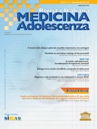
Vol. 1, 1, 2<strong>00</strong>8<br />
Personal approach on tuberous breast correction<br />
using anatomical implants.<br />
Nava MB, Cadenelli PF, Ticozzi A, Spano A<br />
Vertical reduction mammaplasty<br />
using a su<strong>per</strong>o-medial pedicle.<br />
Campiglio G<br />
Lipofilling to treat <strong>per</strong>iprosthesic<br />
capsular contraction after augmentation<br />
mammaplasty.<br />
Baruffaldi Preis FW, Cavallini M, Lanfranchi LA<br />
Periodico quadrimestrale - Spedizione in abbonamento postale 45% - art. 2 comma 20/B legge 662/96 - Milano<br />
In caso di mancata consegna restituire al mittente che si impegna a pagare la relativa tassa.<br />
Is aesthetic surgery a classical model of one day<br />
surgery, as far as anaesthesia is concerned?<br />
Pellanda A, Menasce G, Borroni M, Daviri G, Savoia G, Pollini A<br />
Additive mastoplastic<br />
and exclusion of culpable lesions.<br />
Olini M
A<br />
dieci anni dalla fondazione dell'Associazione Europea di Chirurgia Estetica (ASSECE) è<br />
con grande piacere che porto a battesimo questa nuova rivista di chirurgia estetica. La<br />
pubblicazione vuole essere la rivista della nostra Associazione ma anche un mezzo di<br />
aggiornamento a<strong>per</strong>to alla voce di qualsiasi Società scientifica seria che o<strong>per</strong>a nel campo della<br />
chirurgia plastica ed estetica.<br />
UDPS si propone di guidare i suoi lettori in un affascinante viaggio attraverso i quotidiani aggiornamenti<br />
della chirurgia plastica, estetica e ricostruttiva.<br />
La Rivista pubblicherà le nuove<br />
tecniche, gli studi di follow up a<br />
medio e lungo termine, le complicanze<br />
e le problematiche che<br />
ogni giorno i Colleghi si trovano<br />
ad affrontare sul tavolo o<strong>per</strong>atorio.<br />
Per questo motivo sarà di grande<br />
interesse <strong>per</strong> <strong>tutti</strong> i chirurghi plastici<br />
che amano questa incredibile<br />
branca della chirurgia.<br />
La nostra pubblicazione vuole<br />
anche essere un augurio. Quello di<br />
aprire sincere collaborazioni con<br />
altre Società specialistiche nella<br />
convinzione che solo un onesto<br />
confronto possa aiutare a far crescere<br />
la professionalità di chi pratica<br />
questa disciplina chirurgica.<br />
Ringrazio <strong>per</strong>tanto <strong>tutti</strong> i Colleghi<br />
che con entusiasmo hanno accettato<br />
di aiutarmi in questa impresa.<br />
Ringrazio in particolare gli Editor in<br />
C h i e f, i Coeditors, il Comitato di<br />
Redazione e il Coordinamento<br />
Scientifico. Un grazie anche all'Editore<br />
che, avendo fiducia in me,<br />
mi ha chiesto di dirigere la pubblicazione.<br />
L'o<strong>per</strong>a sarà <strong>per</strong>ò dei lettori<br />
e <strong>per</strong> questo motivo saranno<br />
gradite critiche, suggerimenti e<br />
proposte di collaborazione.<br />
Ten years after the institution of the European<br />
Association of Aesthetic Surgery (ASSECE) I'm really<br />
pleased and proud to start this new scientific journal.<br />
The publication claims to be the journal of our association<br />
but also a means of update open to the entry of any<br />
serious scientific society that works in the field of plastic<br />
and cosmetic surgery.UDPS wants to take us on a<br />
fascinating trip through the daily challenges of plastic,<br />
aesthetic and reconstructive surgery. New techniques,<br />
long-term follow up studies, problems cases and complications<br />
on aesthetic facial and body surgery will be<br />
covered in UDPS. That's the reason why it should be of<br />
interest and a good guide for every plastic surgeon who<br />
loves this incredible branch of surgery.<br />
I take it also as a good omen. I wish other scientific<br />
societies to work togheter writing for this journal in the<br />
belief that only a fair comparison of our ex<strong>per</strong>iences can<br />
grow the professionalism of those who practice our difficult<br />
branch of surgery.<br />
So I thank all colleagues who accepted to help me in<br />
this enterprise with great enthusiasm. Particularly I sincerely<br />
thank the Editors in Chief, the Coeditors, the<br />
Editorial Staff and the Scientific Committee. I thank also<br />
to the Publisher who trust me and asked me to edit the<br />
Journal. We'll work in favour of our reading public and<br />
that's the reason why all criticism, suggestions and proposals<br />
of coo<strong>per</strong>ation is always welcome.<br />
Ecco il primo mattone...<br />
Here's the beginning ....<br />
Ruben Oddenino
Vol. 1, 1, 2<strong>00</strong>8<br />
Editor<br />
UPDATE IN PLASTIC SURGERY<br />
Ruben Oddenino (Italy)<br />
Editor in Chief<br />
Franz Baruffaldi Preis (Italy)<br />
Maurizio Cavallini (Italy)<br />
Co-Editors<br />
Daniel Cassuto (Italy)<br />
Antonello Tateo (Italy)<br />
Editorial Board<br />
Francesco Aji (Italy)<br />
Minami Akihiro (Japan)<br />
Cesare Azzolini (Italy)<br />
Edward Battisti (Italy)<br />
Yousef Bakir (Syria)<br />
Gianfranco Bernabei (Italy)<br />
Corrado Bernasconi (Italy)<br />
Augustin Blanch (Spain)<br />
Giovanni Botti (Italy)<br />
Elio Caccialanza (Italy)<br />
Gianluca Campiglio (Italy)<br />
Artur Carbonell (Spain)<br />
Alessandro Casadei (Italy)<br />
Daniel Cassuto (Italy)<br />
Antonio Di Vincenzo (Italy)<br />
Mohamed El Hadidy (Egypt)<br />
Elena Fasola (Italy)<br />
Alberto Fumagalli (Italy)<br />
Edoardo Garassino (Italy)<br />
Alberto Goldman (Brasil)<br />
Andrzej Ignaciuk (Poland)<br />
Giuseppe Leopizzi (Italy)<br />
Omar Mamoun (Syria)<br />
Maurizio Nava (Italy)<br />
Ahmed Adel Nour El Din (Egypt)<br />
Marino Osellame (Italy)<br />
Josè Palacin (Spain)<br />
Pietro Palma (Italy)<br />
Mario Pelle Ceravolo (Italy)<br />
Alberto Peroni Ranchet (Italy)<br />
Tommasina Polverani (Italy)<br />
Pierluigi Santi (Italy)<br />
Antonio Tateo (Italy)<br />
C. Thomas (Oman)<br />
Marco Klinger (Italy)<br />
Managing Editor<br />
Antonio Di Maio (Italy)<br />
È vietata la riproduzione totale o parziale, con qualsiasi<br />
mezzo, di articoli, illustrazioni e fotografie senza l’autorizzazione<br />
scritta dell’Editore.<br />
L’Editore non risponde dell’opinione espressa dagli Autori<br />
degli articoli.<br />
Ai sensi della legge 675/96 è possibile in qualsiasi momento<br />
opporsi all’invio della rivista comunicando <strong>per</strong> iscritto la propria<br />
decisione a:<br />
Edizioni Scripta Manent s.n.c. - Via Bassini, 41- 20133 Milano<br />
Personal approach on tuberous breast<br />
correction using anatomical implants.<br />
Nava MB, Cadenelli PF, Ticozzi A, Spano A<br />
Vertical reduction mammaplasty<br />
using a su<strong>per</strong>o-medial pedicle.<br />
Direttore Responsabile Pietro Cazzola<br />
Direttore Generale Armando Mazzù<br />
Direttore Marketing Antonio Di Maio<br />
Consulenza grafica Piero Merlini<br />
Impaginazione Clementina Pasina<br />
Campiglio G<br />
Lipofilling to treat <strong>per</strong>iprosthesic<br />
capsular contraction after augmentation<br />
m a m m a p l a s t y.<br />
Baruffaldi Preis FW, Cavallini M, Lanfranchi LA<br />
Is aesthetic surgery a classical model<br />
of one day surgery, as far as anaesthesia<br />
is concerned ?<br />
Pellanda A, Menasce G, Borroni M, Daviri G, Savoia G, Pollini A<br />
Editorial Staff<br />
Additive mastoplastic<br />
and exclusion of culpable lesions.<br />
Olini M<br />
5<br />
1 1<br />
1 7<br />
2 3<br />
29<br />
R e g i s t r. Tribunale di Milano n. 774 del 30/12/2<strong>00</strong>8<br />
Scripta Manent s.n.c. Via Bassini, 41 - 20133 Milano<br />
Tel. 0270608091/0270608060 - Fax 0270606917<br />
E-mail: scriman@tin.it<br />
Abbonamento annuale (3 numeri) Euro <strong>00</strong>,<strong>00</strong><br />
Pagamento: conto corrente postale n. 20350682<br />
intestato a: Edizioni Scripta Manent s.n.c.<br />
via Bassini 41- 20133 Milano<br />
Stampa: Arti Grafiche Bazzi, Milano
Personal approach on tuberous breast<br />
correction using anatomical implants.<br />
Nava MB, Cadenelli PF, Ticozzi A, Spano A<br />
Fondazione IRCCS Istituto Nazionale dei Tumori. S.C. Chirurgia Plastica e Ricostruttiva, Milan, Italy<br />
Personal approach<br />
on tuberous breast correction<br />
using anatomical implants.<br />
Different surgical procedures have been<br />
p e rformed to face the problems related<br />
to tuberous breast deformity: augmentation<br />
m a s t o p l a s t y, mastopexis, combined<br />
augmentation mastoplasty and mastopexis,<br />
glandular reshaping.<br />
Notwithstanding a standard procedure<br />
to correct this deformity hasn’t been<br />
e s t a b l i s h e d .<br />
This pa<strong>per</strong> reports a 15-year-old girl<br />
affected by a bilateral third degree tuberous<br />
breast characterized by serious areola<br />
h y p e rtrophy deeply impairing the relational<br />
life and the psychological growth of the<br />
p a t i e n t .<br />
Three accurate incisions of the mammary<br />
fascia have been <strong>per</strong>formed following a<br />
v e rtical, horizontal and oblique pattern, to<br />
expand the glandular tissue enough to fit<br />
the prostheses.<br />
Anatomical prostheses were insert e d<br />
beneath the muscle in order to realize a<br />
good projection, a bigger volume of the<br />
inferior portion of the breast and the<br />
creation of an anatomical breast shape<br />
which represents a reconstruction following<br />
aesthetic goals.<br />
Posto<strong>per</strong>ative areolar displacement<br />
occurred but the patient denied any<br />
s e c o n d a ry correction because extremely<br />
satisfied of the result.<br />
Combining a correct surgical procedure and<br />
the use of an anatomical implant, applying<br />
a mathematical approach, enables outcomes<br />
not achievable in the past.<br />
Key words: augmentation mastoplasty,<br />
mastopexis glandular re s h a p i n g<br />
INTRODUCTION<br />
The aetiology of Tuberous breast is<br />
still unknown, it is a congenital deformity that<br />
appears during the puberty characterized by<br />
an ample range of malformation.<br />
A variable degree of hypotrophy characterizes<br />
the tuberous breast with glandular tissue herniation<br />
behind the areola and a consequential<br />
areola hy<strong>per</strong>trophy 1 .<br />
A constrictive tissue around the nipple-areola<br />
complex affects the normal breast development;<br />
due to this reason breast tissue<br />
expands itself behind the NAC as resistance is<br />
lower than in the inferior pole.<br />
The most frequent breast anomalies concern<br />
the upward breast tissue development, the<br />
impaired implant base, the submammary fold<br />
position, the areola size and the mammary<br />
asymmetry.<br />
The classification evaluates the different<br />
deformity patterns 2 :<br />
Degree 1: 56%. Development deficit of<br />
i n f e r o - i n t e rnal quadrant and infero-medial areola<br />
deviation. The breast volume is not affected.<br />
Degree 2: 26%. Development deficit of<br />
inferior quadrants and downwards areola<br />
deviation.<br />
1a<br />
Degree 3: 18%. Development deficit of<br />
every quadrant and base retraction of the<br />
breast.<br />
1b<br />
Different surgical procedures have been <strong>per</strong>formed<br />
to face the problems related to this<br />
deformity: augmentation mastoplasty, mastopexis,<br />
combined augmentation mastoplasty<br />
and mastopexis, glandular reshaping 3-6 .<br />
The surgical treatment depends on the deformity<br />
degree.<br />
CASE REPORT<br />
The patient was a 15-year-old girl<br />
affected by a bilateral degree 3 tuberous breast<br />
d e f o rm i t y, characterized by serious areola<br />
h y p e r t r o p h y. The deformity was deeply impairing<br />
the psychological growth of the patient.<br />
Evaluation of the attainable final breast volume<br />
is completed predicting the diameter of the chosen;<br />
selected width was 11,5 cm and height<br />
was 10,6 cm, bilaterally. Considering these<br />
aspects and planning a well projected recons<br />
t ruction, anatomical prostheses (I n a m e d<br />
Corp, Style 410 MF 255) were chosen; new<br />
s u b m a m m a ry folds were signed (Figure 1a-b).<br />
Patient underwent a general anaesthesia.<br />
Figure 1a-b - Preo<strong>per</strong>ative views and markings.<br />
UpDate in Plastic Surgery - Vol. 1, 1, 2<strong>00</strong>8<br />
5
Nava MB, Cadenelli PF, Ticozzi A, Spano A<br />
Personal approach on tuberous breast correction using anatomical implants.<br />
2a<br />
finally a monocryl 5-0 intracuticular suture is<br />
<strong>per</strong>formed and a compressive dressing is<br />
positioned (Figure 2a-b).<br />
Drains were removed after 24 hours while the<br />
transfixed sutures after 7 days (Figure 3a-b).<br />
A minimal delayed recurrence occurred, six<br />
months after the surgical procedure, involving<br />
only the strictly <strong>per</strong>iareolar tissue.<br />
The patient was satisfied from the result<br />
achieved and denied any further surgical correction<br />
(Figure 4a-d).<br />
2b<br />
Figure 2a-b<br />
Intrao<strong>per</strong>ative views.<br />
Figure 3a-b<br />
One month posto<strong>per</strong>ative results.<br />
Figure 4a-d<br />
Follow up: 18 months.<br />
The new areola <strong>per</strong>imeter is marked with a 4,2<br />
cm marker and incised. Then a ring of disepidermization<br />
2 cm wide is created around the<br />
areola.<br />
The dermis is incised at the level of the external<br />
<strong>per</strong>imeter of the incision and the dissection<br />
of the subcutaneous tissue following the<br />
Coo<strong>per</strong> ligaments is carried out.<br />
The inferior portion of the breast is dissected<br />
keeping in place a trophic cutaneous and subcutaneous<br />
tissue following the su<strong>per</strong>ficialis<br />
fascia.<br />
When su<strong>per</strong>ior and inferior limits of the subcutaneous<br />
fold are defined the dissection<br />
reaches the new submammary fold and the<br />
subglandular dissection is <strong>per</strong>formed over the<br />
su<strong>per</strong>ior margin of the areola.<br />
Accurate incisions of the fascia, deepened<br />
about 1 cm, were <strong>per</strong>f o rmed following a vert i-<br />
cal, horizontal and oblique pattern to expand<br />
the glandular tissue enough to fit the prosthes<br />
e s .<br />
Then the dissection of the submuscular surface<br />
is <strong>per</strong>formed and a fold corresponding to<br />
the implant size (Inamed Corp, Style MF 255)<br />
is created.<br />
After a careful emostasis the implant is inserted,<br />
the gland is positioned in the correct way<br />
using 5 nylon 5-0 transfixed sutures passing<br />
through the submammary fold and knotted on<br />
cotton tampons to avoid any decubitus.<br />
A suction drain is positioned in each breast; a<br />
purse suture is done around the new areola<br />
checking the traction and the final shape;<br />
3a<br />
3b<br />
UpDate in Plastic Surgery - Vol. 1, 1, 2<strong>00</strong>8<br />
6
Nava MB, Cadenelli PF, Ticozzi A, Spano A<br />
Personal approach on tuberous breast correction using anatomical implants.<br />
4a<br />
4b<br />
4c<br />
4d<br />
DISCUSSION<br />
The reconstruction of a tuberous<br />
breast represents an important challenge for<br />
the plastic surgeon.<br />
Many Authors faced this surgical challenge in<br />
the past developing different <strong>per</strong>sonal therapeutic<br />
approaches.<br />
In 1976 Williams 7 described a technique to<br />
reduce the areola and Rees et al. 8 used radial<br />
incisions in the breast passing through the<br />
inframammary incision to release the gland.<br />
Few years later Teimurian and Adham 9 were<br />
the first to combine an areolar to a submammary<br />
incision to insert an implant.<br />
Different works illustrated the application of<br />
flaps from the submammary fold 10 and the<br />
use of an inferior flap to reshape the inferior<br />
portions 11 .<br />
Panettiere 12 in his pa<strong>per</strong> reported a combination<br />
of <strong>per</strong>iareolar mastopexy and augmentation<br />
mastoplasty using cross incisions of the<br />
gland; Ribeiro et al. 4-11 explained dividing the<br />
breast into two portions to reshape the lower<br />
mammary quadrants.<br />
Mandrekas et al. 13 described the incision of<br />
the constricting ring creating two breast pillars<br />
applying this procedure mostly to Type II deformities.<br />
Recently Foustanos 14 reported a series of<br />
patients treated by a two stages reconstruction<br />
using temporary expanders.<br />
Our <strong>per</strong>sonal approach, concording to Teimurian,<br />
Panettiere and Mandrekas, combines the<br />
dissection of the gland and the collocation of<br />
a prostheses in the submuscular pocket in<br />
order to correct Type III deformities.<br />
Inferior pole incisions are necessary to<br />
expand the constricted breast tissue and to<br />
obtain enough tissue to cover the implant.<br />
Moreover the tension developed by the<br />
implant placement let the previously molded<br />
tissue expanded avoiding any posto<strong>per</strong>ative<br />
contraction.<br />
Multiple incisions in different direction release<br />
the gland and fixing the chaudal margins of<br />
the glandular tissues over the inferior pole of<br />
the anatomical implants to the inframammary<br />
fold <strong>per</strong>mits to increase the base surface of<br />
the breast.<br />
Recurrencies rate due to the retraction of the<br />
alterated gland is the main problem that follows<br />
the reconstruction.<br />
Combining three directions glandular fascia<br />
incisions to the anatomical prostheses insertion<br />
we manage to achieve a more natural<br />
outcome, even if tissue thickness is scarce,<br />
with an aesthetically pleasant breast profile<br />
and without wrinkling or <strong>per</strong>ceptible implant<br />
b o r d e r.<br />
Using these prostheses it was possible to<br />
realize a good projection and a bigger volume<br />
of the inferior portion which is always insufficiently<br />
represented in this deformity.<br />
Moreover it <strong>per</strong>mitted to us to emphasize the<br />
UpDate in Plastic Surgery - Vol. 1, 1, 2<strong>00</strong>8<br />
7
Nava MB, Cadenelli PF, Ticozzi A, Spano A<br />
Personal approach on tuberous breast correction using anatomical implants.<br />
inferior pole without renouncing to a natural<br />
profile in the su<strong>per</strong>ior pole and without creating<br />
observable margins between the thoracic<br />
wall and the implant.<br />
Finally, combining a correct surgical procedure<br />
to the use of an anatomical implant and<br />
applying a mathematical approach, enables<br />
outcomes not achievable in the past.<br />
CONCLUSIONS<br />
Tuberous breast deformity correction<br />
has undoubtedly a positive effect on the<br />
psychological status of the patients.<br />
Previously described techniques emphasized<br />
the complexity to achieve stable and satisfactory<br />
results especially facing Type III deformities.<br />
Our <strong>per</strong>sonal approach achieved a stable<br />
posto<strong>per</strong>ative fine result in terms of breast<br />
shape, dimension and symmetry.<br />
Nevertheless further technical improvements<br />
can be carried out to minimize any secondary<br />
correction.<br />
REFERENCES<br />
1. Rees TD, Aston SJ. The tuberous breast. Clin<br />
Plast Surg 1976; 3:339<br />
2. Grolleau JL, Lanfrey E, Lavigne B, Chavoin JP,<br />
Costagliola M. Breast base anomalies: treatment<br />
strategy for tuberous breast, minor deformities,<br />
and asimmetry. Plast Reconstr Surg 1999;<br />
104:2040-2048<br />
3. Meara JG, Kolker A, Bartlett G, Theiler R,<br />
Mutimer K, Holmes AD. Tuberous breast deformity:<br />
principles and practice. Ann Plast Surg 2<strong>00</strong>0;<br />
45:607-611<br />
4. Ribeiro L, Canzi W, Buss A Jr, Accorsi A Jr.<br />
Tuberous breast: a new approach. Plast Reconstr<br />
Surg 1998; 101:42-50<br />
5. Argenta LC, Vanderkolk C, Friedman RJ, Marks<br />
M. Refinements in reconstruction of congenital<br />
breast deformities. Plast Reconstr Surg 1985;<br />
75:73-82<br />
6. Elliot LF. Circumareolar mastopexy with augmentation.<br />
Clin Plast Surg 2<strong>00</strong>2; 29 (3):337-47<br />
7. Williams JE. Augmentation mammoplasty:<br />
Inframammary approach. In: Georgiade NG (ed)<br />
Reconstructive breast surgery CV Mosby: St Luis<br />
p 50. 1976<br />
8. Rees TD, Aston SJ. The tuberous breast. Clin<br />
Plast Surg 1976; 3:339<br />
9 Teimourian B, Adham MN. Surgical correction of<br />
the tuberous breast. Ann Plast Surg 1983; 10:190<br />
10. Elliott MP. A musculocutaneous transposition<br />
flap mammaplasty for correction of tuberous<br />
breast. Ann Plast Surg 1988; 20:153<br />
11. Ribeiro L, Accorsi A, Buss A, Castro M,<br />
Pessoa M. Short scar correction of the tuberous<br />
breast. Clin Plast Surg 2<strong>00</strong>2; 29:423-431<br />
12. Panettiere P, Del Gaudio GA, Marchetti L,<br />
Accorsi D, Del Gaudio A. The tuberous breast syndrome.<br />
Aesth Plast Surg 2<strong>00</strong>0; 24:445<br />
13. Mandrekas AD, Zambacos GJ. Aesthetic<br />
reconstruction of the tuberous breast deformity.<br />
Plast Reconstr Surg 2<strong>00</strong>4; 113:2231-2<br />
14. Foustanos A, Zavrides H. Surgical reconstruction<br />
of tuberous breasts. Aesthetic Plast Surg<br />
2<strong>00</strong>6;30:294-3<strong>00</strong><br />
The Authors disclose any commercial associations<br />
or financial relationships that might pose or create<br />
a conflict of interest with information presented in<br />
the submitted manuscript.<br />
UpDate in Plastic Surgery - Vol. 1, 1, 2<strong>00</strong>8<br />
8
Vertical reduction mammaplasty<br />
using a su<strong>per</strong>o-medial pedicle.<br />
Campiglio G<br />
Private Practice, Milan, Italy<br />
Vertical reduction mammaplasty<br />
using a su<strong>per</strong>o-medial pedicle<br />
The author presents a clinical case treated<br />
with a breast reduction technique initially<br />
described by E. Hall Findlay in 1999.<br />
The patient is 39 years-old and presents<br />
a large size hy<strong>per</strong>trophy with asymmetries<br />
for volume and shape between the two<br />
breast.<br />
The post-o<strong>per</strong>ative pictures have been<br />
taken about 9 months post-o<strong>per</strong>atively<br />
and show a nice aesthetic result.<br />
Vertical mammaplasty with a su<strong>per</strong>omedial<br />
pedicle is a simple and reliable<br />
technique.<br />
This technique can be easily learned and<br />
produces reproducible nice results with<br />
a minimal complication rate.<br />
INTRODUCTION<br />
The author presents a clinical case<br />
treated with a breast reduction technique initially<br />
described by E. Hall Findlay in 1999.<br />
This procedure is a modification of the standard<br />
Lejour vertical reduction mammaplasty<br />
which fundamental steps are:<br />
1) creation of a su<strong>per</strong>o-medial dermoglandular<br />
pedicle;<br />
2) no undermining of the skin;<br />
3) liposuction limited to the vertical dog ear.<br />
1<br />
PREOPERATIVE MARKING<br />
1) Breast meridian and inframammary fold<br />
are drawn as usually (Figure 1).<br />
2) New position of the nipple (Point A) is<br />
determined along the breast meridian at<br />
or just below the level of the existing inframammary<br />
fold.<br />
3) Up<strong>per</strong> border of the new areola site is<br />
marked 2.5 cm above the new nipple site<br />
(Point B). The new areola site is marked<br />
as in Lejour technique using a mosquedome<br />
shape or a standard circular one.<br />
Key words: v e rtical reduction mammaplasty<br />
UpDate in Plastic Surgery - Vol. 1, 1, 2<strong>00</strong>8<br />
11
Campiglio G<br />
Vertical reduction mammaplasty using a su<strong>per</strong>o-medial pedicle<br />
4) The vertical limbs are, then, drawn rotating<br />
upward the breast medially and laterally<br />
as in the other vertical techniques.<br />
These vertical limbs are curved inferiorly<br />
to meet each other about 3 cm above the<br />
inframammary fold (Point C).<br />
5 ) Finally the su<strong>per</strong>o-medial pedicle is designed<br />
with a base of 8 cm and extending<br />
for few centimetres into the areola opening.<br />
2<br />
SURGICAL TECHNIQUE<br />
1) The pedicle is deepithelized under tension<br />
in the standard fashion.<br />
2) Skin and glandular resection are then <strong>per</strong>f<br />
o rmed in a one piece (Figure 2), thus creating<br />
the su<strong>per</strong>o-medial derm o g l a n d u l a r<br />
3<br />
pedicle (Figure 3). It is important to leave a<br />
thin layer of tissue above the pectoralis<br />
fascia in order to better maintain the sens<br />
a t i o n .<br />
3) The su<strong>per</strong>o-medial pedicle is upward rotated<br />
without fixing it to the pectoralis fascia<br />
(Figure 4-5). The medial and lateral pillars<br />
are then sutured (Figure 6), starting at an<br />
inferior and dee<strong>per</strong> level and progressing<br />
more su<strong>per</strong>ficially as the sutures are<br />
placed more su<strong>per</strong>iorly (Figure 7).<br />
4) The remaining inferior skin excess is<br />
gathered up using a subcuticular 3/0<br />
resorbable monofilament (Figure 8) and<br />
the old inframammary fold is suctioned<br />
(Figure 9). Drains are not used.<br />
4<br />
2a<br />
5<br />
UpDate in Plastic Surgery - Vol. 1, 1, 2<strong>00</strong>8<br />
12
Campiglio G<br />
Vertical reduction mammaplasty using a su<strong>per</strong>o-medial pedicle<br />
6 7<br />
8 9<br />
10<br />
POSTOPERATIVE CARE<br />
Dressing is removed on the 4 th posto<strong>per</strong>ative day and a<br />
brassaire is used for 4 weeks night and day. Shower is allowed<br />
starting from the 5th post-o<strong>per</strong>ative day.<br />
A light compression is maintained for 6-8 weeks along the old<br />
inframammary fold (Figure 10).<br />
UpDate in Plastic Surgery - Vol. 1, 1, 2<strong>00</strong>8<br />
13
Campiglio G<br />
Vertical reduction mammaplasty using a su<strong>per</strong>o-medial pedicle<br />
CASE PRESENTATION<br />
This patient is 39 years-old and<br />
presents a large size hy<strong>per</strong>trophy with asymmetries<br />
for volume and shape between the<br />
two breast (Figure 11a-c). She also complains<br />
a painful nodule (fibroadenoma) of the<br />
su<strong>per</strong>olateral quadrant of her right breast. This<br />
diagnosis was previously confirmed by an<br />
agobiopsy under ultrasound control.<br />
In this condition the su<strong>per</strong>omedial pedicle<br />
allows a simultaneous reduction of the breast<br />
along with ptossi correction and the excision<br />
of the fibroadenoma.<br />
The post-o<strong>per</strong>ative pictures have been taken<br />
about 9 months post-o<strong>per</strong>atively (Figure 12ac)<br />
and show a nice esthetic result. The vertical<br />
scar healed well and the skin excess present<br />
at the end of the o<strong>per</strong>ation completely disappeared<br />
(Figure 13).<br />
11a<br />
11b<br />
11c<br />
UpDate in Plastic Surgery - Vol. 1, 1, 2<strong>00</strong>8<br />
14
Campiglio G<br />
Vertical reduction mammaplasty using a su<strong>per</strong>o-medial pedicle<br />
CONCLUSION<br />
Vertical mammaplasty with a su<strong>per</strong>o-medial pedicle is a<br />
simple and reliable technique which has been applied in our last 50<br />
consecutive small, medium and large hy<strong>per</strong>trophies.<br />
The su<strong>per</strong>o-medial pedicle allows a safe transposition of the nipple<br />
and areola complex also in patients with a significative ptosis.<br />
This technique can be easily learned and produces reproducible nice<br />
results with a minimal complication rate. Precise preo<strong>per</strong>ative markings<br />
are essential for achieving these results.<br />
In my opinion, considering these advantages it is recommended as a<br />
“four season” method to be taught in the post-graduated schools<br />
instead or along with the classic inferior pedicle technique.<br />
12a<br />
12b<br />
12c<br />
13<br />
UpDate in Plastic Surgery - Vol. 1, 1, 2<strong>00</strong>8<br />
15
Lipofilling to treat <strong>per</strong>iprosthesic capsular<br />
contraction after augmentation mammaplasty.<br />
Baruffaldi Preis FW, Cavallini M, Lanfranchi LA<br />
IRCCS Istituto Ortopedico Galeazzi, Department of Plastic Surgery, Milan, Italy.<br />
Lipofilling to treat <strong>per</strong>iprosthesic<br />
capsular contraction after<br />
augmentation mammaplasty.<br />
Over the last few years, autologous<br />
adipose tissue transfers have been widely<br />
used to treat breast defects over the last<br />
few years as means of restoring volume<br />
after mastectomy or lumpectomy,<br />
or correcting breast hypoplasia<br />
and asymmetry.<br />
Since 2<strong>00</strong>5, we have used purified<br />
adipose tissue together with partial<br />
capsulectomy to treat capsular<br />
contraction in patients ex<strong>per</strong>iencing severe<br />
capsular contraction (Baker grade III-IV)<br />
and rippling after breast augmentation<br />
with moderate to severe breast firmness<br />
and/or distortion, and/or implant<br />
displacement and discomfort<br />
(more frequent after revision than after<br />
primary augmentation).<br />
In this study authors explain the surgical<br />
technique with purified adipose tissue,<br />
partial capsulectomy and breast implant<br />
replacement that was used to treat<br />
capsular contraction in 14 patients.<br />
Considerable improvement was observed<br />
after a follow-up of one year, which shows<br />
that this simple, efficient and reproducible<br />
technique leads to good results<br />
and prevents or diminishes the risk of<br />
severe capsular contraction, particularly<br />
after reo<strong>per</strong>ation.<br />
Key words: Periprosthesic capsular contraction,<br />
Breast augmentation, Lipostructure,<br />
Capsulectomy, breast implant replacement.<br />
INTRODUCTION<br />
Breast augmentation procedures<br />
have a number of delayed complications, of<br />
which the five most frequent are capsular contracture,<br />
implant rupture, implant malpositioning,<br />
infection, and implant rippling. The problems<br />
associated with the first and most common<br />
complication is directly due to the form<br />
and surface of the implant: the first- and second-generation<br />
breast implants (particular the<br />
smooth types) led to very high rates of capsular<br />
contraction, which decreased after the market<br />
introduction of texturised implants. The latest-generation<br />
implants have improved surf a c e<br />
porosity and uniform i t y, which are critical factors<br />
in the development of contracture 1 .<br />
The induction of <strong>per</strong>iprosthetic tissue reaction<br />
is constant and due to the natural foreign body<br />
reaction; however, when the reaction is too<br />
aggressive, it leads to increased thickness<br />
and firmness.<br />
The best known clinical classification system<br />
of capsular contracture in breast augmentation<br />
is that of Baker, who divided the anomalies into<br />
four grades of firmness and evolution 2 :<br />
Grade I: The breast in normally smooth and<br />
looks natural. No palpable capsule<br />
Grade II: The breast is a little firm but<br />
appears natural. The breast is less<br />
soft, and the implant may be palpable<br />
but not visible<br />
Grade III:The breast is firm and beginning to<br />
appear distorted in shape; the<br />
implant can be palpated easily<br />
Grade IV:The breast is hard, painful, tender,<br />
cool, and looks abnormal.<br />
Histologically, capsular contracture normally<br />
shows hy<strong>per</strong>trophic circumferential linear<br />
fibrosis with myofibroblast involvement.<br />
The precise etiology of capsular contracture<br />
remains unknown, but some causes have<br />
been suggested 3-4 :<br />
Hematoma and/or seroma. In order to<br />
prevent these complication, it is important<br />
to control any bleeding that occurs<br />
during surgical dissection and wash the<br />
pocket with cold irrigation saline to<br />
remove blood stains from sub-tissues.<br />
Silicon gel bleed. This is less likely with<br />
the latest generation of breast implants<br />
because the gel is more cohesive.<br />
I n f e c t i o n. Immediately adjacent to the<br />
implant shell, it is possible to observe a<br />
chronic subclinical infection within a<br />
microscopic biofilm that is inaccessible<br />
to cellular and humoral immune function 5 .<br />
The risk of bacterial contamination of<br />
breast implants can be reduced by correctly<br />
handling them, and by using broads<br />
p e c t rum prophylactic antibiotic treatments<br />
during and after surgery.<br />
Glove powder on the implant shell. In<br />
order to to avoid this problem, the gloves<br />
should be washed with sterile saline solution<br />
before touching the implants.<br />
Other foreign bodies such as lint or dust<br />
and, part i c u l a r l y, povidone-iodine part i c l e s .<br />
One of the hypotheses is to control capsular<br />
contracture pharmacologically by means of<br />
immunomodulation as recent evidence indicates<br />
that the leukotriene receptor antagonists<br />
used to treat asthma, such as zafirlukast<br />
(A c c o l a t e) and montelukast (S i n g u l a i r) may<br />
prevent and reverse its induction and form a-<br />
t i o n 6 .<br />
It is also important to stress that capsular<br />
contracture is a risk factor for implant ru p-<br />
ture, and the most frequent reason for repeat<br />
s u r g e ry.<br />
UpDate in Plastic Surgery - Vol. 1, 1, 2<strong>00</strong>8<br />
17
Baruffaldi Preis FW, Cavallini M, Lanfranchi LA<br />
Lipofilling to treat <strong>per</strong>iprosthesic capsular contraction after augmentation mammaplasty.<br />
RATE OF CAPSULAR CONTRACTION<br />
A precise picture of the rate of capsular<br />
contracture can be obtained from two<br />
important ongoing studies by Inamed and<br />
Mentor, the two largest producers of breast<br />
implants for both aesthetic and reconstructive<br />
breast surgery. The Allergan Core Study<br />
(ACS) involved 715 patients (including 455<br />
p r i m a ry and 147 revision augmentation<br />
patients), and the Mentor Core Study (MCS)<br />
involved 1<strong>00</strong>7 (including 551 primary and<br />
146 revision augmentation patients). Both are<br />
10-year studies designed to assess safety,<br />
effectiveness and revision in augmentation<br />
mammoplasty, with posto<strong>per</strong>ative follow-up<br />
visits scheduled after four weeks, six, 12 and<br />
24 months, and then annually for ten years.<br />
The interim results are very interesting as they<br />
indicate that capsular contracture occurs<br />
more frequently after revision than after prim<br />
a ry augmentation. Among the women<br />
receiving breast implants for the first time, the<br />
risk of severe capsular contracture (Baker<br />
grades III-IV) was 13.2% in ACS after four<br />
years, and 8% in MCS after three years,<br />
whereas the corresponding figures among the<br />
women undergoing a reo<strong>per</strong>ation were<br />
respectively 17% and 19%. It is also important<br />
to underline the fact that 39/135 patients<br />
who underwent repeat surgery in ACS, and<br />
40/109 in MCS, did so because of capsular<br />
contracture after their first o<strong>per</strong>ation.<br />
We have therefore revised our own inform e d<br />
consent documents in such a way as to highlight<br />
the greater risk of capsular contraction in<br />
the case of secondary augmentation mammop<br />
l a s t y, which is important for legal purposes.<br />
MATERIALS AND METHODS<br />
1<br />
Figure 1. Baker Grade IV capsular contraction.<br />
2<br />
3<br />
Figure 2. Periprosthetic capsule.<br />
Figure 3. Partial capsulectomy.<br />
Since 2<strong>00</strong>5, we have used purified<br />
adipose tissue together with partial capsulectomy<br />
to treat capsular contraction in patients<br />
ex<strong>per</strong>iencing severe capsular contraction<br />
(Baker grade III-IV) after breast augmentation<br />
with moderate to severe breast firm n e s s<br />
and/or distortion, and/or implant displacement<br />
and discomfort 7 (Figs. 1 and 2).<br />
In 2<strong>00</strong>5 and 2<strong>00</strong>6, we <strong>per</strong>formed breast augmentation<br />
procedures in 106 women, 12 of<br />
whom developed severe capsular contraction<br />
(Baker grade III-IV) and were candidates for<br />
implant replacement and purified adipose tissue<br />
to prevent capsular contraction. The<br />
exclusion criteria included the development of<br />
capsular contraction during the first eight<br />
posto<strong>per</strong>ative months, prosthesis rupture and<br />
total capsulectomy, submuscular pocket,<br />
Figure 4.<br />
Intact capsular flaps in which<br />
purified autologous adipose tissue<br />
is injected<br />
(between each capsular layer<br />
and the breast parenchyma).<br />
4<br />
UpDate in Plastic Surgery - Vol. 1, 1, 2<strong>00</strong>8<br />
18
Baruffaldi Preis FW, Cavallini M, Lanfranchi LA<br />
Lipofilling to treat <strong>per</strong>iprosthesic capsular contraction after augmentation mammaplasty.<br />
mastopexy, and replacement with a bigger<br />
implant. Nine patients were eventually reo<strong>per</strong>ated<br />
(five bilaterally and four monolaterally,<br />
for a total of 14 augmented breasts) and<br />
treated with purified adipose tissue, partial<br />
capsulectomy and breast implant replacement.<br />
Initially, 11 breasts were classified as<br />
Baker grade III and three as Baker grade IV.<br />
5 6<br />
SURGICAL TECHNIQUE<br />
Figure 5. Capsular flaps: intrao<strong>per</strong>ative<br />
design.<br />
This procedure can be carried out<br />
under local anesthesia and sedation, or under<br />
general anesthesia. Local anesthesia (epinephrine<br />
and lidocaine) is injected round the<br />
area in which the adipose tissue will be<br />
placed. Before injecting the local anesthetic, it<br />
is useful to draw the capsular leaves that will<br />
be removed and those which will be kept<br />
(Figs. 3 and 4). A blunt dissection of all the<br />
retroareolar aspect of the capsule is carried<br />
out through a homogeneous <strong>per</strong>iareolar incision<br />
made to the retroareolar aspect of the<br />
<strong>per</strong>iprosthetic capsule. Once this part of the<br />
capsule has been clearly exposed, the (generally<br />
eight) capsular leaves are marked (Figure<br />
5), after which the capsule is incised, the<br />
prosthesis removed, and four leaves completely<br />
dissected from the mammary gland to<br />
the deep part of the capsule and removed<br />
(Figure 6). In patients with severe capsular<br />
contraction (Baker grade IV) and prosthesis<br />
dislocation, it is necessary to enlarge the retroglandular<br />
pocket by means of a capsulotomy<br />
at the inframammary fold (incising the<br />
intact capsular leaves at the base).<br />
Meanwhile, after locally infiltrating the internal<br />
aspect of the knee and/or abdominal region<br />
with Ringer’s lactate solution and epineph-<br />
Figure 6. Partial capsulectomy (flower leaf<br />
technique); generally, four capsular flaps are<br />
r e m o v e d .<br />
rine/lidocaine (1:4<strong>00</strong>,<strong>00</strong>0), approximately<br />
1<strong>00</strong>-180 cc of adipose tissue is aspirated<br />
using a 10 ml luer lock syringe, and then centrifuged<br />
and purified according to Coleman<br />
(3<strong>00</strong>0 rpm for three minutes and refinement).<br />
About 15-20 cc of purified tissue is injected<br />
between each intact capsular layer and the<br />
breast parenchyma while holding and firmly<br />
tensioning the up<strong>per</strong> part of the capsule by<br />
means of an Ellis or Duval clamp in order to<br />
facilitate the maneuver (Figs. 7, 8, 9 and 10).<br />
7 8<br />
Figs. 7, 8, 9 and 10. After partial capsulect<br />
o m y, purified autologous adipose tissue is<br />
injected between each capsular layer (held<br />
by an Ellis clamp in order to facilitate the<br />
maneuver) and the breast parenchyma.<br />
Any visible irregularities are corrected by<br />
means of a surface massage.<br />
9 1 0<br />
UpDate in Plastic Surgery - Vol. 1, 1, 2<strong>00</strong>8<br />
19
Baruffaldi Preis FW, Cavallini M, Lanfranchi LA<br />
Lipofilling to treat <strong>per</strong>iprosthesic capsular contraction after augmentation mammaplasty.<br />
1 1<br />
Figure 11.<br />
A modelled dressing is applied using soft<br />
foam to recreate the inframammary fold.<br />
Between 70 and 80 cc of purified adipose tissue<br />
is generally needed to treat each breast<br />
using Coleman cannulas and a 5 cc syringe.<br />
The cannula is initially placed at the lower part<br />
of the capsule and then retracted while injecting<br />
5 cc of purified tissue. Any visible irr e g u l a r-<br />
ities after tissue injection are corrected by<br />
means of a surface massage. A drain and new<br />
implant are systematically positioned, and the<br />
incisions are then sutured using absorbable<br />
polyglecaprone 25 sutures (3-4/0).<br />
After the procedure, a modelled dressing is<br />
applied using soft foam to recreate the inframammary<br />
fold, and a specific bra is put in<br />
place (Figure 11).<br />
Ceftriaxone 2 g is intravenously administered<br />
intrao<strong>per</strong>atively, and oral antibiotics are continued<br />
for five days after the o<strong>per</strong>ation.<br />
The drainage tubes are usually removed after<br />
3-4 days, or when their 24-hour output is less<br />
than 30 cc of serum.<br />
Fifteen days later, the patients are instructed<br />
to massage their breasts using a circular surface<br />
movement (2-3 kg weight) as in the case<br />
of normal posto<strong>per</strong>ative breast augmentation<br />
treatment.<br />
RESULTS<br />
Considerable improvement was<br />
observed after a follow-up of one year: seven<br />
breasts were Baker grade I, six Baker grade II<br />
and one Baker grade III (Graphic 1) shows<br />
that severe capsular contraction can be treated<br />
with purified adipose tissue in order to<br />
improve and prevent or diminish the risk of<br />
severe capsular contraction. Structural fat<br />
grafting has the same potential complications<br />
as any other surgical procedure 9 .<br />
Although there have been reports of rare complications<br />
such as hematoma, major edema,<br />
seroma, fat necrosis, visible irregularities, calcification,<br />
infection and pigmentation, these<br />
have never occurred in our ex<strong>per</strong>ience.<br />
1 2<br />
CONCLUSIONS<br />
Graphic 1. Capsular contraction after one year.<br />
No surgeon or patient can predict or<br />
control a patient’s wound-healing characteristics<br />
or a patient’s genetic tissue characteristics,<br />
factors that can affect outcomes following<br />
breast augmentation. Each patient has unique,<br />
individual wound-healing and genetic tissue<br />
characteristics that influence the interaction<br />
between a breast implant and the surr o u n d i n g<br />
tissues. Individual wound-healing characteristics<br />
influence the characteristics of the capsule<br />
or lining that forms around every breast implant<br />
and affect the degree to which that capsule<br />
tightens or contracts, which in turn determ i n e s<br />
whether capsular contracture will cause excessive<br />
firmness of the breast or other deform i t i e s .<br />
Figure 12. Bilateral capsular contraction (left), and one year after reo<strong>per</strong>ation.<br />
UpDate in Plastic Surgery - Vol. 1, 1, 2<strong>00</strong>8<br />
20
Baruffaldi Preis FW, Cavallini M, Lanfranchi LA<br />
Lipofilling to treat <strong>per</strong>iprosthesic capsular contraction after augmentation mammaplasty.<br />
1 3<br />
After the reo<strong>per</strong>ation the patients were asked:<br />
“Based on your ex<strong>per</strong>ience, would you undergo<br />
this o<strong>per</strong>ation again to correct capsular<br />
contraction?” All of them answered positively,<br />
highlighting the important improvement of the<br />
cosmetic result, breast natural shape and<br />
softness.<br />
Our ex<strong>per</strong>ience shows that this simple, efficient<br />
and reproducible technique leads to<br />
good results and prevents or diminishes the<br />
risk of severe capsular contraction, particularly<br />
after reo<strong>per</strong>ation.<br />
It has now routinely used in our practice.<br />
Figure 13.<br />
Above: Baker grade III (left)<br />
and grade IV (right) preo<strong>per</strong>atively.<br />
Below: Bilateral Baker grade I 12 months<br />
posto<strong>per</strong>atively.<br />
Figure 14.<br />
Left: Right breast Baker grade III<br />
preo<strong>per</strong>atively.<br />
Right: Baker grade I<br />
12 months posto<strong>per</strong>atively.<br />
All our patients were asked before and after<br />
the reo<strong>per</strong>ation about satisfaction with cosmetic<br />
results and its impact on sexuality and<br />
body image.<br />
Overall satisfaction with cosmetic results was<br />
high, togheter with an increase of satisfaction<br />
in sexual life and body <strong>per</strong>ception, particularly<br />
in patients who had Baker grade III-IV with<br />
severe prosthesis dislocation who sometimes<br />
had more difficulty in looking at themselves<br />
naked and being seen naked by their partners.<br />
1 4<br />
REFERENCES<br />
1) Danino AM, Basmacioglu P, Saito S, et al.<br />
Comparison of the capsular response to the<br />
Biocell RTV AND Mentor 16<strong>00</strong> Siltex breast<br />
implant surface texturing: a scanning electron<br />
microscopy study. Plast Reconstr Surg Dec 2<strong>00</strong>1;<br />
108(7):2047-52<br />
2) Baker JL Jr. Classification of spherical contractures<br />
presented at the Aesthetic Breast<br />
Symposium, Scottsdale, Arizona 1975<br />
3) Barker DE, Retsky MI, Schultz S. Bleeding of<br />
silicone from bag gel breast implants, and its clinical<br />
relation to fibrous capsule reaction. Plast<br />
Reconstr Surg 1978; 61(6):836-41<br />
4) Williams C, Aston S, Rees TD. The effect of<br />
hematoma on the thickness of pseudoheat around<br />
silicon implant. Plast Reconstr Surg 1975; 56:194<br />
5) Virden CP, Dobke MK, Stein P, Parsons CL.<br />
Subclinical infection of the silicone breast implant<br />
surface as a possible cause of capsular contracture.<br />
Aesth Plast Surg 1992; 16:173-79<br />
6) Schlesinger SL, Ellenbogen R, Desvigne MN, et<br />
al. Zafirlukast (Accolate): a new treatment for capsular<br />
contracture. Aesth Surg J 2<strong>00</strong>2,<br />
July/August:329-36<br />
7) Coleman SR, Saboeiro AP. Fat Grafting to the<br />
breast revisited: safety and efficacy. Plast.<br />
Reconstr. Surg. 2<strong>00</strong>7; 175-185<br />
8) Baruffaldi-Preis F. La correzione delle depressioni:<br />
esiti cicatriziali e rippling. Presented at the 30 t h<br />
A n n i v e r s a ry Course of the Foundation of G.<br />
Sanvenero Rosselli, Milan, Italy, 16 September 2<strong>00</strong>5.<br />
9) Coleman SR. Structural fat grafts: the ideal filler?<br />
Clinics in Plastic Surgery 2<strong>00</strong>1; 28:111-119<br />
UpDate in Plastic Surgery - Vol. 1, 1, 2<strong>00</strong>8<br />
21
La chirurgia estetica <strong>per</strong> quanto riguarda<br />
l’anestesia, è un modello classico<br />
di day surgery?<br />
Pellanda A, Menasce G, Borroni M, Daviri G, Savoia G, Pollini A<br />
IRCCS Galeazzi - Servizio di Anestesia, Milano<br />
Is aesthetic surgery a classical<br />
model of one day surgery, as far as<br />
anaesthesia is concerned?<br />
Summary: In this article a detailed<br />
account of factors influencing the anaesthesiologist<br />
point of view, is debated. It<br />
must be pointed out that patients are not<br />
affected by an illness and want to be<br />
discharged soon and in good condition;<br />
most of the patients are ASA 1 or 2 but<br />
older people may require surgery in the<br />
future; aesthetic surgery is generally a<br />
low risk, low bleeding but high incident<br />
somatic pain; great progress has been<br />
fostered by the new techniques of intravenous<br />
sedation; some of the patients<br />
may require a psychological counselling<br />
in advance. Best results can only be<br />
obtained by a very strict estimation and<br />
understanding of the needs of the patient,<br />
the surgeon and the anaesthesiologist,<br />
and their collaboration is therefore<br />
essential. It is concluded that aesthetic<br />
surgery is a classical model of one day<br />
surgery, although with some important<br />
clinical and organisational peculiarities.<br />
K e y w o rd s: Sedation, day surgery and anaesthesia,<br />
ASA classification, total intravenous anaesthesia,<br />
posto<strong>per</strong>ative pain.<br />
INTRODUZIONE<br />
Con il termine “one day surgery”<br />
( c h i rurgia di un giorno) (ods) si intende la possibilità<br />
clinica, organizzativa ed amministrativa<br />
di effettuare interventi chirurgici, o anche procedure<br />
diagnostiche e/o terapeutiche invasive<br />
o semi invasive, in regime di ricovero limitato<br />
alle sole ore del giorno od, al limite, con un solo<br />
p e rn o t t a m e n t o .<br />
Già all’inizio del ‘9<strong>00</strong> in Scozia furono eseguiti<br />
numerosi interventi di chirurgia ambulatoriale<br />
in ambito pediatrico; occorre <strong>per</strong>ò giungere agli<br />
anni ’70 <strong>per</strong>ché le basi del modello organizzativo<br />
della ods vengano consolidate.<br />
Attualmente in Europa la Day Surgery r a p p r e-<br />
senta una sensibile <strong>per</strong>centuale della chiru r g i a<br />
totale, variabile nei vari Paesi anche in rapporto<br />
alla differente espansione dei settori pubblico<br />
e privato: dal 20% in Belgio e Olanda, al 50%<br />
in gran Bretagna, al 60% in Norvegia ed il 70%<br />
negli Stati Uniti d’America fino al 90% in<br />
Finlandia. L’Italia si colloca in posizione intermedia,<br />
come Francia e Spagna, con una <strong>per</strong>centuale<br />
del 30%, <strong>per</strong>altro in notevole rapida<br />
espansione.<br />
L’attenzione alla qualità organizzativa, all’ottimizzazione<br />
dei programmi o<strong>per</strong>atori e alla soddisfazione<br />
dell’utente non deriva soltanto da<br />
spinte provenienti dal diffondersi della cultura<br />
della qualità in medicina ma vuole anche esprimere<br />
la sensibilità dei medici anestesisti verso<br />
le nuove e sempre più diffuse esigenze di ottimizzare<br />
in medicina il rapporto costi/qualità e<br />
di puntare su servizi che erogano cure basate<br />
sul valore.<br />
I vantaggi del regime ods sono:<br />
riduzione dei costi e mantenimento dei livelli<br />
di sicurezza<br />
ridotto <strong>per</strong>iodo di invalidità e rapida reimmissione<br />
nel proprio h a b i t a t<br />
presenza sul “p o s t o” di familiari o amici<br />
abbattimento del rischio di infezioni <strong>per</strong> l’assenza<br />
dei “germi ospedalieri”<br />
rapida mobilizzazione del paziente e riduzione<br />
del rischio di complicanze tromboemb<br />
o l i c h e<br />
I pazienti usufruiscono di una prestazione chirurgica<br />
rapida, mirata e in condizioni di assoluta<br />
sicurezza.<br />
La chirurgia estetica è il classico paradigma di<br />
un intervento espletabile in ods: la gestione<br />
anestesiologica e chirurgica è indirizzata alla<br />
mobilizzazione e dimissione del paziente entro<br />
poche ore dall’intervento.<br />
Sul piano strettamente o<strong>per</strong>ativo è possibile<br />
distinguere quattro momenti nell’attuazione di<br />
un intervento chirurgico in ods:<br />
La selezione del paziente<br />
L’ammissione all’intervento in day surgery,<br />
che prevede le necessarie indagini preo<strong>per</strong>atorie<br />
(esami ematochimici, elettrocardiogramma<br />
e visita anestesiologica).<br />
La cura, ovvero il momento o<strong>per</strong>atorio in<br />
senso stretto.<br />
La dimissione protetta del paziente, che<br />
prevede il controllo chirurgico ed anestesiologico.<br />
Deve essere garantita un’assistenza<br />
d’urgenza domiciliare tramite il contatto<br />
telefonico <strong>per</strong> le 24 ore successive ed il<br />
paziente sarà sottoposto a controllo a 48<br />
ore dall’atto chirurgico.<br />
La stretta collaborazione tra chirurgo e anestesista<br />
è assolutamente fondamentale al fine di<br />
rendere più sicuro, veloce ed agevole il <strong>per</strong>corso<br />
diagnostico-terapeutico del paziente.<br />
L’anestesista <strong>per</strong> sua formazione, non è il delegato<br />
alla sola sedazione o anestesia del paziente,<br />
ma si cura da sempre degli aspetti più<br />
generali e dei problemi medici correlati all’inter-<br />
UpDate in Plastic Surgery - Vol. 1, 1, 2<strong>00</strong>8<br />
23
Pellanda A, Menasce G, Borroni M, Daviri G, Savoia G, Pollini A<br />
La chirurgia estetica <strong>per</strong> quanto riguarda l’anestesia, è un modello classico di day surgery?<br />
vento ed ai rischi ad esso connessi.<br />
Sono atti medici di <strong>per</strong>tinenza e responsabilità<br />
a n e s t e s i o l o g i c a :<br />
la visita preo<strong>per</strong>atoria finalizzata alla selezione<br />
dei pazienti;<br />
prescrizioni terapeutiche e comportamentali;<br />
c o rretta informazione sulle tecniche di<br />
anestesia indicate e relativa sottoscrizione<br />
di consenso;<br />
la scelta e l’esecuzione della tecnica di<br />
anestesia (che andrebbe concordata con il<br />
chirurgo ed il paziente);<br />
l’assistenza posto<strong>per</strong>atoria in relazione al<br />
controllo del dolore posto<strong>per</strong>atorio e degli<br />
altri disturbi posto<strong>per</strong>atori correlati all’anestesia<br />
ed aventi effetti sulle condizioni di<br />
dimissibilità;<br />
la valutazione del paziente ai fini della<br />
dimissione.<br />
La selezione del paziente avviene secondo criteri<br />
di idoneità clinica e socio-anagrafica:<br />
un’età inferiore a 70 anni,<br />
in buone condizioni generali (ASA 1-2)<br />
non obeso (BMI 30)<br />
possibilità di accesso telefonico<br />
possibilità di accompagnamento da parte<br />
di un adulto<br />
domicilio ad 1 ora di distanza dalla sala<br />
o<strong>per</strong>atoria<br />
possibilità di almeno 1 giorno di riposo<br />
assoluto con assistenza di adulto<br />
Le età estreme non costituiscono una controindicazione<br />
assoluta alla dimissione nella<br />
stessa giornata; la possibilità di dimettere i<br />
pazienti anche il mattino successivo va tenuta<br />
presente come misura di sicurezza.<br />
Va notato che nella chirurgia estetica l’età<br />
media dei pazienti raramente è estrema, salvo<br />
casi particolari; inoltre il BMI, che è un indice<br />
di rischio cardiovascolare, può essere elevato,<br />
comportando modificazioni dell’atteggiamento<br />
anestesiologico pur senza controindicare<br />
l’intervento.<br />
La valutazione clinica d’idoneità alla ods deve<br />
tener conto simultaneamente di <strong>tutti</strong> i fattori<br />
clinici ed organizzativi implicati nel decorso<br />
posto<strong>per</strong>atorio, e cioè:<br />
delle possibili anomalie del paziente anche<br />
quando non identificabili come vere e proprie<br />
patologie (ad esempio, apnee notturne)<br />
e della sua costituzione fisica (ad<br />
esempio, obesità);<br />
della eventuale assunzione di farmaci che<br />
potrebbero favorire l’insorgere di complicanze<br />
(ad esempio antiaggreganti o anticoagulanti,<br />
antii<strong>per</strong>tensivi, ipoglicemizzant<br />
i ) ;<br />
della forma clinica in cui si esprime la<br />
patologia che richiede l’intervento;<br />
della tecnica chirurgica adottata;<br />
L’ammissibilità all’intervento chirurgico in<br />
ods, pone il problema della necessità e dell’utilità<br />
clinica degli esami preo<strong>per</strong>atori. Il dibattito<br />
sull’utilità degli esami preo<strong>per</strong>atori, sulla<br />
loro sensibilità e specificità prosegue a tuttora,<br />
anche se le vicende medico-legali influenzano<br />
l’atteggiamento che mantiene valida la<br />
richiesta di esami ematochimici di base,<br />
l’ECG e la radiografia del torace almeno sopra<br />
i 50 anni (se non fumatore). In particolare i<br />
difetti della coagulazione possono avere<br />
importanza <strong>per</strong> un aumento del rischio emorragico,<br />
anche in considerazione degli analgesici<br />
utilizzati. L’eventuale consulenza di un<br />
Centro di ematologia può aiutare a dirimere<br />
casi complessi.<br />
Anche in questa valutazione la coo<strong>per</strong>azione tra<br />
c h i rurgo ed anestesista è preziosa <strong>per</strong> dirimere<br />
le <strong>per</strong>plessità che sorgono <strong>per</strong> esami alterati<br />
che devono quindi ispirare atteggiamenti più<br />
cauti o scelte terapeutiche differenti.<br />
Durante l’intervento la presenza di un medico<br />
anestesista è finalizzata a:<br />
scelta ed esecuzione della tecnica di anestesia;<br />
s o rveglianza e controllo delle funzioni vitali,<br />
durante l’intervento chirurgico e fino al ripristino<br />
di condizioni generali compatibili con<br />
il trasferimento nelle aree di degenza;<br />
nei casi in cui l’anestesia locale sia direttamente<br />
praticata dal chirurgo in base ad<br />
accordi predefiniti,all’anestesista compete<br />
la sorveglianza intrao<strong>per</strong>atoria del paziente<br />
e l’eventuale somministrazione di farmaci<br />
sedativi e analgesici.<br />
In linea di massima nella chirurgia estetica<br />
eseguita in ods si tratta di interventi:<br />
di elezione;<br />
di durata medio-breve;<br />
a bassa incidenza di complicanze (in particolare<br />
<strong>per</strong> emorragia e insufficienza<br />
respiratoria); revisioni di ampie casistiche<br />
confermano che la mortalità e le complicanze<br />
maggiori sono estremamente rare in<br />
questo ambito.<br />
Sono esclusi interventi su lesioni infette.<br />
In realtà essendo la chirurgia estetica una chirurgia<br />
di parete, essa è sempre miniinvasiva e<br />
quindi la durata dell’intervento, così come il<br />
tipo di anestesia effettuato è ininfluente <strong>per</strong> la<br />
dimissibilità, salvo presenza di complicazioni<br />
anestesiologiche o chirurgiche<br />
Sono esclusi i pazienti con turbe psichiche,<br />
etilisti cronici, donne in stato di gravidanza.<br />
Il decorso posto<strong>per</strong>atorio è semplice, relativamente<br />
poco doloroso, senza sequele importanti<br />
e quindi idoneo alla gestione domiciliare.<br />
Assistenza posto<strong>per</strong>atoria<br />
Durante le ore di assistenza posto<strong>per</strong>atoria<br />
dovranno essere istituiti <strong>tutti</strong> i i trattamenti<br />
necessari <strong>per</strong> il recu<strong>per</strong>o delle condizioni<br />
compatibili con l’uscita dalla struttura.<br />
Dovrà cioè essere ottenuto il recu<strong>per</strong>o della<br />
stabilità cardiocircolatoria e respiratoria, dell’orientamento<br />
temporo-spaziale, della comunicazione<br />
e della funzione motoria.<br />
Il riequilibrio idroelettrolitico viene mantenuto<br />
senza grossi problemi, essendo la chirurgia<br />
estetica scarsamente emorragica; quadri di<br />
ipovolemia relativa posto<strong>per</strong>atoria sono relativamente<br />
frequenti e determinati dall’edema<br />
tissutale da trauma chirurgico o dopo una<br />
liposuzione da aspirazione di grasso, sangue<br />
e anestetico; il reintegro si effettua mediante<br />
l’utilizzo di cristalloidi (elettrolitiche e Ringer)<br />
con la regola generale del volume da reinfondere<br />
= ml <strong>per</strong>si o aspirati x 3, eventualmente<br />
con l’aggiunta di colloidi di sintesi (amidi)<br />
privi dei rischi degli emoderivati. La reinfusione<br />
rapida ed eccessiva può <strong>per</strong>altro aumentare<br />
l’edema locale da trauma diretto.<br />
Un discorso particolare lo meritano nausea e<br />
vomito posto<strong>per</strong>atori (PONV da posto<strong>per</strong>ative<br />
nausea and vomiting): essi hanno infatti<br />
un’incidenza alta dal 30 al 70% a seconda<br />
delle casistiche, in scarsa riduzione negli ultimi<br />
10 anni. È <strong>per</strong>tanto un grosso problema<br />
<strong>per</strong> i clinici impegnati in day surgery. Oltre ad<br />
essere uno dei motivi di mancata dimissione<br />
se non trattabile, esso è uno dei fattori più<br />
temuti dai pazienti, oltre ad aumentare il costo<br />
della procedura. Esso va soprattutto prevenuto<br />
identificando i pazienti a rischio; non esistono<br />
atti chirurgici in estetica che hanno un<br />
rischio intrinseco di aumentata PONV.<br />
Tabella 1<br />
Fattori di aumento di rischio <strong>per</strong> PONV<br />
Bambini e giovani<br />
Donne > uomini<br />
BMI > 30<br />
Anamnesi positiva <strong>per</strong> PONV<br />
in precedenti interventi<br />
Storia di cinetosi<br />
UpDate in Plastic Surgery - Vol. 1, 1, 2<strong>00</strong>8<br />
24
Pellanda A, Menasce G, Borroni M, Daviri G, Savoia G, Pollini A<br />
La chirurgia estetica <strong>per</strong> quanto riguarda l’anestesia, è un modello classico di day surgery?<br />
Specie gli ultimi 2 punti in Tabella impongono<br />
l’utilizzo di farmaci antiemetici posto<strong>per</strong>atori.<br />
L’ondasentron in dose di 4-6 mg può venire<br />
somministrato endovena durante la procedura<br />
od alla fine della stessa, in assenza di effetti<br />
collaterali, in particolare sedativi come avviene<br />
se si somministra metoclopramide.<br />
Tabella 2<br />
Misure generali <strong>per</strong> ridurre l’incidenza<br />
di PONV<br />
Alleviare l’ansia<br />
Mantenere il paziente caldo<br />
Mantenere il bilancio idroelettrolitico<br />
Evitare la distensione gastrica<br />
(eventuale maschera laringea)<br />
Evitare aspirazione orofaringea<br />
se non necessaria<br />
Limitare i movimenti non necessari<br />
Si ritiene che il propofol sia l’agente con minor<br />
incidenza di PONV, gli oppioidi “short acting”<br />
hanno una scarsa incidenza rispetto alle<br />
molecole a lunga durata.<br />
Nella PONV consolidata il trattamento è con<br />
ondasentron 4-8 mg, eventualmente dro<strong>per</strong>idolo<br />
come seconda scelta. Viene segnalata<br />
l’utilità di agopuntura, radice di zenzero, cerotti<br />
di scopolamina, ipnosi.<br />
Il controllo del dolore posto<strong>per</strong>atorio inizia fin<br />
dal momento dell’uscita dalla sala o<strong>per</strong>atoria<br />
e <strong>per</strong> la successiva <strong>per</strong>manenza a domicilio,<br />
essendo questo il maggior responsabile di<br />
sofferenza del paziente <strong>per</strong> svariati giorni ed<br />
una delle possibili indicazioni al ricovero ordinario.<br />
Peraltro in chirurgia estetica in ods il<br />
dolore posto<strong>per</strong>atorio è solitamente lieve, sia<br />
<strong>per</strong> la natura su<strong>per</strong>ficiale dell’intervento sia<br />
<strong>per</strong> l’uso di infiltrazione con anestetici locali.<br />
I farmaci di scelta sono quelli di facile re<strong>per</strong>ibilità<br />
sul mercato, con minori effetti collaterali,<br />
facile somministrazione, sicurezza nell’utilizzo.<br />
L’analgesia va adattata sulla severità del dolore<br />
p o s t c h i rurgico e quindi l’es<strong>per</strong>ienza è la variabile<br />
più importante nel trattamento. Le prescrizioni<br />
in merito all’assunzione posto<strong>per</strong>atoria di<br />
alimenti liquidi e solidi, alla ripresa della deambulazione<br />
spontanea e all’analgesia posto<strong>per</strong>atoria<br />
devono essere fornite <strong>per</strong> iscritto.<br />
Nel caso particolare della chirurgia estetica in<br />
ods è raccomandato l’uso di anestetici locali<br />
<strong>per</strong> infiltrazione, in particolare di bupivacaina,<br />
chirocaina e ropivacaina, di cui gli ultimi 2,<br />
oltre ad una maggiore potenza e durata,<br />
hanno una minor incidenza di effetti collaterali,<br />
sia a livello cardiaco che cerebrale.<br />
L’anestesia locale va usata nel maggior numero<br />
di pazienti possibile, avendo il bilancio più<br />
favorevole tra costo ed efficacia.<br />
Il tramadolo è un analgesico centrale; esso ha<br />
una debole affinità <strong>per</strong> i recettori degli oppioidi<br />
ed un meccanismo d’azione non oppioide,<br />
inibendo il reuptake centrale di noradrenalina<br />
e 5 OH-triptamina. La sua potenza analgesica<br />
è 5-10 volte meno della morfina, simile alla<br />
petidina. Più raramente causa depressione<br />
respiratoria, ma aumenta l’incidenza di nausea<br />
e vomito (30-35% specie se somministrato<br />
troppo rapidamente).<br />
Il paracetamolo è un analgesico ad<br />
attività centrale ed un antipiretico, senza effetti<br />
antinfiammatori; è ben assorbito <strong>per</strong> via<br />
orale ed è metabolizzato dal fegato con un’emivita<br />
di 4 ore circa. I suoi effetti collaterali<br />
sono scarsi ai dosaggi abituali di 4 g/die.Il suo<br />
costo è basso ed inoltre <strong>per</strong>mette di risparmiare<br />
15-20% di dose sul morfinico.<br />
L’associazione paracetamolo-codeina (5<strong>00</strong><br />
mg+30 mg) è efficace nel 97% dei pazienti<br />
ods in chirurgia estetica e l’unico effetto collaterale<br />
è la stipsi, senza interferire con la coagulazione.<br />
Gli antinfiammatori non steroidei<br />
(NSAID) danno un’analgesia legata all’inibizione<br />
della formazione dei fattori proinfiammatori<br />
<strong>per</strong>iferici, riducendo l’edema infiammatorio.<br />
Si utilizzano da soli o in combinazione con i<br />
morfinico, potendo ridurre le dosi di questi.<br />
Il loro uso in chirurgia estetica in ods è controverso<br />
<strong>per</strong> il rischio di sanguinamento Per le<br />
controindicazioni ai NSAID vedasi Tabella 3.<br />
Tabella 3<br />
Controindicazioni all’uso di NSAID<br />
>60 anni<br />
Preesistente ulcera peptica, alterazione<br />
funzionalità renale, scompenso cardiaco,<br />
disturbo della coagulazione,<br />
alterazione epatica<br />
Disidratazione<br />
I<strong>per</strong>sensibilità ai NSAID<br />
Asma grave o indotta da spirina<br />
Uso concomitante di NSAID, diuretici,<br />
altri agenti nefrotossici, anticoagulanti<br />
Gravidanza e allattamento<br />
L’omeprazolo è l’agente di scelta <strong>per</strong><br />
il trattamento profilattico dei disturbi gastrointestinali<br />
indotti da NSAID. I pazienti già in trattamento<br />
con NSAID non vanno trattati <strong>per</strong> il dolore<br />
posto<strong>per</strong>atorio con NSAID ma con oppioidi e<br />
anestetici locali ed anzi devono sospendern e<br />
l’assunzione almeno 48 ore prima o più dipendendo<br />
dall’emivita del farmaco utilizzato (<strong>per</strong><br />
l’aspirina è indicata la sospensione 7-10 giorn i<br />
prima di qualunque procedura chirurgica) <strong>per</strong><br />
r i d u rre il rischio di sanguinamento. Il farm a c o<br />
può essere poi ripreso dopo l’interv e n t o.<br />
Il Ketoralac è il farmaco più utilizzato<br />
<strong>per</strong>ché ha la maggiore attività analgesica e la<br />
minore attività antinfiammatoria. Può causare<br />
insufficienza renale. Il dosaggio è di 10 mg 4-6<br />
volte al giorno <strong>per</strong> non più di 72 ore. La dimissione<br />
del paziente dalla struttura è di <strong>per</strong>t i n e n-<br />
za dell’anestesista in accordo con il chirurgo.<br />
Tabella 4<br />
Criteri di dimissione<br />
internazionalmente accettati <strong>per</strong> ods<br />
Segni vitali stabili<br />
Coscienza integra e normale<br />
orientamento temporospaziale<br />
Buone condizioni generali<br />
Assenza di dolore<br />
Deambulazione autonoma<br />
Possibilità di assunzione di liquidi<br />
e solidi con assenza o minima<br />
presenza di nausea e vomito<br />
Assenza di segni di sanguinamento<br />
È indispensabile che sia prevista la possibilità<br />
di ricovero <strong>per</strong> quei pazienti che durante il trattamento<br />
o al momento della dimissione si trovassero<br />
a necessitarne.<br />
Prima della dimissione, il paziente e l’accompagnatore<br />
devono essere informati, possibilmente<br />
<strong>per</strong> iscritto, delle possibili complicanze<br />
posto<strong>per</strong>atorie. In particolare, dovranno essere<br />
chiaramente differenziati tra loro i disagi,<br />
prevedibili e ritenuti inevitabili <strong>per</strong> quel dato<br />
intervento, dalle complicanze impreviste che<br />
possono rappresentare un <strong>per</strong>icolo. Vanno<br />
inoltre consegnate al paziente chiare norme di<br />
comportamento in caso di disturbi, sintomi<br />
abnormi o complicanze. I dati relativi alla valutazione<br />
anestesiologica, il resoconto dell’anestesia<br />
e le condizioni del paziente alla dimissione<br />
vanno registrati sulla cartella clinica.<br />
La struttura che fornisce il servizio di day surgery<br />
deve garantire una re<strong>per</strong>ibilità telefonica<br />
anestesiologica e chirurgica 24 ore su 24 e,<br />
quando necessario, una prestazione di emergenza<br />
diretta o tramite altra struttura di riferimento.<br />
Da quanto detto finora, pur con qualche piccola<br />
differenza, è evidente come la chirurgia<br />
estetica rientri appieno nei criteri di eligibilità<br />
internazionalmente accettati <strong>per</strong> la chirurgia in<br />
regime di ods.<br />
CONDUZIONE ANESTESIOLOGICA<br />
I principi generali dell’anestesia in<br />
c h i rurgia estetica in ods sono assolutamente<br />
identici a quelli applicati al paziente ospedalizzato:<br />
viene richiesta un’alta qualità nell’induzio-<br />
UpDate in Plastic Surgery - Vol. 1, 1, 2<strong>00</strong>8<br />
25
Pellanda A, Menasce G, Borroni M, Daviri G, Savoia G, Pollini A<br />
La chirurgia estetica <strong>per</strong> quanto riguarda l’anestesia, è un modello classico di day surgery?<br />
ne nel mantenimento e nel risveglio, che è<br />
auspicabile senza effetti collaterali. Le tecniche<br />
locoregionali vanno incoraggiate. In ods ed in<br />
p a rticolare in chirurgia estetica, è richiesto un<br />
anestesista part i c o l a rmente es<strong>per</strong>t o.<br />
In <strong>tutti</strong> i pazienti vanno applicate quelle misure<br />
di sicurezza che si attuano anche negli<br />
interventi apparentementi più banali, come<br />
incannulazione venosa e monitoraggio di<br />
ECG, saturimetria O 2 <strong>per</strong>iferico, pressione<br />
arteriosa non invasiva, frequenza respiratoria<br />
e la PcCO 2 (da misurazione transcapillare al<br />
lobo auricolare) o la CO 2 espirata(mediante<br />
campionamento al naso).<br />
Sta inoltre affermandosi <strong>per</strong> i costi contenuti e<br />
la notevole utilità clinica, il Cerebral State<br />
M o n i t o r, che <strong>per</strong>mette di raccogliere un EEG<br />
continuo, consentendo di quantificare su scala<br />
<strong>per</strong>centuale l’entità di <strong>per</strong>iodi silenti o piatti<br />
dell’EEG, tipici dei livelli più profondi di ipnosi.<br />
I farmaci impiegati attualmente hanno una<br />
notevole potenza ed una emivita d’azione<br />
breve o ultrabreve, d’altro canto essi non <strong>per</strong>mettono<br />
più una valutazione solo clinica della<br />
profondità di anestesia, avendone mascherato<br />
i segni classici (ad es. tachicardia, i<strong>per</strong>tensione,<br />
i<strong>per</strong>ventilazione).<br />
L’introduzione negli ultimi anni di nuovi farmaci<br />
ha rivoluzionato la condotta anestesiologica<br />
intrao<strong>per</strong>atoria ed i tempi e la qualità della<br />
recovery posto<strong>per</strong>atoria. L’associazione dell’anestesia<br />
generale o a nostro avviso ora<br />
ancor meglio della sedazione cosciente con<br />
blocchi anestetici <strong>per</strong>iferici può contribuire,<br />
ove sussista l’indicazione, ad un più pronto e<br />
rapido risveglio e ad una migliore analgesia<br />
posto<strong>per</strong>atoria.<br />
Tabella 5<br />
Tecniche anestesiologiche utilizzate<br />
in chirurgia estetica<br />
Anestesia locale<br />
Anestesia locale con sedazione<br />
Sedoanalgesia endovena<br />
Anestesia generale<br />
In chirurgia estetica la posizione del paziente<br />
sul letto o<strong>per</strong>atorio varia da prona a laterale a<br />
supina, anche nel corso dell’intervento: di solito<br />
impone le precauzioni usuali e raramente<br />
richiede l’utilizzo di maschera laringea o di intubazione<br />
orotracheale.<br />
Qualora ritenuto necessario si ricorre alla somministrazione<br />
aggiuntiva di O 2 a t t r a v e r s o<br />
maschera e reservoir <strong>per</strong> mantenere una alta<br />
concentrazione di O 2 .<br />
Si deve tener conto della durata limitata della<br />
sorveglianza posto<strong>per</strong>atoria, quindi l’anestesia<br />
deve essere prontamente reversibile sia<br />
<strong>per</strong> un rapido recu<strong>per</strong>o delle funzioni fisiologiche<br />
e di relazione, sia <strong>per</strong> una ridotta incidenza<br />
di effetti collaterali posto<strong>per</strong>atori. Inoltre,<br />
spesso, la chirurgia estetica viene praticata in<br />
sale o<strong>per</strong>atorie private, parte di ambulatori<br />
medico-chirurgici estetici che non sempre<br />
hanno la disponibilità di mezzi di monitoraggio<br />
sofisticati, specie nelle sale di risveglio.<br />
Tabella 6<br />
Obiettivo<br />
delle varie tecniche anestesiologiche<br />
Ottimizzare il confort del paziente<br />
Minimizzare il dolore e l’ansia<br />
Garantire un campo chirurgico<br />
esangue<br />
Ridurre il movimento<br />
durante la procedura<br />
Mantenere stabilità emodinamica<br />
e respiratoria<br />
Garantire il pronto recu<strong>per</strong>o<br />
dello stato di coscienza<br />
LA SEDAZIONE<br />
C’è una grossa variabilità tra i pazienti<br />
riguardo alla necessità di sedazione versus<br />
anestesia ed è ruolo dell’anestesista cercare<br />
di capire le necessità del paziente rispetto<br />
ai bisogni chirurgici.<br />
Per alcuni pazienti sarà sufficiente il contatto<br />
verbale eventualmente con piccole dosi di<br />
sedativo prima dell’iniezione di anestetico<br />
locale, <strong>per</strong> altri ci sarà necessità di amnesia<br />
completa. Inoltre la situazione può frequentemente<br />
cambiare durante la procedura chirurgica<br />
in base ai differenti stimoli chirurgici od<br />
al confort delle varie posizioni necessarie <strong>per</strong><br />
l’intervento<br />
Per sedazione cosciente si intende una condizione<br />
medica farmacoindotta che comporta<br />
una riduzione graduata dello stato di coscienza<br />
con ansiolisi, amnesia anterograda e<br />
modesta miorisoluzione, finalizzata al raggiungimento<br />
dell’immobilità del paziente, ma<br />
con la capacità di mantenere <strong>per</strong>vie le vie<br />
aeree ed i riflessi di protezione, in respiro<br />
autonomo e di rispondere in modo adeguato<br />
agli stimoli fisici ed ai comandi verbali.<br />
Per sedazione profonda si intende uno stato<br />
farmacoindotto nel quale la depressione di<br />
coscienza è associata a <strong>per</strong>dita parziale dei<br />
riflessi di protezione ivi compresa la capacità<br />
di mantenere continuativamente <strong>per</strong>vie le vie<br />
aeree e quindi di mantenere un respiro autonomo<br />
(con necessità di assistenza respiratoria),<br />
e di rispondere in modo adeguato agli stimoli<br />
fisici ed ai comandi verbali.<br />
Tabella 7<br />
Sedazione e analgesia in ods:<br />
vantaggi <strong>per</strong> il paziente<br />
Ridotta ansia<br />
Meno dolore da distensione<br />
all’iniezione di anestesia locale<br />
Ridotto dolore da trazione<br />
Maggiore tolleranza alla procedura<br />
Annullamento dei rischi<br />
dell’anestesia generale (AG)<br />
Amnesia <strong>per</strong> l’atto chirurgico<br />
Più rapida ripresa dall’anestesia<br />
e precoce dimissione<br />
Minori complicanze posto<strong>per</strong>atorie<br />
rispetto ad AG<br />
Tabella 8<br />
Sedazione e analgesia in ods:<br />
vantaggi <strong>per</strong> il chirurgo<br />
Condizioni o<strong>per</strong>atorie ottimali<br />
Miglior controllo durante<br />
l’intera procedura<br />
Minor risposta simpatica alla chirurgia<br />
Riduzione dei tempi o<strong>per</strong>atori<br />
con paziente coo<strong>per</strong>ante<br />
Benzodiazepine<br />
L’utilizzo delle benzodiazepine, in<br />
particolare del midazolam, trova il suo razionale<br />
nel controllo di ansia e paura che sono<br />
reazioni psico-emotive che diminuiscono la<br />
soglia del dolore.<br />
L’effetto delle benzodiazepine è dose/dipendente,<br />
cioè riferito alla <strong>per</strong>centuale di recettori<br />
gaba impegnati:~20% ansiolitico, 30-50%<br />
sedativo,> 60% ipnotico.<br />
Il midazolam è un potente sedativo, ad azione<br />
ultrabreve, e breve emivita (4 ore), che richiede<br />
una somministrazione lenta ed un aggiustamento<br />
individuale del dosaggio in base alle<br />
esigenze cliniche, allo stato di salute, all’età e<br />
ad eventuali somministrazioni contemporanee<br />
di altri farmaci.<br />
Il midazolam provoca un’amnesia anterograda<br />
dose-dipendente.Se da un lato l’amnesia è<br />
un evento positivo e ricercato, d’altro canto in<br />
rari casi va ricordato che può causare problemi<br />
nei pazienti esterni.<br />
In seguito alla somministrazione <strong>per</strong> via endovena<br />
di midazolam sono stati osservati casi di<br />
depressione respiratoria e <strong>per</strong>sino di arresto<br />
respiratorio. In alcuni casi, in assenza di pronto<br />
intervento, questa complicanza è stata fatale<br />
o ha portato a ipossia cerebrale. Per questo<br />
motivo, il midazolam va somministrato solo<br />
ove siano disponibili le opportune misure <strong>per</strong><br />
la rianimazione. Si fa notare che la scheda tec-<br />
UpDate in Plastic Surgery - Vol. 1, 1, 2<strong>00</strong>8<br />
26
Pellanda A, Menasce G, Borroni M, Daviri G, Savoia G, Pollini A<br />
La chirurgia estetica <strong>per</strong> quanto riguarda l’anestesia, è un modello classico di day surgery?<br />
nica del midazolam recita:”va somministrato<br />
solo in ambiente idoneo ad immediati interv e n-<br />
ti di rianimazione, sia <strong>per</strong> quanto riguarda il<br />
<strong>per</strong>sonale che la strumentazione”.<br />
Sono state riportate reazioni psichiatriche e<br />
“paradosse”, quali inquietudine, movimenti<br />
riflessi (p.es. movimenti tonici e clonici e<br />
tremo-ri muscolari), i<strong>per</strong>attività e movimenti<br />
violenti; queste reazioni possono essere causate<br />
dalla somministrazione inadeguata, eccessiva<br />
o scorretta del midazolam oppure costituire<br />
una reazione idiosincrasica; tuttavia, va considerata<br />
anche la possibilità di un’ipossia cereb<br />
r a l e .<br />
I pazienti che hanno ricevuto il midazolam <strong>per</strong><br />
via parenterale devono essere visitati con cura<br />
prima della dimissione – che in genere non<br />
deve avvenire prima di 3 ore dalla somministrazione<br />
– e devono essere accompagnati da un<br />
adulto responsabile. Il paziente non deve guidare<br />
o manovrare macchine <strong>per</strong> 12 ore.<br />
Il vicendevole potenziamento degli effetti del<br />
midazolam e dell’alcool può portare, in alcuni<br />
casi, a reazioni imprevedibili; <strong>per</strong>ciò i pazienti<br />
non devono assumere bevande alcoliche prima<br />
e almeno 12 ore dopo il trattamento. Interazioni<br />
di rilievo clinico possono verificarsi tra il midazolam<br />
e sostanze che inibiscono enzimi epatici<br />
specifici (soprattutto il citocromo P 450 3A).<br />
Questi prodotti possono influire sulla farm a c o c i-<br />
netica del midazolam e portare ad un’anestesia<br />
più profonda e prolungata. Le seguenti sostanze<br />
sono note <strong>per</strong> causare tali reazioni: cimetidina,<br />
eritromicina, claritromicina, roxitromicina,<br />
inibitori delle proteasi (fialerenavir, indinavir, nelf<br />
i n a v i r, ritonavir, saquinavir), diltiazem, verapamile,<br />
chetoconazolo, itraconazolo, fluossetina,<br />
nefazodone. Se possibile, <strong>per</strong>ciò, la somministrazione<br />
del midazolam va evitata in pazienti<br />
che ricevono contemporaneamente una di queste<br />
sostanze o altri prodotti che inibiscono il<br />
citocromo P 450 3A. Se ciò non è possibile, il<br />
dosaggio va ridotto al 50-75%. Questi pazienti<br />
vanno sottoposti ad accurato monitoraggio.<br />
Uno dei vantaggi che giustifica l’utilizzo del<br />
midazolam, al di là della breve durata d’azione,<br />
è l’esistenza comunque di un antidoto<br />
specifico, il Flumazenil (Anexate) da somministrare<br />
in piccole dosi successive nel caso di<br />
sovradosaggio, in presenza di depressione<br />
respiratoria. Va ricordato <strong>per</strong> sicurezza che<br />
l’emivita del flumazenil è 1 h mentre quella del<br />
midazolam è 2-4 h e quindi esiste il rischio di<br />
risedazione cioè al termine dell’effetto del flumazenil,<br />
ritornano gli effetti del midazolam.<br />
Da un punto di vista pratico è possibile effettuare<br />
piccoli interventi, ben co<strong>per</strong>ti da un’anestesia<br />
locale <strong>per</strong> infiltrazione associata, se<br />
richiesta, ad una sedazione cosciente, tecnica<br />
farmacologica che ottiene uno stato intermedio<br />
fra veglia e ipnosi, sfruttando le proprietà<br />
suddette dei recettori gabaergici.<br />
Obbiettivi della sedazione cosciente sono un’adeguata<br />
sedazione, che attenua l’ansia e produce<br />
un’amnesia anterograda, con mantenimento<br />
della <strong>per</strong>vietà delle vie aree e dei riflessi<br />
protezione (deglutizione, tosse),associati a<br />
minimo rilassamento muscolare ed ad un<br />
aumento della tolleranza del paziente alla procedura<br />
chiru r g i c a .<br />
Propofol<br />
Usato in dosi subanestetiche, il<br />
propofol è diventato molto popolare come<br />
agente sedativo. Esso infatti ha delle caratteristiche<br />
ottimali di rapida redistribuzione, emivita<br />
breve e rapido risveglio con minor impaccio<br />
psicomotorio residuo rispetto al midazolam.<br />
Esso inoltre <strong>per</strong>mette, proprio <strong>per</strong> le sue<br />
caratteristiche farmacocinetiche, un più preciso<br />
aggiustamento posologico, con minor<br />
rischio di sovrasedazione. Il midazolam ed il<br />
propofoll danno una sedazione simile, ma il<br />
primo è più efficiente come amnesizzante.<br />
Peraltro l’uso combinato di propofol e midazolam<br />
<strong>per</strong>mette di ridurre i dosaggi di entrambi,<br />
con un risultato ottimale sulla sedazione,<br />
più rapido recu<strong>per</strong>o e minori rischi di depressione<br />
respiratoria.<br />
Sono segnalati risvegli i<strong>per</strong>erotici in pazienti<br />
donne, dopo iniezioni singole di propofol: qualunque<br />
azione intrapresa <strong>per</strong> contattare la<br />
paziente va eseguita in presenza di testimoni.<br />
Analgesici oppioidi<br />
È utile l’aggiunta ai sedativi di un<br />
oppioide, ad es. il remifentanil, derivato morfinico<br />
a brevissima durata d’azione (emivita di<br />
8-10 min), <strong>per</strong> ottenere un’elevata e ben modulabile<br />
co<strong>per</strong>tura analgesica, specie prima dell’iniezione<br />
di anestetico locale, <strong>per</strong> prevenire il<br />
dolore da trazione. Peraltro questa procedura<br />
aumenta il rischio di depressione respiratoria e<br />
di rigidità muscolare, in fase intrao<strong>per</strong>atoria e di<br />
nausea e vomito posto<strong>per</strong>atori. Anche <strong>per</strong> il<br />
remifentanil esiste un antagonista specifico, il<br />
naloxone (N a r c a n) da somministrare endovena,<br />
ma anche intramuscolo, a dosi refratte,<br />
fino all’ottenimento dell’effetto desiderato.<br />
Anestetici locali<br />
Essendo l’utilizzo degli anestetici<br />
locali molto diversificato tra le varie equipes<br />
chirurgiche, vista l’importanza e l’interesse<br />
che tale argomento riveste sia <strong>per</strong> la fase intra<br />
che <strong>per</strong> quella posto<strong>per</strong>atoria, rimandiamo<br />
tale trattazione ad un articolo successivo.<br />
Tabella 9<br />
Dosaggi medi dei farmaci usati<br />
<strong>per</strong> la sedazione cosciente<br />
Midazolam:<br />
L’anestesia generale<br />
0.05- 0.1 mg/kg<br />
(antagonista Flumazenil)<br />
Remifentanil: 0.05 µg/kg/min<br />
in infusione continua<br />
(antagonista Naloxone)<br />
Propofol:<br />
1-2 mg/kg<br />
e 0.3-4/ mg/kg/ora<br />
in infusione continua<br />
Tabella 10<br />
Interventi effettuabili in sedoanalgesia<br />
Liposuzioni anche importanti<br />
(in ogni posizione)<br />
Mastoplastiche additive<br />
sottoghiandolari<br />
Mastopessi di piccola entità<br />
Blefaroplastica<br />
Lifting (facciale,gambe e braccia)<br />
Rinoplastica<br />
Miniaddominoplastiche<br />
Per quanto possa sembrare strano,<br />
l’Anestesia Generale (AG) non è una controindicazione<br />
assoluta alla ods. In particolare l’utilizzo<br />
di Propofol, Remifentanil, Rocuronio e<br />
Sevoflurano hanno portato una notevole sicurezza<br />
in questo ambito, <strong>per</strong> la loro rapida farmacocinetica,<br />
in assenza di metabolici attivi;<br />
la qualità della recovery dalla AG è tale da<br />
consentire una rapida ripresa del paziente<br />
anche in regime di ods.<br />
Come già sottolineato più sopra, sono le complicazioni<br />
anestesiologiche e quelle chirurgiche<br />
che devono indurre alla cautela nella<br />
dimissione precoce, in presenza di ipovolemia<br />
assoluta o relativa, ipotermia ed ipossiemia,<br />
<strong>tutti</strong> quadri valutabili clinicamente o con l’ausilio<br />
dei comuni sistemi di monitoraggio.<br />
CONSIDERAZIONI CONCLUSIVE<br />
Come più volte sottolineato i principi<br />
della condotta anestesiologica durante chirurgia<br />
estetica in ods sono assolutamente<br />
simili a quelli generali. Ovviamente essi vanno<br />
riletti sulla base delle necessità del paziente e<br />
del chirurgo e dei rischi specifici di questo<br />
tipo di chirurgia, ma soprattutto sono da correlare<br />
alla necessità di una rapida ma sicura<br />
recovery.<br />
UpDate in Plastic Surgery - Vol. 1, 1, 2<strong>00</strong>8<br />
27
Pellanda A, Menasce G, Borroni M, Daviri G, Savoia G, Pollini A<br />
La chirurgia estetica <strong>per</strong> quanto riguarda l’anestesia, è un modello classico di day surgery?<br />
Questo è stato enormemente facilitato dai<br />
progressi farmacologici, ma va <strong>per</strong>altro notato<br />
che la collaborazione che si sviluppa tra l’equipe<br />
anestesiologica e quella chirurgica è la<br />
base assolutamente fondamentale che <strong>per</strong>mette<br />
di o<strong>per</strong>are sul paziente con tranquillità,<br />
minimizzando i rischi e portando al massimo<br />
il confort del paziente.<br />
Anche negli ambulatori privati, l’equipe anestesiologica<br />
autonoma che conosce l’ambiente<br />
ed il chirurgo, ed usa metodiche anestesiologiche<br />
omogenee e dedicate <strong>per</strong> quel tipo di<br />
chirurgia, lavora più in sicurezza e garantisce<br />
co<strong>per</strong>ture di tempo illimitate, potendo rispondere<br />
anche <strong>per</strong> le eventuali urgenze.<br />
Va tenuto presente che la chirurgia estetica<br />
non si rivolge a “m a l a t i” ma a pazienti alla<br />
ricerca di un benessere psicofisico compreso<br />
nella definizione generale che di salute dà<br />
l ’ O.M.S. A volte tale equilibrio psicofisico è<br />
p e rturbato da quadri di nevrosi, che richiedono<br />
una sedazione maggiore anche preo<strong>per</strong>atoria;<br />
in altri casi si tratta di vere dismorf o f o b i e<br />
che sono al limite dell’interesse psichiatrico.<br />
L’anestesista deve quindi adattare il proprio<br />
intervento su una tipologia di paziente varia,<br />
che comprende <strong>tutti</strong> i livelli socioculturali, in<br />
regime di solvenza o con convenzione assicurativa,<br />
particolarmente esigente non solo <strong>per</strong><br />
l’aspetto estetico ma anche molto attento al<br />
risultato globale, proprio <strong>per</strong>ché sottoposto ad<br />
intervento non strettamente necessario, dove<br />
il confine tra l’obbligo di mezzi e di risultato è<br />
molto labile e quindi la valutazione del livello di<br />
anestesia raggiunto, relativamente al tipo di<br />
intervento effettuato, tutt’altro che banale.<br />
È possibile infatti che l’aspettativa soggettiva<br />
di anestesia sia diversa da quella es<strong>per</strong>ita<br />
nella realtà: molta attenzione andrà quindi<br />
posta al consenso informato ed alle delucida-<br />
zioni fornite al riguardo.<br />
In conclusione l’obbiettivo futuro sarà quello di<br />
ottenere un alto grado di soddisfazione dei<br />
pazienti e una precoce e completa ripresa funzionale<br />
una volta che il paziente è ritornato a<br />
casa.<br />
È fondamentale <strong>per</strong>ò non basarsi solo su criteri<br />
numerici, ma anche su giudizio clinico individuale<br />
e di programmare i <strong>per</strong>corsi non solo in<br />
funzione di logica economica, ma anche sulle<br />
convenienze cliniche e di sicurezza.<br />
La chirurgia estetica in one day surgery è un<br />
modello o<strong>per</strong>ativo sostanzialmente uguale<br />
alle altre specialità chirurgiche effettuate in<br />
ods; alcuni problemi relativi al paziente, alle<br />
posizioni, alla sedazione ed all’analgesia,<br />
richiedono un’attenzione ed un’es<strong>per</strong>ienza<br />
p a r ticolari sia <strong>per</strong> ottenere assieme al chiru r-<br />
go “l ’ o p t i m u m”, sia <strong>per</strong> evitare sequele<br />
m e d i c o - l e g a l i .<br />
BIBLIOGRAFIA<br />
1. Commissione SIAARTI/AAROI sull’anestesia in<br />
day surgery. Coordinatore: M. SOLCA, G. BETTELLI,<br />
M. LEUCCI, C. MAT T I A, V. A. PEDUTO, E. RECCHIA,<br />
P. RUJU, I. SALV O, A. TERREVOLI.<br />
RACCOMANDAZIONI CLINICO- O R G A N I Z Z ATIVE PER<br />
L’ANESTESIA IN DAY- S U R G E R Y<br />
2. MINERVA ANESTESIOL 2<strong>00</strong>2; 68:735-50 SIAARTI<br />
recommendations for the treatment of posto<strong>per</strong>ative<br />
pain. G. SAV O I A 1 , F. AMBROSIO 2 , F. PA O L E T T I 3 ,<br />
L. BERTINI 4 , C. MAT T I A 5 , B. AMANTEA 6 , L. BRANCA 7<br />
A. DENICOLA 8 , F. NICOSIA 9 , M. NOLLI 1 0 ,<br />
R. PA G N O N I 1 1 , A. PA O L I C C H I 1 2 , L. ROSS I G N O L I 1 3 ,<br />
A. SANSONE 1 4 , E. SANTA N G E LO 1 5 , R. TUFA N O 1 6 ,<br />
G. VA R R A SS I 1 7 , S. VENUTI 1 8<br />
3. MINERVA ANESTESIOL 2<strong>00</strong>4;70:5-24 Pain control<br />
in day surgery: SIAARTI Guidelines M. SOLCA<br />
Coordinator of Clinical Organizational SIAARTI<br />
Recommendations for Anesthesia in Day Surgery<br />
G. SAVOIA Coordinator of Clinical Organizational<br />
SIAARTI Recommendations for Posto<strong>per</strong>ative Pain<br />
Control. C. MATTIA Coordinator of Clinical<br />
Organizational SIAARTI Recommendations for Pain<br />
Treatment in Day Surgery. F. AMBROSIO,<br />
G. BETTELLI , M. BERTI, L. BERTINI, D. CELLENO,<br />
F. COLUZZI,G. FANELLI, G. FINCO, C. GIORGINI,<br />
F. GIUNTA, M. LORETO, E. MONDELLO, F. PAOLETTI,<br />
F. PAOLICCHI, F. PETRINI, G. PITTONI, G. VA R R A SS I<br />
4. ESRA 2<strong>00</strong>2 Italian Chapter - Relazioni 3 - Day<br />
s u r g e ry: criteri di dimissione ed outcome. Y. Le y k i n ,<br />
M. Lucca, A. Ferramosca* 2° Servizio d’Anestesia e<br />
Rianimazione,* S.O.S. Day Surgery Sacile, Azienda<br />
Ospedaliera “S. Maria degli Angeli”, Po r d e n o n e<br />
5. Crews J C. Multimodal pain management<br />
strategies for office-based and ambulatory procedures.<br />
JAMA 2<strong>00</strong>2; 288:629-632<br />
6. Rawal N. Analgesia for day case surgery. Br J<br />
Anaesth 2<strong>00</strong>1; 87:73-87<br />
7. Schecter W P, Bongard F S, Gainor BJ et al.<br />
Pain control in outpatient surgery. J Am Coll Surg<br />
2<strong>00</strong>2; 195:95-104<br />
8. White P. Anaesthesia for day surgery:past, present,<br />
and future. Curr Anaesth Crit Care 1994; 5:<br />
123-6<br />
9. Korttila K. Post-anaesthetic psychomotor and<br />
cognitive function. Eur J Anaesth 1995; 12 (suppl<br />
10):43-46<br />
10. Chung F. Recovery patterns and home-readiness<br />
after ambulatory surgery. Anesth Analg<br />
1995; 80:896-902<br />
11. Fritz WT, George L, Krill N, Krig J. Utilization of<br />
a home nursing protocol allows ambulatory surgery<br />
patients to be discharged prior to voiding.<br />
Aesth Analg 1997; 84:S6<br />
12.Mulroy MF. Regional anaesthetic techniques.<br />
Int Anesthesiol Clin 1994; 32:81-98<br />
13.Warner MA, Shields SE, Chute CG. Major morbility<br />
and mortality within 1 month of ambulatory<br />
s u r g e ry and anesthesia. JAMA 1993; 270:1437-41<br />
14.Chung F, Mezei G. Factors Contributing to<br />
Prolonged Stay after ambulatory surgery. Aaesth<br />
Analg 1999; 89:1352-9<br />
15.Osborne GA, Rudkin GE. Outcome after daycare<br />
surgery in a major teachin hospital. Anaesth<br />
Intensive Care 1993;21:822-7<br />
16. Fortier J, Chung F, Su J. Unanticipated admission<br />
after ambulatory surgery-a prospective study.<br />
Can J Anaesth 1998;45:612-9<br />
17. Fancourt-Smith PF, Hornstein J, Jenkins LC.<br />
Hospital admissions from the surgical day care<br />
centre of Vancouver General Hospital 1977-1987.<br />
Can J Anaesth 1990; 37:699-704<br />
UpDate in Plastic Surgery - Vol. 1, 1, 2<strong>00</strong>8<br />
28
Mastoplastica additiva<br />
ed esclusione<br />
di lesioni colpose.<br />
Commento alla sentenza 11 aprile 2<strong>00</strong>5 - 567/2<strong>00</strong>5<br />
emessa dal Tribunale Penale di Nola<br />
Olini M<br />
Additive mastoplastic<br />
and exclusion of culpable lesions.<br />
Summary: This article is a comment on<br />
legal judgement (567/2<strong>00</strong>5) of Nola’s<br />
Law Court (Italy).<br />
K e y w o rd s: Additive mastoplastic, Culpable lesion.<br />
Sono sempre più frequenti, ai nostri giorni, i casi di pazienti che convengono in giudizio il<br />
c h i rurgo e la clinica presso la quale l’intervento chirurgico è stato eseguito, adducendo ad<br />
esiti insoddisfacenti degli interventi di chirurgia estetica ai quali si sono sottoposti.<br />
Il significativo fenomeno, in evidente ed esponenziale crescita, si verifica proprio in un<br />
momento storico nel quale le tecniche chirurgiche utilizzate sono quanto di più moderno e tecnologicamente<br />
avanzato possa esistere e ciò sia con riferimento alla massima sicurezza e<br />
comodità, sia con riferimento alla minore e limitata invasività (ovviamente differenziata a seconda<br />
dell’intervento di cui trattasi) delle moderne tecniche.<br />
Ciononostante i casi di contenzioso aumentano ed i nostri tribunali sono sempre più spesso<br />
chiamati a giudicare l’o<strong>per</strong>ato di medici e chirurghi.<br />
Viene da chiedersi quali siano le vere cause di questo fenomeno e, soprattutto, se sia sempre<br />
vero che le doglianze siano da attribuirsi ad errori dei medici.<br />
Ma <strong>per</strong> quanto possano manifestarsi casi di evidente negligenza da parte dei medici, non appare<br />
certo ragionevole pensare ad una così diffusa incapacità degli stessi nell’espletare le funzioni<br />
tipiche del loro lavoro.<br />
Le azioni poste in essere dai pazienti interessano sempre più spesso non solo i Tribunali in sede<br />
civile, che devono giudicare in relazione alla richiesta di risarcimento del danno, ma anche i<br />
Tribunali in sede penale, che devono giudicare in relazione ad ipotesi di reato connesse alla scorretta<br />
esecuzione degli interventi chirurgici, come ad esempio <strong>per</strong> le ipotesi di lesioni colpose.<br />
Infatti la scorretta esecuzione di un intervento chirurgico può determinare lesioni all’integrità fisica<br />
del soggetto e ciò <strong>per</strong>ché la pratica chirurgica scorretta può provocare danni che non si limitano<br />
ad essere di tipo economico ma che compromettono direttamente l’integrità fisica del<br />
paziente: si pensi alla presenza di eccessivi esiti cicatriziali, di asimmetrie o peggio di compromissione<br />
di funzionalità degli organi.<br />
La sentenza in commento, emessa dal Tribunale di Nola, prende in esame proprio un caso trattato<br />
in sede penale e la motivazione della sentenza medesima è sicuramente di rilevante interesse.<br />
Nel dettaglio, di seguito la ricostruzione del fatto storico, così come o<strong>per</strong>ata dalla sentenza<br />
in esame.<br />
Nel febbraio del 1996 la paziente, aspirante attrice<br />
teatrale e televisiva, si rivolgeva ad un medico dietologo,<br />
al fine di ottenere la prescrizione di una<br />
dieta dimagrante in vista della sua partecipazione<br />
a trasmissioni televisive e provini teatrali.<br />
Attraverso il medico dietologo aveva modo di contattare<br />
un chirurgo plastico es<strong>per</strong>to ed affidabile<br />
ben conosciuto dal dietologo, poichè la paziente<br />
aveva necessità di effettuare al più presto, <strong>per</strong><br />
ragioni lavorative artistiche, un intervento chirurgico<br />
di mastoplastica additiva al seno, contestualmente<br />
chiedeva di poter asportare alcuni nei presenti<br />
sulle mammelle.<br />
La paziente si sottoponeva quindi ad una serie di<br />
Avvocato del Foro di Milano<br />
UpDate in Plastic Surgery - Vol. 1, 1, 2<strong>00</strong>8<br />
29
Olini M<br />
Mastoplastica additiva ed esclusione di lesioni colpose<br />
visite ed esami preliminari all’intervento. Al riguardo la<br />
paziente sosteneva, nel corso del giudizio, che il chirurgo<br />
non l’aveva informata circa le difficoltà ed i rischi<br />
connessi a tale tipo di o<strong>per</strong>azione decidendo, in autonomia<br />
anche l’asportazione dei nei senza alcun consenso<br />
effettivo.<br />
Per contro, emergeva dalle testimonianze dei medici e<br />
dalla prova documentale acquisita che la paziente era<br />
stata pienamente informata della metodica dell’intervento<br />
e dei rischi connessi anche con riferimento alla risposta<br />
soggettiva della cicatrizzazione.<br />
Infatti, prima di eseguire l’intervento la paziente aveva<br />
sottoscritto il consenso informato ricevendo precise<br />
spiegazioni in merito alla tecnica chirurgica ed ai rischi<br />
dell’o<strong>per</strong>azione.<br />
Non solo. Il chirurgo aveva del tutto sconsigliato la<br />
contestuale asportazione dei nei a motivo della<br />
dilatazione dei tessuti cutanei.<br />
Tuttavia, poichè non si presentavano particolari controindicazioni,<br />
attese le insistenze della paziente che<br />
intendeva risolvere rapidamente il tutto, veniva deciso di eseguire anche tale<br />
o<strong>per</strong>azione.<br />
La tecnica o<strong>per</strong>atoria prescelta consisteva nel praticare un solco sottomammario<br />
con sutura intradermica.<br />
Si trattava di procedura standard riconosciuta universalmente dalla collettività<br />
scientifica del settore, meno invasiva rispetto ad altre tecniche chirurgiche (quali il taglio <strong>per</strong>iareolare<br />
o in corrispondenza dell’ascella) di maggiore effetto estetico ma più <strong>per</strong>icolose <strong>per</strong> la<br />
ghiandola mammaria ed a rischio di infezioni.<br />
Dopo circa un mese durante il quale la paziente era più volte stata visitata dal chirurgo nel decorso<br />
post-o<strong>per</strong>atorio, essa lamentava la im<strong>per</strong>fetta riuscita dell’o<strong>per</strong>azione i cui esiti non erano<br />
soddisfacenti poiché le mammelle presentavano una evidente divergenza e vistose cicatrici<br />
arrossate.<br />
A fronte di tale obiezione, il chirurgo ribadiva invece la <strong>per</strong>fetta riuscita dell’intervento sostenendo<br />
che era necessario il decorso di un congruo lasso di tempo affinchè le cicatrici potessero<br />
regredire e scomparire e precisava che la risposta cicatriziale dipendeva in ogni caso dalle caratteristiche<br />
<strong>per</strong>sonali della cute del soggetto.<br />
Premesso che a seguito di tale intervento cessava la carriera artistica della paziente, la stessa<br />
effettuava un nuovo intervento a causa della rottura di una delle protesi sostituite con altre più<br />
moderne al silicone.<br />
In tale occasione il chirurgo che aveva eseguito l’intervento praticando incisioni sulle pregresse<br />
cicatrici, la informava della impossibilità di eliminarle.<br />
Nel corso del processo gli es<strong>per</strong>ti del settore esaminati quali testimoni concordemente affermavano<br />
che la tecnica chirurgica utilizzata dal chirurgo consisteva nell’inserimento di protesi attraverso<br />
una incisione cutanea sottomammaria, e si trattava di tecnica di routine universalmente<br />
riconosciuta dalla comunità scientifica, adeguata alla conformazione anatomica della paziente<br />
che presentava una ipoplasìa mammaria marcata bilaterale con leggera asimmetria.<br />
Veniva <strong>per</strong>tanto provato che l’intervento era stato effettuato correttamente e nel rispetto dei<br />
metodi e degli accorgimenti riconosciuti dalla migliore chirurgia estetica, sia con riferimento alle<br />
incisioni cutanee praticate <strong>per</strong> l’inserimento delle protesi ed alla loro localizzazione, sia con rife-<br />
UpDate in Plastic Surgery - Vol. 1, 1, 2<strong>00</strong>8<br />
30
Olini M<br />
Mastoplastica additiva ed esclusione di lesioni colpose<br />
rimento alle suture cutanee eseguite con tecnica intradermica, ottenendo un risultato del tutto<br />
soddisfacente che rispetta le proporzioni e l’unità estetica delle mammelle.<br />
Gli es<strong>per</strong>ti interpellati confermavano altresì che la regressione e la scomparsa delle cicatrici è un<br />
fattore variabile in base alla qualità della cute del paziente ed in ogni caso è collegata al decorso<br />
del tempo che deve essere congruo e, comunque, non inferiore ad un anno.<br />
Nel caso di specie, come si poteva rilevare dalle fotografie, le cicatrici apparivano ancora molto<br />
arrossate ed evidenti in quanto nelle foto erano immortalate le mammelle della paziente a distanza<br />
di breve tempo dall’intervento di talchè le stesse, <strong>per</strong> il naturale processo biologico del trauma<br />
post-o<strong>per</strong>atorio, non potevano ritenersi ancora stabilizzate necessitando di ulteriore tempo<br />
<strong>per</strong> regredire.<br />
Occorreva poi tenere conto che sulla scomparsa delle cicatrici possono incidere sfavorevolmente<br />
anche fattori esterni quali il mancato rispetto delle precauzioni post-o<strong>per</strong>atorie consigliate alla<br />
paziente (a titolo esemplificativo, una esposizione avventata al sole).<br />
Tali esiti cicatriziali costituiscono ad ogni modo l’inevitabile conseguenza di un intervento chirurgico<br />
di mastoplastica additiva, senza che possano essere ritenuti la conseguenza di manovre<br />
chirurgiche errate.<br />
Quanto alla lamentata divergenza ed asimmetria delle mammelle, asserivano gli es<strong>per</strong>ti sentito<br />
nel corso del processo, che l’intervento di mastoplastica additiva comporta semplicemente un<br />
aumento di volume del seno che automaticamente ne migliora la conformazione nel senso che<br />
riduce l’asimmetria delle mammelle.<br />
Non può tuttavia pretendersi che un intervento di mastoplastica additiva possa eliminare totalmente<br />
tale divergenza collegata in buona sostanza ad una conformazione anatomica naturale del<br />
seno, cui potrebbe ovviarsi solo attraverso una tecnica particolarmente invasiva i cui esiti cicatriziali<br />
sarebbero di gran lunga su<strong>per</strong>iori e più gravi.<br />
Infatti, dal momento che nell’inserimento della protesi mammaria si è condizionati dalla costituzione<br />
anatomica di partenza della paziente e dalla collocazione e caratteristiche dell’areola (la<br />
protesi ha forma tonda e deve essere posizionata in modo tale che l’areola costituisca il centro<br />
della protesi), la correzione della divergenza richiederebbe lo spostamento dell’asse della mammella<br />
e della posizione delle areole, intervento di fatto improponibile e mai praticato.<br />
Nè <strong>per</strong>altro, l’adozione delle richiamate tecniche chirurgiche alternative<br />
a quella eseguita avrebbe comportato <strong>per</strong> la paziente un<br />
diverso risultato sotto tale profilo, trattandosi di tecniche finalizzate<br />
meramente all’aumento di volume del seno e non già<br />
a correggere eventuali asimmetrie.<br />
In altri termini, non si ravvisava alcun collegamento tra l’intervento<br />
praticato di mastoplastica additiva e l’asimmetria delle<br />
mammelle dipendente da una mera conformazione anatomica,<br />
certamente attenuata <strong>per</strong> effetto dell’aumento di volume.<br />
Analogamente, nessun risultato soddisfacente si sarebbe<br />
ottenuto aumentando ulteriormente il seno giacchè in tal caso<br />
sarebbe stata evidente una sproporzione che non avrebbe eliminato<br />
comunque l’asimmetria. Infine, nessuna incidenza ha<br />
avuto l’intervento contestuale di asportazione dei nei <strong>per</strong> il<br />
quale non vi era alcuna particolare controindicazione.<br />
Quanto alle cicatrici, le stesse risultavano correttamente localizzate<br />
e confermavano una incisione correttamente eseguita nella zona<br />
sottomammaria.<br />
Precisava inoltre il <strong>per</strong>ito nominato dal Tribunale, che nessuna interferenza sulla<br />
riuscita dell’intervento aveva avuto la asportazione dei nei e che non vi era alcun col-<br />
UpDate in Plastic Surgery - Vol. 1, 1, 2<strong>00</strong>8<br />
31
Olini M<br />
Mastoplastica additiva ed esclusione di lesioni colpose<br />
legamento tra l’intervento di mastoplastica additiva e l’asimmetria e divergenza delle mammelle.<br />
Anche con riferimento alle protesi utilizzate, nessuna censura poteva essere sollevata in merito<br />
al corretto o<strong>per</strong>ato del chirurgo giacchè si trattava, <strong>per</strong> l’epoca, delle migliori protesi conosciute nel<br />
s e t t o r e . Alla luce di tali risultanze processuali, riteneva il Tribunale di Nola che non fosse raggiunta<br />
la prova della sussistenza del reato ascritto al chirurgo.<br />
In punto di diritto si legge nella sentenza che il delitto previsto dall’art. 590 c.p. sanziona la condotta<br />
di chi cagiona ad altri <strong>per</strong> colpa una lesione <strong>per</strong>sonale dalla quale derivi una malattia nel<br />
corpo o nella mente. Ma <strong>per</strong> evidenziare la colpevolezza del chirurgo occorre che al chirurgo stesso<br />
possa muoversi il rimprovero della mancata verifica della prevedibilità e prevenibilità o evitabilità<br />
dell’evento dannoso. Con riferimento all’attività professionale medica dove è tollerato un certo<br />
rischio connesso alla scelta tra interventi terapeutici, deve ritenersi colposa la condotta del professionista<br />
inosservante delle regole dell’arte scritte o non scritte volte a prevenire il su<strong>per</strong>amento<br />
del limite del rischio consentito.<br />
Prima ancora, l’o<strong>per</strong>azione da effettuare è la valutazione del consenso manifestato validamente<br />
da chi dispone del diritto sul proprio corpo, consenso che non ha efficacia completamente assolutoria<br />
laddove il paziente non sia pienamente consapevole delle conseguenze lesive della propria<br />
integrità <strong>per</strong>sonale determinate dall’agire del chirurgo e laddove siano cagionate lesioni non<br />
preventivamente valutabili, mentre solo un imminente <strong>per</strong>icolo di morte o di conseguenze irreparabili<br />
su<strong>per</strong>a l’eventuale dissenso o rifiuto manifestato dal paziente a sottoporsi ad un determinato<br />
trattamento chirurgico, così discolpando il professionista.<br />
Alla luce delle risultanze probatorie acquisite nel corso del processo, il Tribunale ha <strong>per</strong>tanto<br />
escluso l’esistenza di qualunque im<strong>per</strong>izia o negligenza nella condotta professionale del chirurgo,<br />
ritenendo pienamente rispettate nell’esecuzione dell’intervento di mastoplastica additiva, le<br />
regole medico-scientifiche universalmente riconosciute dalla miglior es<strong>per</strong>ienza e conoscenza<br />
del settore della chirurgia estetica.<br />
Il chirurgo veniva, quindi, assolto dal reato <strong>per</strong>ché il Tribunale riteneva non sussistente il fatto.<br />
La sentenza in commento si presenta del tutto condivisibile, ben motivata e priva di vizi logici.<br />
Appare ottimamente ricostruito il fatto nonché altrettanto ottimamente appaiono valutate le risultanze<br />
acquisite nel corso del processo. Ma ciò che balza immediatamente all’occhio dell’interprete<br />
è la conferma dell’ineliminabile principio secondo il quale la responsabilità colposa sussiste<br />
unicamente laddove la condotta del medico sia stata condizione necessaria dell’evento lesivo<br />
e non già quando l’evento dannoso sia ravvisabile unicamente in termini probabilistici.<br />
E se si prescinde, senza tuttavia negarne l’importanza, dagli interventi di chirurgia estetica, il principio<br />
affermato appare di grande rilievo se lo si riferisce alla chirurgia funzionale, laddove cioè l’int<br />
e rvento chirurgico se rende necessario <strong>per</strong> l’eliminazione di un problema o di una malattia.<br />
In quest’ambito, infatti, se il sistema giudiziario non prevedesse adeguate garanzie del medico<br />
in punto di responsabilità, se il sistema non ricollegasse l’esistenza di responsabilità all’effettivo<br />
nesso tra la condotta del chirurgico e l’evento lesivo, se non fosse possibile escludere la responsabilità<br />
qualora l’evento sia ravvisabile in termini probabilistici, si addiverrebbe ad un <strong>per</strong>icolosissimo<br />
circolo vizioso <strong>per</strong> il quale lo spettro della denuncia penale e / o della citazione in sede<br />
civile, porterebbe il chirurgo a sottrarsi dall’esecuzione di una serie di interventi in assenza di<br />
precise garanzie spesso di non possibile assicurazione in caso di necessità ed urgenza.<br />
Occorre, <strong>per</strong>tanto, che si stabilisca il corretto equilibrio tra l’ineliminabile diritto dei pazienti ad<br />
ottenere il risarcimento del danno qualora questo sia inequivocabilmente ricondotto all’inesatta<br />
condotta del medico, e la primaria esigenza di garanzia di corretto e tempestivo intervento del<br />
medico a salvaguardia della salute, della vita e dell’integrità fisica delle <strong>per</strong>sone.<br />
Ma l’intervento del medico non può, in alcun modo, essere privo di condizionamenti se il medico<br />
medesimo non è libero di intervenire senza temere responsabilità che non siano direttamente<br />
ed inequivocabilmente riconducibili alla propria scorretta condotta.<br />
UpDate in Plastic Surgery - Vol. 1, 1, 2<strong>00</strong>8<br />
32
INSTRUCTIONS TO AUTHORS FOR “UpDate in Plastic Surgery - UDPS”<br />
Authors’ responsibilities<br />
Manuscripts are accepted with the understanding that they have not<br />
been published or submitted for publication in any other journ a l .<br />
The Authors must obtain <strong>per</strong>mission to reproduce figures, tables and text<br />
from previously published material.<br />
Written <strong>per</strong>mission must be obtained from the original copyright holder<br />
(generally the Publisher).<br />
Manuscript presentation<br />
Authors can submit the text (MAC and WINDOWS Microsoft Word are<br />
accepted) and the illustrations by e-mail:<br />
u d p s . a s s e c e @ g m a i l . c o m .<br />
As an alternative manuscripts can be submitted by surface mail on disk<br />
with two hard copies of the manuscript and two sets of illustrations.<br />
Manuscripts can be written in Italian or English language in accordance<br />
with the “Uniform Requirements for Manuscripts submitted to<br />
biomedical journals” defined by The International Committees of<br />
Medical Journal Editors (Ann Intern Med 1988; 258). As a general ru l e ,<br />
manuscripts and illustrations are not returned, whether published or not.<br />
Manuscripts must be subdivided into the following sections:<br />
Title page<br />
It must contain:<br />
a) title;<br />
b) a short (no more than 40 characters) running head title;<br />
c) first, middle and last name of each Author without abbreviations;<br />
d) University or Hospital, and Department of each Author;<br />
e) last name, address and e-mail of the corresponding Author;<br />
d) e-mail and/or fax number to facilitate communication;<br />
f ) acknowledgement of financial support;<br />
g) list of abbreviations.<br />
S u m m a ry<br />
The Authors must submit a long English summary (3<strong>00</strong> words, 2<strong>00</strong>0<br />
c h a r a c t e r s ) .<br />
A fter the summary, three to ten key words must appear, taken from the<br />
standard Index Medicus term i n o l o g y.<br />
Te x t<br />
For original articles concerning ex<strong>per</strong>imental or clinical stud-ies and case<br />
reviews, the following standard scheme must be followed: Introduction<br />
- Material and methods - Results - Discussion - Conclusions - Summary<br />
- References - Tables - Legends - Figures.<br />
Size of manuscripts<br />
Literature reviews, Editorials and Original articles concerning ex<strong>per</strong>imental<br />
or clinical studies should not exceed 20 typewritten pages including<br />
figures, tables, and reference list. Case reports and notes on surgical<br />
technique shouid not exceed 10 type written pages (references are to be<br />
limited to 12). Letters to the editors should be not longer than 1<strong>00</strong>0<br />
w o r d s .<br />
Re f e r e n c e s<br />
The Author is responsible for the accuracy of the references. Re f e r e n c e s<br />
must be sorted in order of quotation and numbered with arabic digits<br />
between parentheses. Only the references quoted in the text can be<br />
listed. Journal titles must be abbreviated as in the Index Medicus. Only<br />
studies published on easily retrieved sources can be quoted.<br />
Unpublished studies cannot be quoted, however articles “in press” can<br />
be listed with the pro<strong>per</strong> indication of the journal title, year and possibly<br />
volume. References must be listed as follows:<br />
J o u rnal art i c l e s<br />
All Authors if there are six or fewer, otherwise the first three, followed by<br />
“et al.”. Complete names for Work Groups or Committees. Complete title<br />
in the original language.<br />
Title of the journal following Index Medicus rules. Year of publication;<br />
Volume number: First page.<br />
E x a m p l e : S t a rzl T, Iwatsuki S, Shaw BW, et al. Le ft hepatic trisegmentect<br />
o m y. Surg Gynecol Obstet 1982; 155:21<br />
B o o k s<br />
Authors - Complete title in the original language. Edition number (if later<br />
than the first). City of publication: Publisher, Year of publication.<br />
E x a m p l e : Bergel DIA. Cardiovascular dynamics. 2nd ed. Lo n d o n :<br />
Academic Press Inc., 1974.<br />
Book chapters<br />
Authors of the chapters - Complete chapter title. In: Book Editor, complete<br />
Book Title, Edition number. City of publication: Publisher, Publication<br />
y e a r: first page of chapter in the book.<br />
E x a m p l e : Sagawa K. The use of central theory and system analysis. In:<br />
Bergel DH (Ed), Cardiovascular dynamics. 2nd ed. London: Academic<br />
Press Inc., 1964; 115<br />
Ta b l e s<br />
Tables must be clearly printed and aimed to make comprehension of the<br />
written text easier. They must be numbered in Arabic digits and referr e d<br />
to in the text by progressive numbers. Every table must be typed on a<br />
separate sheet and accompanied by a brief title. The meaning of any<br />
abbreviations must be explained at the bottom of the table itself.<br />
F i g u r e s<br />
(graphics, algorithms, photographs, drawings)<br />
Figures must be numbered and quoted in the text by number. The meaning<br />
of all symbols, abbreviations or letters must be indicated.<br />
Histology photograph legends must include the enlargement ratio and<br />
the staining method. Legends must be collected in one or more separate<br />
pages.<br />
(If sent by surface mail figures must be submitted in duplicate. On the<br />
back side of each figure the following data must appear: figure number,<br />
title of the pa<strong>per</strong>, name of the first Author, an arrow pointing to the<br />
top of the figure).<br />
Please follow these instructions when preparing files:<br />
• Do not include any illustrations as part of your text file.<br />
• Do not prepare any figures in Word as they are not workable.<br />
• Line illustrations must be submitted at 6<strong>00</strong> DPI.<br />
• Halftones and color photos should be submitted at a minimum of<br />
3<strong>00</strong> DPI.<br />
• Power Point files cannot be uploaded.<br />
• Save art as either TIFF or EPS files.<br />
• If at all possible please avoid transmitting electronic files in JPEG<br />
format. If this is unavoidable please be sure to save the JPEG at the<br />
highest quality available and at the correct resolution for the type of<br />
artwork it is<br />
• Color art must be saved as CYMK, not RGB.<br />
• PDF files for individual figures may be uploaded.<br />
Where to send the manuscript<br />
Only manuscript written according to the above mentioned rules will be<br />
considered. All submitted manuscripts are evaluated by the Editorial<br />
Board and/or by two referees designat-ed by the Editors. The Authors are<br />
i n f o rme d in a time as short as possible on whether the pa<strong>per</strong> has been<br />
accepted, rejected or if a revision is deemed necessary. The Editors reserve<br />
the right to make editorial and literary corrections with the goal of<br />
making the article clearer or more concise, without altering its contents.<br />
Submission of a manuscript implies acceptation of all above ru l e s .<br />
Pa<strong>per</strong>s submitted for publication and all other editorial correspondence<br />
should be addressed to:<br />
Edizioni Scripta Manent s.n.c.<br />
Via Bassini 41<br />
20133 Milano<br />
Tel. 0270608091 Fax 0270606917<br />
E-mail: u d p s . a s s e c e @ g m a i l . c o m