Indice - Giornale Italiano di Medicina del Lavoro ed Ergonomia ...
Indice - Giornale Italiano di Medicina del Lavoro ed Ergonomia ...
Indice - Giornale Italiano di Medicina del Lavoro ed Ergonomia ...

You also want an ePaper? Increase the reach of your titles
YUMPU automatically turns print PDFs into web optimized ePapers that Google loves.
G Ital M<strong>ed</strong> Lav Erg 2006; 28:3, Suppl 91<br />
www.gimle.fsm.it<br />
PHOSPHATIDYLSERINE METABOLISM DURING CHROMIUM<br />
(VI)-INDUCED APOPTOSIS IN HUMAN LYMPHOBLASTIC<br />
CELLS<br />
Key words: chromium VI, phosphatidylserine, apoptosis.<br />
ABSTRACT. Hexavalent Chromium (Cr VI) compounds, which are<br />
recogniz<strong>ed</strong> human carcinogens, induce apoptosis with mechanisms that<br />
have not been completely clarifi<strong>ed</strong>.<br />
Phosphatidylserine (PS) exposure toward the external surface of the<br />
membrane is a well-establish<strong>ed</strong> event of programm<strong>ed</strong> cell death; in<br />
ad<strong>di</strong>tion, apoptotic stimuli mo<strong>di</strong>fy PS metabolism in several cell types.<br />
Cr (VI) induces apoptosis in a human lymphoblastic cell line (MOLT-4).<br />
In this study we evaluat<strong>ed</strong> whether PS metabolism is mo<strong>di</strong>fi<strong>ed</strong> during<br />
Cr (VI)-induc<strong>ed</strong> apoptosis in MOLT-4 cells, measuring the<br />
incorporation of [ 3 H]serine into PS and the conversion of newly<br />
synthesis<strong>ed</strong> PS into metabolically relat<strong>ed</strong> glycerophospholipids. The<br />
apoptosis level and PS exposure were measur<strong>ed</strong> in parallel samples<br />
using propi<strong>di</strong>um io<strong>di</strong>de solution and annexin V bin<strong>di</strong>ng, respectively.<br />
Cell treatment with 200 µM Cr (VI) decreas<strong>ed</strong> the incorporation of<br />
[ 3 H]serine into PS before DNA fragmentation and PS exposure. Newly<br />
synthesiz<strong>ed</strong> PS was decarboxylat<strong>ed</strong> to phosphatidylethanolamine (PE);<br />
ra<strong>di</strong>oactive PE/ PS ratio was decreas<strong>ed</strong> 6 and 12 hours after Cr (VI)<br />
treatment causing apoptosis.<br />
In conclusion, this study demonstrates that Cr (VI) treatment increases<br />
PS exposure and mo<strong>di</strong>fies PS metabolism in MOLT-4 cells. These events<br />
appear correlat<strong>ed</strong> with Cr (VI)-induc<strong>ed</strong> apoptosis. These fin<strong>di</strong>ngs could<br />
be relevant in elucidating the mechanism underlying Cr (VI)-induc<strong>ed</strong><br />
apoptosis in MOLT-4 cells.<br />
INTRODUZIONE<br />
Il cromo è un metallo <strong>di</strong> transizione la cui evidenza <strong>di</strong> cancerogenicità<br />
è per la IARC “inadeguata per cromo metallico e composti <strong>del</strong> cromo<br />
trivalente per l’uomo”, “sufficiente per i composti <strong>del</strong> cromo esavalente”quin<strong>di</strong><br />
classificati nel gruppo 1 (1). L’impiego <strong>del</strong> Cromo (Cr) è <strong>di</strong>ffuso<br />
in molti processi industriali. Numerosi stu<strong>di</strong> hanno <strong>di</strong>mostrato che a<br />
livello cellulare il Cr innesca processi ossidativi (2, 3) che danneggiano il<br />
DNA inducendo l’arresto <strong>del</strong> ciclo cellulare, l’apoptosi o la trasformazione<br />
neoplastica. Il meccanismo citotossico alla base <strong>di</strong> tali meccanismi<br />
non è tuttavia completamente chiarito.<br />
È generalmente accettato che le cellule apoptotiche espongono sulla<br />
superficie cellulare la fosfati<strong>di</strong>lserina (PS), normalmente presente sul lato<br />
citosolico <strong>del</strong>la membrana, e che questo evento determini il loro riconoscimento<br />
<strong>ed</strong> eliminazione da parte dei macrofagi. Il meccanismo molecolare<br />
alla base <strong>del</strong>l’esposizione <strong>del</strong>la PS non è ancora definito e potrebbe,<br />
almeno in parte, <strong>di</strong>pendere dal tipo cellulare. In alcune cellule, stimoli<br />
apoptotici causano aumento <strong>del</strong>la sintesi <strong>di</strong> PS (4, 5, 6), che potrebbe contribuire<br />
alla sua esposizione. Il fenomeno non può però essere generalizzato,<br />
in quanto macrofagi indotti all’apoptosi da infezione con streptococco<br />
<strong>di</strong> gruppo B (GBS) espongono la PS pur avendo una <strong>di</strong>minuita sintesi<br />
<strong>del</strong> fosfolipide (7).<br />
Le variazioni nella sintesi <strong>di</strong> PS sopra menzionate potrebbero avere<br />
un ruolo rilevante nell’in<strong>di</strong>rizzare la cellula verso la morte programmata,<br />
in quanto è nota la capacità <strong>del</strong> fosfolipide <strong>di</strong> modulare l’attività <strong>di</strong> numerose<br />
proteine implicate nella trasduzione dei segnali.<br />
Cellule linfocitarie umane (MOLT-4) sono indotte all’apoptosi in<br />
modo dose e tempo-<strong>di</strong>pendente dall’esposizione al Cr VI (8). In questo<br />
stu<strong>di</strong>o, abbiamo verificato se il metabolismo <strong>del</strong>la PS fosse mo<strong>di</strong>ficato,<br />
durante l’apoptosi indotta dal Cr VI, in cellule MOLT-4, misurando l’incorporazione<br />
<strong>del</strong>la [ 3 H]serina nella PS e la conversione <strong>del</strong>la PS <strong>di</strong> nuova<br />
sintesi nei fosfolipi<strong>di</strong> ad essa correlati. Esperimenti paralleli sono stati<br />
condotti per verificare se anche in tale mo<strong>del</strong>lo <strong>di</strong> apoptosi venisse esposta<br />
la PS.<br />
MATERIALI E METODI<br />
Cellule MOLT-4 (ATCC N. CRL-1582) (5x10 5 cellule/pozzetto)<br />
erano poste, in doppio, in 2 ml RPMI-1640 supplementato con 20% FBS,<br />
glutamina (2 mM) e gentamicina (80 mg/l) (me<strong>di</strong>um) e mantenute in atmosfera<br />
al 5% <strong>di</strong> CO 2 a 37°C.<br />
L’effetto <strong>del</strong> Cr VI sul metabolismo <strong>del</strong>la PS è stato stu<strong>di</strong>ato incubando<br />
le cellule per <strong>di</strong>versi tempi (1, 2, 4, 6, 12 ore) nel me<strong>di</strong>um contenente<br />
30 µCi L-[ 3 H(G)]serina (S.R. 25.8 mCi/mmol,; Perkin Elmer, Boston,<br />
USA), in presenza o assenza <strong>di</strong> Cr VI, utilizzato a 2 concentrazioni<br />
(9 e 200 µM). Al termine <strong>del</strong>l’incubazione, le cellule erano centrifugate a<br />
200xg per 10 minuti <strong>ed</strong> era valutata la ra<strong>di</strong>oattività nella PS. L’estrazione<br />
lipi<strong>di</strong>ca, la separazione dei fosfolipi<strong>di</strong> me<strong>di</strong>ante cromatografia bi<strong>di</strong>mensionale<br />
su strato sottile e la misura <strong>del</strong>la ra<strong>di</strong>oattività nella PS, nella fosfati<strong>di</strong>letanolammina<br />
(PE) e nella fosfati<strong>di</strong>lcolina (PC) erano condotte<br />
come prec<strong>ed</strong>entemente riportato (7).<br />
In cellule incubate nelle stesse con<strong>di</strong>zioni ma in assenza <strong>di</strong> serina ra<strong>di</strong>oattiva<br />
veniva valutata l’apoptosi, me<strong>di</strong>ante metodo <strong>del</strong> propi<strong>di</strong>o ioduro<br />
(8), e l’esposizione <strong>del</strong>la PS utilizzando il test per l’annessina V (Vybrant<br />
Apoptosis Assay, Invitrogen).<br />
RISULTATI<br />
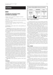
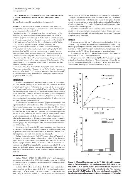
Il trattamento con 200 µM Cr VI causava una <strong>di</strong>minuzione <strong>del</strong>la sintesi<br />
<strong>di</strong> PS (Fig. 1A) che prec<strong>ed</strong>eva nel tempo l’esposizione <strong>di</strong> PS (Fig.<br />
1B) e l’apoptosi. Quest’ultima era infatti trascurabile entro le 4 ore <strong>di</strong> trattamento<br />
<strong>ed</strong> evidente (45%) dopo 6 h <strong>di</strong> trattamento. Tempi lunghi <strong>di</strong> incubazione<br />
con Cr VI (12 ore) determinavano la necrosi cellulare.<br />
Il trattamento con 9 µM Cr VI mo<strong>di</strong>ficava leggermente i parametri<br />
sopra riportati (a 6 e 12 ore <strong>di</strong> incubazione).<br />
L’esposizione per 6 e 12 ore con 200 µM <strong>di</strong> Cr VI, riduceva la capacità<br />
<strong>del</strong>le cellule <strong>di</strong> decarbossilare la PS neosintetizzata, valutata dal rapporto<br />
tra la ra<strong>di</strong>oattività nella PE e quella nella PS, non mo<strong>di</strong>ficata invece<br />
da trattamenti per tempi o concentrazioni inferiori (9 µM).<br />
Figura 1. Effetto <strong>del</strong> trattamento con Cr VI (200 µM) sull’incorporazione<br />
<strong>di</strong> [ 3 H]serina nella PS (A) e sull’esposizione <strong>del</strong>la PS (B) in cellule<br />
MOLT-4. In A, cellule MOLT-4 erano incubate con [ 3 H]serina in<br />
presenza o in assenza <strong>di</strong> Cr VI. La ra<strong>di</strong>oattività nella PS è espressa<br />
come dpmx10 3 /5x10 5 cellule. In B è riportata l’esposizione <strong>del</strong>la PS<br />
in cellule incubate nelle stesse con<strong>di</strong>zioni ma in assenza <strong>di</strong> [ 3 H]serina