complicanze vascolari della chirurgia protesica del ginocchio
complicanze vascolari della chirurgia protesica del ginocchio
complicanze vascolari della chirurgia protesica del ginocchio

Create successful ePaper yourself
Turn your PDF publications into a flip-book with our unique Google optimized e-Paper software.
COMPLICANZE<br />
VASCOLARI DELLA<br />
CHIRURGIA<br />
PROTESICA DEL<br />
GINOCCHIO
TKA: intervento a più alto rischio di<br />
Arteria poplitea:<br />
<strong>complicanze</strong> <strong>vascolari</strong><br />
• Trombosi/Dissezione<br />
• Pseudoaneurisma<br />
• Lacerazione/sezione<br />
completa<br />
• FAV<br />
Vena poplitea:<br />
• lacerazione/sezione<br />
completa<br />
• Trombosi
Incidenza complessiva<br />
di <strong>complicanze</strong> arteriose<br />
0.03% - 0.2%
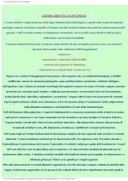
FATTORI DI RISCHIO PER<br />
COMPLICANZE VASCOLARI<br />
• Reintervento<br />
• Arteriosclerosi <strong>del</strong> distretto femoro-popliteo<br />
• Esiti di frattura <strong>del</strong> 1/3 distale <strong>del</strong> femore<br />
• Terapia cortisonica cronica
amputazione dopo complicanza arteriosa ~ 10%<br />
maggior FR di amputazione: ritardo diagnostico
MECCANISMI DI DANNO<br />
• Manipolazione<br />
introperatoria con<br />
dislocazione <strong>del</strong>le<br />
strutture <strong>del</strong> <strong>ginocchio</strong><br />
– Torsione e stiramento<br />
arterioso<br />
• Trauma diretto<br />
– Strumenti chirurgici<br />
– Viti<br />
– Cementi<br />
• Tourniquet<br />
• Lesioni da calore<br />
generato dalla reazione<br />
esotermica prodotta dalla<br />
polimerizzazione <strong>del</strong><br />
cemento
Anatomia
Anatomia
The Journal of Arthroplasty Vol. 14 No. 7 1999<br />
Injury to the Popliteal Artery and Its Anatomic<br />
Location in Total Knee Arthroplasty<br />
James T. Ninomiya, MD, MS* John C. Dean, MD, PE,-]- and<br />
Victor M. Goldberg, MD~-<br />
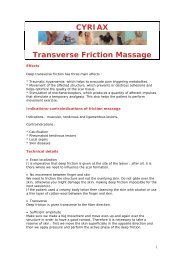
Transverse magnetic resonance imaging scans at joint line (A) and 10 mm below this (B). The medial (M)-lateral<br />
(L) anatomic axis was defined as the line that passes through the greatest medial-lateral dimension of the proximaI tibia<br />
and is parallel to the bisector of the angle formed by lines drawn tangenI to the posterior and anterior surfaces of the tibial<br />
plateau. The perpendicular bisector of this anatomic axis was defined to be the midline of the proximal tibia for both levels.<br />
The location of the artery (A, arrow) is described as a ratio of its position along the length of the analomic axis as measured<br />
from the lateral side. In 95% of the cases, the artery was localed lateral to the midline at both levels.
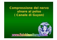
Schematic diagram of<br />
a transverse section of the knee<br />
at the level of the joint. The<br />
artery is lateral to the midline,<br />
lies less than 1 cm from the<br />
capsule with the leg in extension,<br />
and is the most anterior<br />
structure in the neurovascular<br />
bundIe. (ACL, anterior cruciate<br />
ligament; PCL, posterior<br />
cruciate ligament.)<br />
The Journal of Arthroplasty Vol. 14 No. 7 1999<br />
Injury to the Popliteal Artery and Its Anatomic<br />
Location in Total Knee Arthroplasty<br />
James T. Ninomiya, MD, MS* John C. Dean, MD, PE,-]- and<br />
Victor M. Goldberg, MD~-
The Journal of Arthroplasty Vol. 14 No. 7 1999<br />
Injury to the Popliteal Artery and Its Anatomic<br />
Location in Total Knee Arthroplasty<br />
James T. Ninomiya, MD, MS* John C. Dean, MD, PE,-]- and<br />
Victor M. Goldberg, MD~-<br />
Anteroposterior artenogram demonstrating<br />
lateral position of the popliteal artery at the level<br />
of the tibial plateau as it descends obliquely from the<br />
adductor hiatus medially to the interosseous space laterally.
The Journal of Arthroplasty Vol. 14 No. 7 1999<br />
Injury to the Popliteal Artery and Its Anatomic<br />
Location in Total Knee Arthroplasty<br />
James T. Ninomiya, MD, MS* John C. Dean, MD, PE,-]- and<br />
Victor M. Goldberg, MD~-<br />
Lateral arteriograms during total knee<br />
arthroplasty after all femoral osteotomies when<br />
bent knee retractors were placed behind the tibial<br />
plateau to lever it forward. (A) Single-prong retractor<br />
placed lateral to the posterior cruciate ligament<br />
insertion resulting in direct displacement of the<br />
popliteal artery. The retractor was intentionally<br />
placed in a slightly deep position to demonstrate<br />
the proximity of the artery to the posterior joint<br />
line. (B) Single-prong retractor placed medial to<br />
the posterior cruciate ligament insertion, which<br />
never produced displacement of the artery. (C)<br />
Double-prong retractor placed over the intact<br />
posterior cruciate ligament resulting in minimal<br />
displacement of tile artery as its posterior excursion<br />
is limited.
The Journal of Arthroplasty Vol. 14 No. 7 1999<br />
Injury to the Popliteal Artery and Its Anatomic<br />
Location in Total Knee Arthroplasty<br />
James T. Ninomiya, MD, MS* John C. Dean, MD, PE,-]- and<br />
Victor M. Goldberg, MD~-<br />
Lateral arteriogram after<br />
all total knee arthroplasty<br />
osteotomies but before component<br />
implantation with the<br />
knee hyperextended resulting<br />
in severe tenting of the artery<br />
over the sharp posterior edge<br />
of the tibial plateau.
The Journal of Arthroplasty Vol. 14 No. 7 1999<br />
Injury to the Popliteal Artery and Its Anatomic<br />
Location in Total Knee Arthroplasty<br />
James T. Ninomiya, MD, MS* John C. Dean, MD, PE,-]- and<br />
Victor M. Goldberg, MD~-<br />
Lateral arteriograms of hyperflexed knee. (A) Before total knee arthroplasty demonstrating flexural deformity of<br />
the popliteal artery. (B) The tibia has been levered forward with the bent knee retractor further deforming the artery.
Interventi riparativi <strong>vascolari</strong><br />
• Tromboendoarteriectomia<br />
• Innesto o bypass<br />
– popliteo-popliteo<br />
– femoro-popliteo<br />
Materiale utilizzato:<br />
• vena grande safena<br />
• Protesi (PTFE)<br />
Lesioni ostruttive
Interventi riparativi <strong>vascolari</strong><br />
• Sutura arteriosa<br />
• Innesto o bypass<br />
• Stent graft<br />
• Iniezione di Trombina<br />
Pseudoaneurismi
Valutazione<br />
vascolare preoperatoria<br />
• Clinica<br />
anamnesi<br />
esame obiettivo: polsi periferici, trofismo<br />
• Strumentale<br />
– I.W. (indice caviglia braccio)<br />
– ECD arti inferiori<br />
– AngioTC<br />
– AngioRMN
Chirurgia <strong>del</strong> <strong>ginocchio</strong><br />
in paziente arteriopatico<br />
• Valutazione stadio clinico Leriche-<br />
Fontaine (I, IIa, IIb, III, IV)<br />
• Presenza di pregresso bypass vascolare o<br />
di un intervento endovascolare<br />
• Tipo di antiaggregazione/anticoagulazione
STUDIO RETROSPETTIVO DATI MAYO<br />
CLINIC<br />
• 19.808 interventi di artroprotesi totale di<br />
<strong>ginocchio</strong> (TKA) eseguiti fra il 1970 e il<br />
1997<br />
• 9 pazienti sottoposti a TKA dopo<br />
confezionamento di bypass periferico
STUDIO RETROSPETTIVO DATI MAYO<br />
CLINIC<br />
• 2 pazienti presentarono trombosi arteriosa acuta<br />
– 1 amputazione<br />
• Assenza di correlazione statistica fra<br />
- protesi utilizzata<br />
- tourniquet<br />
- intervallo di tempo fra bypass e TKA<br />
- terapia anticoagulante e<br />
- occlusione arteriosa
Valutazione<br />
vascolare postoperatoria<br />
Clinica<br />
– Polsi periferici<br />
– Cute<br />
– Logge muscolari<br />
Strumentale<br />
– I.W. (indice caviglia<br />
braccio)<br />
– ECD arti inferiori<br />
• Attenta sorveglianza<br />
<strong>del</strong>le condizioni<br />
circolatorie <strong>del</strong>l’arto<br />
• Possibile insorgenza<br />
di ischemia anche a<br />
24-48h p.-o.!!!
Valutazione<br />
vascolare postoperatoria<br />
• Un basso livello di sospetto può ritardare<br />
la diagnosi e rendersi responsabile <strong><strong>del</strong>la</strong><br />
comparsa di quadri clinici gravi, fino alla<br />
ischemia irreversibile con perdita <strong>del</strong>l’arto<br />
• Anestesia peridurale !!!
amputazione dopo complicanza arteriosa ~ 10%<br />
maggior FR di amputazione: ritardo diagnostico
COMPLICANZE VENOSE<br />
TVP e artroprotesi<br />
• Rischio elevatissimo di eventi trombo-<br />
embolici<br />
• senza profilassi<br />
– TVP 60-80%<br />
– E.P. clinica 4-10%<br />
– E.P. fatale 1-5%
Il ricorso sistematico alla profilassi<br />
farmacologica e fisica ha drasticamente<br />
ridotto il rischio di TEP<br />
Katharine He Xing e coll.<br />
Thromb. Res. 2008;123:24-34
• EBPM TVP 25-30%<br />
• anticoagulanti orali e<br />
<strong>del</strong>l’eparina non<br />
TVP e artroprotesi<br />
frazionata a dosi fisse<br />
40-45% TVP<br />
• fondaparinux<br />
– sanguinamenti<br />
maggiori (2,1% contro<br />
0,2% P=0,006)<br />
• dabigatran<br />
–orale<br />
– non inferiore a<br />
enoxaparina
TVP e artroprotesi<br />
• EBPM enoxaparina<br />
+ mezzi fisici<br />
• 4-6 settimane<br />
• il grado di<br />
mobilizzazione<br />
• Le recenti linee guida<br />
ACCP <strong>del</strong> 2008<br />
suggeriscono una<br />
profilassi prolungata<br />
come dopo<br />
l’intervento sull’anca.