Skull and Dura - Department of Medical Imaging - University of Toronto
Skull and Dura - Department of Medical Imaging - University of Toronto
Skull and Dura - Department of Medical Imaging - University of Toronto
You also want an ePaper? Increase the reach of your titles
YUMPU automatically turns print PDFs into web optimized ePapers that Google loves.
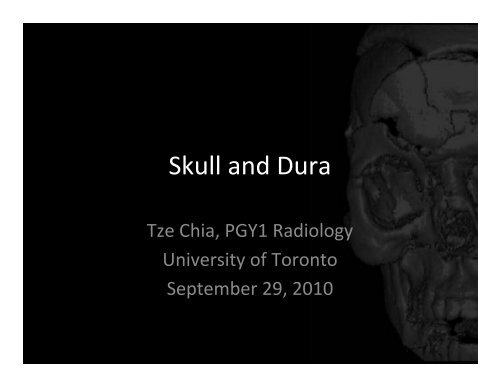
<strong>Skull</strong> <strong>and</strong> <strong>Dura</strong><br />
Tze Chia, PGY1 Radiology<br />
<strong>University</strong> <strong>of</strong> <strong>Toronto</strong><br />
September 29, 2010
Outline<br />
• Embryology<br />
• Anatomy<br />
– Bones <strong>of</strong> cranium<br />
– Cranial foramina<br />
– Scalp <strong>and</strong> meninges<br />
• <strong>Imaging</strong> cases
Neonatal <strong>Skull</strong><br />
• Sutures: Frontal (
Neonatal Brain U/S
• Neurocranium<br />
Adult <strong>Skull</strong> aka Cranium<br />
– Calvaria + Cranial base<br />
– Single: frontal, ethmoidal,<br />
sphenoidal, occipital<br />
– Paired: temporal, parietal<br />
• Viscerocranium (facial)<br />
– Single: m<strong>and</strong>ible, vomer<br />
– Paired: nasal, lacrimal,<br />
maxilla, palatine, zygoma,<br />
inferior nasal conchae
Cranial Fossae<br />
• Anterior:<br />
– Frontal + Ethmoid + Lesser wing <strong>of</strong> Sphenoid<br />
• Middle:<br />
– Body <strong>of</strong> Sphenoid + Temporal<br />
• Posterior:<br />
– Occipital + Temporal
Parietal Bone<br />
• Meningeal grooves<br />
• Parietal eminence<br />
• Parietal foramen for<br />
emissary vein
Parietal Bone<br />
• Meningeal grooves<br />
• Parietal eminence<br />
• Parietal foramen for<br />
emissary vein
Parietal Bone<br />
• Meningeal grooves<br />
• Parietal eminence<br />
• Parietal foramen for<br />
emissary vein
Frontal Bone<br />
• Frontal sinuses<br />
• Ethmoid notch<br />
• Orbital plate<br />
• Supraorbital notch<br />
(supraorbital nerve)<br />
• Glabella
Frontal Bone<br />
• Frontal sinuses<br />
• Ethmoid notch<br />
• Orbital plate<br />
• Supraorbital notch<br />
(supraorbital nerve)<br />
• Glabella
Frontal Bone<br />
• Frontal sinuses<br />
• Ethmoid notch<br />
• Orbital plate<br />
• Supraorbital notch<br />
(supraorbital nerve)<br />
• Glabella
Ethmoid Bone<br />
• Cribiform plate (foramina for<br />
olfactory nerves)<br />
• Crista galli (attaches falx<br />
cerebri)<br />
• Perpendicular plate<br />
• Lateral masses<br />
• Ethmoid sinuses/ethmoid bulla<br />
• Superior <strong>and</strong> middle nasal<br />
conchae (turbinates)<br />
• Nasal meatus<br />
• Uncinate process<br />
• Infundibulum
Sphenoid Bone<br />
• Body, lesser wings, <strong>and</strong> greater wings<br />
• Sella turcica<br />
– Hypophyseal fossa<br />
– Tuberculum sellae<br />
– Dorsum sellae<br />
• Anterior <strong>and</strong> posterior clinoid<br />
processes (attachment for tentorium<br />
cerebelli)<br />
• Sphenoid sinuses<br />
• Optic canal<br />
• Foramina: rotundum, ovale, spinosum<br />
• Superior orbital fissure<br />
• Pterygoid process (pterygoid muscles<br />
attach)<br />
– medial/lateral pterygoid plates,<br />
– pterygoid hamulus (anchor for muscles<br />
that open Eustachian tube)<br />
– pterygoid canal
• Frontal<br />
• Coronal suture<br />
• Parietal
• Ethmoid<br />
sinuses<br />
• Ethmoid bulla<br />
• Perpendicular<br />
plate<br />
• Ant/Post<br />
clinoid<br />
process<br />
• Optic canal
• Crista galli<br />
• Cribiform plate<br />
• Perpendicular plate<br />
• Conchae<br />
• Infundibulum<br />
• Inferior orbital<br />
fissure
• Anteroir clinoid<br />
process<br />
• Optic canal<br />
• Sphenoid sinus<br />
• Medial <strong>and</strong> lateral<br />
pterygoid processes<br />
• Zygomatic arch<br />
• Ramus <strong>of</strong> m<strong>and</strong>ible
Occipital Bone<br />
• Foramen magnum<br />
• Lateral condyles<br />
(atlanto‐occipital joint)<br />
• Clivus<br />
• Squamous portion<br />
• Grooves for sinuses<br />
• Internal/external<br />
occipital protuberance<br />
• Hypoglossal canal
Occipital Bone<br />
• Foramen magnum<br />
• Lateral condyles<br />
(atlanto‐occipital joint)<br />
• Clivus<br />
• Squamous portion<br />
• Grooves for sinuses<br />
• Internal/external<br />
occipital protuberance<br />
• Hypoglossal canal
Temporal Bone<br />
• Squamous<br />
– Zygomatic process/arch<br />
– Articular tubercle<br />
– M<strong>and</strong>ibular fossa (forms TMJ)<br />
– Temporal fossa<br />
– Pterion<br />
• Tympanic<br />
– External auditory meatus<br />
• Mastoid<br />
– Mastoid process, air cells, antrum<br />
(communicates with middle ear)<br />
• Petrous<br />
– Internal auditory canal<br />
– Jugular foramen/fossa<br />
– Carotid canal<br />
– Meckel’s cave (trigeminal ganglion)<br />
– Foramen lacerum<br />
– Styloid process<br />
– Stylomastoid foramen / facial nerve<br />
canal
Facial Bones<br />
• Nasal bone<br />
• Lacrimal bone<br />
• Maxilla<br />
– Infraorbital foramen<br />
– Maxillary sinuses<br />
– Processes: frontal, zygomatic,<br />
alveolar, palatine (anterior ¾<br />
hard palate)<br />
• Palatine bone (posterior ¼<br />
hard palate)<br />
• Zygoma<br />
– Temporal process<br />
• Inferior nasal conchae<br />
• Vomer (inferior bony nasal<br />
septum)
Facial Bones<br />
• M<strong>and</strong>ible<br />
– Head<br />
– Neck<br />
– Ramus<br />
– Angle (gonion)<br />
– Body<br />
– Coronoid process<br />
(attachment <strong>of</strong><br />
temporalis <strong>and</strong> masseter)<br />
– Condylar process<br />
– M<strong>and</strong>ibular notch<br />
– Mental protuberance<br />
– Mental foramen
Foramina<br />
Bone Foramen/fissure Major structures<br />
Frontal Supraorbital notch Supraorbital nerve <strong>and</strong> artery<br />
Sphenoid Optic canal CN II <strong>and</strong> ophthalmic artery<br />
Superior orbital<br />
fissure<br />
Inferior orbital<br />
fissure<br />
CN III, IV, V1, VI, ophthalmic vein<br />
Maxillary <strong>of</strong> CN V<br />
Maxillary Infraorbital foramen Infraorbital <strong>and</strong> maxillary <strong>of</strong> CN V<br />
M<strong>and</strong>ible Mental foramen Mental artery <strong>and</strong> nerve
Foramina<br />
Bone Foramen/fissure Major structures<br />
Ethmoid Cribiform plate CN I<br />
Sphenoid Foramen rotundum Maxillary CN V2<br />
Foramen ovale M<strong>and</strong>ibular CN V3<br />
Foramen spinosum Middle meningeal artery<br />
Occipital Foramen magnum Medulla oblongata <strong>and</strong> CN XI<br />
Hypoglossal canal CN XII<br />
Temporal Carotid canal Internal carotid artery<br />
Temporal <strong>and</strong> occipital bone Jugular foramen Internal jugular vein, CN IX, X, XI<br />
External auditory meatus Air to tympanic membrane<br />
Internal auditory meatus CN VII, VIII<br />
Temporal, spenoid, <strong>and</strong> occipital<br />
bones<br />
Foramen lacerum Internal carotid artery, nerve <strong>of</strong><br />
pterygoid canal, meningeal<br />
branch <strong>of</strong> ascending pharyngeal<br />
artery<br />
Stylomastoid foramen / facial CN VII<br />
nerve canal
Foramina<br />
• Cribiform plate<br />
• Foramen rotundum<br />
• Foramen ovale<br />
• Foramen spinosum<br />
• Optic canal<br />
• Superior orbital fissure<br />
• Foramen magnum<br />
• Hypoglossal canal<br />
• Carotid canal<br />
• Jugular foramen<br />
• Internal auditory meatus<br />
• Foramen lacerum
Foramina<br />
• Foramen ovale<br />
• Foramen spinosum<br />
• Foramen magnum<br />
• Hypoglossal canal<br />
• Carotid canal<br />
• Jugular fossa<br />
• External auditory meatus<br />
• Foramen lacerum<br />
• Stylomastoid foramen / facial<br />
nerve canal
Scalp<br />
• Skin<br />
• Connective tissue<br />
(dense)<br />
• Aponeurosis<br />
• Loose connective<br />
tissue<br />
• Pericranium
Meninges<br />
• <strong>Dura</strong> mater<br />
• Arachnoid mater<br />
• Pia mater
Cases<br />
• Depressed parietal skull fracture
Cases<br />
• Le Fort I fracture
Le Fort Fractures <strong>of</strong> Maxillae<br />
• Type I: Alveolar process <strong>and</strong> hard palate separated from superior<br />
skull<br />
• Type II: Alveolar, zygomatic <strong>and</strong> frontal processes separated from<br />
frontal bone<br />
• Type III: Maxillae, nasal bones, <strong>and</strong> zygomatic bones separate from<br />
frontal bone
Cases<br />
• Le Fort II fracture<br />
• Occipitomental<br />
view
Cases<br />
• Left angle<br />
m<strong>and</strong>ibular fracture
Cases<br />
• Right orbit blowout<br />
fracture
Cases<br />
• Tripod fracture:<br />
– Maxillary sinus<br />
– Zygomatic arch<br />
– Lateral orbital rim
References<br />
• Netter, Atlas <strong>of</strong> Human Anatomy<br />
• Kelley & Petersen, Sectional Anatomy for<br />
<strong>Imaging</strong> Pr<strong>of</strong>essionals<br />
• Moore & Dalley, Clinically Oriented Anatomy


