L'anthropologie du vivant : objets et méthodes - CNRS - Dynamique ...
L'anthropologie du vivant : objets et méthodes - CNRS - Dynamique ... L'anthropologie du vivant : objets et méthodes - CNRS - Dynamique ...
Computed tomography (CT) and magnetic resonance (MR) imaging to better understand and characterize brain and craniumgrowth and development.the same structure, the choice of the most suitable one will depend on thenature of the study that is being performed. For example, in a head CTimage, the bones appears white and very clear because they absorb largequantities of X-rays; grease and other soft tissues absorb less quantitiesof X-rays and appear in a gray scale; and finally, the air absorbs very littleradiation, with hollow structures appearing black (Figure 1).Figure 1: Coronal (upper left), horizontal (upper right) and sagittal (lower left) craniumCT images. Three dimensional (3D) reconstruction of cranium (lower right)On the other hand, in a head MR image, the bone structures areshown black because of their lack of water, but the soft tissues ofthe brain can be clearly recognized with high detail: gray matter,white matter and cerebrospinal fluid (Figure 2).Figure 2: Coronal (upper left), horizontal (upper right) and sagittal (lowerleft) head MR. 3D reconstruction of cortical brain tissue (lower right)generated using an automatic algorithm for brain extraction.L’anthropologie du vivant : objets et méthodes - 2010 110
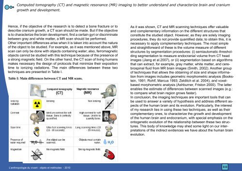
Computed tomography (CT) and magnetic resonance (MR) imaging to better understand and characterize brain and craniumgrowth and development.Hence, if the objective of the research is to detect a bone fracture or todescribe cranium growth, a CT scan should be made. But if the objectiveis to characterize the brain development, find a certain gyri or discriminatebetween gray and white matter, a MR scan should be performed.Other important differences arise when it is taken into account the natureof the object to be studied. For example, as it was mentioned above, MRscan can only be done with objects containing water; also, ferromagneticobjects cannot be studied with this technique because of the presence ofa strong magnetic field. On the other hand, the CT scan of living humansmakes necessary the design of protocols that minimize their expositiontime to ionizing radiations. The main differences between these twotechniques are presented in Table I.Table I: Main differences between CT and MR scans.As it was shown, CT and MR scanning techniques offer valuableand complementary information on the different structures thatconstitute the studied object. However, as they are solely imagingprocedures, they do not provide quantified data; to obtain this, it isnecessary to apply complementary techniques. The most simpleand straightforward of these is the volume measure of differentstructures by segmentation procedures: (i) semiautomatic thresholdingsegmentation to measure endocranial volume from CT headimages (Jiang et al.2007), or (ii) segmentation based on algorithmsthat can extract, for example, gray matter, white matter, and cerebrospinalfluid from MR brain images (Smith, 2002). Another groupof techniques that allows the obtaining of size and shape informationfrom images includes geometric morphometric analysis (Bookstein,1991; Rohlf, Marcus 1993; Zelditch et al. 2004), and voxelbasedmorphometric analysis (Ashburner, Friston 2000). The latterenables the estimate of differences between scanned images (e.g.:to compare what brain region grows faster).In conclusion, the imaging techniques are important tools that canbe used to answer a variety of hypothesis and address different aspectsof the human brain and its evolution. Particularly, the interestof my research lies in using these two techniques, as well as theircomplementary ones, to characterize the growth and developmentof the human brain and endocranium, with special emphasis on theontogenetic evolution of the relationship between these two structures.This body of knowledge may shed some light on our interpretationsof the indirect evidences we have about the human brainevolution.L’anthropologie du vivant : objets et méthodes - 2010111
- Page 61 and 62: ETUDE DES PRATIQUES ALIMENTAIRES ET
- Page 63 and 64: ETUDE DES PRATIQUES ALIMENTAIRES ET
- Page 65 and 66: Les méthodes de l’anthropologie
- Page 67 and 68: Les méthodes de l’anthropologie
- Page 69 and 70: Les méthodes de l’anthropologie
- Page 71 and 72: L’anthropologie biologique :une a
- Page 73 and 74: L’anthropologie biologique :une a
- Page 75 and 76: L’anthropologie biologique :une a
- Page 77: Une approche synchronique du vieill
- Page 80 and 81: Une approche synchronique du vieill
- Page 82 and 83: De l’échelle macro à l’échel
- Page 84 and 85: De l’échelle macro à l’échel
- Page 86 and 87: Activités instrumentales physiques
- Page 88 and 89: Activités instrumentales physiques
- Page 90 and 91: Dialogue interdisciplinaire ? De l
- Page 92 and 93: Femmes marocaines âgées vivant se
- Page 94 and 95: Femmes marocaines âgées vivant se
- Page 96 and 97: La Modélisation Statistique en Ant
- Page 98 and 99: La Modélisation Statistique en Ant
- Page 100 and 101: Méthodologie et intérêts des gro
- Page 102 and 103: Méthodologie et intérêts des gro
- Page 104 and 105: Méthodologie et intérêts des gro
- Page 106 and 107: Méthodologie et intérêts des gro
- Page 108 and 109: Mesure de la composition corporelle
- Page 110 and 111: Mesure de la composition corporelle
- Page 114 and 115: Computed tomography (CT) and magnet
- Page 116 and 117: Le modèle Primate en anthropologie
Computed tomography (CT) and magn<strong>et</strong>ic resonance (MR) imaging to b<strong>et</strong>ter understand and characterize brain and craniumgrowth and development.Hence, if the objective of the research is to d<strong>et</strong>ect a bone fracture or todescribe cranium growth, a CT scan should be made. But if the objectiveis to characterize the brain development, find a certain gyri or discriminateb<strong>et</strong>ween gray and white matter, a MR scan should be performed.Other important differences arise when it is taken into account the natureof the object to be studied. For example, as it was mentioned above, MRscan can only be done with objects containing water; also, ferromagn<strong>et</strong>icobjects cannot be studied with this technique because of the presence ofa strong magn<strong>et</strong>ic field. On the other hand, the CT scan of living humansmakes necessary the design of protocols that minimize their expositiontime to ionizing radiations. The main differences b<strong>et</strong>ween these twotechniques are presented in Table I.Table I: Main differences b<strong>et</strong>ween CT and MR scans.As it was shown, CT and MR scanning techniques offer valuableand complementary information on the different structures thatconstitute the studied object. However, as they are solely imagingproce<strong>du</strong>res, they do not provide quantified data; to obtain this, it isnecessary to apply complementary techniques. The most simpleand straightforward of these is the volume measure of differentstructures by segmentation proce<strong>du</strong>res: (i) semiautomatic thresholdingsegmentation to measure endocranial volume from CT headimages (Jiang <strong>et</strong> al.2007), or (ii) segmentation based on algorithmsthat can extract, for example, gray matter, white matter, and cerebrospinalfluid from MR brain images (Smith, 2002). Another groupof techniques that allows the obtaining of size and shape informationfrom images includes geom<strong>et</strong>ric morphom<strong>et</strong>ric analysis (Bookstein,1991; Rohlf, Marcus 1993; Zelditch <strong>et</strong> al. 2004), and voxelbasedmorphom<strong>et</strong>ric analysis (Ashburner, Friston 2000). The latterenables the estimate of differences b<strong>et</strong>ween scanned images (e.g.:to compare what brain region grows faster).In conclusion, the imaging techniques are important tools that canbe used to answer a vari<strong>et</strong>y of hypothesis and address different aspectsof the human brain and its evolution. Particularly, the interestof my research lies in using these two techniques, as well as theircomplementary ones, to characterize the growth and developmentof the human brain and endocranium, with special emphasis on theontogen<strong>et</strong>ic evolution of the relationship b<strong>et</strong>ween these two structures.This body of knowledge may shed some light on our interpr<strong>et</strong>ationsof the indirect evidences we have about the human brainevolution.L’anthropologie <strong>du</strong> <strong>vivant</strong> : <strong>obj<strong>et</strong>s</strong> <strong>et</strong> méthodes - 2010111


