Create successful ePaper yourself
Turn your PDF publications into a flip-book with our unique Google optimized e-Paper software.
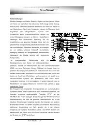
Epifasziale Nerven und Gefäße der <strong>dorsal</strong>en Rumpfwand<br />
Abbildung aus: Tillmann, Atlas der Anatomie, Springer Verlag<br />
N. occipitalis major (R. dors. C2)<br />
N. occipitalis tertius (R. dors. C3)<br />
Plexus cervicalis<br />
N. occipitalis minor<br />
Nn. supraclaviculares<br />
Spinalnerven (Rr. <strong>dorsal</strong>es)<br />
Rr. cutanei mediales<br />
Rr. cutanei laterales<br />
Spinalnerven (Rr. ventrales)<br />
Rr. cutanei laterales (Interkostalnerven)<br />
Plexus lumbalis<br />
R. cutaneus lat. des N. iliohypogastricus<br />
Nn. clunium sup. (Rr. <strong>dorsal</strong>es L1-L3)<br />
Nn. clunium medii (Rr. <strong>dorsal</strong>es S1-S3<br />
Plexus sacralis<br />
Nn. clunium inf.<br />
(Äste des N. cutaneus fem. post.)<br />
D8