

Fonction et régulation de la protéine ICAP-1alpha dans la ...
Fonction et régulation de la protéine ICAP-1alpha dans la ...
Fonction et régulation de la protéine ICAP-1alpha dans la ...

Create successful ePaper yourself
Turn your PDF publications into a flip-book with our unique Google optimized e-Paper software.
tel-00435843, version 1 - 24 Nov 2009<br />
Published January 28, 2008<br />
were obtained with CL: 50% spreading occurred at a coating<br />
concentration of 5 µ g/ml for Icap-1 – null cells compared with<br />
25 µ g/ml for WT cells ( Fig. 2 C ). Icap-1 – null cells appeared to<br />
exhibit a stronger interaction with FN and CL and, consequently,<br />
spread at lower <strong>de</strong>nsities of these ECM substrates. In both migration<br />
and spreading assays, the differences b<strong>et</strong>ween WT and<br />
mutant cells were more pronounced using CL. This probably<br />
re� ects the more restricted use of � 1 integrins on CL. On the<br />
other hand, the expression of <strong>ICAP</strong>-1 in Icap-1 – null MEFs restores<br />
cell spreading and migration simi<strong>la</strong>rly to what was observed<br />
for WT MEFs (Fig. S2, A and B; avai<strong>la</strong>ble at http://www<br />
.jcb.org/cgi/content/full/jcb.200707142/DC1), proving that the<br />
altered migration was in<strong>de</strong>ed caused by the loss of endogenous<br />
<strong>ICAP</strong>-1. A simi<strong>la</strong>r effect was also observed with osteob<strong>la</strong>sts: at<br />
the FN-coating concentration of 0.3 µ g/ml, the totality of Icap-1 –<br />
null osteob<strong>la</strong>sts were fully spread, whereas only 50% of WT<br />
and rescued osteob<strong>la</strong>sts were spread un<strong>de</strong>r i<strong>de</strong>ntical experimental<br />
conditions, <strong>de</strong>monstrating that this <strong>de</strong>fect was still <strong>ICAP</strong>-1<br />
<strong>de</strong>pen<strong>de</strong>nt but not cell speci� c ( Fig. 2 D ). In agreement with<br />
the speci� city of <strong>ICAP</strong>-1 for � 1 integrin, migration and adhesion<br />
behaviors of WT and Icap-1 – null MEFs on vitronectin<br />
(VN) coating were not signi� cantly different. This <strong>de</strong>monstrates<br />
that <strong>ICAP</strong>-1 loss did not alter � 3 integrin – mediated adhesion<br />
(Fig. S3, A and B).<br />
<strong>ICAP</strong>-1 slows down the talin recruitment<br />
into FAs<br />
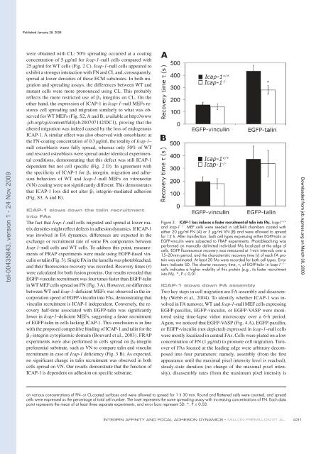
The fact that Icap-1 – null cells migrated and spread at lower matrix<br />
<strong>de</strong>nsities might re� ect <strong>de</strong>fects in adhesion dynamics. If <strong>ICAP</strong>-1<br />
was involved in FA dynamics, differences are expected in the<br />
exchange or recruitment rate of some FA components b<strong>et</strong>ween<br />
Icap-1 – null cells and WT cells. To address this point, measurements<br />
of FRAP experiments were ma<strong>de</strong> using EGFP-fused vinculin<br />
or talin ( Fig. 3 ). Single FA in the <strong>la</strong>mel<strong>la</strong> was photobleached,<br />
and their � uorescence recovery was recor<strong>de</strong>d. Recovery times ( � )<br />
were calcu<strong>la</strong>ted for both fusion proteins. Our results revealed that<br />
EGFP-vinculin recruitment was four times faster than EGFP-talin<br />
in WT MEF cells spread on FN ( Fig. 3 A ). However, no difference<br />
b<strong>et</strong>ween WT and Icap-1 – <strong>de</strong>� cient MEFs was observed in the incorporation<br />
speed of EGFP-vinculin into FAs, <strong>de</strong>monstrating that<br />
vinculin recruitment is <strong>ICAP</strong>-1 in<strong>de</strong>pen<strong>de</strong>nt. Conversely, the recovery<br />
half-time associated with EGFP-talin was signi� cantly<br />
lower in Icap-1 – <strong>de</strong>� cient MEFs, suggesting a faster recruitment<br />
of EGFP-talin in cells <strong>la</strong>cking <strong>ICAP</strong>-1. This conclusion is in line<br />
with the proposed comp<strong>et</strong>itive binding of <strong>ICAP</strong>-1 and talin for the<br />
� 1 -integrin cytop<strong>la</strong>smic domain ( Bouvard <strong>et</strong> al., 2003 ). FRAP<br />
experiments were also performed in cells spread on � 3 -integrin<br />
preferential substrate, such as VN to compare talin and vinculin<br />
recruitment in case of Icap-1 <strong>de</strong>� ciency ( Fig. 3 B ). As expected,<br />
no signi� cant change in talin recruitment was observed in both<br />
cells spread on VN. Our results <strong>de</strong>monstrate that the function of<br />
<strong>ICAP</strong>-1 is <strong>de</strong>pen<strong>de</strong>nt on adhesion on speci� c substrate.<br />
Figure 3. <strong>ICAP</strong>-1 loss induces a faster recruitment of talin into FAs. Icap-1 +/+<br />
and Icap-1 � / � MEF cells were see<strong>de</strong>d in LabTekII chambers coated with<br />
either 20 µ g/ml FN (A) or 5 µ g/ml VN (B) and were allowed to spread<br />
for 12 h. After transfection, both cell types expressing either EGFP-talin or<br />
EGFP-vinculin were subjected to FRAP experiments. Photobleaching was<br />
performed on manually <strong>de</strong>limited individual FAs localized at the edge of<br />
cells. EGFP fl uorescence recovery was measured at 1-min intervals over a<br />
15 – 20-min period, and the characteristic recovery time ( � ) of each FA protein<br />
was estimated. At least 20 FAs were recor<strong>de</strong>d for both cell types. Error<br />
� / �<br />
bars indicate SD. The shorter recovery time, � , of EGFP-talin in Icap-1<br />
cells indicates a higher mobility of this protein (e.g., its faster recruitment<br />
into FA). *, P ≤ 0.01.<br />
<strong>ICAP</strong>-1 slows down FA assembly<br />
Two key steps in cell migration are FA assembly and disassembly<br />
( Webb <strong>et</strong> al., 2004 ). To i<strong>de</strong>ntify wh<strong>et</strong>her <strong>ICAP</strong>-1 was involved<br />
in FA turnover, WT and Icap-1 – null MEF cells expressing<br />
EGFP-paxillin, EGFP-vinculin, or EGFP-VASP were monitored<br />
using time-<strong>la</strong>pse vi<strong>de</strong>o microscopy over a 6-h period.<br />
Again, we noticed that EGFP-VASP ( Fig. 4 A ), EGFP-paxillin,<br />
or EGFP-vinculin (not <strong>de</strong>picted) expressed in Icap-1 – null cells<br />
were mostly localized in central FAs. Cells were p<strong>la</strong>ted on a low<br />
concentration of FN (1 µ g/ml) to promote cell migration. Turnover<br />
of FAs located at the leading edge were arbitrary <strong>de</strong>composed<br />
into four param<strong>et</strong>ers: namely, assembly (from the � rst<br />
appearance until the maximal pixel intensity level is reached),<br />
steady-state duration (no change of the maximal pixel intensity),<br />
disassembly rates (from the maximum pixel intensity is<br />
on various concentrations of FN- or CL-coated surfaces and were allowed to spread for 1 h 30 min. Round and fl attened cells were counted, and spread<br />
cells were expressed as the percentage of total cell number. The ins<strong>et</strong> represents the same spreading assay with increasing concentrations of FN. Each data<br />
point represents the mean of at least three separate experiments, and error bars represent SD. *, P ≤ 0.03.<br />
INTEGRIN AFFINITY AND FOCAL ADHESION DYNAMICS • MILLON-FR É MILLON ET AL.<br />
431<br />
Downloa<strong>de</strong>d from<br />
jcb.rupress.org<br />
on March 30, 2009


