

Fonction et régulation de la protéine ICAP-1alpha dans la ...
Fonction et régulation de la protéine ICAP-1alpha dans la ...
Fonction et régulation de la protéine ICAP-1alpha dans la ...

Create successful ePaper yourself
Turn your PDF publications into a flip-book with our unique Google optimized e-Paper software.
tel-00435843, version 1 - 24 Nov 2009<br />
Published January 28, 2008<br />
disrupts a salt bridge b<strong>et</strong>ween the � and � subunits, leading to<br />
integrin activation ( Luo <strong>et</strong> al., 2004 ; Vinogradova <strong>et</strong> al., 2004 ).<br />
Evi<strong>de</strong>nce suggests that the binding of a complex including<br />
talin, Rap1-GTP – interacting adaptor molecule, Rap1, and<br />
vasodi<strong>la</strong>tor-stimu<strong>la</strong>ted phosphoprotein (VASP) to the integrin<br />
cytop<strong>la</strong>smic tail is a common � nal step in integrin activation<br />
( Han <strong>et</strong> al., 2006 ).<br />
Among integrin partners, integrin cytop<strong>la</strong>smic domain –<br />
associated protein 1 (<strong>ICAP</strong>-1) encompassing a phosphotyrosinebinding<br />
domain interacts speci� cally with the cytop<strong>la</strong>smic tail<br />
of � 1 integrin at the membrane-distal NPXY motif. The increase<br />
of � 1 integrin – <strong>de</strong>pen<strong>de</strong>nt cell motility on � bronectin (FN) upon<br />
<strong>ICAP</strong>-1 overexpression ( Chang <strong>et</strong> al., 1997 , 2002 ; Zhang<br />
and Hemler, 1999 ) and cell rounding up after overexpression<br />
of a phosphomim<strong>et</strong>ic mutant of <strong>ICAP</strong>-1 at the CaMKII site<br />
( Bouvard and Block, 1998 ) suggest that <strong>ICAP</strong>-1 might regu<strong>la</strong>te<br />
� 1 -integrin function. Moreover, talin and <strong>ICAP</strong>-1 comp<strong>et</strong>e in<br />
vitro for integrin binding, and high <strong>ICAP</strong>-1 concentrations disrupt<br />
FA ( Bouvard <strong>et</strong> al., 2003 ). <strong>ICAP</strong>-1 and � 1 integrin are colocalized<br />
at the leading edges of cells during the early stages<br />
of spreading ( Fournier <strong>et</strong> al., 2002 ). This fac<strong>et</strong> of <strong>ICAP</strong>-1 in<br />
FA dynamics, which is obviously regu<strong>la</strong>tory and transitory,<br />
prompted us to d<strong>et</strong>ermine in more d<strong>et</strong>ail the role p<strong>la</strong>yed by<br />
<strong>ICAP</strong>-1 in adhesion site dynamics.<br />
In this study, we show that Icap-1 – <strong>de</strong>� cient mouse embryonic<br />
� brob<strong>la</strong>sts (MEFs) disp<strong>la</strong>y <strong>de</strong>fects in cellu<strong>la</strong>r spreading<br />
and migration corre<strong>la</strong>ting with the redistribution of FA, a modi-<br />
� cation in FA dynamics, and an increase in integrin af� nity.<br />
These <strong>de</strong>fects could be mimicked by increasing either integrin<br />
af� nity or the surface <strong>de</strong>nsity of the un<strong>de</strong>rlying matrix. By modu<strong>la</strong>ting<br />
integrin af� nity, <strong>ICAP</strong>-1 allows the cell to adapt its adhesion<br />
strength and rate of migration to changes in the matrix<br />
surface <strong>de</strong>nsity. These data point not only to the existence of<br />
speci� c molecules involved in FA assembly or disassembly signaling<br />
pathways but also to the control of FA assembly and matrix<br />
sensing through the � ne-tuning of integrin af� nity.<br />
Results<br />
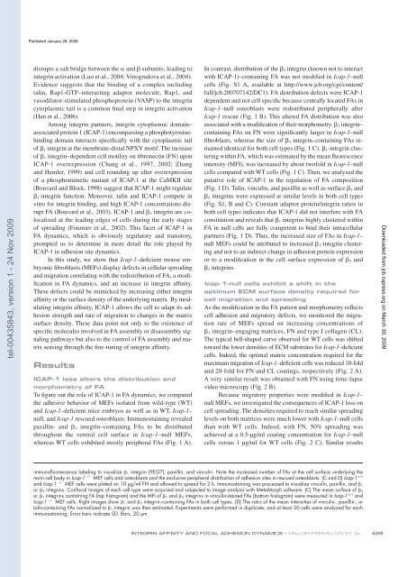
<strong>ICAP</strong>-1 loss alters the distribution and<br />
morphom<strong>et</strong>ry of FA<br />
To � gure out the role of <strong>ICAP</strong>-1 in FA dynamics, we compared<br />
the adhesive behavior of MEFs iso<strong>la</strong>ted from wild-type (WT)<br />
and Icap-1 – <strong>de</strong>� cient mice embryos as well as in WT, Icap-1 –<br />
null, and Icap-1 rescued osteob<strong>la</strong>sts. Immunostaining revealed<br />
paxillin- and � 1 integrin – containing FAs to be distributed<br />
throughout the ventral cell surface in Icap-1 – null MEFs,<br />
whereas WT cells exhibited mostly peripheral FAs ( Fig. 1 A ).<br />
In contrast, distribution of the � 3 integrin (known not to interact<br />
with <strong>ICAP</strong>-1) – containing FA was not modi� ed in Icap-1 – null<br />
cells (Fig. S1 A, avai<strong>la</strong>ble at http://www.jcb.org/cgi/content/<br />
full/jcb.200707142/DC1). FA distribution <strong>de</strong>fects were <strong>ICAP</strong>-1<br />
<strong>de</strong>pen<strong>de</strong>nt and not cell speci� c because centrally located FAs in<br />
Icap-1 – null osteob<strong>la</strong>sts were redistributed peripherally after<br />
Icap-1 rescue ( Fig. 1 B ). This altered FA distribution was also<br />
associated with a modi� cation of their morphom<strong>et</strong>ry. � 1 integrin –<br />
containing FAs on FN were signi� cantly <strong>la</strong>rger in Icap-1 – null<br />
� brob<strong>la</strong>sts, whereas the size of � 3 integrin – containing FAs remained<br />
i<strong>de</strong>ntical for both cell types ( Fig. 1 C ). � 1 -integrin clustering<br />
within FA, which was estimated by the mean � uorescence<br />
intensity (MFI), was increased by about twofold in Icap-1 – null<br />
cells compared with WT cells ( Fig. 1 C ). Then, we analyzed the<br />
putative role of <strong>ICAP</strong>-1 in the regu<strong>la</strong>tion of FA composition<br />
( Fig. 1 D ). Talin, vinculin, and paxillin as well as surface � 1 and<br />
� 3 integrins were expressed at simi<strong>la</strong>r levels in both cell types<br />
(Fig. S1, B and C). Constant adaptor protein/integrin ratios in<br />
both cell types indicates that <strong>ICAP</strong>-1 did not interfere with FA<br />
constitution and reveals that � 1 integrins highly clustered within<br />
FA in null cells are fully comp<strong>et</strong>ent to bind their intracellu<strong>la</strong>r<br />
partners ( Fig. 1 D ). Thus, the increased size of FAs in Icap-1 –<br />
null MEFs could be attributed to increased � 1 -integrin clustering<br />
and not to an indirect change in adhesion protein expression<br />
or to a modi� cation in the cell surface expression of � 3 and<br />
� 1 integrins.<br />
Icap-1 – null cells exhibit a shift in the<br />
optimum ECM surface <strong>de</strong>nsity required for<br />
cell migration and spreading<br />
As the modi� cation in the FA pattern and morphom<strong>et</strong>ry re� ects<br />
cell adhesion and migratory <strong>de</strong>fects, we monitored the migration<br />
rate of MEFs spread on increasing concentrations of<br />
� 1 integrin – engaging matrices, FN and type I col<strong>la</strong>gen (CL).<br />
The typical bell-shaped curve observed for WT cells was shifted<br />
toward the lower <strong>de</strong>nsities of ECM substrates for Icap-1 – <strong>de</strong>� cient<br />
cells. In<strong>de</strong>ed, the optimal matrix concentration required for the<br />
maximum migration of Icap-1 – <strong>de</strong>� cient cells was reduced 10-fold<br />
and 20-fold for FN and CL coatings, respectively ( Fig. 2 A ).<br />
A very simi<strong>la</strong>r result was obtained with FN using time-<strong>la</strong>pse<br />
vi<strong>de</strong>o microscopy ( Fig. 2 B ).<br />
Because migratory properties were modi� ed in Icap-1 –<br />
null MEFs, we investigated the consequences of <strong>ICAP</strong>-1 loss on<br />
cell spreading. The <strong>de</strong>nsities required to reach simi<strong>la</strong>r spreading<br />
levels on both matrices were much lower with Icap-1 – null cells<br />
than with WT cells. In<strong>de</strong>ed, with FN, 50% spreading was<br />
achieved at a 0.5- µ g/ml coating concentration for Icap-1 – null<br />
cells versus 1 µ g/ml for WT cells ( Fig. 2 C ). Simi<strong>la</strong>r results<br />
immunofl uorescence <strong>la</strong>beling to visualize � 1 integrin (9EG7), paxillin, and vinculin. Note the increased number of FAs at the cell surface un<strong>de</strong>rlying the<br />
main cell body in Icap-1 � / � MEF cells and osteob<strong>la</strong>sts and the exclusive peripheral distribution of adhesion sites in rescued osteob<strong>la</strong>sts. (C and D) Icap-1 +/+<br />
and Icap-1 � / � MEF cells were p<strong>la</strong>ted on 10 µ g/ml FN and allowed to spread for 2 h. Immunostaining was processed to visualize vinculin, paxillin, and � 1<br />
or � 3 integrins. Confocal images of each cell type were acquired and subjected to image analysis with M<strong>et</strong>aMorph software. (C) The mean surface of � 3<br />
or � 1 integrins containing FA (top histogram) and the MFI of � 1 and � 3 integrins in vinculin-stained FAs (bottom histogram) were measured in Icap-1 +/+ and<br />
Icap-1 � / � MEF cells. Right images show � 1 and � 3 integrin – containing FAs in both cell types. (D) The ratio of the mean intensities of vinculin-, paxillin-, or<br />
talin-containing FAs normalized to � 1 integrin was then estimated. Experiments were performed in duplicate, and at least 20 cells were analyzed for each<br />
immunostaining. Error bars indicate SD. Bars, 20 µ m.<br />
INTEGRIN AFFINITY AND FOCAL ADHESION DYNAMICS • MILLON-FR É MILLON ET AL.<br />
429<br />
Downloa<strong>de</strong>d from<br />
jcb.rupress.org<br />
on March 30, 2009


