faculte de medecine, de pharmacie et d'odonto-stomatologie
faculte de medecine, de pharmacie et d'odonto-stomatologie faculte de medecine, de pharmacie et d'odonto-stomatologie
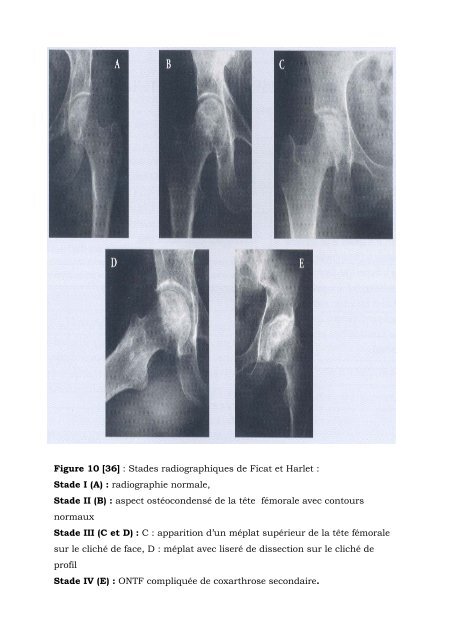
Figure 10 [36] : Stades radiographiques de Ficat et Harlet : Stade I (A) : radiographie normale, Stade II (B) : aspect ostéocondensé de la tête fémorale avec contours normaux Stade III (C et D) : C : apparition d’un méplat supérieur de la tête fémorale sur le cliché de face, D : méplat avec liseré de dissection sur le cliché de profil Stade IV (E) : ONTF compliquée de coxarthrose secondaire.
Figure 11 [36] : Coupes tomodensitométriques axiales transverses mettant en évidence figure A : Un liseré d’ ostéocondensation péri-nécrotique dans le cadre d’un stade II. figure B : Un liseré d’ ostéocondensation péri-nécrotique accompagné d’un tassement modéré de la zone nécrotique dans une ostéonécrose de stade III
- Page 6 and 7: 4. ASSISTANTS Mr Saibou MAIGA Mr Ou
- Page 9 and 10: DEDICACES Je dédie cette thèse: A
- Page 11 and 12: REMERCIEMENTS Mes vifs remerciement
- Page 13 and 14: Fanta, Jolie, Kaman, Mariammondri,
- Page 16 and 17: A Notre Maître et Président du Ju
- Page 18 and 19: A notre Maître et juge : Docteur T
- Page 20 and 21: ABREVIATIONS Hb: Hémoglobine HbS:
- Page 22: III. Méthodologie……………
- Page 25: I. OBJECTIFS A. Objectif général
- Page 28 and 29: C. Epidémiologie Il s’agit d’u
- Page 30 and 31: 3. Les moyens d’union Ils sont cl
- Page 32 and 33: 6. Myologie de la cuisse 6-1. La lo
- Page 34 and 35: Les ONA dysbariques pourraient êtr
- Page 36 and 37: placebo[29]. Une meilleure connaiss
- Page 38 and 39: - La grossesse : L’ostéonécrose
- Page 40 and 41: en bande, arciforme ou par plages v
- Page 42 and 43: . Stade IV : atteinte totale ou sub
- Page 44 and 45: Les paresthésies accompagnatrices
- Page 46 and 47: très précoce. Ceci était déjà
- Page 48 and 49: Figure 1 [35] : L’os du fémur 1=
- Page 50 and 51: Figure 3 [35] : Myologie de l’art
- Page 52 and 53: Figure 5 [35] : Coupe coronale de l
- Page 54 and 55: Figure 8 [35] : vascularisation de
- Page 58 and 59: Figure 12 [36]: Ostéonécrose de l
- Page 61 and 62: III. METHODOLOGIE 1. Cadre d’étu
- Page 63: Stade III : c’est le classique si
- Page 66 and 67: Cas 2 Madame D.O, 37 ans, ménagèr
- Page 68 and 69: Cas 4 Madame S.H, 21 ans, ménagèr
- Page 70 and 71: Cas 6 Monsieur S.B, 27 ans, vétér
- Page 72 and 73: Cas 8 Monsieur M.H, 19 ans, sans em
- Page 74 and 75: Cas 10 Mademoiselle D.K, 15 ans, é
- Page 76 and 77: Tableau III : Répartition des mala
- Page 78 and 79: Tableau VII : Répartition des mala
- Page 80 and 81: Tableau XI : tableau récapitulatif
- Page 82: Tableau XV : Répartition des malad
- Page 85 and 86: conforme au constat de Diarra K .F
- Page 88 and 89: VI. Conclusion L’ONATF est une r
- Page 91 and 92: VIII. REFERENCES BIBLIOGRAPHIQUES 1
- Page 93 and 94: 17. Kawai K, Teak A, Hirohata K Ste
- Page 95: 33. Cotten A. IRM de la hanche: ost
- Page 98 and 99: VII. EXAMEN PHYSIQUE Examen génér
- Page 100 and 101: Autres à préciser ……………
- Page 102 and 103: FICHE SIGNALETIQUE Nom : COULIBALY
Figure 10 [36] : Sta<strong>de</strong>s radiographiques <strong>de</strong> Ficat <strong>et</strong> Harl<strong>et</strong> :<br />
Sta<strong>de</strong> I (A) : radiographie normale,<br />
Sta<strong>de</strong> II (B) : aspect ostéocon<strong>de</strong>nsé <strong>de</strong> la tête fémorale avec contours<br />
normaux<br />
Sta<strong>de</strong> III (C <strong>et</strong> D) : C : apparition d’un méplat supérieur <strong>de</strong> la tête fémorale<br />
sur le cliché <strong>de</strong> face, D : méplat avec liseré <strong>de</strong> dissection sur le cliché <strong>de</strong><br />
profil<br />
Sta<strong>de</strong> IV (E) : ONTF compliquée <strong>de</strong> coxarthrose secondaire.


