PET CO - JLAR
PET CO - JLAR
PET CO - JLAR

You also want an ePaper? Increase the reach of your titles
YUMPU automatically turns print PDFs into web optimized ePapers that Google loves.
Jérôme CUNY<br />
S.A.M.U. Régional de Lille<br />
Pôle de l’Urgence<br />
CHRU de LILLE<br />
2010
INTRODUCTION<br />
<br />
<br />
<br />
1943 : 1 ère mesure infrarouge du <strong>CO</strong> 2 (Luft)<br />
1978 : Utilisation courante lors d’anesthésie générale (Pays-Bas)<br />
Mesure du <strong>CO</strong> 2 dans l’air expiré<br />
Indicateur<br />
- direct des échanges pulmonaires,<br />
- indirect de la production tissulaire de <strong>CO</strong> 2<br />
Technique non invasive, reflète :<br />
La production de <strong>CO</strong> 2 ,<br />
La perfusion pulmonaire,<br />
La ventilation alvéolaire,<br />
Le mode ventilatoire,<br />
La surveillance des circuits de ventilation.
Capnographe : appareil de mesure enregistrant la<br />
forme de l’onde<br />
Capnomètre : Mesure des concentrations expirées de <strong>CO</strong> 2<br />
Capnogramme : représentation graphique<br />
Pa <strong>CO</strong> 2 : pression partielle artérielle en <strong>CO</strong> 2<br />
PA <strong>CO</strong> 2 : Pression partielle alvéolaire en <strong>CO</strong> 2<br />
<strong>PET</strong> <strong>CO</strong> 2 : pression télé expiratoire en <strong>CO</strong> 2<br />
FE <strong>CO</strong> 2 : fraction expiratoire de <strong>CO</strong> 2
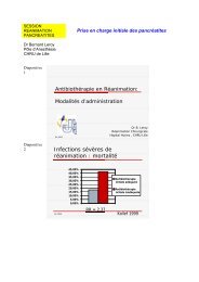
Anesthesiology 1989<br />
• Utilisation de<br />
capnographie + SpO 2 :<br />
prévention de 93% des<br />
incidents<br />
Anesth Intensive Care 1993<br />
• Analyse de 2000<br />
incidents : 52% détectés<br />
par capnographie + SpO 2<br />
Capnography.com<br />
morbi-mortalité
En pré hospitalier, 2002 :<br />
53 % des SMUR équipés d’un capnographe<br />
32 % des polytraumatisés<br />
FREYSZ, le traumatisé grave, actualités en réanimation, 2002-2003. p85-111<br />
GDS fait aux urgences, patient intubé ventilé<br />
= hypercapnie<br />
Par mauvais réglage du ventilateur<br />
Pathologies pulmonaires<br />
JEUR, 2001,14,163-164
Asthme et ET <strong>CO</strong> 2<br />
<br />
<br />
<br />
Capteur nasal<br />
Surveillance possible de l’évolution de la crise : amélioration avec le<br />
traitement<br />
Pente de la Phase III<br />
Angle α<br />
Identique aux données du DEP réalisés aux urgences<br />
Int J Emerg Med. 2009 feb 24;2(2):83-9<br />
Détresse respiratoire et ET <strong>CO</strong> 2<br />
<br />
<br />
Capteur nasal<br />
Détresse respiratorie tout venant, quelque soit l’étiologie<br />
Différence significative entre la Pa <strong>CO</strong> 2 et ET<strong>CO</strong> 2<br />
<br />
Gradient de 8 à 12 mmhg<br />
Pas de reflet de la Pa <strong>CO</strong> 2 Am J Emerg Med. 2009nov;27(9):1056-9
Polytraumatisés et Et <strong>CO</strong> 2<br />
Utilisation prudente<br />
Polytraumatisés graves intubés ventilés<br />
Etco 2 entre 35 et 40<br />
30% des patients : Pa <strong>CO</strong> 2 > 50 mmHg<br />
80 % des patients : Pa <strong>CO</strong> 2 > 40 mmHg<br />
J Trauma. 2009 Jan;66(1):26-31
PHYSIOPATHOLOGIE<br />
<strong>CO</strong> 2 air ambiant = 0,03 % Pa<strong>CO</strong> 2 ~ 0<br />
<br />
Production du <strong>CO</strong> 2 par le métabolisme aérobie :consommation d’ oxygène<br />
= 200 L / j (adulte)<br />
Transport sous 3 formes :<br />
Transformation hépatique et rénale<br />
• Bicarbonates = 60 à 70 %<br />
• Liaisons protidiques (carbamino-hémoglobine) = 20 à 30 %<br />
• Libre dans le plasma (veineux, Pv <strong>CO</strong> 2 ) = 5 à 10 %<br />
<br />
Élimination pulmonaire<br />
élimination par ventilation<br />
<br />
Équilibre RAPIDE au niveau pulmonaire entre<br />
Pressions alvéolaires (PA <strong>CO</strong> 2 ) et pressions artérielle (Pa <strong>CO</strong> 2 )<br />
Pa <strong>CO</strong> 2 reflète la ventilation alvéolaire
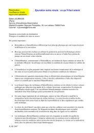
Apex pulmonaire<br />
Ventilation > Perfusion<br />
Base pulmonaire<br />
Perfusion > Ventilation<br />
Capnography.com
GRADIENT P (a – et) <strong>CO</strong> 2<br />
= Gradient artério-alvéolaire<br />
Si poumon homogène : pas de differnece entre Pa et PA<br />
Gaz expiré = Gaz alvéolaire<br />
Mais les rapports V / Q sont inhomogènes<br />
Variations de l’espace mort alvéolaires de 10% à 30%<br />
Provenant d’alvéoles ventilées, perfusées et ventilées non<br />
perfusées<br />
<strong>PET</strong> <strong>CO</strong> 2<br />
< PA <strong>CO</strong> 2 des alvéoles perfusées<br />
< Pa <strong>CO</strong> 2
ET <strong>CO</strong> 2 = 35 à 37 mmHg (ou 5 %)<br />
A poumons sains : (sauf enfant et grossesse)<br />
le gradient Pa <strong>CO</strong> 2 et <strong>PET</strong> <strong>CO</strong> 2<br />
= 2 à 6 mmHg en VS<br />
<br />
Pa <strong>CO</strong> 2<br />
~ PA <strong>CO</strong> 2<br />
ESPACE MORT<br />
=<br />
DETERMINANT PRINCIPAL<br />
DU GRADIENT (a – ET) <strong>CO</strong> 2
PHYSIOPATHOLOGIE<br />
<br />
<strong>PET</strong> <strong>CO</strong> 2<br />
ne reflète l’espace<br />
mort alvéolaire que dans les<br />
conditions normales hors<br />
pathologie cardiaque ou<br />
respiratoire sous-jacente<br />
<br />
Disparité de V/Q dans le<br />
poumon<br />
(a - <strong>PET</strong> <strong>CO</strong> 2<br />
)<br />
de l’espace mort<br />
Inadaptation du rapport V/q<br />
Capnography.com<br />
Dc influence la <strong>PET</strong> <strong>CO</strong> 2<br />
Dc meilleur perfusion<br />
des alvéoles<br />
<strong>PET</strong> <strong>CO</strong> 2
Les variations de la <strong>PET</strong> <strong>CO</strong> 2 ne sont pas proportionnelles<br />
à celles de la Pa <strong>CO</strong> 2<br />
• Neurochirurgie : dans 26 % des cas, Pa <strong>CO</strong>2 et <strong>PET</strong> <strong>CO</strong> 2 varient<br />
en sens opposé<br />
Grenier, Anesth Analg 1999 :88<br />
PHYSIOPATHOLOGIE DE L’AUGMENTATION L<br />
DU GRADIENT<br />
• Anomalie du rapport ventilation / perfusion et espace mort<br />
Embolie Pulmonaire, hypovolémie,<br />
• Absence de vidange alvéolaire complète dans certain mode ventilatoire<br />
Utilisation de la Jet ventilation à haute fréquence<br />
• Erreur technique<br />
défaut de calibrage, fuite du circuit
<strong>PET</strong> <strong>CO</strong> 2 non substituable à la Pa <strong>CO</strong> 2<br />
<strong>PET</strong> <strong>CO</strong> 2 = alerte précoce<br />
<strong>PET</strong> <strong>CO</strong> 2 : pression télé-expiratoire en <strong>CO</strong> 2<br />
déterminée par :<br />
• Production métabolique de <strong>CO</strong> 2<br />
• Transport vers le poumon<br />
• Élimination par la ventilation = ventilation alvéolaire<br />
(en l’absence de trouble de la diffusion pulmonaire)
EN PRE HOSPITALIER : Pause et al, Resuscitation 1997<br />
• Adaptation difficile de la ventilation sur la Pet <strong>CO</strong> 2<br />
Insuffisance circulatoire, EP, asthme, insuffisance respiratoire<br />
• Si insuffisance cardiaque ou insuffisance respiratoire :<br />
le gradient moyen = 17 mm Hg [ e = 1 à 30 ]<br />
• Pas d’évaluation de la Pa <strong>CO</strong> 2 par la Pet <strong>CO</strong> 2<br />
INTERFERENCES :<br />
• H 2 O : présence de piège à eau<br />
• N 2 O<br />
• <strong>CO</strong> : quantifié comme du <strong>CO</strong> 2<br />
• Variations de température : 0,3% par °C<br />
• Pression atmosphérique : utilisation de la fraction de <strong>CO</strong> 2<br />
• Variation du gradient avec l’âge : augmentation de 1,5 mmHg par 10 ans
CAPNOGRAMME<br />
<br />
Capnogramme de temps et capnogramme<br />
de volume<br />
Volume :<br />
• pas de segment inspiratoire<br />
Temps :<br />
• Permet surveillance de patients non<br />
ventilés<br />
• Surveillance de l’inspi- et expiration<br />
• Faible évaluation de l’état de V / Q<br />
• Difficulté d’évaluer l’espace mort
A<br />
B<br />
C<br />
D A • 2 SEGMENTS :<br />
Expiratoire (3 phases)<br />
Inspiratoire<br />
• 2 ANGLES<br />
PHASE I (espace D-A) D<br />
• Fin de l’inspirationl<br />
• <strong>CO</strong> 2 des voies aériennesa<br />
• Vidange de l’espace l<br />
mort anatomique<br />
• Espace mort absolu<br />
• [ <strong>CO</strong> 2 ] = 0
A<br />
B<br />
C<br />
POINT A :<br />
• Expiration initiale de gaz<br />
ne participant pas aux échanges<br />
POINT C :<br />
D A PHASE II (espace A-B) A<br />
• <strong>PET</strong> <strong>CO</strong> 2 (~ PA <strong>CO</strong> 2 )<br />
• Apparition du <strong>CO</strong> 2 dans les gaz<br />
• Mélange des gaz de l’espace l<br />
mort au gaz alvéolaire<br />
• Ascension lente si poumon inhomogène<br />
ne
ANGLE α<br />
A<br />
B<br />
D<br />
C<br />
A<br />
• Normal à 105 °<br />
•Reflète la spasticité<br />
• Varie avec la pente<br />
• Corrélé au DEP<br />
• Indication indirecte<br />
de l’état de V/Q<br />
PHASE III (espace B-C B C )<br />
• Plateau alvéolaire = Expiration du <strong>CO</strong> 2 des alvéoles<br />
• Pente ascendante car :<br />
• élimination du <strong>CO</strong> 2 est régulir<br />
gulière<br />
• vidange tardives des alvéoles distales = rapport V/Q petit<br />
• asynchronisme de vidange<br />
• perfusion et ventilation<br />
• pente si poumon inhomogène<br />
ne
A<br />
B<br />
C<br />
ANGLE ß<br />
• Angle droit<br />
• Etat de réinhalation<br />
D A PHASE 0 (espace C-D) C<br />
• Inspiration<br />
• Inhalation de gaz dépourvu d<br />
de <strong>CO</strong> 2<br />
• Chute rapide de [ <strong>CO</strong> 2 ] vers 0
<strong>CO</strong> 2<br />
Pa<strong>CO</strong> 2<br />
( GDS )<br />
ESPACE MORT<br />
ALVEOLAIRE<br />
<strong>PET</strong> <strong>CO</strong> 2<br />
Espace<br />
Mort<br />
anatomique<br />
ESPACE MORT<br />
PHYSIOLOGIQUE<br />
VENTILATION ALVEOLAIRE<br />
EFFICACE<br />
VOLUME EXPIRE
VARIATION BRUTALE DE <strong>PET</strong> <strong>CO</strong> 2<br />
=<br />
SITUATION CRITIQUE<br />
ANOMALIES<br />
Production de <strong>CO</strong> 2<br />
Transport = débit cardiaque<br />
Ventilation alvéolaire
4 méthodes m<br />
:<br />
– Spectrométrie trie de masse :<br />
méthode de référencer<br />
rence<br />
Sépare les gaz en fonction de<br />
leur poids moléculaires<br />
Analyse de plusieurs gaz<br />
simultanément<br />
ment<br />
Bloc<br />
Techniques<br />
Impossible en médecine m<br />
d’urgenced<br />
– Spectrométrie trie Raman :<br />
Rayon laser argon haute intensité<br />
Analyse multi gaz<br />
Pas en France<br />
Capnography.com
Méthode colorimétrique<br />
:<br />
– Semi quantitative<br />
– Papier filtre<br />
– Marginale<br />
– Pourpre < 0,5 %<br />
– Jaune > 2 %<br />
Spectrophotométrie trie d’absorption<br />
d<br />
infrarouge :<br />
– La plus employée<br />
– Faisceau IR traversant les gaz<br />
– Comparaison avec échantillon<br />
référencerence<br />
– Rapide
<strong>CO</strong> 2 absorbe fortement les Infrarouges (λ = 4,3 µm)<br />
+<br />
-<br />
<br />
<br />
Comparaison de la différence d’absorption entre le mélange<br />
gazeux et un mélange de référence à concentration connue<br />
TYPE ASPIRATIF : (sidestream) Echantillonnage LATERAL<br />
• Permet une étude multi gaz, cellule dans la machine, comparaison à<br />
échantillon de référence<br />
• secrétions = obstruction, fuite, distorsion du signal<br />
+<br />
-<br />
<br />
NON ASPIRATIF : (meanstream) Echantillonnage CENTRAL<br />
• lecture directe, rapide, sans distorsion,<br />
• augmente l’espace mort, fragilité<br />
Précision de 0,2 %<br />
<br />
Mesure faussée si O 2 ou protoxyde d’azote mais correction<br />
automatique
MESURE NASALE<br />
Surveillance d’un patient en ventilation spontanée MAIS :<br />
• Mélange du <strong>CO</strong> 2 expiré avec O 2 inhalé<br />
• Ventilation par la bouche<br />
• Débit O 2<br />
> 4 L / min<br />
• Hypoventilation<br />
<strong>PET</strong> <strong>CO</strong> 2<br />
Surveillance de la variation de la valeur
ABSENCE DE CAPNO<br />
Intubation oesophagienne :<br />
• La SpO 2 peut rester normale pendant<br />
plusieurs dizaines de secondes<br />
Arrêt cardiaque:<br />
• Spiromètrie conservée<br />
Bronchospasme :<br />
• Élévation des pressions dans le circuit<br />
Déconnexion du circuit:<br />
• spirométrie nulle<br />
Dysfonctionnement du<br />
capnomètre, obstruction de la<br />
sonde, fuites
<strong>PET</strong> <strong>CO</strong> 2<br />
Hypothermie<br />
Anesthésie : consommation d’ O 2 , production de <strong>CO</strong> 2<br />
Baisse progressive<br />
de la ventilation alvéolaire, Bronchospasme,<br />
encombrement<br />
Chute du débit cardiaque :<br />
perfusion pulmonaire espace mort alvéolaire<br />
Hypovolémie, EP, AC
Production de <strong>CO</strong> 2<br />
Ventilation alvéolaire<br />
Hypothermie<br />
Perfusion pulmonaire<br />
<br />
<strong>PET</strong> <strong>CO</strong> 2<br />
Hyperventilation (Vt, fr)<br />
Apnée<br />
Obstruction VA<br />
Extubation<br />
Dc<br />
Collapsus<br />
Hypovolémie<br />
EP<br />
ACR<br />
Fuites<br />
Débranchement<br />
Anomalies du ventilateur<br />
Problèmes techniques
<strong>PET</strong> <strong>CO</strong> 2<br />
Hyperthermie maligne<br />
Reperfusion après clampage<br />
Apport exogène de <strong>CO</strong> 2<br />
Injection de bicarbonates<br />
Diminution de la ventilation alvéolaire
Production de <strong>CO</strong> 2<br />
Fièvre<br />
Hyperthermie maligne<br />
Bicarbonates<br />
Ventilation alvéolaire<br />
<strong>PET</strong> <strong>CO</strong> 2<br />
Hypoventilation<br />
(Vt, fr)<br />
Obstruction VA<br />
Perfusion pulmonaire<br />
Dc<br />
HTA<br />
Problèmes techniques<br />
Fuites<br />
Anomalies de valves<br />
Anomalies du ventilateur
PRONOSTIC :<br />
Cantineau et al. Crit Care Med 1996 ; 24 : 791-6.<br />
Asplin et al. Ann Emerg Med 1995 ; 25 : 756-61<br />
Callaham et al. Crit Care Med 1990 ; 18 : 358-62<br />
Valeur pronostique (AC avec activité électrique !) :<br />
Levine et al. N Engl J Med 1997;337 : 301-6<br />
<strong>PET</strong> <strong>CO</strong> 2 > 10 mmHg au cours des 20 premières res min. de réa r<br />
<br />
Probabilité de RACS<br />
<strong>PET</strong> <strong>CO</strong> 2 initiale > 15 mmHg 91% de RACS
<strong>PET</strong> <strong>CO</strong> 2 10 mmHg après s 20 min de réa r<br />
<br />
100% décèsd<br />
ATTENTION :<br />
• ADRENALINE : <strong>PET</strong> <strong>CO</strong> 2<br />
(50% 1 min après s IVD)<br />
• BICARBONATES : <strong>PET</strong> <strong>CO</strong> 2<br />
Ne JAMAIS HYPERVENTILER un patient après s un AC :<br />
Pa <strong>CO</strong>2 2 entraine une de D de perfusion coronaire et cérébralc
B.P.C.O.<br />
40<br />
Augmentation de la pente<br />
du plateau expiratoire<br />
20<br />
Attention à ne pas vouloir obtenir<br />
une ET <strong>CO</strong> 2<br />
normale, mais celle de base<br />
Courbe normale
BRONCHOSPASME<br />
40<br />
Augmentation des résistances<br />
Expiratoires<br />
20<br />
Phase II : prolongée, progressive<br />
Phase III : pente<br />
Courbe normale
EMPHYSEME<br />
Inversion de la pente<br />
40<br />
20<br />
Courbe normale<br />
Destruction du système capillaire alvéolaire
REPRISE DE VENTILATION SPONTANEE<br />
Encoche lors du plateau = cleft<br />
40<br />
20<br />
-Douleur<br />
-Insuffisance de sédation<br />
-Hypercapnie<br />
-Hoquet<br />
Courbe normale
ASYNCHRONISME VENTILATOIRE<br />
DES DEUX POUMONS<br />
Plateau biphasique<br />
40<br />
20<br />
Courbe normale<br />
Compression pulmonaire extrinsèque<br />
Embolie pulmonaire
Fistule Broncho-pleurale<br />
40<br />
Plateau biphasique<br />
+ chute ET <strong>CO</strong>2<br />
20<br />
- Désaturation<br />
-Fuite<br />
-Drain thoracique bulle a nouveau<br />
Courbe normale
Cycle de ventilation mécanique m<br />
pendant une<br />
ventilation spontanée<br />
Courbe normale<br />
40<br />
20
Bronchospasme avec auto-pep<br />
Courbe normale<br />
40<br />
20<br />
-Angle alpha augmenté<br />
-Chute de la pression artérielle<br />
-Pas de retour à la ligne de base = auto pep
Sonde d’intubation d<br />
mobile<br />
Courbe normale<br />
40<br />
20<br />
- Plateau bosselé<br />
- orifice de la sonde au dessus des cordes vocales<br />
-Transmission de l’ecg
AVENIR<br />
EVALUATION DU DEBIT CARDIAQUE :<br />
• Technique de réinhalation, mesure non-invasive<br />
• Mesure de l’espace mort par réinhalation de <strong>CO</strong> 2<br />
• Estimation du shunt par SpO 2 et FiO 2<br />
• Déduction par Equation de Fick du débit cardiaque<br />
• Monitoring du Débit cardiaque<br />
DIAGNOSTIC D’EMBOLIE PULMONAIRE :<br />
• Calcul de surface sous la courbe<br />
• Évaluation de l’espace mort alvéolaire : équation de Bohr<br />
• Calcul d’un ratio, CHOPIN, CCM 1990<br />
• Spécificité : 100 %, sensibilité : 48 %<br />
EVALUATION DU BRONCHOSPASME :<br />
• Mesure de la pente du plateau alvéolaire<br />
• Ajustement de la PeeP<br />
• Guider l’intubation à l’aveugle<br />
• Surveillance d’un patient en VS.