FMC en direct des associations CME from FFFCEDV's associations
FMC en direct des associations CME from FFFCEDV's associations
FMC en direct des associations CME from FFFCEDV's associations

You also want an ePaper? Increase the reach of your titles
YUMPU automatically turns print PDFs into web optimized ePapers that Google loves.
terme de « loupes »), ils sont peu fréqu<strong>en</strong>ts dans<br />
d’autres localisations mais qui, dans ces cas-là,<br />
peuv<strong>en</strong>t être tout à fait ubiquitaires, r<strong>en</strong>dant alors<br />
le diagnostic clinique peu aisé. Ces localisations<br />
inhabituelles sont de plus <strong>en</strong> plus rapportées dans<br />
la littérature, notamm<strong>en</strong>t sur le tronc, les membres,<br />
la pulpe du doigt, la lèvre, la vulve (4-6).<br />
Ils sont souv<strong>en</strong>t multiples et familiaux, se transmettant<br />
<strong>en</strong> dominance autosomique régulière.<br />
C’est une lésion bénigne, dont la transformation<br />
maligne est extrêmem<strong>en</strong>t rare. Elle conti<strong>en</strong>t<br />
de la kératine et ses produits de dégradation,<br />
limitée par un mur ressemblant à la gaine épithéliale<br />
externe du follicule pileux. Elle se développe<br />
préfér<strong>en</strong>tiellem<strong>en</strong>t dans <strong>des</strong> zones à haute d<strong>en</strong>sité<br />
de follicules pileux, sur le cuir chevelu dans<br />
90 %. Elle est solitaire dans 30 % <strong>des</strong> cas et multiple<br />
dans 70 % <strong>des</strong> cas.<br />
Les KT sont plus fréqu<strong>en</strong>ts chez la femme d’âge<br />
moy<strong>en</strong>. Ils peuv<strong>en</strong>t s’<strong>en</strong>flammer. Le cont<strong>en</strong>u peut<br />
occasionnellem<strong>en</strong>t s’extruder pour former une<br />
corne cutanée.<br />
Le diagnostic différ<strong>en</strong>tiel (<strong>en</strong> particulier au cuir<br />
chevelu) peut se poser avec : kyste dermoïde,<br />
acné chéloïdi<strong>en</strong>ne de la nuque, kyste épidermique<br />
d’implantation, pilomatrixome.<br />
À l’exam<strong>en</strong> anatomopathologique, on trouve un<br />
kyste situé dans le derme profond, délimité par<br />
une paroi épithéliale pluristratifiée évoluant selon<br />
la kératinisation trichilemmale, c'est-à-dire sans<br />
le stade intermédiaire de couche granuleuse qui<br />
est abs<strong>en</strong>te. Les cellules pariétales, dont le cytoplasme<br />
est clair et vacuolisé, reproduis<strong>en</strong>t les cellules<br />
de la gaine épithéliale externe du follicule<br />
pileux ou trichilemme. La cavité kystique conti<strong>en</strong>t<br />
un matériel éosinophilique compact ; <strong>des</strong> foyers<br />
de calcification s'observ<strong>en</strong>t dans 25 % <strong>des</strong> cas.<br />
MOTS-CLÉS • Kyste trichilemmal • Kyste du cuir chevelu<br />
• Kyste bénin<br />
KEY WORDS • Pilar cyst • Trichilemmal cyst • Scalp cyst<br />
• B<strong>en</strong>ign cyst<br />
Résumé : Le kyste trichilemmal représ<strong>en</strong>te le deuxième<br />
type de kyste cutané le plus fréqu<strong>en</strong>t sur le cuir chevelu. Ce<br />
type de kyste se r<strong>en</strong>contre le plus souv<strong>en</strong>t chez les femmes<br />
d’âge moy<strong>en</strong> et, surtout, sur le cuir chevelu. Leur paroi est<br />
revêtue par un épithélium malpighi<strong>en</strong> dépourvu de couche<br />
granuleuse. Leur transformation maligne est très rare. Ils<br />
sont souv<strong>en</strong>t familiaux, se transmettant <strong>en</strong> dominance<br />
autosomique régulière. Nous décrivons ici un cas de<br />
<strong>FMC</strong> <strong>en</strong> <strong>direct</strong> <strong>des</strong> <strong>associations</strong><br />
<strong>CME</strong> <strong>from</strong> FFFCEDV’s <strong>associations</strong><br />
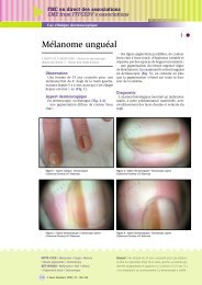
Cas clinique<br />
Facteurs de transformation maligne : certains<br />
facteurs ont été suggérés comme semblant induire<br />
la prolifération <strong>des</strong> KT, il s’agit surtout de traumatismes<br />
précessifs et d’inflammation (7, 8). Une<br />
autre théorie concernant <strong>des</strong> troubles dysembryoblastiques<br />
a été égalem<strong>en</strong>t évoquée.<br />
Conclusion<br />
La localisation <strong>en</strong> dehors du scalp d’un kyste<br />
trichilemmal (dans notre cas <strong>en</strong> l’occurr<strong>en</strong>ce au<br />
poignet) ne doit pas faire éliminer ce diagnostic.<br />
La paroi du kyste trichilemmal peut se rompre,<br />
avec formation de granulome inflammatoire, pouvant<br />
cliniquem<strong>en</strong>t et histologiquem<strong>en</strong>t, dans certaines<br />
rares situations, poser un problème de diagnostic<br />
différ<strong>en</strong>tiel avec un carcinome épidermoïde<br />
bi<strong>en</strong> différ<strong>en</strong>cié. ●<br />
RÉFÉRENCES<br />
1 - Grosshans E, Cribier B. Les kystes et pseudo-kystes cutanés. Ann<br />
Dermatol V<strong>en</strong>ereol 1994 ; 121 : 595-9 ; 647-53.<br />
2 - Rook A, Wilkinson DS, Ebling FJG et al. Trichilemmal cyst. In:<br />
Textbook of Dermatology. 6th ed. Vol 2. London (England): Blackwell<br />
Sci<strong>en</strong>ce 1998 : 1667-8.<br />
3 - Leppard BJ, Sanderson KV. The natural history of trichilemmal<br />
cysts. Br J Dermatol 1976 ; 94 : 379-90.<br />
4 - Yamaguchi J, Irimajiri T, Ohara K. Proliferating trichilemmal cyst arising<br />
in the arm of a young woman. Dermatology 1994 ; 189 : 90-2.<br />
5 - Ikegami T, Kameyama M, Orikasa H et al. Tricholemmal cyst in<br />
the pulp of the index finger: a case report. Hand Surg 2003 ; 8 :<br />
253-5.<br />
6 - Komuro, Takedai T, Tagawa K. Proliferating trichilemmal tumor<br />
on the dorsum of the hand. Ann Plast Surg 1995 ; 34 : 657-9.<br />
7 - Rutty GN, Richman PI, Laing JH. Malignant change in trichilemmal<br />
cysts: a study of cell proliferating and DNA cont<strong>en</strong>t. Histopathology<br />
1992 ; 21 : 465-8.<br />
8 - Saber S, Chiheb S, Jamali M et al. Periungual trichilemmal cyst<br />
of the big toe. Ann Dermatol V<strong>en</strong>ereol 2006 ; 133 : 1011.<br />
localisation non fréqu<strong>en</strong>te au niveau du poignet chez une<br />
femme de 56 ans.<br />
NODULAR LESION OF WRIST: A MISDIAGNOSIS.<br />
Summary: Pilar or trichilemmal cysts are the second most common<br />
keratinizing cyst on the scalp. They are more common in middleaged<br />
wom<strong>en</strong>, 90% of cases occur on the scalp. Histologically, pilar<br />
cysts are characterized by the abs<strong>en</strong>ce of a granular layer in the<br />
lining epithelium. Pilar cysts contain keratin, lined by a wall<br />
resembling the external hair root sheath. Their malignant transformation<br />
is extremely rare. Inheritance may occur in an autosomal<br />
dominant pattern. We report a clinical case about a middleaged<br />
woman with an infrequ<strong>en</strong>t localization on the wrist.<br />
© Nouv. Dermatol. 2008 ; 27 : 183-185<br />
185<br />
Nous remercions le<br />
Pr J-M. Bonnetblanc<br />
et le Pr B. Cribier pour<br />
leurs conseils amicaux.