JADC - Canadian Dental Association
JADC - Canadian Dental Association
JADC - Canadian Dental Association

Create successful ePaper yourself
Turn your PDF publications into a flip-book with our unique Google optimized e-Paper software.
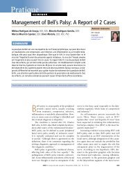
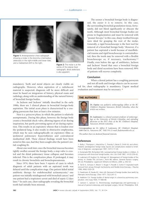
Figure 1: Anteroposterior chest radiograph<br />
showing a molar in the bronchus intermedius;<br />
atelectasis in the right-middle and lower lobe;<br />
and a mediastinal shift to the right.<br />
mandatory. Teeth and metal objects are clearly visible on<br />
radiographs. However, when aspiration of a radiolucent<br />
material is suspected, diagnosis will be more difficult and<br />
must be based on integration of history, physical exam and<br />
radiology, along with an understanding of the natural history<br />
of bronchial foreign bodies.<br />
As Jackson and Jackson 7 initially described in the early<br />
1900s, there are 3 clinical phases in bronchial foreign-body<br />
aspiration. The initial acute phase is characterized by a coughing<br />
paroxysm that lasts at least a few minutes.<br />
Next is a quiescent phase, in which the patient is relatively<br />
asymptomatic. During this phase, however, the foreign body<br />
creates a bronchial check-valve, allowing ingress of air during<br />
inspiration, but partly preventing egress of air during expiration.<br />
This results in an expiratory wheeze that is loudest over<br />
the ipsilateral lung. It also results in obstructive emphysema,<br />
which may be seen radiographically on expiratory films as<br />
ipsilateral pulmonary hyperinflation and contralateral<br />
mediastinal shift. These clinical findings would have been<br />
noted in this case, had they been sought after the patient’s initial<br />
coughing fit.<br />
About one week later, once the bronchial mucosa becomes<br />
tightly swollen around the foreign body, a stop-valve is created<br />
and the distal pulmonary tissue collapses and becomes<br />
infected. This is the complication phase. If prolonged, it may<br />
result in chronic bronchitis and bronchopneumonia.<br />
Since 1974, there have been 3 reports of cases in which<br />
diagnosis of adult patients with an aspirated tooth was<br />
delayed until the complication stage. One patient failed initial<br />
antibiotic therapy for endobronchial actinomycosis, 8 one<br />
patient was initially misdiagnosed with bronchial cancer, 9 and<br />
one patient had a respiratory arrest and died of sepsis 12 days<br />
later. 10 In each case, chest radiographs revealing the bronchial<br />
tooth had initially been misread.<br />
––– Ludemann –––<br />
Figure 2: The molar is at the<br />
centre of this lateral chest<br />
radiograph; distal atelectasis<br />
is also apparent.<br />
582 <strong>JADC</strong> • www.cda-adc.ca/jadc • Septembre 2005, Vol. 71, N o 8 •<br />
The sooner a bronchial foreign body is diagnosed,<br />
the easier it is to remove. In this case,<br />
the surrounding bronchial granulation tissue, fortunately,<br />
did not bleed significantly or obscure the<br />
tooth. Although most bronchial foreign bodies are<br />
prone to fragmentation and must be removed with<br />
“peanut forceps,” in this case, sharp-toothed forceps<br />
were ideal for grasping the root of the molar.<br />
Generally, a rigid bronchoscope is the best tool for<br />
removal of a bronchial foreign body. 3 However, if a<br />
patient has aspirated a tooth because of maxillofacial<br />
trauma and rigid bronchoscopy is contraindicated,<br />
then the tooth may be removed with a flexible<br />
bronchoscope or, if necessary, tracheotomy. 4,5<br />
Finally, even before the age of antibiotics, Jackson<br />
and Jackson 7 found that once a bronchial foreign<br />
body and distal secretions have been removed, 99%<br />
of patients will recover completely.<br />
Conclusion<br />
When a dental patient has a coughing paroxysm<br />
and not all teeth and foreign objects can be accounted<br />
for, chest radiography is mandatory. Urgent medical<br />
evaluation and treatment may be necessary. C<br />
THE AUTHORS<br />
References<br />
Dr. Ospina was pediatric otolaryngology fellow at the BC<br />
Children’s Hospital, Vancouver, British Columbia, when the<br />
article was written.<br />
Dr. Ludemann is a clinical assistant professor of otolaryngology<br />
at the University of British Columbia, and attending<br />
physician at the ENT clinic of the BC Children’s Hospital,<br />
Vancouver, British Columbia.<br />
Correspondence to: Dr. Jeffrey P. Ludemann, BC Children’s Hospital,<br />
4480 Oak St., Vancouver, BC V6H 3V4. E-mail: jludemann@cw.bc.ca.<br />
The authors have no declared financial interests.<br />
1. Reilly J, Thompson J, MacArthur C, Pransky S, Beste D, Smith M, and others.<br />
Pediatric aerodigestive foreign body injuries are complications related to timeliness<br />
of diagnosis. Laryngoscope 1997; 107(1):17–20.<br />
2. Esclamado RM, Richardson MA. Laryngotracheal foreign bodies in children. A<br />
comparison with bronchial foreigh bodies. Am J Dis Child 1987; 141(3):259–62.<br />
3. Ludemann JP, Hughes CA, Holinger LD. Management of foreign bodies of the<br />
airway: In: Shields TW, LoCicero J, Ponn RB, editors. General thoracic surgery.<br />
5th ed. Philadelphia: Lippencott Williams & Wilkens; 2000. p. 853–61.<br />
4. Tal-Or E, Schwarz Y, Bloom Y, Kluger Y, Roodik V, Sorkin P. Aspirated tooth<br />
removal from airway through tracheotomy and flexible bronchoscopy. J Trauma<br />
1996; 40(6):1029–30.<br />
5. Fieselmann JF, Zavala DC, Keim LW. Removal of foreign bodies (two teeth) by<br />
fiberoptic bronchoscopy. Chest 1977; 72(2):241–3.<br />
6. Hedblom CA. Foreign bodies of dental origin in a bronchus pulmonary complication.<br />
Ann Surg 1920; 71:568–80.<br />
7. Jackson C, Jackson CL. Bronchoscopy, esophagoscopy and gastroscopy: a<br />
manual of peroral endoscopy and laryngeal surgery. 3rd ed. Philadelphia: W.B.