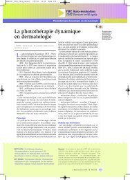
Mélanome unguéal - CEDEF, Collège des Enseignants en ...
Mélanome unguéal - CEDEF, Collège des Enseignants en ...
Mélanome unguéal - CEDEF, Collège des Enseignants en ...

You also want an ePaper? Increase the reach of your titles
YUMPU automatically turns print PDFs into web optimized ePapers that Google loves.
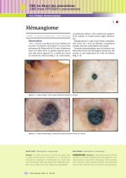
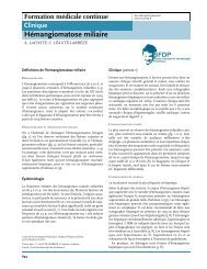
- une pigm<strong>en</strong>tation diffuse de couleur brun clair;<br />
- <strong>des</strong> ban<strong>des</strong> pigm<strong>en</strong>tées parallèles dont la couleur varie du<br />
brun clair au brun foncé et dont l’épaisseur est variable;<br />
- une pigm<strong>en</strong>tation du repli <strong>unguéal</strong> (signe de Hutchinson);<br />
- une pigm<strong>en</strong>tation de l’épaisseur de toute la tablette<br />
<strong>unguéal</strong>e qui est visible au bord libre de l’ongle.<br />
Une exérèse chirurgicale est nécessaire. L’histopathologie a<br />
révélé un mélanome d’origine matricielle <strong>unguéal</strong>e.<br />
NAIL MELANOMA. Summary: A 33-year-old woman<br />
att<strong>en</strong>ded for a melanonychia of the fourth finger of the left<br />
Comm<strong>en</strong>taires<br />
Devant une mélanonychie longitudinale, la prés<strong>en</strong>ce<br />
<strong>en</strong> dermoscopie d’une pigm<strong>en</strong>tation brune<br />
diffuse associée à <strong>des</strong> lignes pigm<strong>en</strong>tées brunes<br />
est <strong>en</strong> faveur d’une lésion mélanocytaire (1).<br />
L’irrégularité de couleur, d’épaisseur, d’espacem<strong>en</strong>t<br />
et de parallélisme de ces lignes pigm<strong>en</strong>tées<br />
sont <strong>des</strong> argum<strong>en</strong>ts <strong>en</strong> faveur d’un mélanome<br />
(1).<br />
La prés<strong>en</strong>ce d’un signe de Hutchinson est un<br />
argum<strong>en</strong>t supplém<strong>en</strong>taire, majeur, mais rare.<br />
Dans notre cas, co-existai<strong>en</strong>t <strong>des</strong> anomalies modérées<br />
<strong>des</strong> lignes pigm<strong>en</strong>tées (quelques lignes<br />
un peu plus foncées, plus épaisses et plus<br />
espacées) et un signe de Hutchinson, l’<strong>en</strong>semble<br />
reflétant probablem<strong>en</strong>t la nette prédominance<br />
matricielle du mélanome.<br />
La dermoscopie du bord libre, <strong>en</strong> montrant une<br />
pigm<strong>en</strong>tation de toute l’épaisseur de la tablette<br />
<strong>unguéal</strong>e, a pour intérêt supplém<strong>en</strong>taire d’indiquer<br />
que l’anomalie <strong>en</strong> cause concerne toute la<br />
matrice (2). Cette constatation exclut toute<br />
possibilité de biopsie partielle et implique la<br />
réalisation, comme dans le cas prés<strong>en</strong>t, d’une<br />
biopsie-exérèse longitudinale (3). ●<br />
RÉFÉRENCES<br />
1 - Ronger S, Touzet S, Ligeron C et al. Dermoscopic examination of<br />
nail pigm<strong>en</strong>tation. Arch Dermatol 2002; 138: 1327-33.<br />
FMC <strong>en</strong> direct <strong>des</strong> associations<br />
CME from FFFCEDV’s associations<br />
Cas clinique dermoscopique<br />
hand. This longitudinal pigm<strong>en</strong>ted band appeared about 5<br />
or 6 years ago. There was rec<strong>en</strong>tly an <strong>en</strong>largem<strong>en</strong>t.<br />
Dermoscopy demonstrated:<br />
- diffuse investigations of light brown color;<br />
- parallel pigm<strong>en</strong>ted bands whose colors varied from light<br />
brown to dark brown; thickness was variable;<br />
- pigm<strong>en</strong>tation of the dorsal nail fold (Hutchinson sign);<br />
- pigm<strong>en</strong>tation of all the nail plate which was visible at<br />
the free edge of the nail.<br />
Surgical removal was required: histopathology revealed a<br />
melanoma from the nail matrix.<br />
Figure 5 : Aspect dermoscopique du rebord <strong>unguéal</strong><br />
Dermoscopic aspect of ungueal edge<br />
(Collection/Courtesy of F. Mantoux)<br />
2 - Braun RP, Baran R, Saurat JH et al. Surgical Pearl: dermoscopy<br />
of the free edge of the nail to determine the level of nail plate pigm<strong>en</strong>tation<br />
and the location of its probable origin in the proximal or<br />
distal nail matrix. J Am Acad Dermatol 2006; 55: 512-3.<br />
3 - Braun RP, Baran R, Le Gal FA et al. Diagnosis and managem<strong>en</strong>t<br />
of nail pigm<strong>en</strong>tations. J Am Acad Dermatol 2007; 56: 835-47.<br />
© Nouv. Dermatol. 2008; 27 : 138-139 139