6 months ++ - Infectiologie
6 months ++ - Infectiologie
6 months ++ - Infectiologie

You also want an ePaper? Increase the reach of your titles
YUMPU automatically turns print PDFs into web optimized ePapers that Google loves.
DESC de Maladies infectieuses<br />
Maladies tropicales<br />
Module « médecine tropicale et des voyages »<br />
2010
Eric CAUMES. SMIT Pitié-Salpêtrière. Paris<br />
Homme de 23 ans<br />
• Voyage au Mali, bain (compliqué d’une dermatite)<br />
15 jours avant le retour<br />
• Fièvre apparue 4 jours après le retour<br />
• Urticaire et toux 10 jours après retour<br />
• Examen clinique : fièvre, urticaire, hémorragies sous<br />
unguéales en flammèches<br />
• Biologie : PE = 750/mm 3
…dermatite après un bain au Mali<br />
(Banini, pays Dogon)
Quel est votre diagnostic devant cette<br />
urticaire ?<br />
1 - Amœbose intestinale chronique<br />
2 - Paludisme viscéral évolutif<br />
3 - Helminthose invasive<br />
4 - Toxocarose<br />
5 - Larva migrans cutanée ankylostomienne<br />
6 - Babésiose
1 - Amœbose intestinale chronique<br />
2 - Paludisme viscéral évolutif<br />
3 - Helminthose invasive<br />
(urticaire aiguë)<br />
4 - Toxocarose<br />
(urticaire chronique)<br />
5 - Larva migrans cutanée<br />
ankylostomienne<br />
6 - Babésiose
Urticaire et parasitoses<br />
= helminthoses<br />
• Urticaire aiguë = phase invasive des<br />
helminthoses : ascaridose, ankylostomose,<br />
strongyloïdose (anguillulose), distomatoses,<br />
bilharzioses, gnathostomose, anisakidose,<br />
kyste hydatique (compliqué), trichinose<br />
• Urticaire chronique = toxocarose et autres causes<br />
du syndrome de larva migrans viscérale
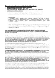
Le diagnostic de bilharziose invasive<br />
repose sur (choix unique) :<br />
1 - L’épidémiologie et l’histoire clinique<br />
2 - L’hyperéosinophilie<br />
3 - La sérologie positive<br />
4 - La séroconversion bilharzienne<br />
5 - La présence d’œufs dans les selles<br />
6 - La présence d’œufs dans les urines
1 - L’épidémiologie et l’histoire clinique<br />
2 - L’hyperéosinophilie<br />
3 - La sérologie positive<br />
4 - La séroconversion bilharzienne<br />
5 - La présence d’œufs dans les selles<br />
6 - La présence d’œufs dans les urines
Bilharziose aiguë chez 14 voyageurs au Mali<br />
(S.haematobium)<br />
Signes<br />
Fièvre<br />
Urticaire<br />
Toux<br />
Eosinophilie<br />
Seroconversion<br />
EPU +<br />
%<br />
93<br />
57<br />
86<br />
100<br />
100<br />
57<br />
Tps > exposition<br />
19 j (15-24)<br />
33 j (21-55)<br />
30 j (18-65)<br />
47 j (25-119)<br />
46 j (27-100)<br />
196 j (124-330)<br />
Durée (jrs)<br />
9 (2-30)<br />
4 (1-8)<br />
61 ( 7-210)<br />
113 (1-190)<br />
NA<br />
NA<br />
Grandière-Perez L et al. Am J Trop Med Hyg 2006; 74: 814-818
Le(s) traitement(s) possible(s) de la<br />
bilharziose invasive est (sont) :<br />
1 - Le praziquantel (Biltricide®)<br />
2 - La prednisone (Cortancyl®)<br />
3 - La prednisolone (Solupred®)<br />
4 - L’association praziquantel-corticoïdes<br />
5 - L’abstention thérapeutique<br />
6 - Le linézolide
1 - Le praziquantel (Biltricide®)<br />
2 - La prednisone (Cortancyl®)<br />
3 - La prednisolone (Solupred®)<br />
4 - L’association praziquantel-corticoïdes<br />
5 - L’abstention thérapeutique<br />
6 - Le linézolide
N =<br />
Efficacité du praziquantel chez 18<br />
voyageurs infectés par S.haematobium<br />
Tt/exposition<br />
Bilharz aiguë<br />
Bilharz chroniq<br />
Guéris**<br />
Tt As<br />
précoce<br />
8<br />
14 (10-15)<br />
8<br />
8<br />
8<br />
* : 2 aggravés; ** : après 1 - 2 PRZQ<br />
Tt As<br />
tardif<br />
4<br />
33 (28-40)<br />
0<br />
3<br />
8<br />
Forme<br />
invasive<br />
6*<br />
26 (20-39)<br />
NA<br />
Grandière-Perez L et al. Am J Trop Med Hyg 2006; 74: 814-818<br />
6<br />
6
Bilharziose invasive<br />
• Pathologie possible au retour de voyage chez les<br />
personnes exposées non immunes<br />
• Dg évoqué sur épidémiologie + clinique<br />
+ hyperéosinophilie (retardée)<br />
• Dg confirmé par la séroconversion bilharzienne<br />
• Tt = abstention thérapeutique (f. mineures), prednisone<br />
(f. graves)<br />
• Praziquantel = f.chroniques (adultes)
Acute neuroschistosomiasis<br />
Magnetic resonance imaging (T2 flair) showing borderzone infarcts suggestive of<br />
cerebral vasculitis in a 21 y old tourist with acute schistosmiasis (S.haematobium),<br />
1 month after bathing in a cascade in the Dogon area (Mali), 2 days after PRZ treatment
Bruno MARCHOU. SMIT. CHU Toulouse.<br />
Claire, 30 ans<br />
• « Safari » en Tanzanie du 02 au 22 Avril 2002<br />
• Vaccins à jour : dTP, FJ, VHA/typhoïde, méningo A+C<br />
• Chimioprophylaxie bien prise par Savarine®<br />
• Pendant le séjour : diarrhée hydrique, modérée, 7 jours<br />
• 15 Mai : fièvre aux environs de 38°C, spontanément résolutive début<br />
juin<br />
• Vers le 20 Juin, réapparition d’une fièvre intermittente<br />
• Hospitalisée le 4 Juillet :<br />
– T° = 39°, perte de poids de 4 kg<br />
– torticolis, douleur à la percussion des épineuses C6-C7<br />
– splénomégalie débordant de 1 à 2 TD
• Hémogramme normal<br />
• VS : 11 mm 1 ère h, CRP : 43 mg/l<br />
• ALAT : 3N, ASAT : 1.5 N ; LDH : 2N<br />
• Frottis sanguin- goutte épaisse : absence<br />
d’hématozoaire<br />
• Sérologies VHB, VHC : négatives<br />
• Radiographies standard du rachis : normales<br />
• Echographie abdominale : normale<br />
• Hémocultures : Streptococcus mitis (1/2)
Quels examens complémentaires<br />
demandez-vous ?<br />
1 - Recherche de Borrelia sur le frottis sanguin<br />
2 - PCR P. falciparum<br />
3 - Echocardiographie<br />
4 - Panoramique dentaire<br />
5 - Scintigraphie osseuse<br />
6 - IRM cervicale
1 - Recherche de Borrelia sur le<br />
frottis sanguin : négative<br />
2 - PCR P. falciparum : négative<br />
3 - Echocardiographie : normale<br />
4 - Panoramique dentaire : normal<br />
5 - Scintigraphie osseuse : +<br />
6 - IRM cervicale : +<br />
Scintigraphie :<br />
discrète hyperfixation en C6<br />
IRM cervicale : spondylite C6 avec discrète<br />
collection pré-rachidienne
Quel est votre diagnostic ?<br />
1 - Mal de Pott<br />
2 - Infection par Salmonella sérotype non-Typhi<br />
3 - Endocardite à S. mitis<br />
4 - Infection par Brucella melitensis<br />
5 - Kyste hydatique cervical
1 - Mal de Pott<br />
2 - Infection par Salmonella<br />
sérotype non-Typhi<br />
3 - Infection par Brucella melitensis<br />
4 - Endocardite à S. mitis ?<br />
5 - Kyste hydatique cervical<br />
Hémocultures + : Brucella melitensis
La brucellose animale a disparu en France depuis Juin 2003
Brucellose<br />
• Rare chez le voyageur : 3 cas publiés (PubMed)<br />
– B. melitensis<br />
– Fièvre isolée, Tchad (J Travel Med 2005, 12, 282-4)<br />
– Spondylite D6, Algérie (Med Trop 1998, 58, 205)<br />
– Sacro-iliite, Inde (Ned Tijdschr Geneedkd 2005, 149, 2810-4)<br />
• Zoonose relativement fréquente dans les PED :<br />
– B.melitensis <strong>++</strong>+<br />
– Prévalence brucellose bovine en zone péri-urbaine au<br />
Burkina Faso : 8% (Acta Trop 2000, 76, 53-7)<br />
– Séroprévalence (com.pers. : Dr Akakpo, Ecole Vétérinaire,<br />
Dakar):<br />
• Bovins : 5 - 15-20% ; petits ruminants : < 0.5%<br />
• Zones humides >> zones sèches<br />
• Élevage en stabulation >>> élevage pastoral
Brucella melitensis<br />
B.melitensis jamais décrite B.melitensis éradiquée<br />
B.melitensis présente B.melitensis rapportée dans le passé<br />
Données non disponibles<br />
(Source : B Garin-bastuji, AFFSA)
Fabrice LEGROS. Centre National de Référence<br />
de l’Epidémiologie du Paludisme d’Importation et<br />
Autochtone. Paris.<br />
French woman, 37 yrs (58 kg)<br />
returning from Mali<br />
• Remained in rural area of Mali (August 1 st - 23 th )<br />
• No chemoprophylaxis for malaria<br />
• 1 st consultation with a GP on August 26 th :<br />
headache, fever, diarrhoea, dry cough<br />
→ symptomatic treatment<br />
→ no improvement
2 nd consultation (hospital) August 31 th :<br />
- Thick & thin smears positive<br />
P. falciparum : 1.7 %<br />
- no criteria of severity<br />
- treatment : atovaquone + proguanil<br />
(Malarone ® ) 4 /day x 3 day<br />
Day 3 control :<br />
- P falciparum parasitaemia 0.01 %<br />
- QBC ® malaria test -<br />
- fever 37.9 °C<br />
- no other symptoms<br />
③ hospitalization & quinine infusion<br />
③ clinical and parasitological recovery
How do you explain this residual<br />
parasitaemia?<br />
(Choose up to 3 answers)<br />
1 - biological misdiagnosis?<br />
2 - resistance of the Plasmodium strain ?<br />
3 - poor absorbtion of atovaquone-proguanil?
How do you explain this residual<br />
parasitaemia?<br />
(Choose up to 3 answers)<br />
1 - biological misdiagnosis?<br />
2 - resistance of the Plasmodium strain ?<br />
3 - poor absorbtion of atovaquone-proguanil?
1- A biological misdiagnosis: possible, but…<br />
- QBC is a reliable method<br />
- - Gay F & al. Evaluation du système QBC ® pour le diagnostic du<br />
paludisme. Cah Santé 1994; 4(4): 289-297<br />
- - Khrisna BV & al. Comparison between conventional and QBC ®<br />
methods for diagnosis of malaria. Indian J Pathol Microbiol 2003; 46(3):<br />
517-520<br />
- This microscopist is experienced<br />
- A subsequent PCR confirmed the<br />
diagnosis<br />
- - Morassin B & al. One year’s experience with the PCR as routine<br />
method for the diagnosis of imported malaria.<br />
- AJTMH 2002; 66(5): 503-508<br />
- - Schindler HC & al. Development and optimization of PCR-based<br />
malaria diagnostic method and their comparison with QBC ® .<br />
- AJTMH 2001; 65(4): 355-361
2- resistance of the Plasmodium strain: possible, but…<br />
- only 7 documented cases<br />
- Kuhn s & al. Emergence of atovaquone-proguanil resistance during the<br />
treatment of P falciparum acquired by a non immune \American traveller<br />
to West Africa. AJTMH 2005;72(4): 407-409<br />
- David KP & al. Atovaquone-proguanil resistance in Africa: A case<br />
report. Scand J Infect Dis 2003; 35(11-12):897-898<br />
- P falciparum subsequently tested as sensitive<br />
- Le Bras J & al. CNRCP www.imea.fr/imea-recherche/imea-rbachimiosenspalu.php<br />
- WHO Monitoring antimalarial drug resistance.<br />
WHO/CDS/CSR/EPH/2002.17<br />
3- poor absorbtion of atovaquoneproguanil:<br />
possible, but…<br />
- subsequent blood PK data have confirmed the<br />
presence of drug above therapeutic concentrations
Is it justifiable to switch from<br />
atovaquone-proguanil to quinine?<br />
1 - Yes<br />
2 - No
Is it justifiable to switch from<br />
atovaquone-proguanil to quinine?<br />
1 - Yes<br />
2 - No
At day 3, from 38 to 50 % of patients show a<br />
persistent residual parasitaemia<br />
Musset L & al. (in press) study of 341 parasitologic surveys<br />
Parasitic clearance between day 4 and day 9<br />
In the absence of criteria of severity or<br />
vomiting, IV infusion is not appropriate
Parasite clearance time is low with atovaquoneproguanil<br />
Radloff PD & al. Atovaquone- proguanil for P falciparum malaria.<br />
Lancet 1996; 347(9014): 1511-1514 [ 72.1 ± 22.7 h ]<br />
Faucher JF & al. Atovaquone- proguanil vs amodiaquine for the treatment<br />
of P falciparum malaria in African infants and youg children.<br />
CID 2003 ; 37(11): 1441-1447 [ 86.2 ± 45.0 h ]<br />
Day 3 = at least 3 complete periods of 24 hours<br />
after the 1 st intake of drug<br />
Sabchareon A & al. Efficacy and pharmacokinetics of atovaquoneproguanil<br />
in children with multidrug resistant P falciparum malaria.<br />
Trans R Soc Trop Med Hyg 1998; 92(2): 201-256
Examination :<br />
A 28th year old caucasian man consults for<br />
dysuria and muscular weakness for 3 years<br />
Absent abdominal reflex and tendon reflexes in the left lower limb<br />
Babinski's reflex and spastic hypertonicity on the left<br />
Painful tactile hypoaesthesia and paraparesis of lower limbs<br />
Dysuria and bladder dysfunction<br />
Anamnesis :<br />
Ludovic de GENTILE. Laboratoire de Parasitologie-Mycologie<br />
CHU d’Angers<br />
The neurological disease has begun 3<br />
years ago with dysuria and constipation<br />
just after a stay in Mali for 2 years
Laboratory :<br />
Hb :18.0 g/dL Viral serology :<br />
Platelets : 383 giga/L HIV -<br />
White cells :7.64 giga/L HBsAg -<br />
NPN : 4.5 giga/L (57%) anti HBs Ab +<br />
EPN : 1.6 giga/L (16%) HCV Ab -<br />
BPN : 0 EBV +<br />
L : 1.45 giga/L (19%) Lyme disease serology -<br />
M : 610 giga/L Treponematosis serology -<br />
Red cells : 5.21 tera/L<br />
Serum creatinine : 93 µmol/L<br />
Magnetic Resonance Imaging:<br />
Lesion in front of T11 :<br />
- enhanced mid-sagittal spin echo T2<br />
- unenhanced mid-sagittal fast spin echo in T1<br />
Two smaller lesions of the cauda equina
What is your diagnosis ?<br />
1 - multiple sclerosis<br />
2 - Lyme disease<br />
3 - medullary schistosomiasis<br />
4 - tuberculous myelitis
What is your diagnosis ?<br />
1 - multiple sclerosis<br />
2 - Lyme disease<br />
3 - medullary schistosomiasis<br />
4 - tuberculous myelitis
Urine filtration : numerous Schistosoma haematobium ova<br />
Specific serology : FITC (S. mansoni Ag) : 1/320<br />
Schistosomal myeloradiculopathy<br />
due to S. haematobium
Treatment :<br />
Praziquantel : 40 mg/Kg 2 doses one month apart<br />
(and ivermectin 12 mg to prevent strongyloidiasis)<br />
Progress:<br />
• Urine samples become negative<br />
• Neurological degradation stopped<br />
• Recovery of muscular strength<br />
• Incomplete recovery of sensation in lower limb<br />
• Major urological after-effects
Schistosomal myeloradiculopathy<br />
• Due to S. mansoni and S. haematobium<br />
• Less frequent than cerebral schistosomiasis but<br />
underdiagnosed, asymptomatic deposition of ova<br />
is frequent at autopsy<br />
• Picture of a lower spinal cord or cauda equina<br />
compression<br />
• Great value of MRI and epidemiological findings<br />
for the diagnosis<br />
de Abreu Ferrari TC. Mem Inst Oswaldo Cruz 2004; 99 (Suppl1) : 59-62<br />
Leite CC et al. Singapore Med J 2000 ; 41 : 417-419
Frédéric MECHAI. SMIT Hôpital Necker. Paris<br />
Infirmière de 33 ans<br />
sans antécédents<br />
coordinatrice de missions MSF depuis 3 ans : 2001 Albanie, 2002 Angola,<br />
2005 Congo, début 2006 Bostwana<br />
• Avril-novembre 2006 : camp au Darfour<br />
• 10/11/06 : retour du Darfour<br />
• Depuis le 09/11/06 : T : 39°, frissons, céphalées frontales, myalgies<br />
poly-arthralgies des poignets, IPP et IPD<br />
• 10/11/06 : automédication par chloroquine<br />
• 11/11/06 : ttt par artémether-luméfantrine (Coartem® : 4 cp x 2)<br />
• Prophylaxie anti-palustre : non prise<br />
• Vaccinations : DTPolio, hépatite A, B, fièvre jaune, typhoïde, rage<br />
méningite A, C, Y, W135 à jour
Parmi les examens complémentaires<br />
suivants, lesquels prescrivez-vous en<br />
première intention ?<br />
(Choix multiple possible)<br />
1 - NFS-plaquettes<br />
2 - frottis-goutte épaisse<br />
3 - radiographie thorax<br />
4 - ponction lombaire<br />
5 - hémocultures<br />
6 - ECBU<br />
7 - Bilan hépatique
1 - NFS-plaquettes : GB à 5800, pas d’hyperéosinophilie,<br />
lympho : 1400, plaquetttes : 81000/mm 3<br />
2 - frottis-goutte épaisse : négatif<br />
3 - radiographie thorax : normale<br />
4 - ponction lombaire<br />
5- hémocultures : stériles<br />
6 - ECBU : stérile<br />
7 - bilan hépatique : ASAT : 253, ALAT : 214, gGT : 254<br />
PAL : 219, bili totale : 21 (échographie abdominale<br />
normale)<br />
Mais aussi : CRP : 135 mg/l, parasito/selles et coproculture négatives
Quel diagnostic évoquez-vous ?<br />
1 - Borréliose<br />
2 - Rickettsiose<br />
3 - Hépatite virale<br />
4 - Arbovirose
1 - Borréliose : frottis GE -<br />
2 - Rickettsiose : sérologies -<br />
3 - Hépatite virale :<br />
vaccinée VHA, VHA IgM : -<br />
HbsAC : + (vacccinée)<br />
sérologies VHC et VHE : -<br />
4 - Arbovirose :<br />
- O’nyong nyong : IgG -, IgM +<br />
(PCR générique alphavirus non faite)<br />
- Chikungunya : sérologie et PCR<br />
spécifique négative<br />
- Dengue : sérologie négative
O’nyong nyong<br />
• Arbovirus genre alphavirus<br />
famille des togaviridae<br />
• ARN, enveloppé<br />
• Génétiquement et<br />
cliniquement proche<br />
du virus Chikungunya<br />
• Réservoir vertébré encore indéterminé<br />
• Vecteurs : Anopheles funestus ± A.gambiae
Bassin méditerranéen<br />
Moyen Orient<br />
Afrique tropicale<br />
Extrême Orient,<br />
pacifique<br />
Europe non<br />
méditerranéene<br />
ex-URSS, Inde<br />
Amérique<br />
Répartition des arbovirus<br />
Alphavirus<br />
Sindbis<br />
Chikungunya,<br />
O’nyong nyong<br />
Chikungunya<br />
Ross River<br />
Méningo-encéphalite<br />
équine type Est,<br />
Ouest ou du<br />
Venezuela<br />
Mayaro<br />
Flavivirus<br />
Virus du Nil occidental<br />
Fievre jaune<br />
West Nile<br />
Wesselsbron<br />
Zika<br />
Encéphalite japonaise, dengue,<br />
encéphalite de la Murray valley<br />
Encéphalites à tiques<br />
Maladie de la forêt de Kyasanur<br />
Fièvre hémorragique d'Omsk<br />
West Nile<br />
Fièvre jaune<br />
Encéphalite de Saint Louis<br />
Dengue<br />
Rocio<br />
Autres<br />
Fièvre à phlébotomes<br />
Fievre hémorragique de<br />
Crimée-Congo<br />
Bunyamwera<br />
Bwamba<br />
Fièvre de la vallé du Rift<br />
Fièvre à phlébotome<br />
Fièvre à phlébotomes<br />
Fièvre hémorragique de<br />
Crimée-Congo<br />
Encéphalite de<br />
Californie<br />
Bunyavirus du groupe C<br />
Oropuche<br />
Groupe Guama<br />
Fièvre à tiques du<br />
Colorado
Epidémies d’O’nyong nyong<br />
• Virus isolé en juillet 1959 (Ouganda) chez 1 homme<br />
de 40 ans avec fièvre, polyarthralgies et céphalées<br />
• 1 ère épidémie (1959-1962) : ± 2 M de cas : Ouganda, Kenya,<br />
Tanzanie, Mozambique, Malawi. Quelques cas : RDC et Sénégal<br />
Johnson B.K et al. Trans R Soc Trop Med Hyg. 1981<br />
• 2 ème épidémie (1996-1997) : principalement en Ouganda<br />
Rwaguma E.B et al. Emerg Infect Dis 1997<br />
• Petite épidémie en 2003 : décrite dans un camp de<br />
réfugiés (Côte d’ivoire occidentale)<br />
Posey D.L et al. Am J Trop Med Hyg. 2005<br />
• Périodes inter-épidémiques : cas sporadiques isolés<br />
(études de séroprévalence)
Clinique :<br />
Fièvre bénigne<br />
ou formes<br />
sévères :<br />
- encéphalites<br />
- hémorragies<br />
- hépatite…<br />
Biologie :<br />
- Sérologie : réaction croisée possible avec Chikungunya<br />
- PCR (virémie) générique alphavirus puis séquençage si positif (1-2 cas/an au<br />
CNR)<br />
- Culture virale rarement faite en pratique<br />
Traitement : symptomatique<br />
Prévention limitée (anti-répulsifs), lutte anti-vectorielle<br />
Kiwanuka N et al. CID 1999
Dr Mike BEADSWORTH<br />
Liverpool School of Tropical Medicine<br />
64 year old Indian man presents in UK<br />
in 2003 as emergency with painful<br />
blistering lesion on legs for 3 days<br />
From Gujarat to UK 1965, last in India<br />
4 years ago<br />
Retired textile worker<br />
Married, 3 children<br />
Diabetes for 2 years<br />
Treatment: gliclazide<br />
Seeing dermatologist for<br />
6 <strong>months</strong>, treatment<br />
hydroxyzine
What is the most likely diagnosis ?<br />
(Choose one)<br />
1 - infected insect bite<br />
2 - allergic reaction to insect bite<br />
3 - Lyell’s disease (staphylococcal)<br />
4 - contact dermatitis<br />
5 - erysipelas (streptococcal)<br />
6 - something else
What is the most likely diagnosis ?<br />
(Choose one)<br />
1 - infected insect bite<br />
2 - allergic reaction to insect bite<br />
3 - Lyell’s disease (staphylococcal)<br />
4 - contact dermatitis<br />
5 - erysipelas (streptococcal)<br />
6 - something else
The cause
Rash for 6 <strong>months</strong>, worse for 1 month
Rash for 6 <strong>months</strong>, worse for 1 month
What is the most likely diagnosis?<br />
(Choose one)<br />
1 - generalised reaction to Ayurvedic medicine<br />
2 - sarcoid<br />
3 - lupus vulgaris<br />
4 - paucibacillary leprosy<br />
5 - multibacillary leprosy<br />
6 - smallpox<br />
7 - something else
What is the most likely diagnosis?<br />
(Choose one)<br />
1 - generalised reaction to Ayurvedic medicine<br />
2 - sarcoid<br />
3 - lupus vulgaris<br />
4 - paucibacillary leprosy<br />
5 - multibacillary leprosy<br />
6 - smallpox<br />
7 - something else
Details<br />
Exam: mild temp 37.5°<br />
no thickened nerves<br />
no sensory neuropathy<br />
no uveitis<br />
Skin smears: 6 sites & nasal scrape:<br />
Positive for AFB: bacterial index 2-3+<br />
morphological index 65%<br />
Skin biopsy: 1 month ago reported to show atypical active<br />
granulomata without caseation, repeat biopsy showed AFB.<br />
Review in London (Prof Sebastian Lucas) borderline lepromatous<br />
leprosy<br />
Inflammatory markers & neutrophils normal
Progress<br />
Diagnoses:<br />
Borderline lepromatous (multibacillary) leprosy<br />
Type 1 leprosy reaction<br />
Diabetes<br />
Treatment with WHO regimen for 2 years (G6PD normal):<br />
Dapsone 100mg daily<br />
Clofazimine 50 mg daily plus DOT 300mg monthly<br />
Rifampicin 600mg monthly<br />
Prednisolone 40mg daily for 2 <strong>months</strong> then slow<br />
reduction over 4 more <strong>months</strong><br />
Skin lesions flat after 1 week
Follow up<br />
Notified to public health authorities<br />
Wife, 3 children and 5 grandchildren examined (normal)<br />
Grandchildren given 6 <strong>months</strong> of monthly rifampicin as daily<br />
household contacts of a multibacillary case<br />
Mild recurrence of reactions at 1 year, treated with steroids for 2<br />
<strong>months</strong><br />
Diabetes not a problem<br />
Skin lesions flat but scaly and brown discolouration from<br />
clofazimine<br />
Skin smears for AFB at 12 <strong>months</strong>:<br />
Bacterial index 0-1+<br />
Morphological index 0%
Points to remember<br />
• Always ask patients about “traditional” and “over the counter”<br />
medicines<br />
• Any skin or neurological problem in immigrant always consider<br />
leprosy, even if diabetes present<br />
• Average delay to diagnosis in UK is 1.5 years after seeing an<br />
average of 2.5 specialists 1<br />
• 75% of British cases (~10/year) are from Indian subcontinent 1,2<br />
• Consult with experts as reactions may be prolonged and can occur<br />
years after eradication of mycobacteria<br />
• Duration of treatment for multibacillary disease ??<br />
WHO says 2 years, but up to 8% relapse if high BI (>3) 3<br />
1 Lockwood DN et al. QJ Med 2001; 94: 207-12. Delays in diagnosis<br />
2 Gill AL et al. QJ Med 2005; 98: 505-11. Liverpool cases<br />
3 Britton WJ, Lockwood DN. Lancet 2004; 363: 1209-19 Review
Bruno Marchou SMIT CHU Toulouse<br />
M elle A, 27 ans<br />
• Originaire de Sierra Leone mais notion d’un court séjour à<br />
Lagos, Nigeria.<br />
• En France depuis juillet 2000.<br />
• Pas de prurit ni d’œdème<br />
• Découverte d’une hyperéosinophilie en octobre 2000 :<br />
1300 /mm 3<br />
• Frottis sanguin : dia. suivante….
Microfilaire type Loa Loa
Quel traitement auriez-vous proposé ?<br />
1 - Diéthylcarbamazine (DEC)<br />
2 - Ivermectine<br />
3 - Ivermectine puis DEC<br />
4 - Albendazole<br />
5 - Pas de traitement
Quel traitement auriez-vous proposé ?<br />
1 - Diéthylcarbamazine (DEC)<br />
2 - Ivermectine<br />
3 - Ivermectine puis DEC<br />
4 - Albendazole<br />
5 - Pas de traitement
Evolution sous traitement<br />
Dates microfilarémie (par ml) traitement<br />
10/10 / 2000 1200 – 4300 ivermectine 1<br />
(12h) - (24h)<br />
23 / 11 / 2000 1200 ivermectine 2<br />
19 / 12 / 2000 1000 ivermectine 3<br />
29 / 01 / 2001 1300 ivermectine 4<br />
08 / 03 / 2001 0<br />
DEC 6 mg/kg/j<br />
4 semaines
Traitement de la loase<br />
• Traitement de référence : DEC<br />
Risque d’encéphalite si mf >3-5 log/ml<br />
Dose progressive<br />
• Ivermectine : prétraitement si mf<strong>++</strong><br />
• Cytaphérèse si mf <strong>++</strong><strong>++</strong><br />
• Albendazole : une molécule d ’avenir ?
Hélène GROS et Olivier BOUCHAUD.<br />
SMIT. Hôpital Avicenne. Bobigny.<br />
Patiente de 21 ans<br />
Originaire de Mauritanie, née en France<br />
Sans antécédent particulier<br />
Hospitalisée pour fièvre, céphalées et diarrhée<br />
Voyage récent de 6 semaines en Afrique<br />
(Mauritanie et Sénégal)<br />
Chimioprophylaxie : chloroquine + proguanil<br />
(mauvaise observance)<br />
Pas d’automédication avant la consultation
• Examen clinique :<br />
Fièvre à 41°C<br />
Examen physique<br />
sans particularité<br />
• Examens<br />
complémentaires :<br />
NFS :<br />
Hb : 9.8 g/dl<br />
Plaquettes : 64.000 / mm 3<br />
Leucocytes : 7700 / mm 3<br />
CRP : 101 mg/l
Parmi les diagnostics suivants,<br />
quel diagnostic doit on évoquer<br />
en première intention ?<br />
1. Gastroentérite aiguë<br />
2. Dengue<br />
3. Accès palustre<br />
4. Grippe<br />
5. Fièvre typhoïde
Parmi les diagnostics suivants,<br />
quel diagnostic doit on évoquer<br />
en première intention ?<br />
1. Gastroentérite aiguë<br />
2. Dengue<br />
3. Accès palustre<br />
4. Grippe<br />
5. Fièvre typhoïde
Le frottis sanguin et la goutte épaisse sont négatifs<br />
(pour P.falciparum et les autres Plasmodium)<br />
Existe-t’il des arguments pour un accès<br />
palustre avec examen microscopique<br />
faussement négatif ?<br />
1. Oui<br />
2. Non
Le frottis sanguin et la goutte épaisse sont négatifs<br />
(pour P.falciparum et les autres Plasmodium)<br />
Existe-t’il des arguments pour un accès<br />
palustre avec examen microscopique<br />
faussement négatif ?<br />
1. Oui<br />
2. Non
→ Oui<br />
- Tableau clinique et biologique compatible<br />
avec un accès palustre<br />
- Décapitation possible par la chimioprophylaxie
…voici le résultat du frottis<br />
Ce frottis peut-il vous donner le<br />
bon diagnostic ?<br />
1. Oui<br />
2. Non
…voici le résultat du frottis<br />
Ce frottis peut-il vous donner le<br />
bon diagnostic ?<br />
1. Oui<br />
2. Non
Quel est votre diagnostic final ?<br />
1. Accès palustre à Plasmodium falciparum<br />
2. Trypanosomose africaine<br />
3. Syphilis<br />
4. Borréliose<br />
5. Filariose à Wuchereria bancrofti
Quel est votre diagnostic final ?<br />
1. Accès palustre à Plasmodium falciparum<br />
2. Trypanosomose africaine<br />
3. Syphilis<br />
4. Borréliose<br />
5. Filariose à Wuchereria bancrofti
Trypanosoma brucei rhodesiense<br />
Borrelia<br />
Microfilaire Loa loa +<br />
Mansonella perstans
Borrelia crocidurae<br />
• Epidémiologie :<br />
Transmission par piqûre de tique (Alectrobius erraticus)<br />
Sahel : Mauritanie, Sénégal, Tchad<br />
Augmentation du nombre de pays touchés<br />
Pas d’immunisation prolongée<br />
• Présentation clinique :<br />
Incubation : 4 à 7 jours<br />
Fièvre et syndrome pseudogrippal<br />
Evolution de la fièvre : 2 ou 3 récurrences<br />
Signes inconstants : ictère, rash, hépato-splénomégalie,<br />
méningite<br />
Trape et al, Am J Trop Med Hyg, 1996
• Diagnostic<br />
Frottis sanguin après coloration au Giemsa<br />
(lecture au microscope à fond noir)<br />
Culture difficile (milieu spécial)<br />
• Traitement<br />
Cyclines (ou ß-Lactamines ou macrolides)<br />
Durée : 5 à 10 jours<br />
Guérison spontanée
Eric PICHARD. SMIT CHU d’Angers.<br />
M. O. M. , 39 ans, hospitalisé au<br />
retour d’un voyage en Thaïlande<br />
pour fièvre à 40°C<br />
• Séjour rural d’1 mois au sud du pays<br />
• Bains : mer et lac<br />
• Contacts avec des rats, piqûres de « moustiques »<br />
• Fièvre 6 jours après le retour<br />
• Gêne pharyngée<br />
• ATCD : MNI<br />
• Vaccins : DTP, HVA, typhoïde<br />
• Atovaquone + proguanil : séjour + 3j
Examen normal sauf petites papules<br />
prurigineuses du dos et conjonctivite<br />
NFS : syndrome mononucléosique
Quelle diagnostic(s) suspectez-vous ?<br />
1- Paludisme<br />
2- Paratyphoïde ?<br />
3- MNI ?<br />
4- Arbovirose ?<br />
5- PI VIH<br />
6- Autre maladie ?
1- Paludisme ? → frottis/GE -<br />
2- Paratyphoïde ? → hémo/coprocultures -<br />
3- MNI ? → ATCD<br />
4- Arbovirose ? → séro. DEN- Chik- EJ-<br />
5- PI VIH → Ag P24-<br />
6- Autre maladie ? → BU- RP nle Ag CMV-<br />
HVE- Lyme- Toxo-<br />
mais….
… le contexte de contact<br />
avec des rats évoque :<br />
1- une haverilose<br />
2- une leptospirose<br />
3- une peste<br />
4- un sodoku<br />
5- une rickettsiose
1 : haverrilose → pas de morsure<br />
2 : leptospirose → monocytose, MAT -<br />
3 : peste → adénopathies- et RP nle<br />
4 : Sodoku → pas de morsure<br />
5 : rickettsiose → sérologie +
mais quelle rickettsie ?<br />
1- Rickettsia prowasekii<br />
2- Rickettsia typhi<br />
3- Rickettsia rickettsii<br />
4- Rickettsia africæ<br />
4- Rickettsia conorii
Rickettsia typhi : spot I/Mérieux 512 (≥ 40)<br />
→ typhus murin (ex R.mooseri)<br />
Rickettsia prowasekii : typhus épidémique<br />
(poux/hautes terres des pays pauvres)<br />
Rickettsia rickettsii : fièvre pourprée des montagnes<br />
Rocheuses (tique/Amériques)<br />
Rickettsia africæ : fièvre à tique africaine (tique/Afrique)<br />
Rickettsia conorii : f. boutonneuse méditerranéenne<br />
(tique/bassin méditerranéen, Afrique, Inde, Géorgie)
• Rickettsiose éruptive, groupe typhus,<br />
• Réservoir : rats<br />
• Transmission par du rat<br />
(Xenopsylla cheopis) ± puce chat/souris<br />
• Déjections de puce → grattage, muqueuse<br />
± inhalation<br />
• Cosmopolite comme le<br />
Incubation : s. grippal ± tbles digestifs,<br />
éruption maculo-papuleuse,<br />
leucopénie, thrombopénie, cytolyse, ins.rénale,<br />
Dg sérologique, durée 10-15j, létalité : 2%<br />
Typhus<br />
murin<br />
TTT : doxycyline 200 mg/j → 2j d’apyrexie ( ± 7j), FQ, josamycine<br />
Destruction des puces puis dératisation
Jean-Marie CHENNEBAULT et Eric PICHARD<br />
SMIT. CHU d’Angers.<br />
Franck, 34 ans<br />
• Consulte pour une lésion érosive<br />
du sillon balano-préputial<br />
• Adénopathies inguinales bilatérales<br />
sensibles, fièvre 38°C, asthénie et<br />
céphalées depuis 1 semaine<br />
• Vaccinations à jour : dTP, FJ, HVA<br />
• Aucun antécédent marquant<br />
• Revenu depuis 20 jours d’un voyage de 7 jours en Afrique<br />
du Sud dont 5 jours dans une réserve … avec son épouse
Quel est votre diagnostic ?<br />
1 - Chancre mou<br />
2 - Leishmaniose cutanée<br />
3 - Syphilis<br />
4 - Ulcère de Buruli<br />
5 - Piqûre/morsure d’insecte
1 - Chancre mou<br />
2 - Leishmaniose cutanée<br />
3 - Syphilis<br />
4 - Ulcère de Buruli<br />
5 - Piqûre/morsure d’insecte
Sous la chemise ….<br />
éruption de macules<br />
disséminées apparue<br />
la veille de la<br />
consultation
Et la biologie est de retour :<br />
• GR : 5,02 téra/L, Hb : 15,8g/dL<br />
• GB : 5,40 giga/L PN : 3,87 giga/L, lympho : 1,09 giga/L<br />
• Plaquettes : 180 giga/L<br />
• CRP : 38 mg/L<br />
• ASAT : 65U/L (N< 45), ALAT : 105U/L (N< 65),<br />
• Gamma GT : 96U/L (N< 70)<br />
• LDH : 531U/L (N< 390)<br />
• bilirubine : 13 mmol/L (N< 17)
Quel est votre diagnostic ?<br />
1 - Trypanosomose africaine<br />
2 - Rickettsiose<br />
3 - Babésiose<br />
4 - Fièvre de la vallée du Rift
1 - Fièvre Q : sérologie -<br />
2 - Rickettsiose : +<br />
3 - Babésiose : frottis-GE -<br />
4 - Fièvre de la vallée du Rift :<br />
sérologies arboviroses -<br />
Séroconversion Rickettsia africae et R. conorii<br />
Guérison sous doxycycline en quelques jours<br />
Sérologie syphilis négative
• Fièvre africaine à<br />
tiques<br />
• Rickettsiose transmise par les<br />
Amblyomma spp.<br />
• Diagnostic est clinique :<br />
– Eruption fébrile<br />
– Escarres souvent multiples<br />
• Diagnostic biologique<br />
– Atteinte hépatique<br />
– Thrombopénie modérée<br />
– Sérologie croisée avec<br />
autres rickettsioses<br />
TTT : doxycycline<br />
Répartition des infections à R. africae<br />
séroprévalence cas prouvés<br />
Fournier CID 1998;27:316-23
Chancre, escarre, ulcère cutané tropical…<br />
Génitaux, bouche, anus = IST avant tout…<br />
Chancre mou<br />
Sypilis primaire Donovanose<br />
…mais gare aux arthropodes, ça gratte<br />
Gale
… et ça<br />
pique<br />
Leishmaniose à L.tropica<br />
Bouton d’Orient<br />
Morsure<br />
d’araignée<br />
Piqûre de tique<br />
Rickettsiose<br />
Piqûre de tique avec ECM<br />
Borréliose de Lyme<br />
Leishmaniose à L.brazilienis<br />
Chicleros<br />
Trypanôme
Mal perforant<br />
Lèpre, tabès…<br />
Charbon<br />
Ulcère de<br />
Buruli<br />
Piqûre de<br />
poisson-pierre<br />
Ulcère tropical<br />
± phagédénique<br />
Hémoglobinoses, plaie…
Pr. Eric CAUMES. GH Pitié-Salpêtrière.<br />
Paris. France.<br />
A 44 year old french woman<br />
returning from Cambodia<br />
• Lag time return/signs : 15 days<br />
• Creeping eruption and papules<br />
1 st episode<br />
lasting for 20 days<br />
failure of ivermectin<br />
plus asthenia and fever<br />
• Hypereosinophilia :<br />
1647/mm 3
What is your diagnosis ?<br />
1. creeping eruption<br />
2. cutaneous larva migrans<br />
3. cutaneous gnathostomiasis<br />
4. (animal) hookworm related cutaneous larva migrans<br />
5. loiasis
What is your diagnosis ?<br />
1. creeping eruption<br />
2. cutaneous larva migrans<br />
3. cutaneous gnathostomiasis<br />
4. (animal) hookworm related cutaneous larva migrans<br />
5. loiasis
Cutaneous gnathostomiasis to be<br />
suspected in a traveller or a migrant…..<br />
- coming from an<br />
endemic country<br />
- consumption of<br />
raw dishes<br />
- complaining of<br />
recurrent symptoms<br />
- with cutaneous<br />
(± viscéral) signs<br />
- and (70%) hypereosinophilia<br />
High endemicity<br />
Imported cases<br />
Low<br />
endemicity<br />
creeping eruption = a sign<br />
cutaneous larva migrans = a syndrom<br />
cutaneous gnathostomiasis = a disease
Cutaneous gnathostomiasis :<br />
positive diagnosis<br />
• Parasitological examination :<br />
•<br />
• - Skin biopsy : 34 % (Diaz - Camacho, Am J Trop Med Hyg<br />
1998) 16 % (5/30) (Kurokawa, Arch Dermatol<br />
1998)<br />
- Cutaneous extraction after treatment :<br />
5 % (ivermectin), 6 % (albendazole)<br />
(Nontasut, South East Asian J Trop Med Hyg 2000)<br />
• Serologic test (Dr P.Dekumoy, Bangkok) :<br />
• - ELISA [cut off > 1/400]<br />
• - Western Blot<br />
The serologic test may be initialy<br />
negative and thus needs to be<br />
repeated to show seroconversion.
What is your treatment for<br />
cutaneous gnathostomiasis ?<br />
1. single dose ivermectin<br />
2. single course albendazole<br />
3. XXX courses of albendazole<br />
4. XXX courses of ivermectin<br />
5. I do not know
What is your treatment for<br />
cutaneous gnathostomiasis ?<br />
1. single dose ivermectin<br />
2. single course albendazole<br />
3. XXX courses of albendazole<br />
4. XXX courses of ivermectin<br />
5. I do not know
Gnathostomiasis : treatment<br />
Thaïland : - albendazole : 400 mg/d x 21d = 400 mg x 2/d x 21d<br />
(Cure rate M6 = 94 %)<br />
(Kraivichian, Trans Roy Soc Trop Med Hyg 1992)<br />
- ivermectin : 12 mg/d = albendazole : 400 mg/d x 21d<br />
(Cure rate M6 = 95 % vs 94 %)<br />
(Nontasut, SEAJTM 2000)<br />
Europe : - albendazole : (800 mg/d, 21d), 1 to 2 courses<br />
(Moore, Emerg Infect Dis 2003)<br />
- Failure of ivermectine; albendazole 1 to 4 courses<br />
recurrence may occur up to 20 <strong>months</strong> after apparent<br />
cure without reinfection (cl. case : 2 recurrences M2<br />
and M20) (Menard, Trans Roy Soc Trop Med Hyg 2003)<br />
Treatment may require multiple courses of albendazole and a<br />
prolonged period of follow up (up to 20 <strong>months</strong>) is necessary
Imported gnathostomiasis : 1980 1999<br />
• N = 10 cases<br />
• Gn. cutaneous (7) ; pulmonary (2) ; oph. (1)<br />
• Thaïlande (6) ; SEA (2) : sub indian cont (2)<br />
• PE : 4 % → 64 % ; 540/mm 3 → 15 500/mm 3<br />
• Treatment = surgery ( → 1986)<br />
= DEC, PRAZ, THIA ( → 1994)<br />
= ALB (1994 → 1999)
Imported gnathostomiasis : 2000 2003<br />
• N = 33 cases [- 16 (Moore et al)] = 17<br />
• Gn cutaneous (13) ; neurological (2) ; oph (1), pleural (1)<br />
• Asia (<strong>++</strong>+), Latin America (4) ; Zambia (2)<br />
• PE : N → 55 %<br />
0 → 11 083/mm 3<br />
• Tt = albendazole<br />
= ivermectine
Cutaneous larva migrans (CLM) is a<br />
syndrome caused by the following disease<br />
1. loiasis<br />
2. strongyloidiasis<br />
3. dracunculiasis<br />
4. (animal) hookworm related cutaneous larva migrans<br />
5. migratory myiasis
Not related to<br />
parasitic<br />
disease : lichen<br />
striatus,<br />
thrombophlebitis,,<br />
Creeping<br />
eruption<br />
and CLM<br />
Related to the<br />
migration of parasite’s<br />
Larvae = CLM<br />
stricto sensu:<br />
hookworm, larva<br />
currens, gnathostomiasis<br />
Related to the<br />
migration of<br />
parasite : scabies,<br />
dracunculiasis,<br />
loiasis, myiasis<br />
Caumes E, Danis M, Lancet Infect Dis 2004; 4: 659-660
F. Botterel, S. Brun, P. Bourée<br />
La Presse Médicale, 2000, 29 :81<br />
Femme de 60 ans :<br />
lésions des cuisses<br />
après un séjour aux<br />
Saintes (Guadeloupe)
Anamnèse<br />
Apparition d’un érythème en bandes suivi d’une<br />
sensation de brûlure, non prurigineuse, non fébrile,<br />
puis d’une éruption vésiculo-bulleuse avec œdème<br />
Notion d’une chute quelques heures avant<br />
l’apparition des lésions, un jour de pluie, alors que<br />
la patiente se promenait jambes nues sous des<br />
arbres<br />
Diagnostic ?
Mancenilliose!
Mancenillier<br />
Hippomane<br />
mancinella<br />
« manzanilla » :<br />
petite pomme
Hippomane mancinella<br />
- Famille des Euphorbiacées (hévéa, manioc, …)<br />
- Antilles, sud des USA, Amérique Centrale et du Sud<br />
Afrique de l’Ouest<br />
- Sols secs et sablonneux (bord de mer <strong>++</strong>+)<br />
- Un des végétaux les plus toxiques du monde tropical<br />
- Toutes les parties de l’arbre sont toxiques (latex)
Hippomane mancinella
- C. Colomb :<br />
Hippomane mancinella<br />
« Leurs figures s’enflaient et il leur venait une si<br />
grande inflammation accompagnée de douleurs si<br />
aiguës qu’ils paraissaient être en rage… »<br />
- Flèches empoisonnées des Indiens
Hippomane mancinella<br />
Clinique<br />
- Peau : - contact direct, indirect <strong>++</strong>+<br />
- brûlures persistantes du 1er et du 2è degré<br />
- Muqueuses : - ingestion des fruits ou du latex<br />
- brûlures oropharyngées (choc)<br />
- Œil : - contact indirect <strong>++</strong>+<br />
- kératoconjonctivite (guérison sans séquelle)
Hippomane mancinella<br />
Traitement<br />
- Peau : retirer les traces cutanées de latex à l’eau<br />
savonneuse ou à l’eau de mer<br />
- Œil : laver à grande eau + antiseptiques locaux
Hippomane mancinella<br />
- Campagnes de coupe<br />
Prévention<br />
- Troncs ceinturés de peinture rouge<br />
- Panneaux signalant sa présence et son danger<br />
- Éviter de toucher les feuilles et les fruits<br />
- Se laver les mains en cas de contact avec le latex<br />
- Ne pas stationner en dessous en cas de pluie
Eric CAUMES. SMIT Hôpital Pitié-Salpêtrière. Paris.<br />
Jeune homme de 19 ans<br />
Séjour au Mali (Ouest) pendant un an<br />
– Ulcération cutanée apparue au 3 eme mois<br />
– Dg supposé : leishmaniose cutanée localisée (LCL)<br />
– Tt : DPA IM (dose ? ; durée ?)<br />
– Evolution non favorable<br />
Retour en France 9 mois plus tard :<br />
– Ulcération persistante + lymphangite nodulaire<br />
– Examen parasitologique direct : leishmaniose +
Quelle espèce de Leishmania est<br />
responsable ?<br />
1- Leishmania infantum<br />
2- Leishmania donovani<br />
3- Leishmania malinensis<br />
4- Leishmania major<br />
5- Leishmania tropica<br />
6- Leishmania tapioca
Quelle espèce de Leishmania est<br />
responsable ?<br />
1- Leishmania infantum<br />
2- Leishmania donovani<br />
3- Leishmania malinensis<br />
4- Leishmania major<br />
5- Leishmania tropica<br />
6- Leishmania tapioca
Classification des leishmanies<br />
(ancien monde AM)<br />
Sous genre Leishmania<br />
. Complexe phylogénique L. donovani<br />
. CP L. infantum L. infantum (Tunisie)<br />
. CP L. major L. major (Algérie)<br />
L. donovani (Inde)<br />
. CP L. tropica L. tropica (Azerbaidjan)<br />
. CP L. killicki L. killicki (Tunisie)<br />
. CP L. aethiopica L. aethiopica (Ethiopie)<br />
L. archibaldi (Soudan)<br />
. CP L. arabica L. arabica (Arabie Saoudite)
Parmi les médicaments suivants, quels<br />
sont ceux classiquement recommandés<br />
dans le traitement de la LCL de l’AM ?<br />
1- Ketoconazole PO<br />
2- Antimoniate de méglumine (Glucantime ® ) IM<br />
3- Itraconazole PO<br />
4- Isethionate de pentamidine IM (Pentacarinat ® )<br />
5- Fluconazole PO<br />
6- Antimoniate de méglumine (Glucantime ® ) IL
Question 2 : parmi les médicaments<br />
suivants, quels sont ceux classiquement<br />
recommandés dans le traitement de la LCL<br />
de l’AM ?<br />
1- Ketoconazole PO<br />
2- Antimoniate de méglumine (Glucantime ® ) IM<br />
3- Itraconazole PO<br />
4- Isethionate de pentamidine IM (Pentacarinat ® )<br />
5- Fluconazole PO<br />
6- Antimoniate de méglumine (Glucantime ® ) IL
Traitement LCL<br />
• DPA (antimoines) IM (NM) ou IL (AM)<br />
• Pentamidine IM (NM ; L.guyanensis)<br />
• Fluconazole oral (AM ; L.major)<br />
• Ketoconazole oral (NM ; L.mexicana)<br />
• Miltéfosine oral (NM ; sauf L.braziliensis complex)<br />
• Paromomycine topique (NM)<br />
• Thermothérapie (AM ; L.tropica)<br />
(Wortmann G. Clin Infect Dis 2005; 40:1156-1158)
Le patient est traité par le fluconazole,<br />
200 mg par jour, pendant 6 semaines.<br />
Trois mois plus tard, les signes cliniques<br />
persistent et l’examen parasitologique<br />
direct est toujours positif.<br />
Vous concluez à un échec.
Tt LCL en Arabie Saoudite par le<br />
fluconazole : 200 mg/j x 6 semaines<br />
Fluco Placebo<br />
n (L. major) 106 103<br />
suivi + 80 65<br />
guérison (M3)* 63 (79%) 22 (34%)<br />
tps de guérison** 8.5 sem 11.2 sem<br />
* OR = 2.33 (1.63-3.33) ** p < 0.001<br />
(Alrajhi et al ; NEJM 2002;346:891-895)
Tt LCL (importée) en France par le<br />
fluconazole : 200 mg/j x 6 sem<br />
(guérison évaluée à J 90)<br />
Espèce<br />
Nombre de<br />
patients<br />
Patients<br />
évaluables<br />
Patients guéris<br />
L.major<br />
27<br />
26<br />
12 (44%)<br />
L.infantum<br />
4<br />
4<br />
0 (0%)<br />
( Morizot G et al. AJTMH sous presse)
Quels traitements proposeriez-vous<br />
1- Ketoconazole PO<br />
maintenant ?<br />
2- Antimoniate de méglumine (Glucantime ® ) IM<br />
3- Itraconazole PO<br />
4- Isethionate de pentamidine IM (Pentacarinat ® )<br />
5- Fluconazole PO<br />
6- Antimoniate de méglumine (Glucantime ® ) IL
Quels traitements proposeriez-vous<br />
1- Ketoconazole PO<br />
maintenant ?<br />
2- Antimoniate de méglumine (Glucantime ® ) IM<br />
3- Itraconazole PO<br />
4- Isethionate de pentamidine IM (Pentacarinat ® )<br />
5- Fluconazole PO<br />
6- Antimoniate de méglumine (Glucantime ® ) IL
Réf<br />
Pays<br />
Nb pts<br />
Espèce<br />
Dose<br />
Nb IM<br />
Guéris<br />
LCL et IP dans les deux mondes<br />
Soto J<br />
AJTMH 1994<br />
Colombie<br />
51<br />
L. panam…<br />
3 mg/kg IP<br />
4 IM<br />
96 %<br />
Lightburn E<br />
Med Trop 2003<br />
Guyane frcse<br />
205 Guy/ 32 Fr<br />
L.guyanensis<br />
4 mg/kg base<br />
2 IM<br />
95 % G/ 75% F<br />
Hellier I<br />
Dermatology 2000<br />
AM<br />
11<br />
Divers AM<br />
4 mg/kg base<br />
3 IM<br />
73 %
Sels de pentamidine<br />
[Methane sulfonate de pentamidine (Lomidine ® )]<br />
Isethionate de pentamidine (Pentacarinat ® )<br />
Tolérance :<br />
Allergie Imm IM ou IV lente<br />
IM : hypoTA, choc<br />
EI tardifs : rein, foie, cœur<br />
Problèmes potentiels : diabète et hypoglycémie,<br />
rhabdomyolyse (élévation des CPK)<br />
Posologie : 4 mg/kg/j d’IP, 1 jr/2<br />
nombre d’injections ??
Leishmaniose cutanée localisée (de l’ancien<br />
Monde) : l’espèce conditionne le traitement
Olivier BOUCHAUD SMIT Avicenne (Bobigny)<br />
Qui s’y frotte s’y pique<br />
• Au retour de 3 semaines de<br />
vacances, Mauricette, 26<br />
ans, vous montre cette<br />
lésion du gros orteil du<br />
pied.<br />
• Le début remonte au<br />
lendemain de son arrivée où<br />
en se baignant et en posant<br />
le pied au fond, elle a<br />
ressenti une douleur sous<br />
le pied.<br />
• Rougeur puis «zone noire»<br />
dés le début avec<br />
aggravation progressive.
Quel est votre diagnostic ?<br />
1 - Plaie par un objet tranchant + surinfection<br />
2 - Lésion par contact avec du corail<br />
3 - Piqûre d’oursin surinfectée<br />
4 - Nécrose secondaire à une piqûre de poisson
Quel est votre diagnostic ?<br />
1 - Plaie par un objet tranchant + surinfection<br />
2 - Lésion par contact avec du corail<br />
3 - Piqûre d’oursin surinfectée<br />
4 - Nécrose secondaire à une piqûre de poisson
Voici le coupable et son arme…
Il s’agit d’un...<br />
1 - poisson pierre<br />
2 - poisson scorpion<br />
3 - poisson lion<br />
4 - star fish
Il s’agit d’un...<br />
1 - poisson pierre<br />
2 - poisson scorpion<br />
3 - poisson lion<br />
4 - star fish
Le fond sur lequel Mauricette a dû<br />
poser le pied…<br />
…poisson pierre (stone fish)
Un sérum anti-venin est recommandé<br />
1 - Dans tous les cas<br />
2 - Dans les formes sévères avec symptômes systémiques<br />
3 - Dans les formes avec nécrose<br />
4 - Jamais car n’existe pas
Un sérum anti-venin est recommandé<br />
1 - Dans tous les cas<br />
2 - Dans les formes sévères avec symptômes systémiques<br />
3 - Dans les formes avec nécrose<br />
4 - Jamais car n’existe pas
Dans quelles zones marines Mauricette<br />
était-elle probablement en vacances ?<br />
(Plusieurs réponses possible)<br />
1 - Zone Caraïbes<br />
2 - Zone indo-océanienne<br />
3 - Zone pacifique américaine<br />
4 - Zone méditerranéenne<br />
5 - Zone ouest-africaine
Dans quelles zones marines Mauricette<br />
était-elle probablement en vacances ?<br />
1 - Zone Caraïbes<br />
2 - Zone indo-océanienne<br />
3 - Zone pacifique américaine<br />
4 - Zone méditerranéenne<br />
5 - Zone ouest-africaine
C’était à l’Ile Maurice<br />
(zone indo-océanienne)<br />
Mauricette a une arthrite du pouce…
Dr David LALLOO<br />
Liverpool School of Tropical Medicine<br />
45 year old female South African nurse<br />
in May 2005 with 4 seizures in 6 hours<br />
Brought up in Rural Transkei<br />
Arrived UK 1987, last visit home Jan 2005<br />
Generally well with 4 children<br />
4 weeks: minor cervical adenopathy<br />
3 days: mild swelling of wrist<br />
Presented as emergency with generalised convulsions
Initial progress<br />
Exam: reduced conscious level, Glasgow Coma Score 7/15<br />
No neck stiffness or focal neurological signs<br />
Two small cervical nodes<br />
FBC: Hb 12.5 g/dl<br />
WBC 13 x 10 6 /l<br />
Neuts 8 x 10 6 /l<br />
Urea & elecrolytes: normal<br />
Liver function ALT 52 u/l (norm
Next day<br />
Extubated and alert<br />
MR scan reviewed :<br />
Right frontal lesion
What is the most likely diagnosis?<br />
1 - tuberculoma<br />
2 - neurocysticercosis<br />
3 - histoplasmosis<br />
4 - toxoplasmosis<br />
5 - coccidioidomycosis<br />
6 - tumour metastasis
What is the most likely diagnosis?<br />
1 - tuberculoma<br />
2 - neurocysticercosis<br />
3 - histoplasmosis<br />
4 - toxoplasmosis<br />
5 - coccidioidomycosis<br />
6 - tumour metastasis
Progress<br />
Radiologist thought<br />
TB or cysticercosis*<br />
FBC : no eosinophilia<br />
HIV and syphilis serology<br />
negative<br />
Patient alert, on phenytoin<br />
Consult Tropical & ID unit to ask for advice<br />
*Pretell EJ et al. J Comput Assis Tomogr 2005; 29: 112-4
What would you have advised?<br />
(Choose one)<br />
1. nothing more<br />
2. add TB therapy<br />
3. add dexamethasone<br />
4. add steroids & TB therapy<br />
5. add anthelmintic therapy<br />
6. add dexamethasone & anthelmintics<br />
7. something else
What would you have advised?<br />
(Choose one)<br />
1. nothing more<br />
2. add TB therapy<br />
3. add dexamethasone<br />
4. add dexamethasone & TB therapy<br />
5. add anthelmintic therapy<br />
6. add dexamethasone & anthelmintics<br />
7. something else
Transfer to Tropical & ID Unit + 48 hours<br />
No cutaneous nodules<br />
X rays of femurs normal (no cysticerci)<br />
Mantoux strongly positive (>15 mm)<br />
Toxoplasma serology neg<br />
Lumbar puncture not performed<br />
Started on dexamethasone<br />
Cervical node biopsy : non specific histology (not TB)<br />
Cysticercosis serology (EITB) sent to London HTD
Radiology and cysticercosis<br />
CT scan of muscles more sensitive than x ray of femurs :<br />
X ray positive 6/25<br />
CT positive in 13/25<br />
Number of muscle cysts did not correlate with number in brain<br />
Bustos JA et al. Trans R Soc TMH 2005; 99: 775-9
What would you give now?<br />
(Choose one)<br />
1. reduce dexamethasone rapidly, nothing more<br />
2. add TB therapy<br />
3. add albendazole<br />
4. add praziquantel<br />
5. something else
What would you give now?<br />
(Choose one)<br />
1. reduce dexamethasone rapidly, nothing more<br />
2. add TB therapy<br />
3. add albendazole<br />
4. add praziquantel<br />
5. something else
Treatment<br />
Praziquantel: effective as 1 day or 15 day course<br />
absorption reduced by steroids<br />
possibly inferior to albendazole<br />
Albendazole in patients with few brain parenchymal cysts (
Progress and summary<br />
Cysticercosis serology (EITB) sent to London HTD:<br />
Weakly positive immunoblot - single 50kDa band<br />
10 days albendazole 400 mg twice daily<br />
Dexamethasone withdrawn after 7 days<br />
Phenytoin for 6 <strong>months</strong> (MR due this November)<br />
No convulsions since discharge<br />
Do South American data apply to African or Indian<br />
cysticercosis?<br />
Current reviews:<br />
Garcia HH et al. Am J TMH 2005; 72: 3-9<br />
Hawk MW et al . Surg Neurol 2005; 63: 123-132
Dr Nick BEECHING<br />
Liverpool School of Tropical Medicine<br />
28 year old British soldier with lesion<br />
on leg for 3 <strong>months</strong>, starting in Belize<br />
Lesion itching<br />
Mild pain<br />
No response to flucloxacillin<br />
No other symptoms<br />
General examination normal
What is the most likely diagnosis?<br />
1 - infected insect bite<br />
2 - pyoderma gangrenosum<br />
3 - cutaneous myiasis<br />
4 - cutaneous leishmaniasis<br />
5 - Dracunculus medinensis<br />
6 - cutaneous larva migrans
What is the most likely diagnosis?<br />
1 - infected insect bite<br />
2 - pyoderma gangrenosum<br />
3 - cutaneous myiasis<br />
4 - cutaneous leishmaniasis<br />
5 - Dracunculus medinensis<br />
6 - cutaneous larva migrans
What is the most likely type of myiasis?<br />
1 - Cordylobia anthropophaga<br />
2 - Cochliomyia hominivorax<br />
3 - Phaenicia sericata<br />
4 - Dermatobia hominis<br />
5 - Wohlfartia magnifica<br />
6 - Musca domestica
What is the most likely type of myiasis?<br />
1 - Cordylobia anthropophaga<br />
2 - Cochliomyia hominivorax<br />
3 - Phaenicia sericata<br />
4 - Dermatobia hominis<br />
5 - Wohlfartia magnifica<br />
6 - Musca domestica
Tumbu fly<br />
Cordylobia<br />
anthropophaga<br />
Lays eggs on<br />
clothes etc<br />
Africa
Tumbu fly
Cochliomyia hominivorax Venezuela
How would you treat him?<br />
1 - squeeze it out<br />
2 - suffocate with petroleum jelly<br />
3 - dig it out with a needle<br />
4 - suck it out with vacuum pump<br />
5 - minor surgery<br />
6 - leave it to hatch by itself
How would you treat him?<br />
1 - squeeze it out<br />
2 - suffocate with petroleum jelly<br />
3 - dig it out with a needle<br />
4 - suck it out with vacuum pump<br />
5 - minor surgery<br />
6 - leave it to hatch by itself
Surgery was required
Dermatobia hominis<br />
“Human botfly”<br />
Central & S America<br />
Transferred by mosquitoes when biting<br />
May be removed after occlusion, but often need<br />
surgery<br />
Video images on web: Marty FM, Whiteside KR NEJM 2005; 352:e21<br />
Review: Maier H, Honigsmann H. Am Acad Dermatol 2004; 50 (Suppl 2): S25-30
Prof David A WARRELL, Nuffield Department of Clinical<br />
Medicine, University of Oxford UK<br />
A 31-year-old forest worker “chiclero”, Amapá, N<br />
Brazil, returned from dense jungle<br />
• Noticed itching and<br />
burning pain on both arms<br />
and right leg<br />
• after 1-2 hours hot<br />
swelling, bruising, nausea,<br />
red urine<br />
• after 12 hours bleeding<br />
gums, decreasing volumes<br />
of red urine
Hospital (+ 24 hours)<br />
• weak, arthralgias, oliguria,<br />
haematuria<br />
right leg<br />
• haemoglobin 7 g/dl<br />
• leucocytes 15 x 10 9 /l (75% N)<br />
• platelets 140 x 10 9 /l<br />
• incoagulable blood,<br />
• creatinine 455 µm/l (5.0 mg/dl) urine DA Warrell<br />
JL Cardoso<br />
JL Cardoso JL Cardoso
What was the cause?<br />
1 - liver failure (alcohol, viral hepatitis)<br />
2 - leech bites (Hirudinae)<br />
3 - snake (pit-viper) bite (Crotalus, Bothrops, Lachesis)<br />
4 - haemolytic uraemic syndrome (E.coli 0157)<br />
5 - acute leukaemia (promyelocytic)<br />
6 - venomous stings by caterpillars (Lepidoptera larvae)<br />
7 - vampire bat bites (Desmodus rotundus)
Diagnosis: venomous stings by<br />
caterpillars (“erucism”)<br />
• History, evolution of symptoms<br />
• Geographical area<br />
• Multiple skin contact lesions (excludes snake bite)<br />
• Caterpillar brought: Lonomia achelous Saturniidae<br />
camouflaged moth<br />
caterpillars<br />
Mauricio Jaramillo<br />
Habibi Fraiha Neto
Haemorrhagic erucism (caterpillar<br />
envenoming) in S America<br />
• Stings by caterpillars’ bristles<br />
• Reported from forests of Venezuela, Colombia, Brazil, Perú,<br />
Paraguay, Guyane Française etc.<br />
• 1,000s of cases/year; untreated mortality ~1.6%<br />
• Local burning, erythema, swelling, inflammation, headache,<br />
nausea+vomiting, malaise, bleeding (+1-12 h) (nose, gums, gut,<br />
GU tract, partly-healed scars), polyarthralgia, acute renal failure<br />
• Cause of death usually cerebral haemorrhage<br />
• Laboratory tests: incoagulable blood; PT, APTT, thrombin times<br />
prolonged, FDPs raised (DIC); platelet count usually normal/mildly<br />
reduced
Haemorrhagic erucism (caterpillar<br />
envenoming) in South America<br />
• Venom contains fibrinolytic (F XIII-activator); anticoagulant;<br />
procoagulant (prothrombin-,<br />
F X-, F V- activators), kallikrein-like,<br />
metalloproteinase, phospholipase A 2 activities<br />
• Treatment: antivenom<br />
developed at Instituto<br />
Butantan (Dra. Eva Kelen)
Pr. Albert SAME-EKOBO.<br />
University of Yaounde I. Cameroon.<br />
A 40 year old man<br />
from Douala, Cameroon<br />
with a big nose<br />
for several <strong>months</strong><br />
Physical exam : normal<br />
No immunosuppression
What is your diagnosis ?<br />
1 - rhinosporidiosis<br />
2 - mucocutaneous leishmaniasis<br />
3 - lepromatous leprosy<br />
4 - rhinoentomophtoromycosis<br />
5 - centro-facial filarial elephantiasis<br />
6 - rhinoscleroma
What is your diagnosis ?<br />
1 - rhinosporidiosis<br />
2 - mucocutaneous leishmaniasis<br />
3 - lepromatous leprosy<br />
4 - rhinoentomophtoromycosis<br />
5 - centro-facial filarial elephantiasis<br />
6 - rhinoscleroma
Rhinoentomophtoromycosis<br />
or rhinophycomycosis<br />
• Conidiobolus coronatus (Zygomycetes)<br />
• Saprophytic mould of the environment<br />
• Tropical humid zones of Africa (Cameroon, Nigeria), exceptional on other<br />
continents<br />
• Secondary to microtraumatisms of nasal cavity<br />
• Young adults ; M/F : 8/2<br />
• Endonasal mucocutaneous lesions : nasal discharge, epistaxis ; then slow<br />
infiltration of the nose, upper lip extending to the whole face<br />
• Muscles, lymphatics and bones remain unaffected for a long period of time
Diagnosis<br />
Biopsy (HES staining) :<br />
Broad sparsely septate hyphae<br />
surrounded by an eosinophilic sheath :<br />
Splenodore-Hoeppli phenomenon<br />
Culture at 25°C on Sabouraud's<br />
medium without antibiotic :<br />
Flat, cream coloured, glabrous, radially folded<br />
colony covered by a fine,<br />
powdery, white surface mycelium<br />
Mature, spherical sporangiola (conidia) with hair-like<br />
appendages, called villae and prominent papillae,<br />
marking the site of former attachment to the<br />
sporangiophore<br />
Dr R. Garrison, V.A. Medical Centre, KansasCity, U.S.A.<br />
Mycology Online. The University of Adelaide Australia.
Treatment<br />
• Classical treatment : iodine (Lugol) : 30 mg/kg/d<br />
for 3 <strong>months</strong> (variable efficacy)<br />
• Azoles for 4 - 6 <strong>months</strong> <strong>++</strong><br />
– Ketoconazole : 200 - 400 mg/d, or<br />
– Itraconazole : 100 - 200 mg/d, or<br />
– Fluconazole : 200 mg/d<br />
• Plastic surgery for sequellae
Treatment : itraconazole, then ketoconazole and surgery
Treatment : ketoconazole alone
FIN