Néoplasie chez la mye - CIRE - Université du Québec à Rimouski
Néoplasie chez la mye - CIRE - Université du Québec à Rimouski
Néoplasie chez la mye - CIRE - Université du Québec à Rimouski
You also want an ePaper? Increase the reach of your titles
YUMPU automatically turns print PDFs into web optimized ePapers that Google loves.
La néop<strong>la</strong>sie de de <strong>la</strong> <strong>la</strong> <strong>mye</strong><br />
<strong>mye</strong><br />
commune ( (Mya ( Mya y arenaria arenaria) )<br />
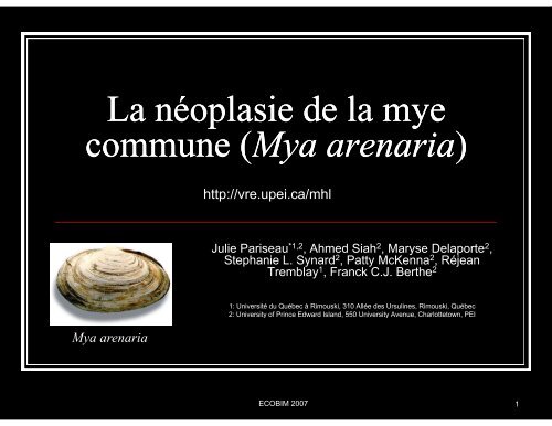
Mya arenaria<br />
http://vre.upei.ca/mhl<br />
Julie Pariseau *1,2 , Ahmed Siah2 , Maryse De<strong>la</strong>porte2 ,<br />
Stephanie L. Synard2 , Patty McKenna2 p y , y , Réjean j<br />
Tremb<strong>la</strong>y1 , Franck C.J. Berthe2 1: <strong>Université</strong> <strong>du</strong> <strong>Québec</strong> <strong>à</strong> <strong>Rimouski</strong>, 310 Allée des Ursulines, <strong>Rimouski</strong>, <strong>Québec</strong><br />
2: University of Prince Edward Is<strong>la</strong>nd, 550 University Avenue, Charlottetown, PEI<br />
ECOBIM 2007 1
Intro<strong>du</strong>ction<br />
<strong>Néop<strong>la</strong>sie</strong>s<br />
20 espèces de bivalves<br />
Deux principaux types: disséminée et<br />
gonadale<br />
Une espèce p pparticulièrement<br />
susceptible<br />
Mye commune: les 2 types sont<br />
présents<br />
La néop<strong>la</strong>sie disséminée est une<br />
néop<strong>la</strong>sie hémique<br />
EEn 1999 1999, mortalité t lité massive i <strong>à</strong> l’IPE (McG<strong>la</strong>derry<br />
et al. 2001)<br />
Jusqu’<strong>à</strong> 95% des indivi<strong>du</strong>s atteints<br />
ECOBIM 2007 2
Aspect fonctionnel des<br />
cellules néop<strong>la</strong>siques<br />
Hémocytes néop<strong>la</strong>siques<br />
Cellu<strong>la</strong>ire<br />
Ont per<strong>du</strong> leurs pseudopodes<br />
Ratio noyau/cytop<strong>la</strong>sme ↑<br />
Molécu<strong>la</strong>ire<br />
125<strong>à</strong>205f 1,25 <strong>à</strong> 2,05 fois i plus l d’ADN<br />
(4N)<br />
↑ chromosomes (44-80 au lieu<br />
de 26 26-39) 39)<br />
Fonctionnel<br />
Ont per<strong>du</strong> leurs fonctions<br />
Adhésion phagocytose et<br />
Adhésion, phagocytose et<br />
défense
N<br />
Hémocytes<br />
Longueur- 10.9 µm<br />
Largeur- 11.8 µm<br />
Longueur <strong>du</strong> noyau- 4.2 µm<br />
Ratio N/C: 0.38
Cellules néop<strong>la</strong>siques<br />
A<br />
N<br />
Longueur- 11.9 µm<br />
Largeur- 13µm<br />
LLongueur noyau- 99 9.9µm<br />
Ratio N/C: 0. 83<br />
B<br />
N<br />
Longueur- 9.4 µm<br />
Largeur- 10µm<br />
Longueur noyau- 8.8µm<br />
Ratio N/C: 0.93
Méthode de diagnostic<br />
Cytométrie en flux<br />
•Analyse de l’hémolymphe<br />
•Coloration de l’ADN avec io<strong>du</strong>re de propidium<br />
Nb d'hémocyte<br />
Nb d'hémocyte<br />
1200<br />
1000<br />
800<br />
600<br />
400<br />
200<br />
180<br />
160<br />
140<br />
120<br />
100<br />
80<br />
60<br />
40<br />
0<br />
20<br />
0<br />
2N<br />
Échantillon #216<br />
G G0-G 0 G 1 S<br />
4N<br />
4% 4N<br />
0 256 512<br />
contenu en ADN<br />
768 1024<br />
2N<br />
Échantillon #1638<br />
4N<br />
60% 4N<br />
0 256 512<br />
contenu en ADN<br />
768 1024<br />
normal<br />
2N<br />
néop<strong>la</strong>sique<br />
4N<br />
2N
Tétraploïdie et phagocytose<br />
% de phagocyttose<br />
30<br />
25<br />
20<br />
15<br />
10<br />
5<br />
0<br />
0,4<br />
0,5<br />
0,5<br />
0,5<br />
0,6<br />
0,7<br />
0,8<br />
0,9<br />
1<br />
1<br />
1<br />
1,2<br />
2<br />
4<br />
4,6<br />
5,7<br />
6<br />
6,7<br />
7,2<br />
10<br />
Tétraploïdie<br />
10<br />
11,8<br />
13,4<br />
17<br />
20<br />
23<br />
ECOBIM 2007 7<br />
26<br />
30<br />
35<br />
47<br />
56<br />
65<br />
71<br />
84
Cycle cellu<strong>la</strong>ire<br />
Cycle cellu<strong>la</strong>ire<br />
G1: croissance 2N<br />
S: synthèse de l’ADN<br />
G2: croissance 4N<br />
M: mitose<br />
Point clé, sous contrôle p53<br />
Permet le bon<br />
fonctionnement cellu<strong>la</strong>ire<br />
p53<br />
p53<br />
ECOBIM 2007 8
Dommage <strong>à</strong> l’ADN<br />
Séquestration cytop<strong>la</strong>smique de <strong>la</strong> p53<br />
(Walker et al 2006)<br />
Mortaline<br />
Ribosome<br />
X<br />
Noyau<br />
Membrane cellu<strong>la</strong>ire<br />
Mutation <strong>du</strong> gène et protéine p53<br />
(Barker et al 1997)<br />
ADN<br />
Arrêt <strong>du</strong><br />
Cycle cellu<strong>la</strong>ire<br />
Apoptose
Mutation de <strong>la</strong> p53 <strong>chez</strong> <strong>la</strong> <strong>mye</strong> commune<br />
Selon Barker et al. 1997<br />
Mutation de type transversion pour le gène<br />
yp p g<br />
Au niveau de l’exon 6 (site non fonctionnel)<br />
Substitution d’une cytosine par une guanine<br />
Proline au lieu de l’a<strong>la</strong>nine l a<strong>la</strong>nine<br />
2 échantillons ont <strong>la</strong> mutation sur 11<br />
Mutation de <strong>la</strong> protéine<br />
Par technique d’immunofluorescence (IFAT)<br />
Anticorps monoclonal PAb 240<br />
Mutation de <strong>la</strong> protéine<br />
MMutation t ti entre t les l acides id aminés i é 213 213-217 217<br />
5 indivi<strong>du</strong>s ont montré <strong>la</strong> mutation sur 11<br />
ECOBIM 2007 10
Expression et mutation <strong>du</strong> gène<br />
30 <strong>mye</strong>s (5%-80%)<br />
RT RT-PCR PCR (E (Expression) i )<br />
Hémocytes<br />
RFLP (Mutation)<br />
Enzymes de restriction<br />
SSCP ?<br />
Hae III, Taq I<br />
421<br />
386<br />
292<br />
264<br />
150<br />
126<br />
HaeIII<br />
TaqI<br />
p53<br />
ECOBIM 2007 11
Expression de <strong>la</strong> protéine<br />
Mise au point p de <strong>la</strong> technique q<br />
Western blotting<br />
Conditions dénaturantes<br />
PAb 240 (type sauvage et muté<br />
de p53)<br />
HCC 70 53<br />
Western blotting (4 <strong>mye</strong>s exprimant<br />
↑niveau <strong>du</strong> gène p53 par Q-RT-<br />
PCR)<br />
Conditions dénaturantes<br />
Branchies<br />
PAb 421 (type sauvage p53)<br />
HCC 70<br />
150<br />
100<br />
75<br />
HCC 70<br />
Hémocyte<br />
Mye<br />
Western blotting (4 <strong>mye</strong>s exprimant + +++ ++ ++ HCC70 Marqueur q<br />
53
Re<strong>la</strong>tive expression e Mortalin/18S<br />
Séquestration de <strong>la</strong> p53 (Walker et al. 2006)<br />
35<br />
30<br />
25<br />
20<br />
15<br />
10<br />
5<br />
0<br />
Mort=0.5p53+0.764<br />
R 2 R =0 =0.498 498<br />
0 10 20 30 40<br />
Re<strong>la</strong>tive expression p53/18S<br />
RELATTIVE<br />
EXPRESSSION<br />
40<br />
30<br />
20<br />
10<br />
0<br />
p53/18S<br />
p73/18S<br />
Mort/18S<br />
0-5 5-15 15-50 50-90<br />
% TETRAPLOIDY
Identification des acteurs<br />
molécu<strong>la</strong>ires lé l i associés ié <strong>à</strong> <strong>la</strong> l néop<strong>la</strong>sie é l i<br />
hémique <strong>chez</strong> Mya arenaria<br />
ECOBIM 2007 14
Principe p de comparaison p <strong>du</strong> transcriptome p<br />
NNormal m l MMa<strong>la</strong>de l d<br />
Transcrit<br />
Down-régulés g<br />
Transcrit<br />
Non-régulés<br />
Transcrit<br />
Up-régulés<br />
Up régulés
« Subtractive PCR<br />
Suppression Hybridization »<br />
Adaptator 1<br />
Tester-Tester<br />
Heterodimers<br />
Tester<br />
Ligation<br />
Driver<br />
Hybridization<br />
Hybridization<br />
PCR<br />
Amplification<br />
Tester-Driver<br />
Tester Driver<br />
Heterodimers<br />
Driver<br />
ds/ss<br />
Tester<br />
ss<br />
Cloning/Sequencing/Bioinformatic Cloning/Sequencing/Bioinformatic g q g Analysis y<br />
Adaptator 2<br />
Tester-Tester<br />
Homodimers<br />
Primer<br />
Amplification<br />
No Amplification<br />
Hairpin Structure
Subtractive Suppression Hybridization<br />
RELAATIVE<br />
EXPREESSION<br />
40<br />
30<br />
p53/18S<br />
P2<br />
p73/18S<br />
Mort/18S<br />
20 P1 P3<br />
10<br />
0<br />
0-5 5-15 15-50 50-90<br />
% TETRAPLOIDY<br />
ECOBIM 2007 17
Branched DNA<br />
Capture Extender Probe<br />
Label Extender Probe<br />
Bl Blocking ki P Probe<br />
b<br />
Système de quantification d’expression <strong>du</strong> gène<br />
sur microp<strong>la</strong>que (simplex)
Système de quantification d’expression <strong>du</strong> gène<br />
sur billes (Multiplex)
Perspectives<br />
<strong>Néop<strong>la</strong>sie</strong> Vibrio spp spp.<br />
SSH SSH<br />
Gènes impliqués Gènes impliqués<br />
N V
Remerciements<br />
ECOBIM 2007 21


