Calcofluor White Reagent Droppers - BD
Calcofluor White Reagent Droppers - BD
Calcofluor White Reagent Droppers - BD

You also want an ePaper? Increase the reach of your titles
YUMPU automatically turns print PDFs into web optimized ePapers that Google loves.
Revisions<br />
Rev from Rev to ECO #<br />
0205 2010/06 5347-10<br />
Notes:<br />
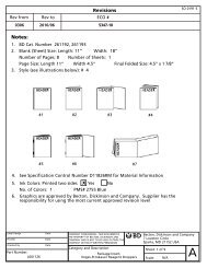
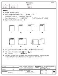
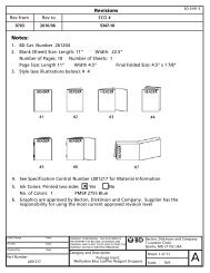
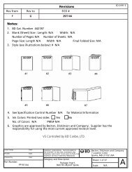
1. <strong>BD</strong> Cat. Number 261195<br />
2. Blank (Sheet) Size : Length: 11” Width: 22.5”<br />
Number of Pages: 12 Number of Sheets: 1<br />
Page Size: Length 11” Width 4.5” Final Folded Size: 4 1/2” x 1 7/8”<br />
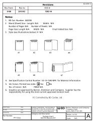
3. Style (see illustrations below): # 4<br />
#1 #2 #3 #4<br />
#5 #6 #7<br />
4. See Specification Control Number L001206 for Material Information<br />
X<br />
5. Ink Colors: Printed two sides Yes No<br />
No. of Colors: 1 PMS# 2755 Blue<br />
6. Graphics are approved by Becton, Dickinson and Company. Supplier has the<br />
responsibility for using the most current approved revision level.<br />
Label Design Date<br />
Proofer Date<br />
Checked By Date<br />
Part Number:<br />
L001206<br />
COMPANY CONFIDENTAL. THIS DOCUMENT IS<br />
THE PROPERTY OF BECTON, DICKINSON AND<br />
COMPANY AND IS NOT TO BE USED OUTSIDE THE<br />
COMPANY WITHOUT WRITTEN PERMISSION<br />
SO 0191-5<br />
Category and Description<br />
Package Insert,<br />
Becton, Dickinson and Company<br />
7 Loveton Circle<br />
Sparks, MD 21152 USA<br />
Sheet: 1 of 13<br />
<strong>Calcofluor</strong> <strong>White</strong> <strong>Reagent</strong> <strong>Droppers</strong> Scale: N/A<br />
A
<strong>Calcofluor</strong> <strong>White</strong> <strong>Reagent</strong><br />
<strong>Droppers</strong><br />
English: pages 1 – 3 Italiano: pagine 6 – 8<br />
L001206<br />
Français : pages 3 – 4 Español: páginas 8 – 10 2010/06<br />
Deutsch: Seiten 4 – 6<br />
INTENDED USE<br />
<strong>BD</strong> <strong>Calcofluor</strong> <strong>White</strong> <strong>Reagent</strong> <strong>Droppers</strong> are used in the rapid fluorescent microscopic detection<br />
of fungi in direct smears.<br />
SUMMARY AND EXPLANATION<br />
<strong>Calcofluor</strong> white is a nonspecific fluorochrome stain that binds to fungi and, depending upon<br />
the filter system employed, fluoresces either an apple green or blue white color when exposed<br />
to light of the appropriate wavelength. 1 It may be used on fresh, frozen, fixed, paraffinimbedded,<br />
and clinical specimens. 2 It has been reported that calcofluor white can be used in the<br />
detection of parasitic Pneumocystis jiroveci (formerly Pneumocystis carinii) 3 and other<br />
opportunistic fungal organisms in bronchoalveolar lavage (BAL) specimens and aspirates from<br />
immunosuppressed individuals. 4,5<br />
PRINCIPLES OF THE PROCEDURE<br />
<strong>Calcofluor</strong> white is a disodium salt of 4,4’-bis-[4-anilino-bis-diethylamino-5-triazin-2-ylamino]-<br />
2,2’-stilbene-disulfonic acid, a colorless dye that is used in the textile and paper industry as a<br />
whitening agent. It has the ability to bind to β 1-3, β 1-4 polysaccharides (i.e., cellulose and<br />
chitin), and exhibits fluorescence when exposed to long-wavelength ultraviolet and shortwavelength<br />
visible light. It has been used as a biological marker to stain the cell wall of plants,<br />
and is therefore valuable in delineating fungal elements. 6<br />
REAGENTS<br />
<strong>Calcofluor</strong> <strong>White</strong> <strong>Reagent</strong> <strong>Droppers</strong> contain 0.5 mL of a 0.05% solution of calcofluor white in<br />
distilled water.<br />
Warnings and Precautions:<br />
For in vitro Diagnostic Use.<br />
Avoid contact with skin. Rinse thoroughly with water if spilled.<br />
Pathogenic microorganisms, including hepatitis viruses and Human Immunodeficiency Virus,<br />
may be present in clinical specimens. “Standard Precautions” 7-10 and institutional guidelines<br />
should be followed in handling all items contaminated with blood and other body fluids. Prior<br />
to discarding, specimen containers and other contaminated materials must be sterilized by<br />
autoclaving.<br />
Storage Instructions: Store at room temperature 15 – 30°C (59 – 85ºF). Protect from light.<br />
Product Deterioration: This reagent is hermetically sealed in an ampule, which affords<br />
protection of the solution from chemical instability until the expiration date. The staining<br />
solution should be clear with a slight green tint. Do not use if a heavy white precipitate is<br />
evident. Each dropper is good for one day’s use after ampule has been broken. Do not use after<br />
the expiration date.<br />
PROCEDURE<br />
Material Provided: <strong>Calcofluor</strong> <strong>White</strong> <strong>Reagent</strong> <strong>Droppers</strong>. Each dropper contains sufficient<br />
reagent to stain one slide.<br />
Materials Required But Not Provided: Ancillary culture media, reagents, 10% Potassium<br />
Hydroxide <strong>Reagent</strong> Dropper, Cat. No. 261191, quality control organisms, a fluorescent<br />
microscope and other laboratory equipment as required for this procedure.<br />
Test Procedure:<br />
GENERAL SPECIMENS (hair, nails, skin, other tissue, culture, etc.)<br />
1. Slides used should be clean and free of oils and debris.<br />
2. Prepare a smear of the specimen to be stained.<br />
3. Add 1 to 2 drops of a 10% potassium hydroxide solution and gently mix to spread.<br />
4. Hold <strong>Calcofluor</strong> <strong>White</strong> <strong>Reagent</strong> dropper upright and POINT TIP AWAY FROM YOURSELF.<br />
Grasp the middle with thumb and forefinger and squeeze gently to break ampule inside<br />
the dropper. Caution: Break ampule close to its center one time only. Do not manipulate<br />
the dropper any further as the plastic may puncture and injury may occur. Tap the bottom<br />
of the dropper on the tabletop a few times. Invert for convenient drop-by-drop dispensing<br />
of the reagent.<br />
5. Add 1 to 2 drops of <strong>Calcofluor</strong> <strong>White</strong> <strong>Reagent</strong>. Wait for 1 to 2 min.<br />
6. Mount specimen with a cover slip.<br />
7. Using a fluorescent microscope, examine smears initially at 10X, then confirm findings at a<br />
higher magnification.<br />
8. Tissue sections showing fungi can be rinsed in distilled water and restained with periodic<br />
acid-Schiff stain or by the Gomori methenamine silver (GMS) technique. 6<br />
ASPIRATE OR BRONCHOALVEOLAR LAVAGE (BAL) SPECIMENS<br />
1. Slides used should be clean and free of oils and debris.<br />
2. Specimens may be fresh or centrifuged and resuspended to the appropriate concentration<br />
in Phosphate Buffered Saline (PBS), pH 7.4.
3. Mark off a well-defined square on the slide with a wax marking pencil.<br />
4. Apply the specimen to the area within the wax square on the slide; allow to air dry.<br />
5. Cover the area within the square with 100% methanol; allow to air dry.<br />
6. Add sufficient 10% potassium hydroxide solution to just cover the specimen. Do not<br />
saturate. Wait for 5 s.<br />
7. Hold <strong>Calcofluor</strong> <strong>White</strong> <strong>Reagent</strong> dropper upright and POINT TIP AWAY FROM YOURSELF.<br />
Grasp the middle with thumb and forefinger and squeeze gently to break ampule inside<br />
the dropper. Caution: Break ampule close to its center one time only. Do not manipulate<br />
the dropper any further as the plastic may puncture and injury may occur. Tap the bottom<br />
of the dropper on the tabletop a few times. Invert for convenient drop-by-drop dispensing<br />
of the reagent.<br />
8. Add 1 to 2 drops of <strong>Calcofluor</strong> <strong>White</strong> <strong>Reagent</strong> to the area within the square. Wait for 2 min.<br />
9. Drain excess <strong>Calcofluor</strong> <strong>White</strong> <strong>Reagent</strong> from the slide.<br />
10. Using a fluorescent microscope view the specimen:<br />
a. To view as a wet mount slide, add <strong>Calcofluor</strong> <strong>White</strong> <strong>Reagent</strong>, as needed, a cover slip and<br />
view with appropriate objective.<br />
- or -<br />
b. To view under oil immersion, after air-drying, view directly.<br />
User Quality Control:<br />
1. Examine the staining solution for signs of deterioration (see “Product Deterioration”).<br />
2. Check the performance of the stain with fresh cultures of Candida albicans ATCC 10231<br />
(positive) and Escherichia coli ATCC 25922 (negative).<br />
Quality control requirements must be performed in accordance with applicable local, state<br />
and/or federal regulations or accreditation requirements and your laboratory’s standard<br />
Quality Control procedures. It is recommended that the user refer to pertinent NCCLS guidance<br />
and CLIA regulations for appropriate Quality Control practices.<br />
RESULTS<br />
When exposed to light of the suggested wavelength (300 – 412 nm), fungi will fluoresce a<br />
bright apple green or blue white color. Bacteria will fluoresce weakly or not at all.<br />
LIMITATIONS OF THE PROCEDURE<br />
Many clinical laboratories now use fluorescence microscopy for the examination of other<br />
stains. For best results with <strong>Calcofluor</strong> <strong>White</strong> <strong>Reagent</strong>, a mercury vapor rather than a quartz<br />
halogen lamp, and UV/BV excitation are recommended. 1 Microscopes fitted with selective<br />
filters for the excitation of fluorescein (KP490 interference filter) are not recommended since<br />
these filters prevent the proper wave length from striking the specimen. 1<br />
Certain debris may fluoresce and should be distinguished from fungal elements on the basis<br />
of morphology.<br />
<strong>Calcofluor</strong> <strong>White</strong> <strong>Reagent</strong> <strong>Droppers</strong> are used for the presumptive identification of fungi. All<br />
positive smears should be confirmed by culture.<br />
Although the cells themselves of Cryptococcus will stain with calcofluor white, it has been<br />
reported that the capsule will not; further testing for confirmation is recommended.<br />
Bacteria may fluoresce, although to a lesser degree than fungi and may be confused with<br />
calcofluor white-stained artifacts. The presence of fungi should be determined on the basis<br />
of morphology.<br />
Fluorescent staining intensity may vary depending on the organism being stained.<br />
PERFORMANCE CHARACTERISTICS<br />
<strong>BD</strong> conducted a study of the <strong>BD</strong> <strong>Calcofluor</strong> <strong>White</strong> <strong>Reagent</strong> using the following organisms:<br />
Aspergillus sp. (ATCC 36607) Candida albicans (ATCC 10231)<br />
Candida guilliermondii (ATCC 14242) Cladosporium sp. (ATCC 13026)<br />
Microsporum canis (ATCC 10214) Penicillium roquefortii (ATCC 9295)<br />
Candida glabrata (ATCC 2001)<br />
All slides were prepared in triplicate with the wet mount method as described in the "GENERAL<br />
SPECIMENS" section of the Test Procedure (including the use of 10% potassium hydroxide<br />
reagent). There was one high positive control slide (cellulose via cotton fibers) and two negative<br />
control slides (Escherichia coli ATCC 25922 and an uninoculated slide).<br />
Slides were examined with a fluorescence microscope (Olympus, BH-2) equipped with a 100w<br />
mercury lamp, exciter filter (490 nm) and barrier filter (545 nm).<br />
The organisms evaluated in this study exhibited appropriate fluorescent staining characteristics.<br />
AVAILABILITY<br />
Cat. No. Description<br />
261195 <strong>BD</strong> <strong>Calcofluor</strong> <strong>White</strong> <strong>Reagent</strong> <strong>Droppers</strong>, packaged 50 droppers/carton.<br />
261191 <strong>BD</strong> 10% Potassium Hydroxide <strong>Reagent</strong> <strong>Droppers</strong> packaged 50 droppers/carton.<br />
REFERENCES<br />
1. Harrington, B.J., and G.J. Hageage, Jr. 1991. <strong>Calcofluor</strong> white: tips for improving its use.<br />
Clin. Microbiol. Newsl. 13:3-5.<br />
2. Monheit, J.E., D.F. Cowan, and D.G. Moore. 1984. Rapid detection of fungi in tissues using<br />
calcofluor white and fluorescence microscopy. Arch. Pathol. Lab. Med. 108:616-618.<br />
3. Stringer, J.R., C.B. Beard, R.F. Miller, and A.E. Wakefield. 2002. A new name (Pneumocystis<br />
jiroveci) for Pneumocystis from humans. Emerg. Infect. Dis. 8(9):891-6.<br />
4. Baselski, V.S., M.K. Robinson, L.W. Pifer, and D.R. Woods. 1990. Rapid detection of<br />
Pneumocystis carinii in bronchoalveolar lavage samples by using Cellufluor staining. J. Clin.<br />
Microbiol. 28:393-394.<br />
5. Kim, Y.K., S. Parulekar, P.K.W. Yu, R.J. Pisani, T.F. Smith, and J.P. Anhalt. 1990. Evaluation of<br />
calcofluor white stain for detection of Pneumocystis carinii. Diagn. Microbiol. Infect. Dis.<br />
13:307-310.<br />
6. Hageage, G.J., and B.J. Harrington. 1984. Use of calcofluor white in clinical mycology. Lab.<br />
Med. 15:109-112.<br />
7. National Committee for Clinical Laboratory Standards. 2001. Approved Guideline M29-A2.<br />
Protection of laboratory workers from occupationally acquired infections, 2nd ed. NCCLS,<br />
Wayne, Pa.<br />
8. Garner, J.S. 1996. Hospital Infection Control Practices Advisory Committee, U.S. Department<br />
of Health and Human Services, Centers for Disease Control and Prevention. Guideline for<br />
isolation precautions in hospitals. Infect. Control Hospital Epidemiol. 17:53-80.<br />
2
9. U.S. Department of Health and Human Services. 1999. Biosafety in microbiological and<br />
biomedical laboratories, HHS Publication (CDC), 4th ed. U.S. Government Printing Office,<br />
Washington, D.C.<br />
10. Directive 2000/54/EC of the European Parliament and of the Council of 18 September 2000<br />
on the protection of workers from risks related to exposure to biological agents at work<br />
(seventh individual directive within the meaning of Article 16(1) of Directive 89/391/EECP).<br />
Official Journal L262, 17/10/2000, p. 0021-0045.<br />
Calcoflour <strong>White</strong> <strong>Reagent</strong><br />
<strong>Droppers</strong><br />
Français<br />
APPLICATION<br />
<strong>BD</strong> <strong>Calcofluor</strong> <strong>White</strong> <strong>Reagent</strong> <strong>Droppers</strong> (compte-gouttes de reactif au calcofluor blanc) servent à la<br />
détection rapide en microscopie fluorescente de champignons dans des frottis directs.<br />
RESUME ET EXPLICATION<br />
Le calcofluor blanc est un fluorochrome colorant non spécifique qui se lie aux champignons et<br />
suivant le système de filtres employé, émet une fluorescence de couleur vert pomme ou bleu<br />
blanc lorsqu'il est exposé à une lumière de la longueur d'onde appropriée. 1 Il peut être utilisé<br />
sur des échantillons frais, congelés, fixés, inclus dans la paraffine et cliniques. 2 Il a été rapporté<br />
que le calcofluor blanc peut être utilisé pour la détection de parasites Pneumocystis jiroveci<br />
(anciennement Pneumocystis carinii) 3 et d'autres champignons opportunistes dans les<br />
échantillons de lavage broncho-alvéolaire (BAL) et les aspirats d'individus immunodéprimés. 4,5<br />
PRINCIPES DE LA METHODE<br />
Le calcofluor blanc est un sel disodique de l'acide 4,4'-bis-[4-anilino-bis-diéthylamino-<br />
5-triazine-2-ylamino]-2,2'-stilbène-disulfonique, un colorant incolore qui est utilisé dans<br />
l'industie textile et papetière comme agent de blanchiment. Il a la propriété de se lier aux<br />
polysaccharides β 1-3 et β 1-4 (par ex., cellulose et chitine) et présente une fluorescence<br />
lorsqu'il est exposé aux longues longueurs d'onde ultraviolettes et aux courtes longueurs<br />
d'ondes du spectre visible. Il a servi de marqueur biologique pour colorer les parois cellulaires<br />
végétales et est par conséquent particulièrement utile pour délimiter les contours des éléments<br />
fongiques. 6<br />
REACTIFS<br />
Les <strong>Calcofluor</strong> <strong>White</strong> <strong>Reagent</strong> <strong>Droppers</strong> contiennent 0,5 mL d'une solution à 0,05 % de<br />
calcofluor blanc dans de l'eau distillée.<br />
Avertissements et précautions :<br />
Pour usage diagnostique in vitro.<br />
Eviter tout contact avec la peau. Rincer soigneusement à l'eau en cas de déversement.<br />
Des microorganismes pathogènes, notamment les virus de l'hépatite et de l'immunodéficience<br />
humaine, sont susceptibles d'être présents dans les échantillons cliniques. Respecter les<br />
" Précautions standard " 7-10 et les consignes en vigueur dans l'établissement pour manipuler<br />
tout objet contaminé avec du sang ou d'autres liquides organiques. Stériliser à l'autoclave les<br />
récipients contenant les échantillons et les autres matériaux contaminés avant de les éliminer.<br />
Instructions pour la conservation : Conserver à température ambiante de 15 à 30 °C et à l'abri<br />
de la lumière.<br />
Détérioration du produit : Ce réactif est hermétiquement scellé dans une ampoule assurant la<br />
protection contre l'instabilité chimique de la solution jusqu'à la date de péremption. La<br />
solution colorante devrait être claire avec une légère teinte verte. Ne pas utiliser si un épais<br />
précipité blanc est présent. Chaque compte-gouttes reste bon à utiliser pendant 24 h une fois<br />
l'ampoule brisée. Ne pas utiliser au delà de la date de péremption.<br />
METHODE<br />
Matériaux fournis : <strong>Calcofluor</strong> <strong>White</strong> <strong>Reagent</strong> <strong>Droppers</strong>. Chaque compte-gouttes contient<br />
suffisamment de réactif pour colorer une lame.<br />
Matériaux requis mais non fournis : Milieux de culture auxilliaires, réactifs, 10% Potassium<br />
Hydroxide <strong>Reagent</strong> Dropper, Cat. N° 261191, souches de contrôle de qualité, microscope à<br />
fluorescence et autres équipements de laboratoire comme requis par cette procédure.<br />
Mode opératoire du test :<br />
ECHANTILLONS GENERAUX (cheveux, ongles, peau, autres tissus, culture, etc.)<br />
1. Les lames utilisées doivent être propres et exemptes de graisses et de débris.<br />
2. Préparer un frottis de l'échantillon à colorer.<br />
3. Ajouter 1 à 2 gouttes d'une solution à 10 % d'hydroxyde de potassium et mélanger<br />
doucement pour étaler.<br />
4. Tenir le <strong>Calcofluor</strong> <strong>White</strong> <strong>Reagent</strong> dropper verticalement et POINTER L'EXTREMITE A<br />
L'OPPOSE DE SOI. Saisir le milieu entre le pouce et l'index et presser doucement afin de<br />
casser l'ampoule à l'intérieur du compte-gouttes. Attention : Briser l'ampoule en son<br />
centre une fois seulement. Ne pas manipuler davantage le compte-gouttes pour ne pas<br />
risquer de perforer le plastique et de se blesser. Tapoter plusieurs fois le fond du comptegouttes<br />
sur la paillasse. Renverser le compte-gouttes pour que le réactif s'écoule goutte à<br />
goutte.<br />
5. Ajouter 1 à 2 gouttes de <strong>Calcofluor</strong> <strong>White</strong> <strong>Reagent</strong> (réactif au calcofluor blanc). Attendre<br />
1 à 2 min.<br />
6. Monter l'échantillon sur la lame avec une lamelle.<br />
7. Au moyen d'un microscope à fluorescence, examiner le frottis d'abord au grossissement<br />
10X puis confirmer les résultats au plus fort grossissement.<br />
8. Les sections de tissus présentant des champignons peuvent être rincées à l'eau distillée et<br />
re-colorées avec l'acide périodique de Schiff ou grâce à la technique à la méthénamine<br />
d'argent de Gomori (GMS). 6<br />
ECHANTILLONS D'ASPIRAT OU DE LAVAGE BRONCHO-ALVEOLAIRE (BAL)<br />
1. Les lames utilisées doivent être propres et exemptes de graisses et de débris.<br />
2. Les échantillons peuvent être frais ou centrifugés et remis en suspension à la concentration<br />
correcte dans du sérum physiologique tamponné au phosphate (PBS), pH 7,4.<br />
3. Marquer un carré bien défini sur la lame avec un crayon marqueur à la cire.<br />
4. Appliquer l'échantillon sur la surface à l'intérieur du carré tracé à la cire sur la lame et<br />
laisser sécher à l'air.<br />
3
5. Recouvrir la surface à l'intérieur du carré avec du méthanol pur (100%) et laisser sécher à<br />
l'air.<br />
6. Ajouter une quantité suffisante de solution à 10 % d'hydroxyde de potassium pour couvrir<br />
l'échantillon. Ne pas saturer. Attendre 5 sec.<br />
7. Tenir le <strong>Calcofluor</strong> <strong>White</strong> <strong>Reagent</strong> dropper verticalement et POINTER L'EXTREMITE A<br />
L'OPPOSE DE SOI. Saisir le milieu entre le pouce et l'index et presser doucement afin de<br />
casser l'ampoule à l'intérieur du compte-gouttes. Attention : Briser l'ampoule en son<br />
centre une fois seulement. Ne pas manipuler davantage le compte-gouttes pour ne pas<br />
risquer de perforer le plastique et de se blesser. Tapoter plusieurs fois le fond du comptegouttes<br />
sur la paillasse. Renverser le compte-gouttes pour que le réactif s'écoule goutte à<br />
goutte.<br />
8. Ajouter 1 à 2 gouttes de réactif au calcofluor blanc sur la surface à l'intérieur du carré.<br />
Attendre 2 min.<br />
9. Drainer l'excédent de réactif au calcofluor blanc de la lame.<br />
10. Examiner l'échantillon avec un microscope à fluorescence.<br />
a. Pour examiner un montage humide sur lame, ajouter du réactif au calcofluor blanc,<br />
comme requis, poser une lamelle et observer avec l'objectif adéquat.<br />
- ou -<br />
b. Examiner avec l'objectif à l'huile d'immersion, après le séchage à l'air, observer<br />
directement.<br />
Contrôle de qualité par l’utilisateur :<br />
1. Inspecter la solution colorante pour vérifier l'absence de traces de détérioration (voir<br />
" Détérioration du produit ").<br />
2. Vérifier les performances du colorant avec des cultures fraîches de Candida albicans ATCC<br />
10231 (positif) et Escherichia coli ATCC 25922 (négatif).<br />
Effectuer les contrôles de qualité conformément aux réglementations nationales et/ou<br />
internationales, aux exigences des organismes d'homologation concernés et aux procédures de<br />
contrôle de qualité en vigueur dans l'établissement. Il est recommandé à l'utilisateur de<br />
consulter les directives NCCLS et la réglementation CLIA correspondantes pour plus<br />
d'informations sur les modalités de contrôle de qualité.<br />
RESULTATS<br />
Lorsque exposé à la lumière de la longueur d'onde suggérée (300 - 412 nm), les champignons<br />
émettront une fluorescence de couleur vert pomme brilliant ou bleu-blanc. Les bactéries ne<br />
fluoresceront que faiblement ou pas du tout.<br />
LIMITES DE LA PROCEDURE<br />
De nombreux laboratoires d'analyses utilisent désormais la microscopie à fluorescence pour<br />
examiner d'autres colorations. Pour obtenir les meilleurs résultats avec le réactif au calcofluor<br />
blanc, une lampe à vapeur de mercure plutôt qu'une lampe quartz-halogène et une excitation<br />
UV/BV sont recommandées. 1 Les microscopes équipés de filtres sélectifs propres à l'excitation<br />
de la fluorescéine (filtre d'interférence KP490) sont déconseillés parce que ces filtres<br />
empêchent la longueur d'onde adéquate de frapper (exciter) l'échantillon. 1<br />
Certains débris peuvent fluorescer et doivent être différencier des éléments fongiques en se<br />
basant sur la morphologie.<br />
Les <strong>Calcofluor</strong> <strong>White</strong> <strong>Reagent</strong> <strong>Droppers</strong> servent à l'identification présomptive de champignons.<br />
Tous les frottis positifs doivent être confirmés par la culture.<br />
Bien que les cellules elles-mêmes de Cryptococcus se coloreront avec le calcofluor blanc, la<br />
capsule ne se colorerait pas d'après certains rapports ; par conséquent des tests<br />
supplémentaires de confirmation sont recommandés.<br />
Les bactéries peuvent fluorescer quoique à un degré moindre que les champignons et elles<br />
peuvent être confondues avec certains artéfacts colorés par le calcofluor blanc. La présence de<br />
champignons doit toujours être établie sur la base de la morphologie.<br />
L'intensité de la coloration fluorescente peut varier en fonction de l'organisme coloré.<br />
CARACTERISTIQUES DE PERFORMANCES<br />
<strong>BD</strong> a effectué une étude du <strong>BD</strong> <strong>Calcofluor</strong> <strong>White</strong> <strong>Reagent</strong> au moyen des organismes suivants :<br />
Aspergillus sp. (ATCC 36607) Candida albicans (ATCC 10231)<br />
Candida guilliermondii (ATCC 14242) Cladosporium sp. (ATCC 13026)<br />
Microsporum canis (ATCC 10214) Penicillium roquefortii (ATCC 9295)<br />
Candida glabrata (ATCC 2001)<br />
Toutes les lames ont été préparées en triple exemplaire selon la méthode de montage humide<br />
décrite dans la section " ECHANTILLONS GENERAUX " du Mode opératoire du test (incluant<br />
l'emploi du réactif à 10 % d'hydroxyde de potassium). Il y avait une lame de contrôle hautement<br />
positive (cellulose de fibres de cotton) et deux lames de contrôle négatives (Escherichia coli<br />
ATCC 25922 et une lame non inoculée).<br />
Les lames ont été examinées avec un microscope à fluorescence (Olympus, BH-2) équipé d'une<br />
lampe au mercure de 100 w, d'un filtre d'excitation (490 nm) et d'un filtre d'émission (d'arrêt)<br />
(545 nm).<br />
Les organismes utilisés dans cette étude ont montré les caractéristiques de coloration<br />
fluorescente appropriées.<br />
CONDITIONNEMENT<br />
No réf. Description<br />
261195 <strong>BD</strong> <strong>Calcofluor</strong> <strong>White</strong> <strong>Reagent</strong> <strong>Droppers</strong>, 50 compte-gouttes emballés/carton.<br />
261191 <strong>BD</strong> 10% Potassium Hydroxide <strong>Reagent</strong> <strong>Droppers</strong>, 50 compte-gouttes<br />
emballés/carton<br />
REFERENCES: voir la rubrique “References” du texte anglais.<br />
Calcoflour <strong>White</strong> <strong>Reagent</strong><br />
<strong>Droppers</strong><br />
Deutsch<br />
VERWENDUNGSZWECK<br />
<strong>BD</strong> <strong>Calcofluor</strong> <strong>White</strong> <strong>Reagent</strong> <strong>Droppers</strong> (<strong>BD</strong> <strong>Calcofluor</strong> <strong>White</strong>-Reagenz-Tropfpipetten) werden<br />
bei der schnellen fluoreszenzmikroskopischen Identifizierung von Pilzen in direkten<br />
Abstrichen verwendet.<br />
4
ZUSAMMENFASSUNG UND ERKLÄRUNG<br />
<strong>Calcofluor</strong> <strong>White</strong> ist ein nicht spezifischer fluorochromer Stamm, der an Pilze bindet und, je<br />
nach verwendetem Filtersystem, bei Einwirkung von Licht der entsprechenden Wellenlänge<br />
entweder apfelgrün oder blauweiß fluoresziert. 1 Es kann auf frischen, gefrorenen, fixierten,<br />
paraffineingebetteten und klinischen Proben eingesetzt werden. 2 Berichten zufolge kann<br />
<strong>Calcofluor</strong> <strong>White</strong> zum Nachweis von parasitischem Pneumocystis jiroveci (ehemals<br />
Pneumocystis carinii) 3 sowie von anderen opportunistischen Pilzorganismen in<br />
bronchoalveolären Spülungen (BAL) und Aspiraten aus immunsupprimierten Individuen<br />
dienen. 4,5<br />
VERFAHRENSGRUNDLAGEN<br />
<strong>Calcofluor</strong> white ist ein Dinatrium-Salz der 4,4'-Bis-[4-anilino-bis-diethylamino-5-triazin-2ylamino]-2,2'-stilben-disulfonsäure,<br />
ein farbloser Farbstoff, der in der Textil- und<br />
Papierindustrie als Weißmacher verwendet wird. Es kann an β 1-3, β 1-4 Polysaccharide binden<br />
(z. B. Cellulose und Chitin) und fluoresziert, wenn es langwelligem ultravioletten und<br />
kurzwelligem sichtbaren Licht ausgesetzt wird. Es wurde als biologischer Marker bei der<br />
Färbung von Pflanzenzellwänden eingesetzt und ist daher bei der Umreißung von<br />
Pilzstrukturen nützlich. 6<br />
REAGENZIEN<br />
<strong>Calcofluor</strong> <strong>White</strong> <strong>Reagent</strong> <strong>Droppers</strong> enthalten 0,5 mL einer 0,05 % Lösung von <strong>Calcofluor</strong><br />
<strong>White</strong> in destilliertem Wasser.<br />
Warnungen und Vorsichtsmaßnahmen:<br />
In-vitro-Diagnostikum.<br />
Kontakt mit der Haut vermeiden. Bei Verschütten gründlich mit Wasser spülen.<br />
Klinische Proben können pathogene Mikroorganismen, darunter auch Hepatitis-Viren und HIV<br />
enthalten. Beim Umgang mit allen durch Blut oder andere Körperflüssigkeiten kontaminierten<br />
Artikeln sind die "Allgemeinen Vorsichtsmaßnahmen" 7-10 sowie die einschlägigen<br />
Institutionsrichtlinien zu beachten. Vor der Entsorgung müssen Probenbehälter und andere<br />
kontaminierte Materialien im Autoklaven sterilisiert werden.<br />
Aufbewahrung: Bei Raumtemperatur (15–30 °C) lagern. Vor Licht geschützt aufbewahren.<br />
Produktverfall: Dieses Reagenz ist in einer Ampulle hermetisch versiegelt, wodurch die Lösung<br />
bis zum Verfallsdatum vor chemischer Instabilität geschützt wird. Die Färbelösung muss klar<br />
und leicht grün gefärbt sein. Bei Anwesenheit eines dicken weißen Niederschlags nicht<br />
verwenden. Jede Tropfpipette kann nach Aufbrechen der Ampulle einen Tag lang verwendet<br />
werden. Nach Ablauf des Verfallsdatums nicht benutzen.<br />
VERFAHREN<br />
Mitgeliefertes Material: <strong>Calcofluor</strong> <strong>White</strong> <strong>Reagent</strong> <strong>Droppers</strong>. Jede Tropfpipette enthält<br />
ausreichend Reagenz für die Anfärbung eines Objektträgers.<br />
Benötigte, aber nicht mitgelieferte Materialien: Hilfskulturmedien, Reagenzien, 10% Potassium<br />
Hydroxide <strong>Reagent</strong> Dropper (10 % Kaliumhydroxidreagenz-Tropfpipetten) Kat. Nr. 261191,<br />
Qualitätskontrollorganismen. ein Fluoreszenzmikroskop und weitere Laborausstattung, die für<br />
dieses Verfahren benötigt wird.<br />
Testverfahren:<br />
ALLGEMEINE PROBEN (Haare, Nägel, Haut, andere Gewebe, Kultur usw.)<br />
1. Die benutzten Objektträger müssen sauber und fettfrei sein und dürfen keine<br />
Fremdkörper enthalten.<br />
2. Fertigen Sie einen Abstrich der zu färbenden Probe an.<br />
3. 1 bis 2 Tropfen einer 10 % Kaliumhydroxidlösung zufügen und vorsichtig vermischen und<br />
ausstreichen.<br />
4. <strong>Calcofluor</strong> <strong>White</strong> <strong>Reagent</strong> dropper aufrecht halten und DIE SPITZE VOM KÖRPER WEG<br />
RICHTEN. Die Mitte mit Daumen und Zeigefinger ergreifen und vorsichtig<br />
zusammendrücken, um die Ampulle in der Tropfpipette aufzubrechen. Vorsicht: Die<br />
Ampulle nur ein einziges Mal in der Nähe der Mitte brechen. Die Pipette nicht weiter<br />
handhaben, da das Plastik durchgestochen werden kann und Verletzungen möglich sind.<br />
Mit dem Boden der Pipette einige Male leicht auf die Tischoberfläche klopfen. Für eine<br />
komfortable tropfenweise Dispensierung des Reagenzes die Pipette umkehren.<br />
5. 1 bis 2 Tropfen <strong>Calcofluor</strong> <strong>White</strong> Reagenz zugeben. 1 bis 2 Min. warten.<br />
6. Probe mit einem Deckglas abdecken.<br />
7. Die Abstriche zunächst mit Hilfe eines Fluoreszenzmikroskops mit 10X-Vergrößerung<br />
untersuchen und die Ergebnisse sodann bei stärkerer Vergrößerung bestätigen.<br />
8. Gewebeschnitte mit Pilzzellen können in destilliertem Wasser abgespült werden und mit<br />
Perjodsäure-Schiff-Reagenz oder mittels der Methenamin-Silber-Methode nach Gomorri<br />
(GMS) erneut gefärbt werden. 6<br />
AUS ASPIRATEN ODER BRONCHOALVEOLÄRER LAVAGE (BAL) ERHALTENE PROBEN<br />
1. Die benutzten Objektträger müssen sauber und fettfrei sein und dürfen keine<br />
Fremdkörper enthalten.<br />
2. Proben können frisch sein oder nach Zentrifugation mit phosphatgepufferter<br />
Kochsalzlösung (PBS), pH 7,4, auf die erwünschte Konzentration resuspendiert sein.<br />
3. Mit einem Wachsmalstift auf dem Objektträger ein scharf umrissenes Quadrat abgrenzen.<br />
4. Die Probe auf das Feld innerhalb des Wachsquadrates aufbringen; an der Luft trocknen<br />
lassen.<br />
5. Die Fläche innerhalb des Quadrats mit 100 % Methanol bedecken; an der Luft trocknen<br />
lassen.<br />
6. Ausreichend 10 % Kaliumhydroxidlösung zugeben, um die Proben gerade eben zu<br />
bedecken. Nicht durchtränken. 5 s. warten.<br />
7. <strong>Calcofluor</strong> <strong>White</strong> <strong>Reagent</strong> dropper aufrecht halten und DIE SPITZE VOM KÖRPER WEG<br />
RICHTEN. Die Mitte mit Daumen und Zeigefinger ergreifen und vorsichtig<br />
zusammendrücken, um die Ampulle in der Tropfpipette aufzubrechen. Vorsicht: Die<br />
Ampulle nur ein einziges Mal in der Nähe der Mitte brechen. Die Pipette nicht weiter<br />
handhaben, da das Plastik durchgestochen werden kann und Verletzungen möglich sind.<br />
Mit dem Boden der Pipette einige Male leicht auf die Tischoberfläche klopfen. Für eine<br />
komfortable, tropfenweise Dispensierung des Reagenzes die Pipette umkehren.<br />
8. 1 bis 2 Tropfen <strong>Calcofluor</strong> <strong>White</strong> Reagenz auf die Fläche innerhalb des Quadrates geben.<br />
2 Min. warten.<br />
9. Überschüssiges <strong>Calcofluor</strong> <strong>White</strong>-Reagenz vom Objektträger ablaufen lassen.<br />
5
10. Die Proben mit Hilfe eines Fluoreszenzmikroskops betrachten:<br />
a. Für die Ansicht als Wet Mount-Präparat nach Bedarf <strong>Calcofluor</strong> <strong>White</strong> Reagenz zugeben,<br />
mit einem Deckglas abdecken und mit dem entsprechenden Objektiv betrachten.<br />
- oder -<br />
b. Für die Ansicht unter Ölimmersion nach Lufttrocknung direkt betrachten.<br />
Qualitätskontrolle durch den Benutzer:<br />
1. Die Färbelösung auf Anzeichen einer Zersetzung hin überprüfen (siehe "Produktverfall").<br />
2. Die Qualität der Anfärbung mit frischen Kulturen von Candida albicans ATCC 10231<br />
(positiv) und Escherichia coli ATCC 25922 (negativ) überprüfen.<br />
Die Qualitätskontrollen müssen unter Einhaltung der örtlich, landesweit und/oder bundesweit<br />
geltenden Bestimmungen oder der Auflagen der Akkreditierungsorganisationen sowie der<br />
Standard-Qualitätskontrollverfahren Ihres Labors erfolgen. Anwendern wird geraten, die<br />
relevanten NCCLS-Richtlinien und CLIA-Vorschriften über geeignete Maßnahmen zur<br />
Qualitätskontrolle einzusehen.<br />
ERGEBNISSE<br />
Unter Licht der empfohlenen Wellenlänge (300–412 nm) fluoreszieren Pilze grell apfelgrün<br />
oder blauweiß. Bakterien fluoreszieren nur schwach oder überhaupt nicht.<br />
VERFAHRENSBESCHRÄNKUNGEN<br />
In vielen klinischen Labors wird Fluoreszenzmikroskopie mittlerweile zur Untersuchung<br />
anderer Färbungen eingesetzt. Zum Erzielen der besten Ergebnisse mit dem <strong>Calcofluor</strong> <strong>White</strong>-<br />
Reagenz wird Quecksilberdampf anstelle einer Quartz-Halogenleuchte und eine UV/BV-<br />
Anregung empfohlen. 1 Mikroskope, die mit selektiven Filtern für die Anregung von<br />
Fluorescein (KP490 Interferenzfilter) ausgestattet sind, werden nicht empfohlen, da diese Filter<br />
verhindern, dass die geeignete Wellenlänge die Probe trifft. 1<br />
Manche Fremdkörper können ebenfalls fluoreszieren und müssen morpohologisch von<br />
Pilzelementen unterschieden werden.<br />
<strong>Calcofluor</strong> <strong>White</strong> <strong>Reagent</strong> <strong>Droppers</strong> werden zur Identifizierung vermuteter Pilze verwendet.<br />
Alle positiven Abstriche müssen durch Kultivierung bestätigt werden.<br />
Obwohl im Fall von Cryptococcus die Zellen selbst mit <strong>Calcofluor</strong> <strong>White</strong> gefärbt werden, wird<br />
die Kapsel Berichten zufolge nicht gefärbt; daher werden zur Bestätigung weitere Tests<br />
empfohlen.<br />
Bakterien können fluoreszieren, jedoch zu einem geringeren Grad als Pilze und können mit<br />
durch Calofluor <strong>White</strong> gefärbten Artefakten verwechselt werden. Die Anwesenheit von Pilzen<br />
muss morphologisch bestimmt werden.<br />
Die Intensität der Fluoreszenzfärbung kann je nach zu färbendem Organismus variieren.<br />
LEISTUNGSMERKMALE<br />
<strong>BD</strong> führte eine Studie über das <strong>BD</strong> <strong>Calcofluor</strong> <strong>White</strong>-Reagenz unter Verwendung der<br />
folgenden Organismen durch:<br />
Aspergillus sp. (ATCC 36607) Candida albicans (ATCC 10231)<br />
Candida guilliermondii (ATCC 14242) Cladosporium sp. (ATCC 13026)<br />
Microsporum canis (ATCC 10214) Penicillium roquefortii (ATCC 9295)<br />
Candida glabrata (ATCC 2001)<br />
Alle Objektträger wurden mittels der Wet-Mount-Methode dreifach präpariert, wie im<br />
Abschnitt "ALLGEMEINE PROBEN" des Testverfahrens beschrieben (einschließlich des<br />
Gebrauchs von 10 % Kaliumhydroxid-Reagenz). Ein Kontrollobjektträger war stark positiv<br />
(Zellulose via Baumwollfasern) und zwei waren negativ (Escherichia coli ATCC 25922 und ein<br />
nicht beimpfter Objektträger).<br />
Die Objektträger wurden mit einem Fluoreszenzmikroskop (Olympus, BH-2) ausgewertet, das<br />
mit einer 100 Watt Quecksilberlampe, einem Anregungsfilter (490 nm) und einem<br />
Barrierefilter (545 nm) ausgestattet war.<br />
Die in dieser Studie ausgewerteten Organismen wiesen adäquate Fluoreszenzfärbungseigenschaften<br />
auf.<br />
LIEFERBARE PRODUKTE<br />
Best.- Nr. Beschreibung<br />
261195 <strong>BD</strong> <strong>Calcofluor</strong> <strong>White</strong> <strong>Reagent</strong> <strong>Droppers</strong>, 50 Pipetten pro Karton.<br />
261191 <strong>BD</strong> 10 % Potassium Hydroxide <strong>Reagent</strong> <strong>Droppers</strong> , 50 Pipetten pro Karton.<br />
LITERATUR S. “References” im englischen Text.<br />
Calcoflour <strong>White</strong> <strong>Reagent</strong><br />
<strong>Droppers</strong><br />
Italiano<br />
USO PREVISTO<br />
I <strong>BD</strong> <strong>Calcofluor</strong> <strong>White</strong> <strong>Reagent</strong> <strong>Droppers</strong> (dropper di reagente calcofluor white) sono usati in<br />
microscopia a fluorescenza per la rilevazione di funghi in strisci diretti.<br />
SOMMARIO E SPIEGAZIONE<br />
<strong>Calcofluor</strong> white è un colorante fluorocromatico non specifico che si lega ai funghi e, a<br />
seconda del sistema di filtro utilizzato, emette fluorescenza di colore verde mela o bianco-blu<br />
allorché esposto a una luce di lunghezza d'onda appropriata. 1 Può essere usato su campioni<br />
freschi, congelati, fissati, paraffinati e clinici. 2 È stato documentato che il calcofluor white può<br />
essere usato nella rilevazione del parassita Pneumocystis jiroveci (in precedenza Pneumocystis<br />
carinii) 3 e di altri miceti opportunisti in campioni di lavaggio broncoalveolare (BAL) e aspirati<br />
di soggetti immunosoppressi. 4,5<br />
PRINCIPI DELLA PROCEDURA<br />
<strong>Calcofluor</strong> white è un sale disodico dell'acido 4,4'-bis-[4-anilino-bis-dietilamino-5-triazin-2ilamino]-2,2'-stilbene-disolfonico,<br />
un colorante incolore utilizzato nell'industria tessile e<br />
cartaria come agente sbiancante. Ha la capacità di legarsi a β 1-3, β 1-4 polisaccaridi (ossia<br />
cellulosa e chitina) ed emette fluorescenza allorché esposto a luce ultravioletta a lunghezza<br />
d'onda lunga e luce visibile a lunghezza d'onda corta. È stato usato come marker biologico per<br />
colorare la parete cellulare di piante ed è pertanto estremamente utile nel delineare elementi<br />
fungini. 6<br />
6
REAGENTI<br />
I <strong>Calcofluor</strong> <strong>White</strong> <strong>Reagent</strong> <strong>Droppers</strong> contengono 0,5 mL di soluzione allo 0,05% di calcofluor<br />
white in acqua distillata.<br />
Avvertenze e precauzioni<br />
Per uso diagnostico in vitro.<br />
Evitare il contatto con la pelle. In caso di versamenti accidentali, sciacquare abbondantemente<br />
con acqua.<br />
I campioni clinici possono contenere microrganismi patogeni, inclusi i virus dell'epatite e il virus<br />
dell'immunodeficienza umana. Manipolare tutti i materiali e gli articoli contaminati con<br />
sangue e altri fluidi biologici in conformità alle norme dell'istituto e alle "Precauzioni<br />
standard". 7-10 Prima dello smaltimento, sterilizzare in autoclave i contenitori dei campioni e gli<br />
altri materiali contaminati.<br />
Modalità di conservazione - Conservare a temperatura ambiente (15 – 30 °C). Proteggere dalla<br />
luce.<br />
Deterioramento del prodotto - Questo reagente è ermeticamente sigillato in una fiala che<br />
protegge la soluzione da instabilità chimiche fino alla data di scadenza. La soluzione colorante<br />
deve essere trasparente con una leggera tonalità verde. Non usare se è evidente un precipitato<br />
bianco macroscopico. Ogni dropper può essere usato per un giorno dopo la rottura della fiala.<br />
Non usare oltre la data di scadenza.<br />
PROCEDURA<br />
Materiale fornito - <strong>Calcofluor</strong> <strong>White</strong> <strong>Reagent</strong> <strong>Droppers</strong>. Ogni dropper contiene reagente<br />
sufficiente per colorare un vetrino.<br />
Materiali necessari ma non forniti - Terreni di coltura accessori, 10% Potassium Hydroxide<br />
<strong>Reagent</strong> Dropper, n. di cat. 261191, microrganismi per controllo di qualità, microscopio a<br />
fluorescenza e altre apparecchiature di laboratorio necessarie per questa procedura.<br />
Procedura del test<br />
CAMPIONI GENERALI (capelli, unghie, pelle, altri tessuti, coltura, ecc.)<br />
1. I vetrini usati devono essere puliti e privi di oli e detriti.<br />
2. Preparare uno striscio del campione da colorare.<br />
3. Dispensare 1 - 2 gocce di soluzione di idrossido di potassio al 10% e mescolare<br />
delicatamente per distribuire.<br />
4. Tenere il <strong>Calcofluor</strong> <strong>White</strong> <strong>Reagent</strong> Dropper in posizione verticale RIVOLGENDONE LA<br />
PUNTA IN DIREZIONE OPPOSTA A SÉ. Stringere delicatamente la parte centrale con il<br />
pollice e l'indice per rompere la fiala all'interno del contagocce. Attenzione: spezzare la<br />
fiala in prossimità del centro una volta sola. Non manipolare ulteriormente il contagocce<br />
in quanto la plastica potrebbe perforarsi e provocare lesioni. Picchiettare alcune volte il<br />
fondo del contagocce sul piano di lavoro. Capovolgere quindi il contagocce per facilitare<br />
la dispensazione goccia a goccia del reagente.<br />
5. Dispensare 1 - 2 gocce di reagente calcofluor white. Attendere 1 - 2 min.<br />
6. Allestire il campione con un vetrino coprioggetti.<br />
7. Con l'ausilio di un microscopio a fluorescenza, esaminare gli strisci, inizialmente a 10X,<br />
quindi confermare i riscontri a un ingrandimento superiore.<br />
8. Le sezioni di tessuto che presentano funghi possono essere risciacquate in acqua distillata<br />
e ricolorate con colorazione PAS (acido periodico-Schiff) o con la tecnica GMS (argentometenamina<br />
secondo Gomori). 6<br />
CAMPIONI DI ASPIRATO O LAVAGGIO BRONCOALVEOLARE (BAL)<br />
1. I vetrini usati devono essere puliti e privi di oli e detriti.<br />
2. I campioni possono essere freschi o centrifugati e risospesi alla concentrazione appropriata<br />
in tampone fosfato isotonico (PBS), pH 7,4.<br />
3. Contrassegnare un quadrato ben definito sul vetrino usando una matita a cera.<br />
4. Dispensare il campione sull'area all'interno del quadrato in cera sul vetrino e lasciare<br />
asciugare all'aria.<br />
5. Coprire l'area all'interno del quadrato con metanolo al 100% e lasciare asciugare all'aria.<br />
6. Dispensare soluzione di idrossido di potassio al 10% in quantità appena sufficiente a<br />
coprire il campione. Non saturare. Attendere 5 s.<br />
7. Tenere il <strong>Calcofluor</strong> <strong>White</strong> <strong>Reagent</strong> Dropper in posizione verticale RIVOLGENDONE LA<br />
PUNTA IN DIREZIONE OPPOSTA A SÉ. Stringere delicatamente la parte centrale con il<br />
pollice e l'indice per rompere la fiala all'interno del contagocce. Attenzione: spezzare la<br />
fiala in prossimità del centro una volta sola. Non manipolare ulteriormente il contagocce<br />
in quanto la plastica potrebbe perforarsi e provocare lesioni. Picchiettare alcune volte il<br />
fondo del contagocce sul piano di lavoro. Capovolgere quindi il contagocce per facilitare<br />
la dispensazione goccia a goccia del reagente.<br />
8. Dispensare 1 - 2 gocce di reagente calcofluor white nell'area all'interno del quadrato.<br />
Attendere 2 min.<br />
9. Rimuovere il reagente calcofluor white in eccesso.<br />
10. Con l'ausilio di un microscopio a fluorescenza, esaminare il campione.<br />
a. In caso di esame di un vetrino allestito in immersione, dispensare il reagente calcofluor<br />
white in base alle necessità, aggiungere un vetrino coprioggetti e analizzare con un<br />
obiettivo appropriato.<br />
- oppure -<br />
b. In caso di esame in immersione di olio, analizzare direttamente dopo aver lasciato<br />
asciugare all'aria.<br />
Controllo di qualità a cura dell'utente<br />
1. Verificare che la soluzione di colorazione non presenti segni di deterioramento (vedere<br />
"Deterioramento del prodotto").<br />
2. Controllare le prestazioni della colorazione con colture fresche di Candida albicans ATCC<br />
10231 (positive) e Escherichia coli ATCC 25922 (negative).<br />
Le procedure prescritte per il controllo di qualità devono essere effettuate in conformità alle<br />
norme vigenti o ai requisiti di accreditazione e alla prassi di controllo di qualità in uso nel<br />
laboratorio. Per una corretta esecuzione delle procedure relative al controllo di qualità, si<br />
consiglia di consultare le linee guida NCCLS e le norme CLIA in materia.<br />
RISULTATI<br />
Allorché esposti a una luce della lunghezza d'onda suggerita (300 - 412 nm), i funghi emettono<br />
fluorescenza di colore verde mela o bianco-blu. I batteri emettono fluorescenza scarsa o non<br />
ne emettono affatto.<br />
7
LIMITAZIONI DELLA PROCEDURA<br />
Numerosi laboratori clinici usano ora la microscopia a fluorescenza per l'esame di altre<br />
colorazioni. Per ottenere i risultati migliori con il reagente calcofluor white, si raccomanda una<br />
lampada a vapore di mercurio anziché alogena al quarzo ed eccitazione UV/BV. 1 I microscopi<br />
provvisti di filtri selettivi per l'eccitazione della fluoresceina (filtro interferenza KP490) non<br />
sono raccomandati perché tali filtri impediscono alla lunghezza d'onda appropriata di investire<br />
il campione. 1<br />
Alcuni detriti possono emettere fluorescenza e devono essere distinti dagli elementi fungini in<br />
base alla morfologia.<br />
I <strong>Calcofluor</strong> <strong>White</strong> <strong>Reagent</strong> <strong>Droppers</strong> sono usati per l'identificazione presuntiva di funghi. Tutti<br />
gli strisci positivi devono essere confermati mediante coltura.<br />
È stato documentato che con calcofluor white le cellule di Cryptococcus in sé si colorano,<br />
mentre invece la capsula non si colora; si raccomandano ulteriori test di conferma.<br />
I batteri possono emettere fluorescenza, sebbene in misura inferiore ai funghi e possono essere<br />
confusi con artefatti colorati con calcofluor white. La presenza di funghi deve essere<br />
determinata in base alla morfologia.<br />
L'intensità della colorazione fluorescente può variare a seconda del microrganismo colorato.<br />
CARATTERISTICHE PRESTAZIONALI<br />
<strong>BD</strong> ha condotto uno studio di <strong>BD</strong> <strong>Calcofluor</strong> <strong>White</strong> <strong>Reagent</strong> usando i microrganismi seguenti:<br />
Aspergillus sp. (ATCC 36607) Candida albicans (ATCC 10231)<br />
Candida guilliermondii (ATCC 14242) Cladosporium sp. (ATCC 13026)<br />
Microsporum canis (ATCC 10214) Penicillium roquefortii (ATCC 9295)<br />
Candida glabrata (ATCC 2001)<br />
Tutti i vetrini sono stati allestiti in triplicato con il metodo di allestimento in immersione,<br />
descritto nella sezione "CAMPIONI GENERALI" della Procedura del test (incluso l'utilizzo del<br />
reagente idrossido di potassio al 10%). Sono stati inclusi un vetrino di controllo altamente<br />
positivo (cellulosa via fibre di cotone) e due vetrini di controllo negativo (Escherichia coli ATCC<br />
25922 e un vetrino non inoculato).<br />
I vetrini sono stati esaminati con un microscopio a fluorescenza (Olympus, BH-2) corredato di<br />
lampada al mercurio da 100 W, filtro eccitatore (490 nm) e filtro barriera (545 nm).<br />
I microrganismi valutati in questo studio hanno evidenziato caratteristiche di colorazione<br />
fluorescente appropriate.<br />
DISPONIBILITÀ<br />
N. di cat. Descrizione<br />
261195 <strong>BD</strong> <strong>Calcofluor</strong> <strong>White</strong> <strong>Reagent</strong> <strong>Droppers</strong>, 50 dropper/confezione.<br />
261191 <strong>BD</strong> 10% Potassium Hydroxide <strong>Reagent</strong> <strong>Droppers</strong>, 50 dropper/confezione.<br />
BIBLIOGRAFIA Vedere “References” nel testo inglese.<br />
Calcoflour <strong>White</strong> <strong>Reagent</strong><br />
<strong>Droppers</strong><br />
Español<br />
USO PREVISTO<br />
Los <strong>BD</strong> <strong>Calcofluor</strong> <strong>White</strong> <strong>Reagent</strong> <strong>Droppers</strong> (cuentagotas con reactivo blanco de calcoflúor <strong>BD</strong>)<br />
se utilizan para la detección rápida de hongos con microscopio de fluorescencia en frotis<br />
directos.<br />
RESUMEN Y EXPLICACIÓN<br />
El blanco de calcoflúor es un colorante de fluorocromo inespecífico que se fija a los hongos y,<br />
en función del sistema de filtración utilizado, emite fluorescencia de color verde manzana o<br />
blanco azulado cuando se expone a una luz de longitud de onda oportuna1 . Se puede utilizar<br />
en muestras frescas, congeladas, fijadas, incluidas en parafina y clínicas2 . Se ha indicado que el<br />
blanco de calcoflúor puede utilizarse para la detección de Pneumocystis jiroveci (antes<br />
Pneumocystis carinii) 3 parásitos y otros organismos fúngicos oportunistas en muestras y<br />
aspirados de lavado broncoalveolar (LBA) de individuos inmunosuprimidos4,5. PRINCIPIOS DEL PROCEDIMIENTO<br />
El blanco de calcoflúor es una sal disódica del ácido 4,4'-bis-[4-anilino-bis-dietilamino-5-triazín-<br />
2-ilamino]-2,2'-estilbeno-disulfónico, un colorante incoloro utilizado en la industria textil y<br />
papelera como agente blanqueador. Tiene la facultad de fijarse a los polisacáridos β 1-3, β 1-4<br />
(esto es, celulosa y quitina) y presenta fluorescencia cuando se expone a la luz ultravioleta de<br />
longitud de onda larga y a la luz visible de longitud de onda corta. Se ha utilizado como<br />
marcador biológico para teñir las paredes celulares de las plantas y por tanto tiene valor para<br />
delinear los elementos fúngicos6 .<br />
REACTIVOS<br />
Los <strong>Calcofluor</strong> <strong>White</strong> <strong>Reagent</strong> <strong>Droppers</strong> contienen 0,5 mL de una solución al 0,05% de blanco<br />
de calcoflúor en agua destilada.<br />
Advertencias y precauciones:<br />
Para uso diagnóstico in vitro.<br />
Evitar el contacto con la piel. Si se derrama, enjuagar con abundante agua.<br />
En las muestras clínicas puede haber microorganismos patógenos, como los virus de la hepatitis<br />
y el virus de la inmunodeficiencia humana. Para la manipulación de todos los elementos<br />
contaminados con sangre u otros líquidos corporales, deben seguirse las "Precauciones<br />
estándar" 7-10 y las directrices del centro. Esterilizar en autoclave los recipientes para muestras<br />
y cualquier otro material contaminado antes de desecharlos.<br />
Instrucciones de conservación: Conservar a temperatura ambiente 15-30 °C. Proteger de la luz.<br />
Deterioro del producto: Este reactivo está sellado herméticamente en una ampolla, la cual<br />
protege la solución contra la inestabilidad química hasta la fecha de caducidad. La solución de<br />
tinción debe ser transparente con un ligero tono verde. No utilizar si se observa un precipitado<br />
blanco denso. Cada cuentagotas tiene un período de validez de un día después de romper la<br />
ampolla. No utilizar después de la fecha de caducidad.<br />
PROCEDIMIENTO<br />
Material suministrado: <strong>Calcofluor</strong> <strong>White</strong> <strong>Reagent</strong> <strong>Droppers</strong>. Cada cuentagotas contiene<br />
suficiente reactivo para teñir un portaobjetos.<br />
8
Materiales necesarios pero no suministrados: Medios de cultivo auxiliares, reactivos, 10%<br />
Potassium Hydroxide <strong>Reagent</strong> Dropper (cuentagotas con reactivo de hidróxido potásico al<br />
10%), Nº de cat. 261191, organismos de control de calidad, un microscopio de fluorescencia y<br />
demás equipo de laboratorio necesario para este procedimiento.<br />
Procedimiento del análisis:<br />
MUESTRAS GENERALES (pelo, uñas, piel, otros tejidos, cultivo, etc.)<br />
1. Los portaobjetos utilizados deben estar limpios y libres de aceites y residuos.<br />
2. Preparar un frotis de la muestra a teñir.<br />
3. Añadir 1 a 2 gotas de solución de hidróxido potásico al 10% y mezclar suavemente para<br />
extenderla.<br />
4. Sostener el <strong>Calcofluor</strong> <strong>White</strong> <strong>Reagent</strong> dropper en posición vertical CON LA PUNTA<br />
DIRIGIDA HACIA FUERA. Sujetar el envase por su parte central entre los dedos pulgar e<br />
índice y apretarlo con cuidado para romper la ampolla dentro del cuentagotas. Precaución:<br />
Romper la ampolla una sola vez cerca de su parte central. No manipular más el<br />
cuentagotas porque puede perforarse el plástico y causar lesiones. Golpear suavemente la<br />
parte inferior del cuentagotas sobre la mesa unas cuantas veces. Invertir para que el<br />
reactivo pueda ser dispensado cómodamente gota a gota.<br />
5. Agregar 1 a 2 gotas del reactivo blanco de calcoflúor. Esperar 1 a 2 min.<br />
6. Montar la muestra con un cubreobjetos.<br />
7. Utilizando un microscopio de fluorescencia, examinar los frotis inicialmente a 10X y<br />
confirmar después los hallazgos con mayor ampliación.<br />
8. Las secciones de tejido que presentan hongos pueden lavarse con agua destilada y volverse<br />
a teñir con colorante de ácido peryódico de Schiff o mediante la técnica de metenamina y<br />
plata de Gomori (GMS) 6 .<br />
MUESTRAS DE ASPIRADO O LAVADO BRONCOALVEOLAR (LBA)<br />
1. Los portaobjetos utilizados deben estar limpios y libres de aceites y residuos.<br />
2. Las muestras pueden ser frescas o estar centrifugadas y resuspendidas a la concentración<br />
oportuna en suero salino tamponado con fosfato (PBS), pH 7,4.<br />
3. Señalar un cuadrado bien definido en el portaobjetos con un marcador de cera.<br />
4. Aplicar la muestra a la zona dentro del cuadrado de cera en el portaobjetos; dejar secar al<br />
aire.<br />
5. Cubrir la zona dentro del cuadrado con metanol al 100%; dejar secar al aire.<br />
6. Agregar cantidad suficiente de solución de hidróxido potásico al 10% para cubrir la<br />
muestra. No saturar. Esperar 5 s.<br />
7. Sostener el <strong>Calcofluor</strong> <strong>White</strong> <strong>Reagent</strong> dropper en posición vertical CON LA PUNTA<br />
DIRIGIDA HACIA FUERA. Sujetar el envase por su parte central entre los dedos pulgar e<br />
índice y apretarlo con cuidado para romper la ampolla dentro del cuentagotas. Precaución:<br />
Romper la ampolla una sola vez cerca de su parte central. No manipular más el<br />
cuentagotas porque puede perforarse el plástico y causar lesiones. Golpear suavemente la<br />
parte inferior del cuentagotas sobre la mesa unas cuantas veces. Invertir para que el<br />
reactivo pueda ser dispensado cómodamente gota a gota.<br />
8. Agregar 1 a 2 gotas del reactivo blanco de calcoflúor a la zona dentro del cuadrado.<br />
Esperar 2 min.<br />
9. Escurrir el exceso de reactivo blanco de calcoflúor del portaobjetos.<br />
10. Examinar la muestra con un microscopio de fluorescencia:<br />
a. Para examinarla como muestra de preparación húmeda, añadir reactivo blanco de<br />
calcoflúor en la medida necesaria y un cubreobjetos, y examinar con el objetivo adecuado.<br />
- o -<br />
b. Para examinarla bajo inmersión en aceite, después del secado al aire, examinarla<br />
directamente.<br />
Control de calidad del usuario:<br />
1. Observar si la solución colorante presenta signos de deterioro (véase "Deterioro del<br />
producto").<br />
2. Comprobar el rendimiento de la tinción con cultivos recientes de Candida albicans ATCC<br />
10231 (positivos) y Escherichia coli ATCC 25922 (negativos).<br />
El control de calidad debe llevarse a cabo conforme a la normativa local y/o nacional, a los<br />
requisitos de los organismos de acreditación y a los procedimientos estándar de control de<br />
calidad del laboratorio. Se recomienda consultar las instrucciones pertinentes del NCCLS y la<br />
normativa de la CLIA para obtener información acerca de las prácticas adecuadas de control de<br />
calidad.<br />
RESULTADOS<br />
Cuando se exponen a una luz de la longitud de onda recomendada (300–412 nm), los hongos<br />
emiten fluorescencia de color verde manzana vivo o blanco azulado. Las bacterias emiten una<br />
fluorescencia débil o nula.<br />
LIMITACIONES DEL PROCEDIMIENTO<br />
Muchos laboratorios clínicos utilizan actualmente microscopía fluorescente para examinar<br />
otras cepas. Para obtener resultados óptimos con el reactivo blanco de calcoflúor se<br />
recomienda el uso de una lámpara de vapor de mercurio en lugar de una lámpara halógena de<br />
cuarzo, y excitación UV/BV1 . No se recomiendan los microscopios dotados de filtros selectivos<br />
para la excitación de la fluoresceína (filtro de interferencia KP490) ya que estos filtros impiden<br />
que llegue a la muestra la longitud de onda adecuada1 .<br />
Ciertos residuos pueden emitir fluorescencia y deben diferenciarse de los elementos fúngicos<br />
con base en la morfología.<br />
Los <strong>Calcofluor</strong> <strong>White</strong> <strong>Reagent</strong> <strong>Droppers</strong> se utilizan para la identificación provisional de los<br />
hongos. Todos los frotis positivos deben confirmarse mediante cultivo.<br />
Aunque las propias células de Cryptococcus se tiñen con el blanco de calcoflúor, se ha<br />
registrado de la cápsula no lo hace; se recomienda efectuar más pruebas para confirmarlo.<br />
Las bacterias pueden emitir fluorescencia en menor grado que los hongos y pueden<br />
confundirse con artefactos teñidos con blanco de calcoflúor. La presencia de hongos debe<br />
determinarse con base en la morfología.<br />
La intensidad de la tinción fluorescente puede variar en función del organismo teñido.<br />
CARACTERÍSTICAS DE RENDIMIENTO<br />
<strong>BD</strong> llevó a cabo un estudio del reactivo blanco de calcoflúor <strong>BD</strong> utilizando los siguientes<br />
organismos:<br />
Aspergillus sp. (ATCC 36607) Candida albicans (ATCC 10231)<br />
Candida guilliermondii (ATCC 14242) Cladosporium sp. (ATCC 13026)<br />
Microsporum canis (ATCC 10214) Penicillium roquefortii (ATCC 9295)<br />
Candida glabrata (ATCC 2001)<br />
9
Todas las muestras se prepararon por triplicado con el método de preparación húmeda del<br />
modo descrito en la sección de "MUESTRAS GENERALES" del Procedimiento de análisis<br />
(incluyendo el uso del reactivo de hidróxido potásico al 10%). Hubo una muestra de control<br />
altamente positiva (celulosa en fibras de algodón) y dos muestras de control negativas<br />
(Escherichia coli ATCC 25922 y una muestra no inoculada).<br />
Las muestras fueron examinadas con un microscopio de fluorescencia (Olympus, BH-2) dotado<br />
de una lámpara de mercurio de 100 w, filtro excitador (490 nm) y filtro de barrera (545 nm).<br />
Los organismos evaluados en dicho estudio presentaron características adecuadas de tinción<br />
fluorescente.<br />
DISPONIBILIDAD<br />
Nº de cat. Descripción<br />
261195 <strong>BD</strong> <strong>Calcofluor</strong> <strong>White</strong> <strong>Reagent</strong> <strong>Droppers</strong>, paquete de 50 cuentagotas/caja.<br />
261191 <strong>BD</strong> 10% Potassium Hydroxide <strong>Reagent</strong> <strong>Droppers</strong>, paquete de 50 cuentagotas/caja.<br />
REFERENCIAS Ver “References” en el texto en inglés.<br />
10
BBL <br />
<br />
Becton, Dickinson and Company<br />
7 Loveton Circle Sparks, MD 21152 USA<br />
800-638-8663<br />
www.bd.com/ds<br />
Benex Limited<br />
Rineanna House<br />
Shannon Free Zone<br />
Shannon, County Clare, Ireland<br />
ATCC is a trademark of the American Type Culture Collection.<br />
<strong>BD</strong>, <strong>BD</strong> Logo and BBL are trademarks of Becton, Dickinson and Company. © 2010 <strong>BD</strong>.