SÍNDROME DE PAGET-SCHROETTER - 30 Congreso de la ...
SÍNDROME DE PAGET-SCHROETTER - 30 Congreso de la ...
SÍNDROME DE PAGET-SCHROETTER - 30 Congreso de la ...

You also want an ePaper? Increase the reach of your titles
YUMPU automatically turns print PDFs into web optimized ePapers that Google loves.
CASO 1• Varón <strong>de</strong> 28 años• No R.A.M. <strong>de</strong> interés• Tras actividad <strong>de</strong>portiva (natación): calor,dolor,tumefacción e impotencia funcional <strong>de</strong> todo elmiembro superior <strong>de</strong>recho (dominante)• 1 er día: Ecografía Doppler• 2º día: Flebografía<strong>SÍNDROME</strong> <strong>DE</strong> <strong>PAGET</strong>-<strong>SCHROETTER</strong>
2º día<strong>SÍNDROME</strong> <strong>DE</strong> <strong>PAGET</strong>-<strong>SCHROETTER</strong>
6º día<strong>SÍNDROME</strong> <strong>DE</strong> <strong>PAGET</strong>-<strong>SCHROETTER</strong>
<strong>SÍNDROME</strong> <strong>DE</strong> <strong>PAGET</strong>-<strong>SCHROETTER</strong>
10 mg rtpa<strong>SÍNDROME</strong> <strong>DE</strong> <strong>PAGET</strong>-<strong>SCHROETTER</strong>
<strong>SÍNDROME</strong> <strong>DE</strong> <strong>PAGET</strong>-<strong>SCHROETTER</strong>
8 mm 10 mm7º día, 100mg rtpa<strong>SÍNDROME</strong> <strong>DE</strong> <strong>PAGET</strong>-<strong>SCHROETTER</strong>
3a 3 m <strong>de</strong>spués<strong>SÍNDROME</strong> <strong>DE</strong> <strong>PAGET</strong>-<strong>SCHROETTER</strong>
3a 3 m <strong>de</strong>spués<strong>SÍNDROME</strong> <strong>DE</strong> <strong>PAGET</strong>-<strong>SCHROETTER</strong>
3a 3 m <strong>de</strong>spués<strong>SÍNDROME</strong> <strong>DE</strong> <strong>PAGET</strong>-<strong>SCHROETTER</strong>
CASO 13 AÑOS 10 MESES <strong>DE</strong>SPUÉS:• A los 6 meses <strong>de</strong> <strong>la</strong> TVASE dcha : tras episodio <strong>de</strong> TVASEbrazo izquierdo, tratado exclusivamente con heparinasistémica.• No I. Q.• Bien el brazo <strong>de</strong>recho• Limitación funcional brazo izquierdo<strong>SÍNDROME</strong> <strong>DE</strong> <strong>PAGET</strong>-<strong>SCHROETTER</strong>
CASO 2• Varón <strong>de</strong> 42 años• No R.A.M. <strong>de</strong> interés• Tras actividad <strong>de</strong>portiva (competición pesca con caña):calor,dolor, tumefacción e impotencia funcional <strong>de</strong> todoel miembro superior <strong>de</strong>recho (dominante)• 20 días: Ecografía Doppler• 25 días: TAC• <strong>30</strong> días: Flebografía y Tratamiento Percutáneo<strong>SÍNDROME</strong> <strong>DE</strong> <strong>PAGET</strong>-<strong>SCHROETTER</strong>
<strong>SÍNDROME</strong> <strong>DE</strong> <strong>PAGET</strong>-<strong>SCHROETTER</strong>
<strong>SÍNDROME</strong> <strong>DE</strong> <strong>PAGET</strong>-<strong>SCHROETTER</strong>
<strong>SÍNDROME</strong> <strong>DE</strong> <strong>PAGET</strong>-<strong>SCHROETTER</strong>
<strong>SÍNDROME</strong> <strong>DE</strong> <strong>PAGET</strong>-<strong>SCHROETTER</strong>
<strong>SÍNDROME</strong> <strong>DE</strong> <strong>PAGET</strong>-<strong>SCHROETTER</strong>
<strong>SÍNDROME</strong> <strong>DE</strong> <strong>PAGET</strong>-<strong>SCHROETTER</strong>
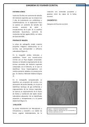
CASO 2En este caso no se pudo atravesar <strong>la</strong> obstrucción, apesar <strong>de</strong> haber empleado un doble abordaje, venasbasílica y femoral y <strong>de</strong> lo frustrante <strong>de</strong> <strong>la</strong> imagen.Por tanto fracasamos en el tratamiento, no siendoconsi<strong>de</strong>rada susceptible reparación quirúrgica por elServicio <strong>de</strong> Cirugía Vascu<strong>la</strong>r.<strong>SÍNDROME</strong> <strong>DE</strong> <strong>PAGET</strong>-<strong>SCHROETTER</strong>
MATERIAL Y METODOSDes<strong>de</strong> enero <strong>de</strong> 1998 hemos tratado 9 pacientescon síndrome <strong>de</strong> Paget-Schroetter. 6 varones y 3mujeres, <strong>de</strong> eda<strong>de</strong>s comprendidas entre 17 y 42 años(m= 27).Se empezó atravesando <strong>la</strong> obstrucción, seguido<strong>de</strong> trombectomía farmacomecánica, con dosis altas entiempos cortos <strong>de</strong> uroquinasa (3 casos) o rtpa (6casos).Posteriormente se di<strong>la</strong>tó <strong>la</strong> estenosis venosa conbalones <strong>de</strong> 8 a 12 mm <strong>de</strong> diámetro y en 4 casospreviamente con balones cortantes.<strong>SÍNDROME</strong> <strong>DE</strong> <strong>PAGET</strong>-<strong>SCHROETTER</strong>
RESULTADOSÉxito en 7 pacientes (78%). Ninguno intervenidoquirúrgicamente con posterioridad.Intervalo <strong>de</strong>s<strong>de</strong> <strong>la</strong> aparición <strong>de</strong> los síntomas y eltratamiento entre 2 y 60 días (m = 27). Fracaso en 2pacientes, que no se pudo atravesar <strong>la</strong> obstrucción,síntomas <strong>30</strong> y 60 días.Seguimiento <strong>de</strong> 1’2 a 9’5 años (m = 6’5 a) libres <strong>de</strong>síntomas y sin limitación funcional.<strong>SÍNDROME</strong> <strong>DE</strong> <strong>PAGET</strong>-<strong>SCHROETTER</strong>
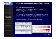
<strong>SÍNDROME</strong> <strong>DE</strong> <strong>PAGET</strong>-<strong>SCHROETTER</strong>• Afecta predominantemente a hombres (4/1)• En el brazo dominante (70%)• Los síntomas suelen ir precedidos <strong>de</strong>:ejercicio infrecuente <strong>de</strong>l brazo oabducción <strong>de</strong>l hombro afecto• Aunque un significativo porcentaje <strong>de</strong> pacientes norefieren ninguna actividad previa.<strong>SÍNDROME</strong> <strong>DE</strong> <strong>PAGET</strong>-<strong>SCHROETTER</strong>
<strong>SÍNDROME</strong> <strong>DE</strong> <strong>PAGET</strong>-<strong>SCHROETTER</strong>SINTOMATOLOGÍA MÁS FRECUENTE :• Dolor• E<strong>de</strong>ma• Congestión venosa <strong>de</strong>l brazo• Parestesias• Fatigabilidad• Cianosis<strong>SÍNDROME</strong> <strong>DE</strong> <strong>PAGET</strong>-<strong>SCHROETTER</strong>
<strong>SÍNDROME</strong> <strong>DE</strong> <strong>PAGET</strong>-<strong>SCHROETTER</strong>El S. <strong>de</strong> Paget-Schroetter se ha atribuido a una anomalía en el<strong>de</strong>sarrollo, que provoca una compresión externa dinámica <strong>de</strong> <strong>la</strong> venasubc<strong>la</strong>via a nivel <strong>de</strong>l estrecho torácico superior. La compresión se acentúapor <strong>la</strong> abducción <strong>de</strong>l brazo y otras posturas forzadas, resultando unpinzamiento por <strong>la</strong>s estructuras óseas y músculotendinosas en el triángulocostoc<strong>la</strong>vicu<strong>la</strong>r.El sustrato anatómico <strong>de</strong>l pinzamiento incluye hipertrofia einserciones excesivamente amplias <strong>de</strong> los tendones escalenoanterior,medio y subc<strong>la</strong>vio. Estos ligamentos y bandas fibromuscu<strong>la</strong>rescongénitas son los que contribuyen a <strong>la</strong> compresión neurovascu<strong>la</strong>r en elestrecho torácico superior, habiendo sido c<strong>la</strong>sificadas en 14 tipos porRoos DB, siendo los tipos 3 y 5 los habitualmente implicados en <strong>la</strong>compresión vascu<strong>la</strong>r.<strong>SÍNDROME</strong> <strong>DE</strong> <strong>PAGET</strong>-<strong>SCHROETTER</strong>
<strong>SÍNDROME</strong> <strong>DE</strong> <strong>PAGET</strong>-<strong>SCHROETTER</strong>
<strong>SÍNDROME</strong> <strong>DE</strong> <strong>PAGET</strong>-<strong>SCHROETTER</strong>Las flebografías practicadas en los pacientes con TVASE, habitualmente<strong>de</strong>muestran oclusiones <strong>de</strong> longitu<strong>de</strong>s variables y <strong>de</strong>sarrollo <strong>de</strong> co<strong>la</strong>teralesalre<strong>de</strong>dor <strong>de</strong>l hombro y pared torácica. Tras <strong>la</strong> disolución <strong>de</strong>l trombo confibrinolisis química o farmacomecánica, se aprecia una estenosis fija ouna oclusión corta en <strong>la</strong> zona <strong>de</strong> <strong>la</strong> vena subc<strong>la</strong>via próxima a <strong>la</strong> unión con<strong>la</strong> vena yugu<strong>la</strong>r.Los escasos cirujanos que, tras los hal<strong>la</strong>zgos flebográficos, hanintervenido estas estenosis, han <strong>de</strong>scrito que consisten en estrechecesfijas fibróticas, a menudo con bandas endoluminales, estando el segmentovenoso afecto adyacente a <strong>la</strong> cabeza <strong>de</strong> <strong>la</strong> c<strong>la</strong>vícu<strong>la</strong>.<strong>SÍNDROME</strong> <strong>DE</strong> <strong>PAGET</strong>-<strong>SCHROETTER</strong>
<strong>SÍNDROME</strong> <strong>DE</strong> <strong>PAGET</strong>-<strong>SCHROETTER</strong>TRATAMIENTOFibrinolisis y/o trombectomía mecánicaLa precocidad <strong>de</strong> <strong>la</strong> fibrinolisis-trombectomía esimportante para evitar <strong>la</strong>s lesiones <strong>de</strong> <strong>la</strong> íntima ysubsecuente fibrosis• Descompresión quirúrgica tardía• Cirugía precoz <strong>de</strong>spués <strong>de</strong> <strong>la</strong> fibrinolisis*Opiniones controvertidas sobre <strong>la</strong> necesidad y el momento <strong>de</strong> realizar<strong>la</strong> cirugía <strong>de</strong>scompresiva.En general <strong>la</strong> indicación y el momento <strong>de</strong> <strong>la</strong> cirugía se pue<strong>de</strong>n fijaren función <strong>de</strong> <strong>la</strong>s características <strong>de</strong>l flujo en el segmento venosoaxilosubc<strong>la</strong>vio y <strong>la</strong> severidad <strong>de</strong> <strong>la</strong> sintomatología residual.<strong>SÍNDROME</strong> <strong>DE</strong> <strong>PAGET</strong>-<strong>SCHROETTER</strong>
<strong>SÍNDROME</strong> <strong>DE</strong> <strong>PAGET</strong>-<strong>SCHROETTER</strong>TRATAMIENTOLa Angiop<strong>la</strong>stia con Balón no corrige <strong>la</strong> compresión extrínsecamúsculotendinosa,en cambio es muy útil para tratar <strong>la</strong> estenosisfibrótica y el trombo organizado, que subyacen tras <strong>la</strong> fibrinolisis y que<strong>de</strong> no tratarse suelen provocar una retrombosis.Existen referencias <strong>de</strong> permeabilidad a <strong>la</strong>rgo p<strong>la</strong>zo y ausencia<strong>de</strong> sintomatología tras angiop<strong>la</strong>stia con balón <strong>de</strong>spués <strong>de</strong> <strong>la</strong> fibrinolisisen pacientes con TVASE, sin cirugía posterior. Esto ha llevado aalgunos autores a recomendar un manejo conservador, reservando <strong>la</strong>indicación quirúrgica sólo para aquellos pacientes con marcadasalteraciones flebográficas y sintomatología acompañante.<strong>SÍNDROME</strong> <strong>DE</strong> <strong>PAGET</strong>-<strong>SCHROETTER</strong>
<strong>SÍNDROME</strong> <strong>DE</strong> <strong>PAGET</strong>-<strong>SCHROETTER</strong>TRATAMIENTOEl objetivo <strong>de</strong> <strong>la</strong> Descompresión Quirúrgica es corregir elpinzamiento postural por el músculo subc<strong>la</strong>vio y <strong>la</strong>s fibras inferiores <strong>de</strong>lescaleno anterior contra <strong>la</strong> primera costil<strong>la</strong>.La intervención consiste en <strong>la</strong> exéresis <strong>de</strong> <strong>la</strong> primera costil<strong>la</strong> y <strong>la</strong>escisión <strong>de</strong>l tendón subc<strong>la</strong>vio así como <strong>de</strong> <strong>la</strong> inserción <strong>de</strong>l escalenoanterior; el abordaje pue<strong>de</strong> ser subc<strong>la</strong>vicu<strong>la</strong>r anterior, suprac<strong>la</strong>vicu<strong>la</strong>r otransaxi<strong>la</strong>r. El Trombo Residual es limpiado y, cuando se encuentra unafibrosis significativa, se pue<strong>de</strong> realizar una Venolisis circu<strong>la</strong>r asi comoVenop<strong>la</strong>stia con parche en el momento <strong>de</strong> <strong>la</strong> <strong>de</strong>scompresión.<strong>SÍNDROME</strong> <strong>DE</strong> <strong>PAGET</strong>-<strong>SCHROETTER</strong>
<strong>SÍNDROME</strong> <strong>DE</strong> <strong>PAGET</strong>-<strong>SCHROETTER</strong>TRATAMIENTO• La Angiop<strong>la</strong>stia con Balón pue<strong>de</strong> tener utilidad en <strong>la</strong>sestenosis residuales• Las Endoprótesis, que no <strong>de</strong>ben emplearse <strong>de</strong> entrada enaquellos pacientes no sometidos a cirugía, por los dudososresultados a <strong>la</strong>rgo p<strong>la</strong>zo, podrían tener alguna aplicación en loscasos postquirúrgicos que no respon<strong>de</strong>n a <strong>la</strong> angiop<strong>la</strong>stia conbalón.<strong>SÍNDROME</strong> <strong>DE</strong> <strong>PAGET</strong>-<strong>SCHROETTER</strong>
CONCLUSIÓNEl tratamiento endovascu<strong>la</strong>r (Trombectomía+ATP)aunque no corrige <strong>la</strong> compresión extrínseca, es muy útil paratratar <strong>la</strong> estenosis fibrótica y el trombo organizado.La permeabilidad a <strong>la</strong>rgo p<strong>la</strong>zo sin sintomatologíaapoyan un manejo conservador, reservando <strong>la</strong> indicaciónquirúrgica para aquellos pacientes con sintomatología ymarcadas alteraciones flebográficas.<strong>SÍNDROME</strong> <strong>DE</strong> <strong>PAGET</strong>-<strong>SCHROETTER</strong>
BIBLIOGRAFÍA1. Hughes ESR. Venous obstruction in the upper extremity (Paget-Schroetter's syndrome): a review of 320 casesabstracted. Int Abst Surg 88:89, 1949.2. McCarthy WJ, Vogelzang RL, Bergan JJ. Changing concepts and present-day etiology of upper extremity venousthrombosis. p. 407. In Bergan JJ, Yao JST ( eds): Venous Disor<strong>de</strong>rs. WB Saun<strong>de</strong>rs, Phi<strong>la</strong><strong>de</strong>lphia, 1991.3. Becker GJ, Hol<strong>de</strong>n RW, Rabe FE et al. Local thrombolytic therapy for subc<strong>la</strong>vian andaxil<strong>la</strong>ry vein thrombosis. Radiology 149:419, 1983.4. Aziz S, Straehley CJ, Whe<strong>la</strong>n TJ Jr. Effort-re<strong>la</strong>ted axillo- subc<strong>la</strong>vian vein thrombosis: a new theory ofpathogenesis and a plea for direct surgical intervention. Am J Surg 152:57, 1986.5. Lindb<strong>la</strong>d, Tengbom L, Bergqvist D. Deep vein thrombosis of the axil<strong>la</strong>ry-subc<strong>la</strong>vian veins:epi<strong>de</strong>miologic data, effects of different types of treatment and <strong>la</strong>te seque<strong>la</strong>e.Eur J Vasc Surg 2:161,1988 .6. Kunkel JM, Mach1e<strong>de</strong>r HI. Treatment of Paget-Schroetter syndrome. Arch Surg 125:1153, 1989.7. Strange-Vognsen HH, Hauch O, An<strong>de</strong>rson J, Struckmann J. Resection of the first rib following <strong>de</strong>ep arm veinthrombolysis in patients with thoracic outlet syndrome. J Cardiovasc Surg <strong>30</strong>:4<strong>30</strong>, 1989.8. DeWeese JA. Results of surgica1 treatment of axil<strong>la</strong>ry- subc<strong>la</strong>vian venous thrombosis. p. 421. In Bergan JJ, YaoJST (eds): Venous Disor<strong>de</strong>rs. WB Saun<strong>de</strong>rs, Phi<strong>la</strong><strong>de</strong>lphia, 1991.9. Hingorani A, Ascher E, Lorenson E, et al. Upper extremity <strong>de</strong>ep venous thrombosis and its impact on morbidityand mortality rates in a hospital- based popu<strong>la</strong>tion. J Vasc Surg 1997; 26:853-860.10. Ellis MH, Manor Y, Witz M. Risk factors and management of patients with upper limb <strong>de</strong>ep vein thrombosis. Chest2000; 117:43-46.<strong>SÍNDROME</strong> <strong>DE</strong> <strong>PAGET</strong>-<strong>SCHROETTER</strong>
BIBLIOGRAFÍA11.Chang R, Home MK, Mayo DJ, Doppman JL. Pulse-spray treatment of subc<strong>la</strong>vian and jugu<strong>la</strong>r venous thrombi with recombinant tissuep<strong>la</strong>sminogen activator. J Vasc Interv Radiol l996; 7:845-851.12.A<strong>de</strong>lman MA, Stone DH, Riles TS, Lamparello PJ, Giango<strong>la</strong> G, Rosen RJ. A multidisciplinary approach to the treatment of Paget-Schroetter syndrome. Ann Vasc Surg 1997; 11:149- 154.13.Sheeran S, Hallisey M, Murphy T, Faberman R, Sherman S. Local thrombolytic therapy as part of a multidisciplinaryapproach to acute axillosubc<strong>la</strong>vian vein thrombosis (Paget- Schroetter syndrome). J Vasc Interv Radiol 1997; 8:253-260.14.Rutherford RB. Primary subc<strong>la</strong>vian- axil<strong>la</strong>ry vein thrombosis: the re<strong>la</strong>tive roles of thrombolysis, percutaneous angiop<strong>la</strong>sty, stents,and surgery. Semin Vasc Surg 1998; 11:91-95.15.Angle N, Ge<strong>la</strong>bert HA, Farooq MM, et al. Safety and efficacy of early surgical <strong>de</strong>compression of the thoracic outlet for Paget-Schroettersyndrome. Ann Vasc Surg 2001; 15:37-42.16. Urschel HC, Jr., Razzuk MA. Paget- Schroetter syndrome: what is the best management?Ann Thorac Surg 2000; 69:1663-1668; discussion, 1668-1669.17. Kreienberg PB, Chang BB, Darling RC III, et al. Long-term results in patients treated withthrombolysis, thoracic inlet <strong>de</strong>compression, and subc<strong>la</strong>vian vein stenting for Paget- Schroetter syndrome. J Vasc Surg 2001;33(suppl):Sl00-S105.18. Lokanathan R, Salvian AJ, Chen JC, Morris C, Taylor DC, Hsiang YN. Outcome afterthrombolysis and selective thoracic outlet <strong>de</strong>compression for primary axil<strong>la</strong>ry vein thrombosis. J Vasc Surg 2001; 33:783-788.19. Lee W A, Hill BB, Harris EJ Jr, Semba CP, Olcott CI. Surgical intervention is not requiredfor all patients with subc<strong>la</strong>vian vein thrombosis. J Vasc Surg 2000; 32:57-67.20. Hall LD, Murray JD, Boswell GE. Venous stent p<strong>la</strong>cement as an adjunct to the staged, multimodal treatment of Paget-Schroettersyndrome. J Vasc Interv Radiol l995; 6:565-569; discus- sion,569-570.21. Lee JT, Karwowski JK, Harris EJ, Haukoos JS, Olcott C. Long-term thrombotic recurrence after nonoperative management ofPaget-Schroetter syndrome. J Vasc Surg 2006;43:1236-43<strong>SÍNDROME</strong> <strong>DE</strong> <strong>PAGET</strong>-<strong>SCHROETTER</strong>