Sindrome de Dyke Davidoff Masson - Hospital General de Culiacán
Sindrome de Dyke Davidoff Masson - Hospital General de Culiacán
Sindrome de Dyke Davidoff Masson - Hospital General de Culiacán

Create successful ePaper yourself
Turn your PDF publications into a flip-book with our unique Google optimized e-Paper software.
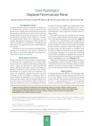
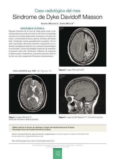
Caso radiológico <strong>de</strong>l mes<strong>Sindrome</strong> <strong>de</strong> <strong>Dyke</strong> <strong>Davidoff</strong> <strong>Masson</strong>HISTORIA CLÍNICAPaciente femenino <strong>de</strong> 41 años <strong>de</strong> edad, quien acu<strong>de</strong> a consultapor presentar crisis convulsivas. Sus crisis son parcialesmotoras con marcha Jacksoniana, iniciando en la mano <strong>de</strong>rechay continuando en brazo, pierna, hemicara <strong>de</strong>l mismolado y finalmente ocurre generalización secundaria. El examenneurológico <strong>de</strong>muestra <strong>de</strong>terioro cognoscitivo leve ydiscreta hemiparesia <strong>de</strong>recha. Los exámenes hematológicosson normales. A pesar <strong>de</strong> múltiples esquemas <strong>de</strong> tratamientodurante varios años (Fenitoína, Valproato <strong>de</strong> magnesio,Carbamazepina, Topiramato), la paciente presenta <strong>de</strong>scontrol<strong>de</strong> sus crisis, requiriendo frecuentes internamientos.Ga x i o l a-Hi g u e r a L 1 , Ló p e z-Me z a E 2HALLAZGOS por RM. Ver figuras 1-5.Figura 2. Imagen RM Axial FLAIR.Figura 1. Imagen RM Axial T2 .Atrofia <strong>de</strong> hemisferio cerebral izquierdo.Figura 3. Imagen <strong>de</strong> RM Sagital en T1. Hemisferio <strong>de</strong>recho1Médico adscrito al servicio <strong>de</strong> radiología e imagen <strong>de</strong>l <strong>Hospital</strong> <strong>General</strong> <strong>de</strong> Culiacán2Neurología clínica <strong>de</strong>l <strong>Hospital</strong> <strong>General</strong> <strong>de</strong> Culiacán.Enviar correspon<strong>de</strong>ncia, observaciones y sugerencias a Gaxiola Higuera al teléfono (667)228 20 78,correo electrónico: logaxiola@hotmail.comEste artículo pue<strong>de</strong> ser visto en www.hgculiacan.comSociedad Médica <strong>de</strong>l <strong>Hospital</strong> <strong>General</strong> <strong>de</strong> Culiacán “Dr. Bernardo J. Gastélum”A S Sin Vol.II No.3 p.113-114, 2008113
Ga x i o l a y c o l s.<strong>Sindrome</strong> <strong>de</strong> <strong>Dyke</strong> <strong>Davidoff</strong> <strong>Masson</strong>DIAGNÓSTICOSíndrome <strong>de</strong> <strong>Dyke</strong> <strong>Davidoff</strong> <strong>Masson</strong>.Figura 4. Imagen <strong>de</strong> RM Sagital en T1.Hemisferio izquierdo con atrofia.DISCUSIÓNEn 1933 <strong>Dyke</strong>, <strong>Davidoff</strong> y <strong>Masson</strong> <strong>de</strong>scribieron los cambiosneumoencefalográficos y radiográficos en 9 pacientes con hemicrisis,hemiparesia y hemiatrofia corporal. 1 El diagnóstico <strong>de</strong>este cuadro suele realizarse ante la presencia <strong>de</strong> hemiatroafia<strong>de</strong> un hemisferio cerebral, se acompaña <strong>de</strong> hiperostosis subyacentey se manifiesta por crisis parciales <strong>de</strong> difícil control.Se ha propuesto como etiología el trauma, inflamación,insultos vasculares in-utero y crisis febriles. 2 Desafortunadamentese trata <strong>de</strong> una forma <strong>de</strong> epilepsia refractaria atratamiento y se asocia a una baja calidad <strong>de</strong> vida.Figura 5. Imagen <strong>de</strong> RM coronal T2.Hemisferio izquierdo con atrofia.Bibliografía1.2.3.<strong>Dyke</strong> C G, <strong>Davidoff</strong> L M and <strong>Masson</strong> C B, Cerebral Hemiatrophy and homolateral hypertrophy of the skull and sinuses. Surg Gynecol Obstet1933; 57: 588-600.Garg RK, Karak B. Cerebral hemiatrophy: a possible etiological relation with febrile seizures. Indian Pediatr 1998;35:79-81.Tas<strong>de</strong>mir HA, Incesu L, Yazicioglu AK, Belet U, Gungor L. <strong>Dyke</strong>-<strong>Davidoff</strong>- <strong>Masson</strong> syndrome. Clin Imaging 2002;26: 13-7Artículo disponible en www.imbiomed.com.A S Sin Vol.II No.3 p.113-114, 2008114