Aca - Departamento de Física - Universidad Técnica Federico Santa ...
Aca - Departamento de Física - Universidad Técnica Federico Santa ...
Aca - Departamento de Física - Universidad Técnica Federico Santa ...

You also want an ePaper? Increase the reach of your titles
YUMPU automatically turns print PDFs into web optimized ePapers that Google loves.
V Encuentro Sud Americano <strong>de</strong> Colisiones Inelásticas en la Materia<br />
Elemental analysis of the Chaitén volcano ash 2008-2009 eruptions<br />
Shimrit Elimelech, Jorge E. Valdés, P. Vargas<br />
Depto. <strong>de</strong> <strong>Física</strong>, <strong>Universidad</strong> <strong>Técnica</strong> Fe<strong>de</strong>rico <strong>Santa</strong> María, Casilla 110-V, Valparaíso, Chile<br />
Shimrit.elimelech@usm.cl<br />
The Chaitén volcano eruptions in 2008 and 2009<br />
have provi<strong>de</strong>d a significant opportunity to study<br />
the typical heavy elemental content and microstructure<br />
of the ash emitted from one of the most<br />
critical volcanoes in Chile. Recent studies of<br />
Chaitén offer insights about the environmental<br />
effects of the 2008 eruption in the water, vegetation<br />
and air. In this study, we offer new insights<br />
about typical properties of the ash such as composition<br />
and microstructure. This information<br />
allows us un<strong>de</strong>rstanding of the process leading<br />
the eruption phenomena, explaining its effects<br />
and predicting the eruption future consequences.<br />
Microstructure Characterization was done using<br />
scanning electron microscopy (SEM) with<br />
mo<strong>de</strong>s of backscattered electrons (BSE) and<br />
secondary electrons (SE). The elements' content<br />
was found using Scanning electron microscopy–<br />
energy dispersive x-ray spectroscopy (SEM-<br />
EDS) and x-ray diffraction (XRD).<br />
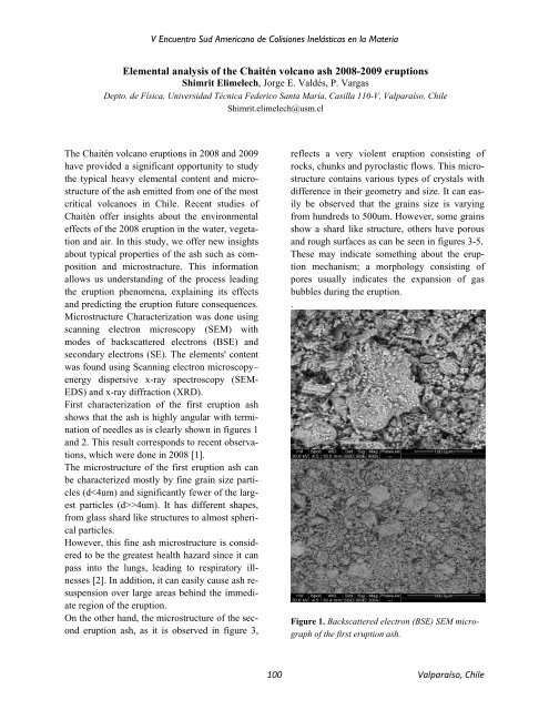
First characterization of the first eruption ash<br />
shows that the ash is highly angular with termination<br />
of needles as is clearly shown in figures 1<br />
and 2. This result corresponds to recent observations,<br />
which were done in 2008 [1].<br />
The microstructure of the first eruption ash can<br />
be characterized mostly by fine grain size particles<br />
(d>4um). It has different shapes,<br />
from glass shard like structures to almost spherical<br />
particles.<br />
However, this fine ash microstructure is consi<strong>de</strong>red<br />
to be the greatest health hazard since it can<br />
pass into the lungs, leading to respiratory illnesses<br />
[2]. In addition, it can easily cause ash resuspension<br />
over large areas behind the immediate<br />
region of the eruption.<br />
On the other hand, the microstructure of the second<br />
eruption ash, as it is observed in figure 3,<br />
reflects a very violent eruption consisting of<br />
rocks, chunks and pyroclastic flows. This microstructure<br />
contains various types of crystals with<br />
difference in their geometry and size. It can easily<br />
be observed that the grains size is varying<br />
from hundreds to 500um. However, some grains<br />
show a shard like structure, others have porous<br />
and rough surfaces as can be seen in figures 3-5.<br />
These may indicate something about the eruption<br />
mechanism; a morphology consisting of<br />
pores usually indicates the expansion of gas<br />
bubbles during the eruption.<br />
.<br />
Figure 1. Backscattered electron (BSE) SEM micrograph<br />
of the first eruption ash.<br />
100 Valparaíso, Chile