Revista Veterinaria Zacatecas 2010 - Universidad Autónoma de ...
Revista Veterinaria Zacatecas 2010 - Universidad Autónoma de ...
Revista Veterinaria Zacatecas 2010 - Universidad Autónoma de ...
Create successful ePaper yourself
Turn your PDF publications into a flip-book with our unique Google optimized e-Paper software.
M CFranciscoJavierDomínguezGaray<br />
Rector<strong>de</strong>la<strong>Universidad</strong>Autónoma<strong>de</strong><strong>Zacatecas</strong><br />
IQArmandoSilvaChairez<br />
SecretarioGeneral<br />
M enCJesúsOctavioEnríquezRivera<br />
SecretarioAcadémico<br />
CPEmilioMoralesVera<br />
SecretarioAdministrativo<br />
PhDJoséManuelSilvaRamos<br />
Director<strong>de</strong>laUnidadAcadémica<strong>de</strong>Medicina<strong>Veterinaria</strong>yZootecnia<br />
DrenCRomanaMelbaRincónDelgado<br />
Responsable<strong>de</strong>lPrograma<strong>de</strong>Licenciatura<br />
PhDCarlosFernandoAréchigaFlores<br />
Responsable<strong>de</strong>lPrograma<strong>de</strong>Doctorado<br />
PhDHéctorGutiérezBañuelos<br />
Responsable<strong>de</strong>lPrograma<strong>de</strong>Maestría<br />
DrenCRómuloBañuelosValenzuela<br />
Coordinador<strong>de</strong>Investigación<br />
M enCJuanIgnacioDávilaFélix<br />
SecretarioAdministrativo<br />
DrenCFranciscoJavierEscobarMedina<br />
DirectorTécnicoyEditor<strong>de</strong>la<strong>Revista</strong><strong>Veterinaria</strong><strong>Zacatecas</strong>
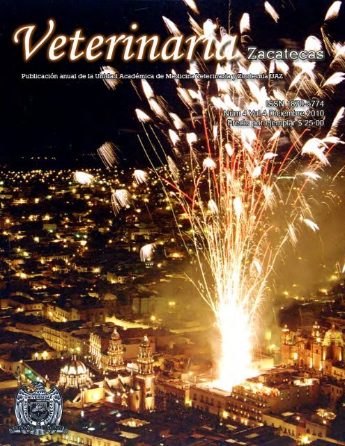
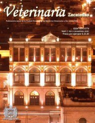
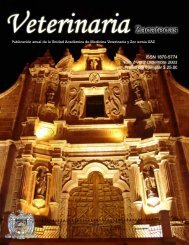
NuestraPortada<br />
Vista<strong>de</strong>LaCatedralyPlaza<strong>de</strong>Armas.<strong>Zacatecas</strong>,Zac.<br />
Fotografía:SalvadorRomoGalardo,tel8992429,e-mailsrmfoto@tera.com<br />
<strong>Veterinaria</strong> <strong>Zacatecas</strong> es una publicación anual <strong>de</strong> la Unidad Académica <strong>de</strong> Medicina<br />
<strong>Veterinaria</strong> y Zootecnia <strong>de</strong> la <strong>Universidad</strong>Autónoma <strong>de</strong> <strong>Zacatecas</strong>.ISSN:1870-5774.Sólo<br />
se autoriza la reproducción <strong>de</strong> artículos en los casos que se cite la fuente.<br />
Corespon<strong>de</strong>ncia dirigirla a: <strong>Revista</strong> <strong>Veterinaria</strong> <strong>Zacatecas</strong>. <strong>Revista</strong> <strong>de</strong> la Unidad<br />
Académica <strong>de</strong> Medicina <strong>Veterinaria</strong> y Zootecnia <strong>de</strong> la <strong>Universidad</strong> Autónoma <strong>de</strong><br />
<strong>Zacatecas</strong>.Caretera Panamericana,tramo <strong>Zacatecas</strong>-Fresnilo Km 31.5.Apartados Postales<br />
9y11,Calera<strong>de</strong>VíctorRosales,Zac.CP98500.Teléfono01(478)9851255.Fax:01<br />
(478) 9 85 02 02.E-mail:vetzac@uaz.edu.mx.URL htp:/www.uaz.edu.mx/publicaciones<br />
Precioporcadaejemplar$25.00<br />
Distribución:UnidadAcadémica<strong>de</strong>Medicina<strong>Veterinaria</strong>yZootecnia<strong>de</strong>la<strong>Universidad</strong><br />
Autónoma<strong>de</strong><strong>Zacatecas</strong>.
COMPORTAMIENTO REPRODUCTIVO DE LA YEGUA<br />
María <strong>de</strong>l Sol Mén<strong>de</strong>z Bernal, Francisco Guadalupe Mén<strong>de</strong>z Bernal, Fe<strong>de</strong>rico <strong>de</strong> la Colina Flores, Francisco<br />
Javier Escobar Medina<br />
Unidad Académica <strong>de</strong> Medicina <strong>Veterinaria</strong> y Zootecnia <strong>de</strong> la <strong>Universidad</strong> Autónoma <strong>de</strong> <strong>Zacatecas</strong><br />
E-mail: fescobar@uaz.edu.mx<br />
RESUMEN<br />
La yegua presenta comportamiento reproductivo estacional, con intervalos entre ovulaciones durante los días<br />
con mayor cantidad <strong>de</strong> horas luz y anestro en otoño e invierno. El ciclo reproductivo se compone <strong>de</strong> intervalo<br />
entre ovulaciones, gestación y período posparto. En los intervalos entre ovulaciones se presenta el crecimiento<br />
folicular, la ovulación, formación <strong>de</strong>l cuerpo lúteo con producción <strong>de</strong> progesterona y <strong>de</strong>strucción <strong>de</strong>l cuerpo<br />
lúteo e inicio <strong>de</strong> un nuevo intervalo. La mayor fertilidad con monta natural se presenta 1 a 2 días antes <strong>de</strong> la<br />
ovulación y con inseminación artificial alre<strong>de</strong>dor <strong>de</strong> la ovulación. Las yeguas nuevamente ovulan poco<br />
tiempo <strong>de</strong>spués <strong>de</strong>l parto.<br />
Palabras clave: comportamiento reproductivo, yeguas, fertilidad<br />
<strong>Veterinaria</strong> <strong>Zacatecas</strong> <strong>2010</strong>; 3: 135-147<br />
INTRODUCCIÓN<br />
La reproducción en la yegua se realiza por medio<br />
<strong>de</strong> interacción entre ambiente y organismo. La<br />
influencia <strong>de</strong>l ambiente se transmite a través <strong>de</strong><br />
los sentidos y el animal las recibe en el sistema<br />
nervioso central, particularmente en hipotálamo<br />
para, por un lado, la secreción <strong>de</strong> hormona<br />
liberadora <strong>de</strong> las gonadotropinas (GnRH); 1-3 y<br />
<strong>de</strong>senca<strong>de</strong>nar el concierto hormonal que conduce<br />
a la ovulación, y, por otro, estimular la secreción<br />
<strong>de</strong> melatonina para programar la estacionalidad<br />
reproductiva. 4-6<br />
La GnRH se dirige al lóbulo anterior <strong>de</strong><br />
la hipófisis para promover la síntesis y secreción<br />
<strong>de</strong> gonadotropinas: hormonas folículo estimulante<br />
(FSH) y luteinizante (LH). 1-3<br />
Las gonadotropinas promueven el<br />
<strong>de</strong>sarrollo folicular, la FSH hasta la <strong>de</strong>sviación y<br />
LH al nivel preovulatorio. 7 Los folículos producen<br />
estradiol e inhibina. El estradiol ejerce<br />
retroalimentación negativa sobre las<br />
gonadotropinas 8 y la inhibina sobre FSH. 9 La LH,<br />
a<strong>de</strong>más, se relaciona con la ovulación. 5 En las<br />
yeguas sin gestación y bajo condiciones<br />
a<strong>de</strong>cuadas para la reproducción, las ovulaciones<br />
(acompañadas <strong>de</strong> celo) se repiten para constituir el<br />
período entre ovulaciones o intervalo<br />
interovulatorio.<br />
Después <strong>de</strong> la ovulación se <strong>de</strong>sarrolla el<br />
cuerpo lúteo, estructura que produce progesterona.<br />
Ésta ejerce retroalimentación negativa sobre<br />
gonadotropinas. 10 El endometrio secreta<br />
prostaglandina F 2 para <strong>de</strong>struir el cuerpo lúteo al<br />
final <strong>de</strong>l intervalo entre ovulaciones en yeguas sin<br />
gestación. 11,12<br />
Las yeguas pue<strong>de</strong>n recibir monta natural<br />
o inseminación artificial en el momento apropiado<br />
<strong>de</strong>l celo y concebir. El producto se <strong>de</strong>sarrolla<br />
durante la gestación y, <strong>de</strong>spués <strong>de</strong> 11 meses, se<br />
presenta el parto.<br />
Con base en lo anterior, el ciclo<br />
reproductivo <strong>de</strong> la yegua se compone <strong>de</strong> intervalo<br />
entre ovulaciones, gestación y período posparto.<br />
El fotoperiodo a través <strong>de</strong> la secreción <strong>de</strong><br />
melatonina, programa el intervalo entre<br />
ovulaciones en una temporada específica, lo que<br />
en el presente documento se conoce como<br />
temporada reproductiva, y establece el<br />
comportamiento reproductivo estacional.<br />
TEMPORADA REPRODUCTIVA<br />
Las yeguas son poliéstricas estacionales,<br />
presentan celos acompañados <strong>de</strong> ovulación en los<br />
días con mayor cantidad <strong>de</strong> horas luz y<br />
permanecen en anestro o período anovulatorio en<br />
los días con reducción <strong>de</strong>l fotoperiodo. 5,13-16 El<br />
anestro se ha observado sistemáticamente en las<br />
yeguas jóvenes entre 2 y 3 años <strong>de</strong> edad y en las<br />
adultas con previo amamantamiento. No todas las<br />
hembras mayores <strong>de</strong> 3 años <strong>de</strong> edad, sin lactancia<br />
135
previa, han mostrado anestro en el otoño y en el<br />
invierno; el 50% <strong>de</strong> éstas pue<strong>de</strong>n presentar<br />
ovulaciones acompañadas <strong>de</strong> celo durante los días<br />
con reducción <strong>de</strong>l fotoperiodo. 17,18 El mayor<br />
porcentaje <strong>de</strong> yeguas concibieron <strong>de</strong> abril –<br />
agosto, en un estudio realizado <strong>de</strong>spués <strong>de</strong>l<br />
sacrificio <strong>de</strong> yeguas proce<strong>de</strong>ntes <strong>de</strong> los Estados <strong>de</strong><br />
Coahuila, Nuevo León, Durango, Tamaulipas, San<br />
Luis Potosí, Jalisco, Aguascalientes y <strong>Zacatecas</strong>.<br />
Por lo tanto, los partos ocurrieron <strong>de</strong> marzo a<br />
julio. 14<br />
Las yeguas utilizan el fotoperiodo para<br />
programar sus partos en primavera, temporada<br />
más favorable para la supervivencia <strong>de</strong> su<br />
<strong>de</strong>scen<strong>de</strong>ncia. 5,19 La señal probablemente se<br />
realiza como en otras especies: las horas luz se<br />
registren en retina y el estímulo se transmita<br />
sucesivamente al núcleo supraquiasmático <strong>de</strong>l<br />
hipotálamo, ganglio cervical superior y glándula<br />
pineal. La glándula pineal actúa como traductor<br />
neuroendocrino, convierte la señal neural en<br />
secreción hormonal. 20-22 Esta glándula produce<br />
melatonina durante las horas oscuras <strong>de</strong>l día, 5 y la<br />
melatonina, <strong>de</strong>pendiendo <strong>de</strong>l período <strong>de</strong> su<br />
secreción, actúa sobre el eje hipotálamo-hipófisisgónada<br />
4,6 Por ejemplo, el período <strong>de</strong> secreción <strong>de</strong><br />
melatonina es más largo durante otoño e invierno<br />
coinci<strong>de</strong> con la temporada <strong>de</strong> anestro en la yegua.<br />
Por lo tanto, períodos largos <strong>de</strong> secreción <strong>de</strong><br />
melatonina inhiben la presentación <strong>de</strong> celos. 23<br />
En algunas explotaciones los partos se<br />
prefieren al inicio <strong>de</strong>l año, antes <strong>de</strong> la época<br />
natural en estos animales. Para modificarles la<br />
temporada se han utilizado principalmente<br />
tratamientos con luz artificial, adicional al<br />
fotoperiodo natural. 24-27 En un estudio las cópulas<br />
se programaron <strong>de</strong>l 15 <strong>de</strong> febrero al 30 <strong>de</strong> junio<br />
para esperar los partos en los 4 primeros meses <strong>de</strong>l<br />
año (enero-mayo). Las hembras sin concepción se<br />
trataron con 16 horas luz <strong>de</strong>l 1 <strong>de</strong> diciembre al 31<br />
<strong>de</strong> enero. El 35.7%, 17.8%, 19.4%, 14.7% y<br />
12.4% <strong>de</strong> los partos se presentaron en enero,<br />
febrero, marzo, abril y mayo, respectivamente. 28<br />
INTERVALO INTEROVULATORIO<br />
Este intervalo inicia en una ovulación<br />
asociada al estro y termina con la ovulación <strong>de</strong>l<br />
estro siguiente, anteriormente se conocía como<br />
ciclo estral. Se <strong>de</strong>cidió utilizar el término <strong>de</strong><br />
intervalo entre ovulaciones para i<strong>de</strong>ntificar con<br />
mayor exactitud los días en que se realiza el<br />
crecimiento folicular y eliminar la ambigüedad<br />
asociada con los métodos utilizados para<br />
<strong>de</strong>tección y <strong>de</strong>finición <strong>de</strong>l estro. El intervalo entre<br />
ovulaciones, en yeguas dura en promedio 21 a 22<br />
días y 24 días en las ponis. 5<br />
En el intervalo interovulatorio se realiza<br />
crecimiento folicular con producción <strong>de</strong> estradiol,<br />
ovulación, formación <strong>de</strong> cuerpo lúteo con<br />
producción <strong>de</strong> progesterona y <strong>de</strong>strucción <strong>de</strong>l<br />
cuerpo lúteo e inicio <strong>de</strong> un nuevo intervalo. Los<br />
niveles hormonales <strong>de</strong>l intervalo se presentan en<br />
la Figura 1.<br />
LH (ng/ml), Estradiol (pg/ml x 2)<br />
16<br />
14<br />
12<br />
10<br />
8<br />
6<br />
4<br />
2<br />
0<br />
Figura 1. Nivel <strong>de</strong> estradiol, 29 LH 29 y progesterona 30 en el intervalo<br />
interovulatorio <strong>de</strong> la yegua<br />
-3 -2 -1 0 1 2 3 4 5 6 7 8 9 10 11 12 13 14 15 16 17 18 19 20 0 1 2<br />
Día <strong>de</strong>l intervalo con relación al día <strong>de</strong> la ovulación<br />
12<br />
10<br />
8<br />
6<br />
4<br />
2<br />
0<br />
Progesterona (ng/ml)<br />
LH (ng/ml) Estradiol Progesterona (ng/ml)<br />
136
CRECIMIENTO FOLICULAR<br />
El crecimiento folicular en la yegua se<br />
lleva a cabo en oleadas u ondas. Las oleadas<br />
inician su <strong>de</strong>sarrollo en la parte media <strong>de</strong>l<br />
intervalo interovulatorio y culmina con la<br />
ovulación (oleada ovulatoria; Figura 2), y por lo<br />
general sólo se presenta una onda en cada<br />
intervalo. 31 Sin embargo, el 24% <strong>de</strong> las yeguas<br />
cuarto <strong>de</strong> milla 33 y 25% <strong>de</strong> Bretón Brasileño 33 han<br />
presentado una onda sin ovulación antece<strong>de</strong>nte a<br />
la ovulatoria (oleada anovulatoria mayor; Figura<br />
3), se <strong>de</strong>sarrolla durante la primera parte <strong>de</strong>l<br />
intervalo entre ovulaciones. 34 A<strong>de</strong>más, se ha<br />
i<strong>de</strong>ntificado la baja inci<strong>de</strong>ncia (≤25%) <strong>de</strong> oleadas<br />
menores (folículos 22 a 23 mm) en diferentes<br />
partes <strong>de</strong>l intervalo interovulatorio, <strong>de</strong> folículos<br />
que no llegar a la etapa dominante 32-34 (Figura 4).<br />
Diámetro folicular (mm)<br />
50<br />
45<br />
40<br />
35<br />
30<br />
25<br />
20<br />
15<br />
10<br />
5<br />
0<br />
Figura 2. Intervalo entre ovulaciones en la yegua con una oleada <strong>de</strong><br />
crecimiento folicular 31<br />
-5 -4 -3 -2 -1 0 1 2 3 4 5 6 7 8 9 10 11 12 13 14 15 16 17 18 19 20 21 0 1 2 3 4 5 6 7 8<br />
Días con relación a la ovulación<br />
Figura 3. Folículos dominantes 33 y concentración <strong>de</strong><br />
progesterona 30 en el intervalo interovulatorio <strong>de</strong> la yegua<br />
Diámetro folicular (mm)<br />
60<br />
50<br />
40<br />
30<br />
20<br />
10<br />
12<br />
10<br />
8<br />
6<br />
4<br />
2<br />
Progesterona (ng/ml)<br />
0<br />
-6 -5 -4 -3 -2 -1 0 1 2 3 4 5 6 7 8 9 10 11 12 13 14 15 16 17 18 19 20 0<br />
Día <strong>de</strong>l intervalo con relación a la ovulación<br />
0<br />
Oleada ovulatoria ciclo anterior<br />
Oleada ovulatoria<br />
Celo<br />
Oleada anovulatoria mayor<br />
Progesterona<br />
137
40<br />
Figura 4. Crecimiento folicular con una oleada menor antes <strong>de</strong> la<br />
ovulatoria 32 y concentración <strong>de</strong> progesterona 30 en el intervalo<br />
interovulatorio <strong>de</strong> la yegua<br />
12<br />
Folículos (mm)<br />
35<br />
30<br />
25<br />
20<br />
15<br />
10<br />
5<br />
10<br />
8<br />
6<br />
4<br />
2<br />
Progesterona (ng/ml)<br />
0<br />
-3 -2 -1 0 1 2 3 4 5 6 7 8 9 10 11 12 13 14 15 16 17 18 19 20 21 0 1 2<br />
0<br />
Día <strong>de</strong>l intervalo con relación al día <strong>de</strong> la ovulación<br />
Oleada ovulatoria ciclo anterior Oleada anovulatoria menor<br />
Oleada ovulatoria<br />
Progesterona<br />
Celo<br />
Cada oleada, pese a tratarse <strong>de</strong> un<br />
proceso continuo, para su estudio se pue<strong>de</strong> dividir<br />
en cuatro fases o períodos: común <strong>de</strong> crecimiento,<br />
<strong>de</strong>sviación o selección, dominancia y ovulación.<br />
La fase común <strong>de</strong> crecimiento se<br />
<strong>de</strong>sarrolla <strong>de</strong> la i<strong>de</strong>ntificación <strong>de</strong> los folículos<br />
mediante ultrasonografía, generalmente con 6 mm<br />
<strong>de</strong> diámetro, hasta la <strong>de</strong>sviación. En esta parte <strong>de</strong>l<br />
proceso, los folículos aumentan su tamaño <strong>de</strong><br />
manera uniforme, 2.8 mm/día, y ninguno influye<br />
sobre el crecimiento <strong>de</strong> sus compañeros. 35 Todos<br />
los folículos <strong>de</strong> cada oleada presentan la<br />
capacidad para continuar su crecimiento y<br />
participar en la siguiente fase <strong>de</strong>l <strong>de</strong>sarrollo<br />
folicular. Sin embargo, sólo uno (u<br />
ocasionalmente más) 36 lo hará; los <strong>de</strong>más pier<strong>de</strong>n<br />
esta capacidad aproximadamente 48 horas <strong>de</strong>spués<br />
<strong>de</strong>l inicio <strong>de</strong> la <strong>de</strong>sviación y sufren atresia. 37 Los<br />
primeros en aparecer, <strong>de</strong>bido al crecimiento<br />
uniforme durante esta etapa, alcanzan antes el<br />
tamaño para la <strong>de</strong>sviación. 35 Por lo tanto,<br />
presentan mayor probabilidad <strong>de</strong> continuar su<br />
crecimiento; la probabilidad aumenta conforme se<br />
aproxima el diámetro esperado para el inicio <strong>de</strong> la<br />
<strong>de</strong>sviación. En el 60% <strong>de</strong> las oleadas, el folículo<br />
<strong>de</strong> mayor tamaño continúa su crecimiento; en los<br />
casos restantes, el folículo mayor <strong>de</strong>tiene (o<br />
ligeramente reduce) su incremento <strong>de</strong> tamaño<br />
durante la fase <strong>de</strong> crecimiento común y lo<br />
remplaza el segundo más gran<strong>de</strong>, en ocasiones lo<br />
pue<strong>de</strong> reemplazar uno <strong>de</strong> menor tamaño. 37<br />
La FSH estimula el crecimiento folicular<br />
durante la fase común, su concentración sanguínea<br />
se incrementa paulatinamente <strong>de</strong>l período previo a<br />
la i<strong>de</strong>ntificación <strong>de</strong> las oleadas por<br />
ultrasonografía 31 a 3 días anteriores a la fecha<br />
esperada para la <strong>de</strong>sviación. 7 Los folículos en este<br />
día llegan a medir 13 mm <strong>de</strong> diámetro. 35,38<br />
Posteriormente, la concentración sanguínea <strong>de</strong><br />
FSH disminuye pero con nivel suficiente para<br />
impulsar el <strong>de</strong>sarrollo <strong>de</strong>l futuro folículo<br />
dominante hasta el diámetro esperado para la<br />
<strong>de</strong>sviación (22 mm) pero incapaz <strong>de</strong> promover el<br />
crecimiento <strong>de</strong> los <strong>de</strong>más folículos, los cuales<br />
sufren atresia <strong>de</strong>bido a la falta <strong>de</strong> apoyo<br />
hormonal. 31,39 Este ambiente endocrino es la base<br />
<strong>de</strong> la <strong>de</strong>sviación folicular. La inhibina y el<br />
estradiol producidos en el folículo dominante<br />
ejercen retroalimentación negativa sobre la<br />
secreción <strong>de</strong> FSH, 7,9,,40,41 incluso en forma<br />
sinérgica. 8,42,43 La continua reducción <strong>de</strong> FSH,<br />
como suce<strong>de</strong> en esta parte <strong>de</strong>l proceso, conduce a<br />
daño morfológico y funcional <strong>de</strong> los folículos<br />
subordinados. 10,38 Aplicación <strong>de</strong> FSH exógena 44 o<br />
inmunización contra inhibina 45 conllevan a la<br />
supresión <strong>de</strong> la <strong>de</strong>sviación folicular y por<br />
consiguiente al <strong>de</strong>sarrollo <strong>de</strong> múltiples folículos<br />
ovulatorios.<br />
138
En la <strong>de</strong>sviación o selección folicular un<br />
miembro <strong>de</strong> la oleada (ocasionalmente dos)<br />
continúa su crecimiento, los <strong>de</strong>más se<br />
atresian. 10,31,35,46 Al folículo seleccionado se le<br />
conoce como dominante, 33 el cual mantiene su<br />
crecimiento constante hasta uno a dos días antes<br />
<strong>de</strong> la ovulación y ovula (oleada ovulatoria) o sufre<br />
atresia (oleada anovulatoria mayor). Los folículos<br />
restantes (subordinados) sufren atresia. 33,47,48 El<br />
folículo dominante incrementa su tamaño <strong>de</strong> 2.5 a<br />
3 mm por día <strong>de</strong>spués <strong>de</strong> la luteólisis, velocidad<br />
<strong>de</strong> crecimiento similar al registrado en la fase<br />
común. Por consiguiente, el folículo llega a medir<br />
<strong>de</strong> 40 a 45 mm el día previo a la ovulación. 31,32<br />
Existe alta correlación en el diámetro folicular<br />
durante 3 días previos a la ovulación, así como en<br />
la concentración <strong>de</strong> FSH y LH en el mismo<br />
individuo, en oleadas espontáneas 49 e inducidas<br />
consecutivamente. 36 La tasa <strong>de</strong> crecimiento <strong>de</strong>l<br />
folículo ovulatorio disminuye la víspera <strong>de</strong> la<br />
ovulación, en yeguas con una y dos<br />
ovulaciones. 47,48,50 Según los estudios realizados<br />
con aplicación ovulatoria <strong>de</strong> hCG, 47,48 el inicio <strong>de</strong><br />
la reducción <strong>de</strong>l diámetro folicular ha coincidido<br />
con el mayor nivel <strong>de</strong> LH <strong>de</strong>l pulso ovulatorio.<br />
La FSH únicamente apoya el <strong>de</strong>sarrollo<br />
folicular hasta la <strong>de</strong>sviación, la tarea <strong>de</strong> promover<br />
el crecimiento <strong>de</strong>l folículo dominante correspon<strong>de</strong><br />
a LH. 7,35 La concentración sanguínea <strong>de</strong> LH se<br />
incrementa antes <strong>de</strong> la <strong>de</strong>sviación en yeguas con<br />
períodos interovulatorios, 7 y su incremento<br />
también se ha relacionado con estimulación <strong>de</strong><br />
folículos dominantes al final <strong>de</strong>l anestro<br />
estacional. 51 Los folículos dominantes suplen el<br />
efecto <strong>de</strong> FSH para la síntesis <strong>de</strong> estradiol<br />
mediante receptores para LH en las células <strong>de</strong> la<br />
granulosa. 52 Por lo tanto, <strong>de</strong>pen<strong>de</strong>n <strong>de</strong> esta<br />
hormona para su crecimiento.<br />
Los factores <strong>de</strong> crecimiento parecido a la<br />
insulina-I (IGF-I) y vascular endotelial (VEGF)<br />
también participan en la <strong>de</strong>sviación folicular. El<br />
IGF-I estimula proliferación en las células <strong>de</strong> la<br />
granulosa y realiza sinergia con gonadotropinas<br />
para promover diferenciación <strong>de</strong> células<br />
foliculares. 53 La concentración <strong>de</strong> IGF-I libre se<br />
incrementa diferencialmente en el futuro folículo<br />
dominante antes <strong>de</strong>l inicio <strong>de</strong> la <strong>de</strong>sviación 54 e<br />
incluso estimula su <strong>de</strong>sarrollo en animales con<br />
bajo nivel <strong>de</strong> gonadotropinas. 55 Aplicación <strong>de</strong><br />
IGF-I al folículo subordinado más gran<strong>de</strong> (o a otro<br />
pequeño) le cambia su <strong>de</strong>stino, lo transforma en<br />
co-dominante, y por consiguiente promueve el<br />
<strong>de</strong>sarrollo <strong>de</strong> múltiples folículos ovulatorios. 33,56-58<br />
A<strong>de</strong>más, su nivel disminuye en los folículos<br />
dominantes en el período <strong>de</strong> transición al anestro<br />
en primavera. 59,60 El proceso contrario se presenta<br />
con aplicación <strong>de</strong> proteína ligadora <strong>de</strong> IGF-3<br />
(IGFBP-3) <strong>de</strong>ntro <strong>de</strong>l futuro folículo dominante al<br />
inicio <strong>de</strong> la <strong>de</strong>sviación, también le cambia su<br />
<strong>de</strong>stino, sufre regresión y lo remplaza el<br />
subordinado más gran<strong>de</strong>. 61 Las IGFBPs (en la<br />
yegua -2, -4 y -5) se unen a la IGF-I para<br />
impedirle la unión a su receptor e inactivarla, 62<br />
por consiguiente regulan negativamente la función<br />
<strong>de</strong> IGF en el <strong>de</strong>sarrollo folicular. 63,64<br />
El VEGF se incrementa en el folículo<br />
dominante y su aumento parece en parte mediado<br />
por IGF-I. 65 Se cree que VEGF se involucra en el<br />
incremento <strong>de</strong> vascularización <strong>de</strong>l futuro folículo<br />
dominante antes <strong>de</strong>l inicio <strong>de</strong> la <strong>de</strong>sviación, lo<br />
cual presumiblemente aumenta la disponibilidad<br />
<strong>de</strong> gonadotropinas circulantes al folículo. 59 El<br />
nivel <strong>de</strong> VEGF en el folículo y la vascularización<br />
en la pared <strong>de</strong>l folículo dominante se reducen<br />
durante la transición hacia la temporada <strong>de</strong><br />
anestro. 66 Ocasionalmente se <strong>de</strong>sarrollan varios<br />
folículos dominantes con una o dos ovulaciones.<br />
En el caso <strong>de</strong> una ovulación, se <strong>de</strong>sarrollan dos<br />
folículos dominantes <strong>de</strong> 28 a 30 mm <strong>de</strong> diámetro,<br />
uno <strong>de</strong> los cuales ovula y el otro reduce su<br />
crecimiento y se atresia. 32,35,46 En el caso <strong>de</strong> dos<br />
ovulaciones, dos folículos dominantes ovulan, 33<br />
sin alterar la duración <strong>de</strong>l intervalo<br />
inerovulatorio. 50,67 La inci<strong>de</strong>ncia <strong>de</strong> dos folículos<br />
dominantes se ha encontrado en el 20% <strong>de</strong> yeguas<br />
Breton, 29,33 30% en ponis gran<strong>de</strong>s, 36 no se ha<br />
observado su presencia en yeguas poni miniatura 68<br />
y no disponemos información en yeguas<br />
Warmblood y Pura Sangre Inglés. La presencia <strong>de</strong><br />
dos folículos dominantes no se ha relacionado con<br />
el nivel hormonal. La concentración plasmática <strong>de</strong><br />
LH, estradiol e inhibina inmunoreactiva no ha<br />
variado en las yeguas con uno o dos folículos<br />
dominantes (pero sin ovulaciones dobles) en el<br />
mismo intervalo interovulatorio, aunque se ha<br />
encontrado menor nivel <strong>de</strong> FSH en yeguas con<br />
dos folículos dominantes. 29<br />
También se han informado casos con<br />
múltiples folículos ovulatorios. 36 Estos folículos<br />
mi<strong>de</strong>n ≥20 mm al inicio <strong>de</strong> la <strong>de</strong>sviación y las<br />
yeguas en ese día presentan LH más elevada y<br />
FSH con menor concentración. La presencia <strong>de</strong><br />
varios folículos ovulatorios conduce a mayor<br />
producción <strong>de</strong> estradiol dos días posteriores a la<br />
<strong>de</strong>sviación. Durante el período periovulatorio, se<br />
registra estradiol con mayor nivel antes <strong>de</strong> la<br />
<strong>de</strong>sviación, FSH con menor concentración antes y<br />
139
<strong>de</strong>spués <strong>de</strong> la ovulación, menor nivel <strong>de</strong> LH, así<br />
como progesterona con mayor secreción <strong>de</strong>l día<br />
posterior a la ovulación en a<strong>de</strong>lante. Estos<br />
hallazgos coinci<strong>de</strong>n con la teoría que mayor nivel<br />
<strong>de</strong> LH previo a la <strong>de</strong>sviación favorece el<br />
crecimiento <strong>de</strong> múltiple folículos<br />
estrogénicamente activos (≥20 mm <strong>de</strong> diámetro)<br />
en la <strong>de</strong>sviación y, como consecuencia, <strong>de</strong>sarrollo<br />
<strong>de</strong> ovulaciones múltiples. 36<br />
OVULACIÓN<br />
La ovulación en la yegua se realiza <strong>de</strong> 24<br />
a 48 horas antes <strong>de</strong>l fin <strong>de</strong>l estro, con variación <strong>de</strong>l<br />
diámetro folicular entre 35 a 55 mm. 32,69 Es <strong>de</strong>cir,<br />
tiene más relación con el fin que con el inicio <strong>de</strong>l<br />
estro.<br />
La concentración hormonal alre<strong>de</strong>dor <strong>de</strong><br />
la ovulación es como sigue: el pulso ovulatorio <strong>de</strong><br />
LH se incrementa lentamente y presenta un<br />
aumento consi<strong>de</strong>rable <strong>de</strong> 48 horas antes a un día<br />
<strong>de</strong>spués <strong>de</strong> la ovulación (Figura 1); registra su<br />
máximo nivel el día posterior a la ovulación. 49 La<br />
FSH muestra un incremento ligero que coinci<strong>de</strong><br />
con el inicio <strong>de</strong>l aumento consi<strong>de</strong>rable <strong>de</strong> LH y la<br />
reducción <strong>de</strong> estradiol, dos días previos a la<br />
ovulación. 49 El incremento <strong>de</strong> gonadotropinas se<br />
<strong>de</strong>be a la reducción <strong>de</strong> la retroalimentación<br />
negativa <strong>de</strong>l estradiol. 4,42,43,47,50,70-72 El estradiol<br />
presenta su mayor concentración dos días antes <strong>de</strong><br />
la ovulación y <strong>de</strong>spués disminuye<br />
significativamente (Figura 1). 49 La inhibina<br />
también ejerce retroalimentación negativa sobre<br />
FSH, su liberación en cavidad abdominal durante<br />
la ruptura folicular, interrumpe <strong>de</strong> 12 horas antes a<br />
12 horas <strong>de</strong>spués <strong>de</strong> la ovulación, el incremento<br />
<strong>de</strong> FSH iniciado previamente. 8,73 Después <strong>de</strong> esta<br />
ligera interrupción, la concentración <strong>de</strong> FSH se<br />
continúa incrementando. 49 El máximo nivel <strong>de</strong><br />
inhibina coinci<strong>de</strong> con la ovulación. 8,73-75 El<br />
estradiol y la inhibina presentan efecto sinérgico<br />
sobre la supresión <strong>de</strong> FSH. 8,42,43 La progesterona<br />
se incrementa paulatinamente <strong>de</strong>spués <strong>de</strong> la<br />
ovulación y ejerce retroalimentación negativa<br />
sobre LH, por consiguiente el nivel <strong>de</strong> LH se<br />
reduce <strong>de</strong>spués <strong>de</strong> haber alcanzado su máxima<br />
concentración un día posterior a la ovulación. 49<br />
FORMACIÓN DEL CUERPO LÚTEO<br />
Después <strong>de</strong> la ovulación, en el lugar<br />
don<strong>de</strong> se realizó la ruptura folicular se forma el<br />
cuerpo lúteo.<br />
El pulso ovulatorio <strong>de</strong> LH, a<strong>de</strong>más <strong>de</strong><br />
provocar la ruptura folicular, también luteiniza las<br />
células <strong>de</strong> la pared folicular para la formación <strong>de</strong>l<br />
cuerpo lúteo. El cuerpo lúteo produce<br />
progesterona y su vida es limitada. La<br />
prostaglandina F 2 lo <strong>de</strong>struye al final <strong>de</strong>l<br />
intervalo entre ovulaciones. La producción<br />
hormonal <strong>de</strong>l cuerpo lúteo se inicia al principio<br />
<strong>de</strong>l intervalo y cuando la concentración <strong>de</strong> esta<br />
hormona es superior a 1 ng/ml en la circulación<br />
sanguínea inhibe la manifestación <strong>de</strong>l estro. 13,76<br />
Esto ocurre <strong>de</strong> 1 a 2 días <strong>de</strong>spués <strong>de</strong> la ovulación.<br />
La concentración hormonal continúa<br />
incrementándose hasta alcanzar <strong>de</strong>l día 6 al 14 o<br />
15 su nivel más elevado, 8 a 10 ng/ml (Figura<br />
1). 77-80 La progesterona promueve la secreción <strong>de</strong>l<br />
endometrio, con lo cual prepara el útero para la<br />
gestación, presenta retroalimentación negativa<br />
sobre la secreción hipotalámica <strong>de</strong> GnRH (a través<br />
<strong>de</strong> opioi<strong>de</strong>s endógenos) 81 e inhibe el<br />
comportamiento <strong>de</strong>l estro.<br />
DESTRUCCIÓN DEL CUERPO LÚTEO<br />
La prostaglandina F 2 se produce en el<br />
endometrio uterino y <strong>de</strong>struye el cuerpo lúteo. 82,83<br />
Por lo tanto, se reduce rápidamente la<br />
concentración <strong>de</strong> progesterona en la circulación<br />
sanguínea y con esto la yegua pue<strong>de</strong> presentar<br />
celo y por consiguiente tener otra oportunidad <strong>de</strong><br />
concebir en el inicio <strong>de</strong> un nuevo intervalo entre<br />
ovulaciones. 84 La yegua es muy sensible a la<br />
acción <strong>de</strong> la prostaglandina F 2 , la luteólisis se<br />
inicia con la más pequeña secreción <strong>de</strong> esta<br />
hormona. 85<br />
GESTACIÓN<br />
La duración <strong>de</strong> la gestación en la yegua<br />
varía <strong>de</strong> 340 a 345 días. El mayor porcentaje <strong>de</strong><br />
concepciones se ha encontrado en servicios con<br />
monta natural 1 a 2 días antes <strong>de</strong> la ovulación. El<br />
porcentaje se reduce ligeramente en servicios<br />
realizados el día <strong>de</strong> la ruptura folicular y<br />
disminuye aún más 3 días antes a la ovulación. 5<br />
En estudios realizados con semen<br />
refrigerado, la mayor fertilidad en la yegua<br />
(57.8% <strong>de</strong> concepciones) se ha localizado con<br />
inseminaciones realizadas el día previo a la<br />
ovulación. 86 El porcentaje <strong>de</strong> concepción se<br />
reduce al 28.6% y 18.2% en servicios llevados a<br />
cabo 24-36 y 36-48 horas posteriores a la ruptura<br />
folicular, respectivamente. 86 En otro estudio más<br />
<strong>de</strong>tallado, realizado con semen refrigerado, 87 el<br />
29.4% y 60% <strong>de</strong> las yeguas concibieron con<br />
140
servicios realizados entre 36-24 y 24-0 horas antes<br />
<strong>de</strong> la ovulación, y 66.7% y 70.1% entre 0-8 y 8–<br />
16 horas posteriores a la ruptura folicular,<br />
respectivamente. Se encontró buena fertilidad en<br />
las yeguas inseminadas <strong>de</strong>spués <strong>de</strong> la ovulación<br />
pero se incrementó la mortalidad embrionaria:<br />
11.8% en las hembras inseminadas entre 8 – 16<br />
horas <strong>de</strong>spués <strong>de</strong>l servicio. Porcentaje<br />
estadísticamente diferente (P < 0.05) en<br />
comparación con el obtenido (7.1%) <strong>de</strong> las yeguas<br />
inseminadas entre 0–8 horas posteriores a la<br />
ruptura folicular. Otros autores también han<br />
observado mortalidad embrionaria en yeguas<br />
inseminadas <strong>de</strong>spués <strong>de</strong> la ovulación. 88,89<br />
El embrión llega al útero 5 a 6 días<br />
posteriores a la ovulación para realizar el<br />
reconocimiento materno <strong>de</strong> la gestación. Lo<br />
realiza para evitar la función <strong>de</strong> la prostaglandina<br />
F 2 y por consiguiente impedir la <strong>de</strong>strucción <strong>de</strong>l<br />
cuerpo lúteo. 90-93 La progesterona <strong>de</strong> origen<br />
ovárico, particularmente <strong>de</strong>l cuerpo lúteo, es<br />
necesaria para mantener la primera parte <strong>de</strong> la<br />
gestación. La yegua pue<strong>de</strong> ovular en los primeros<br />
meses <strong>de</strong> la preñez y formar cuerpos lúteos<br />
secundarios. La progesterona <strong>de</strong> estas estructuras<br />
contribuye al mantenimiento <strong>de</strong> la gestación. 95<br />
En la yegua, a<strong>de</strong>más, se forman las copas<br />
endometriales a los 35 días <strong>de</strong> la preñez 94 y<br />
producen gonadotropina coriónica equina. La<br />
concentración sérica <strong>de</strong> esta gonadotropina<br />
registra su mayor nivel entre los días 60 y 65 <strong>de</strong> la<br />
gestación; posteriormente reduce su concentración<br />
y <strong>de</strong>saparece <strong>de</strong> la circulación alre<strong>de</strong>dor <strong>de</strong>l día<br />
150. 95 La gonadotropina coriónica equina es<br />
luteotrópica, promueve la secreción <strong>de</strong><br />
progesterona en los cuerpos lúteos (primario y<br />
secundarios); 96,97 su ausencia en la parte media <strong>de</strong><br />
la preñez conduce a la <strong>de</strong>saparición <strong>de</strong> los cuerpos<br />
lúteos y por consiguiente al cese <strong>de</strong> la producción<br />
ovárica <strong>de</strong> progesterona, 77,98 y la producción <strong>de</strong><br />
progesterona para mantener la gestación queda a<br />
cargo <strong>de</strong> la unida feto-placenta. 99<br />
La unidad feto-placenta, a<strong>de</strong>más,<br />
produce estrógenos a partir <strong>de</strong>l día 60, éstos<br />
alcanzan su mayor concentración sérica el día 210<br />
<strong>de</strong> la gestación y posteriormente disminuyen. 100<br />
También produce relaxina; esta hormona<br />
incrementa su concentración dos ocasiones. La<br />
primera comienza <strong>de</strong>l día 75 al 80, con duración<br />
<strong>de</strong> 160 días y mayor nivel en el 180 <strong>de</strong> la<br />
gestación. La segunda <strong>de</strong>l 240 al parto. 101 La<br />
gestación culmina con el parto, el cual<br />
generalmente se presenta en la primavera, como<br />
ocurre en las <strong>de</strong>más especies <strong>de</strong> reproducción<br />
estacional. 102 PERÍODO POSPARTO<br />
La yegua <strong>de</strong>spués <strong>de</strong>l parto involuciona<br />
el útero y reanuda su actividad interovulatoria. La<br />
involución uterina, proceso en que el útero<br />
disminuye <strong>de</strong> volumen y recupera el tamaño que<br />
tenía en el período pre-grávido, se <strong>de</strong>sarrolla en<br />
corto tiempo <strong>de</strong>spués <strong>de</strong>l parto. Doce horas<br />
<strong>de</strong>spués, el cuerno uterino grávido presenta<br />
únicamente el 50% <strong>de</strong> tamaño que el opuesto, y la<br />
reducción <strong>de</strong> sus dimensiones se comparan con el<br />
tamaño previo a la gestación alre<strong>de</strong>dor <strong>de</strong>l primer<br />
celo pospato. 103 En el intervalo entre parto y<br />
primer celo (conocido como celo <strong>de</strong>l potro) se<br />
observa hiperemia intensa y secreciones oscuras<br />
en el canal <strong>de</strong>l cérvix, las cuales pue<strong>de</strong>n persistir<br />
incluso <strong>de</strong>spués <strong>de</strong>l primer estro posparto. 104 El<br />
24% <strong>de</strong> las yeguas eliminan exudado vaginal<br />
sanguinolento y mucopurulento <strong>de</strong>spués <strong>de</strong>l celo<br />
<strong>de</strong>l potro. 105 El crecimiento folicular <strong>de</strong>spués <strong>de</strong>l<br />
parto incrementa la concentración plasmática <strong>de</strong><br />
estrógenos, los cuales estimulan leucocitosis y<br />
particularmente acción <strong>de</strong> neutrófilos y fagocitos<br />
en el lumen uterino. 103 La involución anatómica se<br />
completa en el día 14 posparto, pero la histológica<br />
hasta el 32. 104,106 La involución uterina se ha<br />
intentado acelerar con aplicación <strong>de</strong><br />
prostaglandina F 2 107,108 y oxitocina 109,110 para<br />
incrementar la frecuencia <strong>de</strong> sus contracciones,<br />
aunque no siempre con resultados exitosos.<br />
Las yeguas reanudan su actividad<br />
ovulatoria con el primer estro <strong>de</strong> 4 a 8 días<br />
<strong>de</strong>spués <strong>de</strong>l parto. 111 La inci<strong>de</strong>ncia <strong>de</strong><br />
concepciones en este celo es más baja que en los<br />
subsiguientes, 112 lo cual se <strong>de</strong>be a la incompleta<br />
involución y regeneración uterina en el momento<br />
<strong>de</strong>l primer celo posparto. 113,114<br />
REFERENCIAS<br />
1. Alexan<strong>de</strong>r SL, Irvine CHG. Secretion<br />
rates and short-term patterns of<br />
gonadotropin-releasing hormone, FSH<br />
and LH throughout the periovulatory<br />
period in the mare. J Endocrinol 1987;<br />
114: 351-362.<br />
2. Irvine CHG, Alexan<strong>de</strong>r SL. Secretory<br />
patterns and rates of gonadotropinreleasing<br />
hormone, follicle stimulating<br />
hormone, and luteinizing hormone<br />
revealed by intensive sampling of<br />
pituitary venous blood in the luteal phase<br />
141
mare. Endocrinology 1993; 132: 212-<br />
218.<br />
3. Irvine CHG, Alexan<strong>de</strong>r SL. The<br />
dynamics of gonadotropin-releasing<br />
hormone, LH and FSH secretion during<br />
the spontaneous ovulatory surge of the<br />
mare as revealed by intensive sampling<br />
of pituitary venous blood. J Endocrinol<br />
1994; 140: 283-295.<br />
4. Strauss SS, Chen CL, Karla SP, Sharp<br />
DC. Depletion of hypothalamic<br />
gonadotropin-releasing hormone (GnRH)<br />
in ovariectomized mares following<br />
melatonin implants. Fed Proc 1978; 37:<br />
225.<br />
5. Ginther OJ. Reproductive Biology of the<br />
Mare: Basic and Applied Aspects 2nd ed.<br />
Cross Plains, WI: Equiservices, 1992.<br />
6. Sharp DC, Grubaugh W, Berglund LA,<br />
Seamans KW, McDowell KJ, Kilmer<br />
DM, Peck LS. The interaction of<br />
photoperiod and pineal gland on seasonal<br />
reproductive patterns in mares. In:<br />
Photoperiod and Reproduction. Brussels:<br />
INRA Publ 2002: 201-212.<br />
7. Bergfelt DR, Gastal EL, Ginther OJ.<br />
Response of estradiol and inhibin to<br />
experimental reduced luteinizing<br />
hormone during follicle <strong>de</strong>viation in<br />
mares. Biol Reprod 2001; 65: 426-432.<br />
8. Ginther OJ, Gastal EL, Gastal MO, Beg<br />
MA. Passage of postovulatory follicular<br />
fluid into the peritoneal cavity and the<br />
effect on concentrations of circulating<br />
hormones in mares. Anim Reprod Sci<br />
2008; 107: 1-8.<br />
9. Bergfelt DR, Ginther OJ. Delayed<br />
follicular <strong>de</strong>velopment and ovulation<br />
following inhibition of FSH with equine<br />
follicular fluid in the mare.<br />
Theriogenology 1985; 24: 99-108.<br />
10. Gastal EL, Gastal MO, Wiltbank MC,<br />
Ginther OJ. Follicular <strong>de</strong>viation and<br />
intrafollicular and systemic estradiol<br />
concentrations in mares. Biol Reprod<br />
1999; 61: 31-39.<br />
11. Ginther OJ, Rodrigues BL, Ferreira JC,<br />
Araujo RR, Beg MA. Characterization of<br />
pulses of 13,14-dihydro-15-keto-PGF2<br />
(PGFM) and relationships between<br />
PGFM pulses and luteal blood flow<br />
before, during, and after luteolysis in<br />
mares. Reprod Fertil Dev 2008; 20: 684-<br />
693.<br />
12. Ginther OJ, Beg MA. Concentrations of<br />
circulating hormones normalized to<br />
pulses of prostaglandin F2 metabolite<br />
during spontaneous luteolysis in mares.<br />
Theriogenology 2009; 72: 1111-1119.<br />
13. Ginther OJ. Occurrence of anestrus,<br />
estus, diestrus and ovulation over 12<br />
months period in mares. Am J Vet Res<br />
1974; 35: 1173-1179.<br />
14. Orozco JL, Escobar FJ, <strong>de</strong> la Colina F.<br />
Actividad reproductiva <strong>de</strong> la yegua y la<br />
burra durante los días con menor<br />
cantidad <strong>de</strong> horas luz. <strong>Veterinaria</strong><br />
México 1992; 23: 47-50.<br />
15. Escobar FJ. Estacionalidad reproductiva<br />
<strong>de</strong> la yegua. <strong>Veterinaria</strong> <strong>Zacatecas</strong> 2002;<br />
2: 29-39.<br />
16. Escobar MFJ. Comportamiento<br />
reproductivo <strong>de</strong> la yegua y la burra.<br />
<strong>Veterinaria</strong> <strong>Zacatecas</strong> 1997; 1: 22-26.<br />
17. Palmer E, Driancourt MA. Some<br />
interactions of seasonal of foaling,<br />
photoperiod and ovarian activity in the<br />
equine. Livest Prod Sci 1983; 10: 197-<br />
210.<br />
18. Koskinen E, Katila T. Onset of luteal<br />
activity in non-foaling mares during the<br />
early breeding season in Finland. Acta<br />
Vet Scand 1991; 32: 319-325.<br />
19. Palmer E, Guillaume D. Photoperiodism<br />
in the equine species – what is a long<br />
night? Anim Reprod Sci 1992; 28: 21-30.<br />
20. Bittman EL, Karsh FJ, Hopkins JW. Role<br />
of the pineal gland in ovine<br />
photoperoidism: regulation of seasonal<br />
breeding and negative feedback effects<br />
on estradiol upon luteinizing hormone<br />
secretion. Endocrinology 1983; 113: 329-<br />
336.<br />
21. Lincoln GA. The pineal gland. In Austin<br />
CR, Short RV, editors. Hormonal Control<br />
of Reproduction. Cambridge: Cambridge<br />
University Press 1984: 52-75.<br />
22. Malpaux B, Delgadillo JA, Chemineau P.<br />
Neuroendocrinología <strong>de</strong>l fotoperiodo en<br />
el control <strong>de</strong> la actividad reproductiva.<br />
Memorias <strong>de</strong>l Seminario Internacional:<br />
Tópicos Avanzados en Reproducción<br />
Animal; 1997 septiembre 12; Montecillo<br />
(Edo Mex) México; Colegio <strong>de</strong><br />
Postgraduados 1997: 23-42.<br />
23. Irvine CHG, Alexan<strong>de</strong>r SL. The role of<br />
environmental factor in reproduction in<br />
the mare. Arch Vet 1994; 10: 33-41.<br />
142
24. Oxen<strong>de</strong>r WD, No<strong>de</strong>n PA, Hafs HD.<br />
Estrus, ovulation and serum<br />
progesterone, estradiol and LH<br />
concentrations in mares after an<br />
increased photoperiod during winter. Am<br />
J Vet Res 1977; 38: 203-207.<br />
25. Malinowski K, Johnson AL, Scaners CG.<br />
Effects of interrupted photoperiods on<br />
the induction of ovulation in anestrous<br />
mares. J Anim Sci 1985; 61: 951-955.<br />
26. Kooistra LH, Ginther OJ. Effect of<br />
photoperiod on reproductive activity and<br />
hair in mares. Am J Vet Res 1975; 36:<br />
1413-1419.<br />
27. Nequin LG, King SS, Matt KS, Jurak<br />
RC. The influence of photoperiod on<br />
gonadotrophin-releasing hormone<br />
stimulated luteinizing hormone release in<br />
the anestrous mares. Equine Vet J 1990;<br />
22: 356-358.<br />
28. Vázquez-Dueñas S, Escobar-Medina FJ,<br />
<strong>de</strong> la Colina-Flores F, Hay<strong>de</strong>n-Valles S.<br />
Comportamiento reproductivo <strong>de</strong> yeguas<br />
Pura Sangre Inglés en un cria<strong>de</strong>ro con<br />
partos al principio <strong>de</strong>l año. Rev Biomed<br />
2004; 15: 27-31.<br />
29. Jacob JC, Gastal EL, Gastal MO, Berg<br />
MA, Ginther OJ. Follicle <strong>de</strong>viation in<br />
ovulatory follicular waves with one or<br />
two dominant follicles in mares. Reprod<br />
Domest Anim 2009; 44: 248-254.<br />
30. Neely DP. Reproductive endocrinology<br />
and fertility in the mare. In: Neely DP,<br />
Liu IKM, Hillman RB. Equine<br />
reproduction. New Jersey: Hoffmann-La<br />
Roche Inc, 1983: 12-22.<br />
31. Ginther OJ, Beg MA, Dona<strong>de</strong>u FX,<br />
Bergfelt DR. Mechanism of follicle<br />
<strong>de</strong>viation in monovular farm species.<br />
Anim Reprod Sci 2003; 78: 239-257.<br />
32. Ginther OJ. Major and minor follicular<br />
waves during the equine estrous cycle. J<br />
Equine Vet Sci 1993; 13: 18-25.<br />
33. Ginther OJ, Gastal EL, Gastal MO,<br />
Bergfelt DR, Baerwald AR, Pierson RA.<br />
Comparative study of dynamics of<br />
follicular waves in mares and women.<br />
Biol Reprod 2004; 71: 1195-1201.<br />
34. Bergfelt DR, Ginther OJ. Relationships<br />
between FSH surges and follicular waves<br />
during the estrous cycle in mares.<br />
Theriogenology 1993; 39: 781-796.<br />
35. Gastal EL, Gastal MO, Bergfelt DR,<br />
Ginther OJ. Role of diameter differences<br />
among follicles in selection of a future<br />
dominant follicle in mares. Biol Reprod<br />
1997; 57: 1320-1327.<br />
36. Ginther OJ, Jacob JC, Gastal MO, Gastal<br />
EL, Beg MA. Development of one versus<br />
multiple ovulatory follicles and<br />
associated systemic hormone<br />
concentrations in mares. Reprod Domest<br />
Anim 2009; 44: 441-449.<br />
37. Gastal EL, Gastal MO, Beg MA, Ginther<br />
OJ. Interrelationships among follicles<br />
during common-growth phase of a<br />
follicular wave and capacity of individual<br />
follicles for dominance in mares.<br />
Reproduction 2004; 128: 417-422.<br />
38. Dona<strong>de</strong>u FX,Ginther OJ. Effect of<br />
number and diameter of follicles on<br />
plasma concentrations of inhibin and<br />
FSH in mares. Reproduction 2001; 121:<br />
897-903.<br />
39. Checura CM, Beg MA, Gastal EL, Gastal<br />
MO, Wiltkbank MC, Parrish JJ, Ginther<br />
OJ. Effect of suppression of FSH with a<br />
GnRH antagonist (acyline) before and<br />
during follicle <strong>de</strong>viation in the mare.<br />
Reprod Domest Anim 2009; 44: 504-511.<br />
40. Miller KF, Wesson JA, Ginther OJ.<br />
Changes in concentration of circulating<br />
gonadotropins following administration<br />
of equine follicular fluid to<br />
ovariectomized mares. Biol Reprod<br />
1979; 21: 869-872.<br />
41. Watson ED, Thomassen R, Steele M,<br />
Herld M, Leask R, Groome NP, Riley<br />
SC. Concentrations of inhibin,<br />
progesterone and estradiol in fluid form<br />
dominant and subordinate follicles from<br />
mares during spring transition and the<br />
breeding season. Anim Reprod Sci 2002;<br />
74: 55-67.<br />
42. Miller KF, Wesson JA, Ginther OJ.<br />
Interaction of estradiol and a nonsteroidal<br />
follicular fluid substance in regulation of<br />
gonadotropin secretion in the mare. Biol<br />
Reprod 1981; 24: 354-358.<br />
43. Dona<strong>de</strong>u FX, Ginther OJ. Suppression of<br />
circulating concentrations of FSH and<br />
LH by inhibin and estradiol during the<br />
initiation of follicle <strong>de</strong>viation in mares.<br />
Theriogenology 2003; 60: 1423-1434.<br />
44. Squires EL. Suprovulation in mares, Vet<br />
Clin N Am: Eq Pract 2006; 22: 819-830.<br />
45. McCue PM, Carney NJ, Hughes JP,<br />
Ricier J, Vale W, Lasley BL. Ovulation<br />
143
and embryo recovery rates following<br />
immunization of mares against an inhibin<br />
-subunit fragment. Theriogenology<br />
1992; 38: 823-831.<br />
46. Ginther OJ, Gastal EL, Gastal OM,<br />
Bergfelt DR, Baerwald AR, Pierson RA.<br />
Comparative study of the dynamics of<br />
follicular waves in mares and women.<br />
Biol Reprod 2004; 71: 1195-1201.<br />
47. Gastal EL, Gastal MO, Ginther OJ.<br />
Relationships of changes in B-mo<strong>de</strong><br />
echotexture and colour-Doppler signals<br />
in the wall of the preovulatory follicle to<br />
changes in systemic oestradiol<br />
concentrations and the effects of human<br />
chorionic gonadotrophin in mares.<br />
Reproduction 2006; 131: 699-709.<br />
48. Gastal EL, Silva LA, Gastal MO, Evans<br />
MJ. Effect of different doses of hCG on<br />
diameter of the preovulatory follicle and<br />
interval to ovulation in mares. Anim<br />
Reprod 2006; 94: 186-190.<br />
49. Jacob JC, Gastal EL, Gastal MO,<br />
Carvalho GR, Beg MA, Ginther OJ.<br />
Temporal relationships and repeatability<br />
of follicle diameters and hormone<br />
concentrations within individuals in<br />
mares. Reprod Dom Anim 2009; 44: 92-<br />
99.<br />
50. Ginther OJ, Gastal EL, Rodrigues BL,<br />
Gastal MO, Beg MA. Follicle diameters<br />
and hormone concentrations in the<br />
<strong>de</strong>velopment of single and double<br />
ovulation in mares. Theriogenology<br />
2008; 69: 583-590.<br />
51. Dona<strong>de</strong>u FX y Watson ED. Seasonal<br />
changes in ovarian activity: lessons learnt<br />
from horse. Anim Reprod Sci 2007; 100:<br />
225-242.<br />
52. Gou<strong>de</strong>t G, Belin F, Bezard J, Gerard N.<br />
Intrafollicular content of luteinizing<br />
hormone receptor, -inhibin, and<br />
aromatase in relation to follicular growth,<br />
estrous cycle stage, and oocyte<br />
competence for in vitro maturation in the<br />
mare. Biol Reprod 1999; 60: 1120-1127.<br />
53. Spicer LJ, Echternkamp SE. The ovarian<br />
insulin and insulin-like growth factor<br />
system with emphasis on domestic<br />
animals. Domest Anim Endocriol 1995;<br />
12: 223-245.<br />
54. Dona<strong>de</strong>u FX, Ginther OJ. Changes in<br />
concentration of follicular fluid factors<br />
during follicle selection in mares. Biol<br />
Repord 2002; 66: 1111-1118.<br />
55. Checura CM, Beg MA, Parrish JJ,<br />
Ginther OJ. Functional relationships<br />
among intrafollicular insulin-like growth<br />
factor 1, circulatory gonadotropins, and<br />
the <strong>de</strong>velopment of the dominant follicle<br />
in mares. Anim Reprod Sci <strong>2010</strong>; 118:<br />
270-278.<br />
56. Ginther OJ, Bergfelt DR, Beg MA, Meira<br />
C, Kot K. In vivo effects of an<br />
intrafollicular injection of insulin-like<br />
growth factor-I on the mechanism of<br />
follicle <strong>de</strong>viation in heifers and mares.<br />
Biol Reprod 2004; 70: 99-105.<br />
57. Gastal EL, Gastal MO, Dona<strong>de</strong>u FX,<br />
Acosta TJ, Beg MA, Ginther OJ.<br />
Temporal relationships among LH,<br />
estradiol, and follicle vascularization<br />
preceding the first compared with later<br />
ovulations during the year in mares.<br />
Anim Reprod Sci 2007; 102: 314-321.<br />
58. Ginther OJ, Gastal EL, Gastal MO, Beg<br />
MA. Intrafollicular effect of IGF-1 on<br />
<strong>de</strong>velopment of follicle dominance in<br />
mares. Anim Reprod Sci 2008; 105: 417-<br />
423.<br />
59. Acosta TG, Beg MA, Ginther OJ.<br />
Aberrant blood flow and plasma<br />
gonadotropins concentrations during the<br />
<strong>de</strong>velopment of dominant-sized<br />
transitional anovulatory follicles in<br />
mares. Biol Reprod 2004; 71: 637-642.<br />
60. Watson ED, Bae SE, Thomassen R,<br />
Thomassen SR, Woad K, Armstrong DG.<br />
Insulin-like growth factor-I and –II and<br />
insulin-like growth factor-binding<br />
protein-2 in dominant equine follicles<br />
during spring transition and the ovulatory<br />
season. Reproduction 2004; 128: 321-<br />
329.<br />
61. Ginther OJ, Gastal EL, Gastal MO,<br />
Checura CM, Beg MA. Dose-response<br />
study of intrafollicular injection of<br />
insulin-like growth factor-I on follicular<br />
fluid factors and follicle dominance in<br />
mares. Biol Reprod 2004; 70: 1063-<br />
1069.<br />
62. Armstrong BG, Webb R. Ovarian<br />
follicular dominance: the role of<br />
intraovarian growth factors and novel<br />
proteins. Rev Reprod 1997; 2: 139-146.<br />
144
63. Gerard N, Monget P. Intrafollicular<br />
insulin-growth factor-binding protein<br />
levels in equine ovarian follicles during<br />
preovulatory maturation and regression.<br />
Biol Reprod 1998; 58: 1508-1514.<br />
64. Bridges TS, Davidson TR, Chamberlain<br />
CS, Geisert RD, Spicer LJ. Changes in<br />
follicular fluid steroids, insulin-like<br />
growth factors (IGF) and IGF-binding<br />
protein concentration, and proteolytic<br />
activity during equine follicular<br />
<strong>de</strong>velopment. J Anim Sci 2002; 80: 179-<br />
190.<br />
65. Ginther OJ, Gastal EL, Gastal MO,<br />
Checura CM, Beg MA. Dose-response<br />
study of intrafollicular injection of<br />
insulin-like growth factor-I on follicular<br />
fluid factors and follicle dominance in<br />
mares. Biol Reprod 2004; 70: 1063-<br />
1069.<br />
66. Watson ED, Al-zi’abi MO.<br />
Characterization of morphology and<br />
angiogenesis in follicles of mares during<br />
spring transition and the breeding season.<br />
Reproduction 2002; 124: 227-234.<br />
67. Urbin VE, Allen WR. Follicle<br />
stimulating hormone, luteinizing<br />
hormone and progesterone<br />
concentrations in the blood of<br />
Thoroughbred mares exhibiting single<br />
and twin ovulations. Equine Vet J 1983;<br />
15: 325-329.<br />
68. Gastal EL, Neves AP, Mattos RC,<br />
Petrucci BPL, Gastal MO, Ginther OJ.<br />
Miniature ponies: I. Follicular, luteal, and<br />
endometrial dynamics during the<br />
oestrous cycle. Reprod Fertil Dev 2008;<br />
20: 376-385.<br />
69. Ginther OJ, Bergfelt DR. Growth of<br />
small follicles and concentration of FSH<br />
during the equine estrous cycle. J Reprod<br />
Fertil 1993; 99: 105-111.<br />
70. Ginther OJ, Utt MD, Beg MA, Gastal<br />
EL, Gastal MO. Negative effect of<br />
estradiol on LH throughout the ovulatory<br />
LH surge in mares. Biol Reprod 2007;<br />
77: 543-550.<br />
71. Ginther OJ, Beg MA, Gastal EL, Gastal<br />
MO, Cooper DA. Treatment with human<br />
chorionic gonadotropin (hCG) for<br />
ovulation induction is associated with a<br />
immediate 17-estradiol <strong>de</strong>crease and<br />
more rapid LH increase in mares. Anim<br />
Reprod Sci 2009; 114: 311-317.<br />
72. Ginther OJ, Almamun M,<br />
Shahiduzzaman AK, Beg MA.<br />
Disruption of the priovulatory LH surge<br />
by a transient increase in circulating 17estradiol<br />
at the time of ovulation in<br />
mares. Anim Reprod Sci <strong>2010</strong>; 117: 178-<br />
182.<br />
73. Nambo Y, Nagaoka K, Tanaka Y,<br />
Nagamine N, Shinbo H, Nagata S,<br />
Yoshihara T, Watanabe G, Groome N,<br />
Taya K. Mechanisms responsible for<br />
increase in circulating inhibin levels at<br />
the time of ovulation in mares.<br />
Theriogenology 2002; 57: 1707-1717.<br />
74. Bergfelt DR, Mann BG, Schwartz NB,<br />
Ginther OJ. Circulating concentrations of<br />
immunoreactive inhibin and FSH during<br />
the estrous cycle of mares. J Equine Vet<br />
Sci 1991; 11: 319-322.<br />
75. Roser JF, McCue PM, Hoye E. Inhibin<br />
activity in the mare and stallion. Domest<br />
Anim Endocrinol 1994; 11: 87-100.<br />
76. Hughes JP. The oestrus cycle in the<br />
mare. J Reprod Fertil 1975; 23 (Suppl):<br />
161-166.<br />
77. Evans MJ, Irvine CHG. Serum<br />
concentrations of FSH, LH and<br />
progesterone during the oestrus cycle and<br />
early pregnancy in the mare. J Reprod<br />
Fertil 1975; 23 (Suppl): 193-200.<br />
78. Beules G, Holdworth RJ. Progesterone in<br />
mares’ milk. Brit Vet J 1978; 134: 214-<br />
121.<br />
79. Hunt B, Lein DH, Foote RH. Monitoring<br />
of plasma and milk progesterone for<br />
evaluation of postpartum estrous cycles<br />
and early pregnancy in mares. J Amer<br />
Vet Med Assoc 1978; 172: 1298-1302.<br />
80. Gunther JD, Foley CW, Gaverick HA,<br />
Plotka ED. Comparison of milk and<br />
blood plasma progesterone<br />
concentrations in cycling and pregnant<br />
mares. J Anim Sci 1980; 51: 1131-1138.<br />
81. Irvine CHG, Alexan<strong>de</strong>r SL. Secretory<br />
patterns and rates of gonadotropinreleasing<br />
hormone, follicle stimulating<br />
hormone, and luteinizing hormone<br />
revealed by intensive sampling of<br />
pituitary venous blood in the luteal phase<br />
mare. Endocrinology 1993; 132: 212-<br />
218.<br />
82. Ginther OJ, First NL. Maintenance of the<br />
corpus luteum in hysterectomized mares.<br />
Am J Vet Res 1971; 32: 1687-1691.<br />
145
83. Stabenfeldt GH, Hughes JP, Wheat JD,<br />
Evans JW, Kennedy PC, Cupps PT. The<br />
role of the uterus in ovarian control of the<br />
mare. J Reprod Fertil 1974; 37: 343-351.<br />
84. Neely DP, Kindahl H, Sabenfeldt GH,<br />
Edquist LE, Hughes JP. Prostaglandin<br />
release patterns in the mare:<br />
Physiological, patho-physiological, and<br />
therapeutics responses. J Reprod Fertil<br />
1979; 27 (Suppl): 181-189.<br />
85. Stabenfeldt GH, Kindahl H, Hughes JP,<br />
Neely DP, Liu I, Pascoe D. Control of<br />
luteolysis in the mare. Acta Vet Scand<br />
1981; 77 (Suppl): 159-170.<br />
86. Sieme H, Schäfer T, Stout TA, Klug E,<br />
Waberski D. The effect of different<br />
insemination regimens on fertility in<br />
mares. Theriogenology 2003; 1153-1164.<br />
87. Newcome JR, Cuervo-Arango J. The<br />
effect of time of insemination with fresh<br />
cooled transported semen and natural<br />
mating relative to ovulation on<br />
pregnancy and embryo loss rate. Reprod<br />
Dom Anim (In press).<br />
88. Woods J, Bergfelt DR, Ginther OJ.<br />
Effects of time of insemination relative to<br />
ovulation on pregnancy rate and early<br />
embryonic loss in mares. Equine Vet J<br />
1990; 22: 410-415.<br />
89. Barbacini S, Gul<strong>de</strong>n P, Marchi V,<br />
Zavaglia G. Inci<strong>de</strong>nce of embryo loss in<br />
mares inseminated before or after<br />
ovulation. Equine Vet Educ 1999; 11:<br />
251-254.<br />
90. Van Niekerk CH. The early diagnosis of<br />
pregnancy, the <strong>de</strong>velopment of foetal<br />
membranes and nidation in the mare. J S<br />
Afr Vet Med Ass 1965; 36; 483-488.<br />
91. Ginther OJ. Mobility of the early equine<br />
conceptus. Theriogenology 1983; 19:<br />
603-611.<br />
92. Watson ED, Bjorkstein TS, Buckingham<br />
J, Nikilakopoulos E. Immunolocalization<br />
of oxytocin in the uterus of the mare. J<br />
Reprod Fertil Abstract Series 1997; 20:<br />
31.<br />
93. Stout TAE, Lamming GE, Allen WR.<br />
The uterus as a source of oxytocin in<br />
cyclic mares. J Reprod Fertil 2000; 56<br />
(Suppl): 281-287.<br />
94. Allen WR, Hamilton DW, More RM.<br />
The origin of equine endometrial cups. II.<br />
Invasion of the endometrium by<br />
trophoblast. Ant Rec 1973; 117: 475-501.<br />
95. Stewart F, Allen WR, Moor RM.<br />
Pregnant mare serum gonadotrophin:<br />
ratio of follicle-stimulating hormone and<br />
luteinizing hormone activities measured<br />
by radioreceptor assay. J Endocrinol<br />
1976; 71: 371-382.<br />
96. Murphy BD, Martinuk SD. Equine<br />
corionic gonadotropin. Endocr Rev 1991;<br />
12: 27-44.<br />
97. Murphy BD. Mo<strong>de</strong>ls of luteinization.<br />
Biol Reprod 2000; 63: 2-11.<br />
98. Allen WE. Ovarian changes during early<br />
pregnancy in pony mares in relation to<br />
PMSG production. J Reprod Fertil 1975;<br />
23 (Suppl): 425-428.<br />
99. Pashen RL, Allen WR. The role of the<br />
feto gonads and placenta in steroid<br />
production in the mare. J Reprod Fertil<br />
1979; 27 (Suppl): 499-509.<br />
100. Cox JE. Oestrone and equilin in the<br />
plasma of the pregnant mare. J Reprod<br />
Fertil 1975; 23 (Suppl): 463-468.<br />
101. Klonisch T, Mathias S, Hombach-<br />
Klonisch S, Ryan PL, Allen WR.<br />
Placental localization of relaxin inn the<br />
pregnant mare. Placenta 1997; 18: 121-<br />
128.<br />
102. Bronson FH, Hei<strong>de</strong>man PD. Seasonal<br />
regulation of reproduction in mammals.<br />
In: Knobil E, Neil JD, editors. The<br />
Physiology of Reproduction. New York:<br />
Raven Press, 1994: 541-584.<br />
103. Van<strong>de</strong>plassche M, Bouters R,<br />
Spincemaille J, Boute P, Coryn M.<br />
Observations on involution and perpueral<br />
endometritis in mares. Irish Vet J 1983;<br />
37: 126-132.<br />
104. Gygax AP, Ganjam VQ, Kenny RM.<br />
Clinical, microbiological and histological<br />
changes associated with uterine<br />
involution in the mare. J Reprod Fertil<br />
1979; 27 (Suppl): 559-571.<br />
105. Koskinen E, Katila T. Uterine involution,<br />
ovarian activity and fertility in the<br />
postpartum mare. J Reprod Fertil 1987;<br />
35 (Suppl): 704-705.<br />
106. Kotilainen T, Kuusela E, Virtanen K,<br />
Katila T. Changes in the composition of<br />
uterine flushings in the postpartum<br />
mares. J Reprod Fertil 1991; 44 (Suppl):<br />
704-705.<br />
107. Ley WB, Purswell BJ. The effects of<br />
prostalene and alfaprostol as uterine<br />
myotonics, and the effect on the<br />
146
postpartum pregnancy rate in the mare<br />
followed daily treatment with prostalene.<br />
Theriogenology 1988; 29: 1113-1121.<br />
108. Jones DM, Fiel<strong>de</strong>n ED, Carr DH. Some<br />
physiological and pharmacological<br />
factors affecting uterine motility as<br />
measured by electromyography in the<br />
mare. J Reprod Fertil 1991; 44 (Suppl):<br />
357-358.<br />
109. Blanchard TL, Varner DD, Brinsko SP,<br />
Quirk K, Rugula JN, Boehnke L. Effect<br />
of ecobolic agents on measurements of<br />
uterine involution in the mare.<br />
Theriogenology 1991; 36: 559-571.<br />
110. Gündüz MC, Kasikci G, Kaya HH. The<br />
effect of oxytocin and prostaglandin F2<br />
on the uterine involution and pregnancy<br />
rates in Arabian mares. Anim Reprod Sci<br />
2008; 104: 257-263.<br />
111. Matthews RG, Ropiha RT, Butterfield<br />
RM. The phenomenon of foal heat in<br />
mares. Aust Vet J 1967; 43: 579-582.<br />
112. Merkt H, Günzel AR. A survey of early<br />
pregnancy loses in West German<br />
Thoroughbred mares. Equine Vet J 1979;<br />
11: 256-258.<br />
113. Tolksdorff E. Induction of ovulation<br />
during the postpartum period in the<br />
Thoroughbred mare with prostaglandin<br />
analogue Synchrocept. Theriogenology<br />
1976; 6: 405-512.<br />
114. Davies Morel MC, Newcombe JR,<br />
Hinchliffe J. The relationship of<br />
consecutive pregnancies in<br />
Thoroughbred mares. Does localization<br />
of one pregnancy affect the localization<br />
of the next, is this affected by mare age<br />
and foal heat to conception interval or<br />
related to pregnancy success.<br />
Theriogenology 2009; 71: 1072-1078.<br />
ABSTRACT<br />
Mén<strong>de</strong>z-Bernal MS, Mén<strong>de</strong>z-Bernal FG, <strong>de</strong> la Colina-Flores F, Escobar-Medina FJ. Reproductive<br />
behavior in the mare. Mares show a seasonal breeding behavior, with inter-ovulations periods during long<br />
days, and anestrous falls and winters. The reproductive cycle encompasses ovulations, pregnancy and postpartum<br />
period. Follicular growth, ovulation, the appearance of the corpus luteum, progesterone secretion and<br />
the <strong>de</strong>struction of the corpus luteum take place during the inter-ovulations periods. The best fertility results<br />
are achieved when natural mating occurs one or two days before ovulation, whereas artificial insemination<br />
attains its best figures around the ovulation time. Mares usually ovulate shortly after <strong>de</strong>livery. <strong>Veterinaria</strong><br />
<strong>Zacatecas</strong> <strong>2010</strong>; 3: 135-147<br />
Keywords: Reproductive behavior, mares, fertility<br />
147
ESTUDIO RECAPITULATIVO SOBRE FRACTURAS EN PEQUEÑAS ESPECIES Y SU<br />
RESOLUCIÓN<br />
Mirna Elizabeth Aguilar Faz, Sergio Vázquez Salinas, Francisco Javier Escobar Medina<br />
Unidad Académica <strong>de</strong> Medicina <strong>Veterinaria</strong> y Zootecnia <strong>de</strong> la <strong>Universidad</strong> Autónoma <strong>de</strong> <strong>Zacatecas</strong><br />
E-mail: fescobar@uaz.edu.mx<br />
RESUMEN<br />
El esqueleto proporciona soporte estructural al organismo y le permite movimiento y locomoción. A<strong>de</strong>más,<br />
protege sus órganos vitales internos y sus estructuras; mantiene la homeostasis mineral y el balance ácidobásico;<br />
sirve como reservorio <strong>de</strong> factores <strong>de</strong> crecimiento y citocinas; a<strong>de</strong>más, permite la hematopoyesis. En el<br />
perro se compone <strong>de</strong> 321 huesos, todos con el riesgo <strong>de</strong> sufrir fracturas, por lo cual es importante su estudio y<br />
actualización <strong>de</strong> conocimientos. La reparación <strong>de</strong> fracturas se lleva a cabo <strong>de</strong> la misma manera como se<br />
realiza el <strong>de</strong>sarrollo embriológico <strong>de</strong>l hueso, participan <strong>de</strong> manera coordinada osteoblastos y osteoclastos, a<br />
través <strong>de</strong> señales moleculares. Los osteoblastos forman los huesos, producen y secretan la proteína más<br />
abundante <strong>de</strong> la matriz ósea (colágeno tipo I) y <strong>de</strong>spués la mineralizan; también producen fosfatasa alcalina y<br />
la mayoría <strong>de</strong> las proteínas no colágenas constitutivas <strong>de</strong> la matriz. Los osteoblastos pue<strong>de</strong>n evolucionar hacia<br />
osteocitos, transformarse en osteoblastos inactivos, permanecer como células <strong>de</strong> revestimiento o sufrir<br />
apoptosis. Como osteocitos se incorporan a la matriz mineralizada en el proceso <strong>de</strong> osificación. Los<br />
osteoclastos disuelven el mineral y <strong>de</strong>gradan la matriz orgánica <strong>de</strong> los huesos, durante la renovación ósea, en<br />
este proceso se elimina la matriz orgánica en múltiples sitios para sustituirse por tejido nuevo. La resorción<br />
ósea es necesaria para muchos procesos <strong>de</strong>l esqueleto, particularmente durante el crecimiento óseo, resolución<br />
<strong>de</strong> fracturas y mantenimiento <strong>de</strong>l nivel apropiado <strong>de</strong> calcio en la circulación sanguínea. Las señales<br />
moleculares como citocinas, miembros <strong>de</strong>l factor <strong>de</strong> crecimiento transformante (TGF-) y factores<br />
angiogénicos participan en la resolución <strong>de</strong> las fracturas. Las citocinas inician la cascada <strong>de</strong> eventos para su<br />
resolución, reducen la respuesta al daño por medio <strong>de</strong>l reclutamiento <strong>de</strong> otras células inflamatorias,<br />
incrementan la síntesis <strong>de</strong> la matriz extracelular y estimulan la angiogénesis. El <strong>de</strong>sarrollo <strong>de</strong> vasos<br />
sanguíneos se relaciona con la remoción <strong>de</strong>l cartílago, por lo cual es importante para la resolución <strong>de</strong><br />
fracturas. El factor <strong>de</strong> crecimiento endotelial vascular (VEGF) y las angiopoyetinas participan en el <strong>de</strong>sarrollo<br />
<strong>de</strong> la angiogénesis; ambas funcionales durante la resolución <strong>de</strong> fracturas. Los osteoblastos expresan elevados<br />
niveles <strong>de</strong> VEGF y, por consiguiente, se involucran en la regulación primaria <strong>de</strong> angiogénesis y reparación <strong>de</strong><br />
fracturas.<br />
Palabras clave: Fracturas, resolución, pequeñas especies<br />
<strong>Veterinaria</strong> <strong>Zacatecas</strong> <strong>2010</strong>; 3: 149-173<br />
INTRODUCCIÓN<br />
El esqueleto es el conjunto <strong>de</strong> piezas<br />
óseas dispuestas en forma organizada para<br />
proporcionar soporte estructural al organismo,<br />
permitir movimiento y locomoción mediante la<br />
función orquestada con los músculos, proteger los<br />
órganos vitales internos y sus estructuras,<br />
mantener la homeostasis mineral y el balance<br />
ácido-básico, servir <strong>de</strong> reservorio <strong>de</strong> factores <strong>de</strong><br />
crecimiento y citocinas, así como permitir la<br />
hematopoyesis. 1,2<br />
En el perro se compone<br />
aproximadamente <strong>de</strong> 321 huesos, con variaciones<br />
que <strong>de</strong>pen<strong>de</strong>n <strong>de</strong>l número <strong>de</strong> porciones óseas <strong>de</strong> la<br />
cola y la presencia <strong>de</strong>l <strong>de</strong>do vestigial, 2 todos con<br />
el riesgo <strong>de</strong> sufrir fracturas, por lo cual es<br />
importante su estudio y actualización <strong>de</strong><br />
conocimientos.<br />
Las fracturas se pue<strong>de</strong>n resolver por<br />
medio <strong>de</strong> inmovilización <strong>de</strong> la parte afectada y<br />
permitir la formación <strong>de</strong>l callo y posteriormente<br />
osificación por medio <strong>de</strong> señales moleculares.<br />
La inmovilización se realiza mediante<br />
estructuras tubulares <strong>de</strong> yeso, férulas y vendajes,<br />
los cuales se aplican con base en el tipo <strong>de</strong><br />
fractura, así como localización y características<br />
<strong>de</strong>l hueso fracturado. 3-5<br />
149
La reparación <strong>de</strong> las fracturas se lleva a<br />
cabo <strong>de</strong> la misma manera como se realiza el<br />
<strong>de</strong>sarrollo embriológico <strong>de</strong>l hueso, participan <strong>de</strong><br />
manera coordinada osteoblastos y osteoclastos, 6 a<br />
través <strong>de</strong> señales moleculares. 1,7<br />
Los osteoblastos forman los huesos y los<br />
osteoclastos disuelven el mineral y <strong>de</strong>gradan su<br />
matriz orgánica. 8 La participación <strong>de</strong> estas células<br />
óseas permite la formación y remo<strong>de</strong>lación <strong>de</strong>l<br />
callo óseo en el lugar <strong>de</strong> la fractura y con esto<br />
regresan su funcionalidad al hueso.<br />
Entre las citocinas involucradas en la<br />
resolución <strong>de</strong> las fracturas se encuentran las<br />
interleucinas-1 y -6 (IL-1 e IL-6), así como el<br />
factor <strong>de</strong> necrosis tumoral (TNF-) 9,10<br />
En el presente trabajo se revisó y analizó<br />
la información correspondiente a las<br />
características <strong>de</strong>l sistema óseo, osteoblastos y<br />
osteoclastos, así como la resolución <strong>de</strong> fracturas.<br />
.<br />
CARACTERÍSTICAS DEL SISTEMA ÓSEO<br />
El esqueleto es el armazón <strong>de</strong><br />
consistencia dura encargado <strong>de</strong> soportar y proteger<br />
los tejidos blandos en los animales. Se pue<strong>de</strong><br />
dividir en axial, apendicular y esplácnico. El axial<br />
compren<strong>de</strong> la columna vertebral, costillas,<br />
esternón y huesos <strong>de</strong> la cabeza; el apendicular los<br />
huesos <strong>de</strong> los miembros; y esplácnico o visceral,<br />
las estructuras óseas que se <strong>de</strong>sarrollan en el<br />
parénquima <strong>de</strong> algunas vísceras u órganos<br />
blandos, el os penis <strong>de</strong>l perro y el os cardis <strong>de</strong><br />
buey y la oveja son algunos ejemplos. 2<br />
El hueso se pue<strong>de</strong> consi<strong>de</strong>rar como tejido<br />
y órgano. Como lo primero, como una variedad<br />
<strong>de</strong> tejido conjuntivo rígido, don<strong>de</strong> se insertan<br />
músculos, <strong>de</strong>termina la forma <strong>de</strong> las partes<br />
blandas y protege órganos vitales. Como órgano<br />
se compone por diversos tejidos (vasos, grasa,<br />
tejido hematopoyético, tejido fibroso) cuya<br />
función principal servir <strong>de</strong> reservorio mineral<br />
(sobretodo calcio y fósforo) y garantizar la<br />
homeostasis <strong>de</strong> estos minerales bajo la influencia<br />
<strong>de</strong> distintas hormonas como calcitonina y<br />
paratormona, así como vitaminas A y D. 11<br />
El tejido óseo pue<strong>de</strong> ser cortical o<br />
compacto, y trabecular o esponjoso. El cortical es<br />
duro con un rango en su <strong>de</strong>nsidad entre 1.6 y 2.4<br />
g/cm 3 y en él se insertan los músculos. 12,13 El<br />
esponjoso compren<strong>de</strong> una serie <strong>de</strong> espículas óseas<br />
llamadas trabeculas y se localiza en el interior <strong>de</strong>l<br />
hueso. 13 En el compacto, las laminillas se<br />
disponen concéntricamente alre<strong>de</strong>dor <strong>de</strong> un canal<br />
vascular para formar unida<strong>de</strong>s estructurales<br />
cilíndricas (sistemas haversianos u osteonas). Las<br />
laminillas <strong>de</strong>l trabecular forman trabéculas<br />
(retículo tridimensional <strong>de</strong> espículas óseas<br />
ramificadas), las cuales constituyen el límite <strong>de</strong> un<br />
sistema con espacios intercomunicados don<strong>de</strong> se<br />
aloja la médula ósea. 11,14<br />
Los huesos pue<strong>de</strong>n ser largos, cortos,<br />
planos y sesamoi<strong>de</strong>os o irregulares (como el<br />
coxal), todos con riesgo <strong>de</strong> sufrir fracturas. Los<br />
largos se encuentran en los miembros don<strong>de</strong><br />
actúan como columnas <strong>de</strong> soporte y palanca. 2<br />
A<strong>de</strong>más, constan <strong>de</strong> diáfisis, metáfisis y epífisis.<br />
La diáfisis es un cilindro óseo compacto con<br />
excavación en la parte central don<strong>de</strong> se aloja la<br />
médula ósea y vasos centromedulares. 11 La<br />
médula en los perros correspon<strong>de</strong><br />
aproximadamente al 2% <strong>de</strong> su peso adulto. 14<br />
Metáfisis es la zona <strong>de</strong> transición entre diáfisis y<br />
epífisis, compuesta principalmente por hueso<br />
esponjoso y una capa <strong>de</strong> tejido óseo compacto, en<br />
los animales jóvenes una placa <strong>de</strong> crecimiento la<br />
separa <strong>de</strong> la epífisis. 15,16 Epífisis correspon<strong>de</strong> a las<br />
extremida<strong>de</strong>s y se constituye por abundante tejido<br />
esponjoso recubierto por una capa fina <strong>de</strong> tejido<br />
compacto que soporta el cartílago articular. 11<br />
Los planos forman las costillas, escápula,<br />
ilion, la mayoría <strong>de</strong> los huesos <strong>de</strong>l cráneo y pelvis.<br />
Se constituyen por dos láminas <strong>de</strong> hueso<br />
compacto, separadas por una fina capa <strong>de</strong> tejido<br />
óseo esponjoso. 11 Reciben inserciones musculares<br />
y protegen los órganos que recubren. 2<br />
Los huesos cortos forman el carpo y el<br />
tarso, se constituyen por tejido esponjoso<br />
<strong>de</strong>limitado por una capa cortical fina <strong>de</strong> tejido<br />
compacto 11 y su principal función es amortiguar<br />
choques. 2 El tejido óseo se compone <strong>de</strong> 4 tipos<br />
celulares (células osteoprogenitoras, osteoblastos,<br />
osteocitos y osteoclastos), matriz orgánica, parte<br />
mineral y osteonas (laminillas <strong>de</strong> tejido<br />
mineralizado).<br />
Las células osteoprogenitoras (células<br />
mesenquimales embrionarias) originan el tejido<br />
óseo en la vida embrionaria, pero persisten hasta<br />
la vida posnatal y participan en la reparación <strong>de</strong><br />
fracturas y otras lesiones óseas. Estas células<br />
originan los osteoblastos. 17<br />
Los osteoblastos constituyen las células<br />
principales involucradas en la formación <strong>de</strong>l<br />
hueso, sintetizan la mayoría <strong>de</strong> las proteínas<br />
extracelulares <strong>de</strong> la matriz ósea 18-21 y regulan la<br />
mineralización. 22-25 El proceso <strong>de</strong> cristalización <strong>de</strong><br />
calcio y fósforo (mineralización) se realiza por<br />
150
medio <strong>de</strong> la formación <strong>de</strong> pequeñas vesículas con<br />
fosfatasa alcalina en su interior, la cual fracciona<br />
el pirofosfato inorgánico (potente inhibidor <strong>de</strong> la<br />
mineralización) y libera fosfato. 24 Los<br />
osteoblastos tienen permanencia limitada, su vida<br />
media es <strong>de</strong> 1 a 10 semanas; su <strong>de</strong>stino se <strong>de</strong>fine<br />
por medio <strong>de</strong> apoptosis, 26 también pue<strong>de</strong>n<br />
transformarse en células <strong>de</strong> revestimiento y<br />
ostecitos. 27,28<br />
Los osteocitos son las principales y más<br />
abundantes células <strong>de</strong>l hueso, participan en el<br />
remo<strong>de</strong>lado óseo, así como en la maduración y<br />
remo<strong>de</strong>lación <strong>de</strong> la matriz. 29 Su cuerpo celular se<br />
localiza en lagunas osteoplasmáticas <strong>de</strong> la<br />
sustancia intersticial calcificada. Emiten<br />
prolongaciones que transitan por los conductos<br />
calcóforos y se comunican con otros osteocitos,<br />
así como con osteoblastos y células <strong>de</strong><br />
revestimiento. 30-32 Lo cual les permite respon<strong>de</strong>r a<br />
estímulos hormonales pese a localizarse en la<br />
parte profunda <strong>de</strong> la matriz calcificada <strong>de</strong>l<br />
hueso. 29,33 También respon<strong>de</strong>n a estímulos<br />
mecánicos y se consi<strong>de</strong>ran las células sensoriales<br />
más importantes <strong>de</strong>l hueso. 29,34<br />
Los osteoclastos proce<strong>de</strong>n <strong>de</strong> las células<br />
madre hematopoyéticas medulares (Unida<strong>de</strong>s<br />
Formadoras <strong>de</strong> Colonias <strong>de</strong> Granulocitos y<br />
Macrófagos, CFU-GM) 35 y se encargan <strong>de</strong> la<br />
reabsorción ósea 8 durante el proceso <strong>de</strong><br />
renovación <strong>de</strong>l hueso en don<strong>de</strong> se elimina la<br />
matriz ósea en múltiples sitios y se substituye por<br />
tejido nuevo. 36 La reabsorción ósea es necesaria<br />
para muchos procesos <strong>de</strong>l esqueleto,<br />
particularmente durante el crecimiento óseo y<br />
resolución <strong>de</strong> fracturas; también es necesaria para<br />
el mantenimiento <strong>de</strong>l nivel apropiado <strong>de</strong> calcio en<br />
la circulación sanguínea.<br />
Los osteoblastos secretan el factor<br />
estimulante <strong>de</strong> las colonias <strong>de</strong> macrófagos para<br />
promover la función <strong>de</strong> los osteoclastos. La<br />
resorción se realiza por medio <strong>de</strong>l bor<strong>de</strong> en forma<br />
<strong>de</strong> cepillo en los osteoclastos. 36-38<br />
La Matriz Orgánica (sustancia osteoi<strong>de</strong>)<br />
se compone principalmente <strong>de</strong> colágeno y otras<br />
proteínas como proteoglicanos, proteínas con<br />
ácido -carboxiglutámico, glicoproteínas<br />
(fosfatasas alcalinas, osteopontina, osteonectina y<br />
sialoproteína ósea, entre otras), proteínas <strong>de</strong>l<br />
plasma y factores <strong>de</strong> crecimiento; constituye el<br />
mol<strong>de</strong> para la sedimentación <strong>de</strong> los cristales <strong>de</strong><br />
fosfato cálcico que se encuentran en el medio<br />
extracelular en solución sobresaturada. En la<br />
síntesis ósea el fosfato cálcico se <strong>de</strong>posita sobre la<br />
malla <strong>de</strong> colágeno <strong>de</strong> la matriz orgánica que sirve<br />
<strong>de</strong> mol<strong>de</strong>. 39-41<br />
La parte mineral se compone <strong>de</strong> calcio,<br />
fósforo y carbonato, en una proporción <strong>de</strong> 10:6:1.<br />
Éstos forman pequeños cristales hexagonales<br />
compuestos principalmente <strong>de</strong> hidroxiapatita y<br />
con menor contenido <strong>de</strong> burshita, magnesio,<br />
sodio, potasio, manganeso y flúor. Los cristales <strong>de</strong><br />
hidroxiapatita se colocan en la disposición <strong>de</strong> la<br />
malla formada por las moléculas <strong>de</strong> colágeno en la<br />
matriz orgánica; se fijan por la acción <strong>de</strong><br />
glicoproteínas producidas en los osteoblastos y<br />
contenidas en la matriz orgánica como: fosfatasas<br />
alcalinas, osteopontina, osteonectina y<br />
sialoproteína ósea. Estas proteínas presentan<br />
capacidad adhesiva y se encargan <strong>de</strong> favorecer la<br />
mineralización. El colágeno <strong>de</strong> la matriz orgánica<br />
no tiene afinidad por el calcio, estas proteínas son<br />
las encargadas <strong>de</strong>l <strong>de</strong>pósito mineral. Por otro lado,<br />
la matriz orgánica, como se anotó anteriormente,<br />
también contiene proteoglicanos y pirofosfato,<br />
proteínas inhibidoras <strong>de</strong> la mineralización. Los<br />
proteoglicanos forman espacios ausentes <strong>de</strong><br />
sustancia osteoi<strong>de</strong> e impi<strong>de</strong>n el <strong>de</strong>pósito <strong>de</strong>l<br />
mineral, 42 y las moléculas <strong>de</strong> pirofosfato se unen a<br />
la superficie <strong>de</strong> los fosfatos <strong>de</strong> calcio para<br />
dificultar la formación y crecimiento <strong>de</strong> los<br />
cristales <strong>de</strong> hidroxiapatita. 43<br />
El proceso <strong>de</strong> mineralización se compone<br />
<strong>de</strong> dos partes, la primera (65%) <strong>de</strong>pen<strong>de</strong> <strong>de</strong> los<br />
osteoblastos y la segunda es acelular, se presenta<br />
el crecimiento <strong>de</strong> los cristales y se realiza durante<br />
algunos meses. 44<br />
El tejido óseo en general se recubre por el<br />
periostio en la parte externa y endostio en la<br />
interna. El periostio es una capa <strong>de</strong> tejido<br />
conjuntivo muy vascularizada, fibrosa y resistente,<br />
constituida por dos láminas, la interna <strong>de</strong> éstas con<br />
potencial osteógeno y la externa rica en fibras<br />
colágenas. 45-47 El periostio no reviste los extremos<br />
<strong>de</strong> los huesos largos don<strong>de</strong> se insertan los<br />
tendones y se recubren por tejido articular; por lo<br />
tanto, en esta parte <strong>de</strong> los huesos no se dispone <strong>de</strong><br />
actividad osteogénica y por consiguiente no<br />
participa en la resolución <strong>de</strong> fracturas. 47,48 El<br />
endostio, por su parte, es una fina capa <strong>de</strong> tejido<br />
conjuntivo con potencial oesteógeno y<br />
hematopoyético; reviste la cavidad medular <strong>de</strong> las<br />
diáfisis y cavida<strong>de</strong>s <strong>de</strong>l hueso esponjoso. 13<br />
El esqueleto experimenta dos procesos<br />
principales: mo<strong>de</strong>lación y remo<strong>de</strong>lación. 49 La<br />
mo<strong>de</strong>lación se realiza durante su <strong>de</strong>sarrollo y<br />
crecimiento, remueve componentes óseos <strong>de</strong> un<br />
151
lugar y los <strong>de</strong>posita en otro diferente, para esculpir<br />
los huesos y alcancen su forma y tamaño final. 49<br />
La remo<strong>de</strong>lación regenera<br />
constantemente el tejido óseo, mediante el<br />
reemplazo <strong>de</strong> hueso añejo por nuevo en el mismo<br />
lugar, y se realiza <strong>de</strong>spués <strong>de</strong> haber alcanzado su<br />
forma y tamaño adulto. 50 La remo<strong>de</strong>lación se<br />
realiza por el equilibro <strong>de</strong> activida<strong>de</strong>s <strong>de</strong> las<br />
células óseas encargadas para ello, los<br />
osteoclastos realizan la resorción ósea y los<br />
osteoblastos <strong>de</strong>positan tejido nuevo. 31,51,52 Estos<br />
dos tipos <strong>de</strong> células forman la unidad básica<br />
multicelular (BMU); estructura temporal con<br />
osteoclastos en el frente y osteoblastos en la parte<br />
posterior. 53<br />
El proceso <strong>de</strong> remo<strong>de</strong>lación se inicia con<br />
la fase activa <strong>de</strong> resorción, que se realiza <strong>de</strong> la<br />
manera siguiente: se reclutan precursores<br />
mononucleares <strong>de</strong> osteoclastos <strong>de</strong> la circulación<br />
sanguínea, posteriormente se infiltran células <strong>de</strong><br />
revestimiento óseas y se fusionan los precursores<br />
<strong>de</strong> osteoclastos con pre-osteoclastos<br />
multinucleares. 54 Los pre-osteoclastos se unen a la<br />
matriz ósea por medio <strong>de</strong> podosomas, estructuras<br />
especializadas en adhesión para formar la zona <strong>de</strong><br />
sellado. 55 Durante la fase <strong>de</strong> resorción, se activan<br />
los pre-osteoclastos y se transforman en<br />
osteoclastos activos y funcionales para iniciar la<br />
resorción, la cual finaliza con apoptosis <strong>de</strong> estas<br />
células; 56 y se continua con la fase <strong>de</strong> formación<br />
ósea don<strong>de</strong> las lagunas <strong>de</strong> resorción liberan<br />
señales apropiadas para reclutar células<br />
osteoprogenitoras hacia su superficie y estimular<br />
la diferenciación <strong>de</strong> las células osteoprogenitoras<br />
en osteoblastos funcionales, los que mineralizan la<br />
matriz para formar hueso nuevo y rellenar las<br />
lagunas <strong>de</strong> resorción. 57<br />
OSTEOBLASTOS<br />
Los osteoblastos son células<br />
mononucleares, con citoplasma granular basófilo;<br />
el aspecto granular se <strong>de</strong>be al elevado número <strong>de</strong><br />
ribosomas asociados al ARNm. A<strong>de</strong>más,<br />
contienen prominente aparato <strong>de</strong> Golgi y retículo<br />
endoplásmico rugoso muy <strong>de</strong>sarrollado; extien<strong>de</strong>n<br />
protrusiones celulares hacia el osteoi<strong>de</strong> y carecen<br />
<strong>de</strong> capacidad para dividirse. 58<br />
Los osteoblastos forman los huesos y se<br />
<strong>de</strong>rivan <strong>de</strong> las células mesenquimales<br />
indiferenciadas; el mismo origen <strong>de</strong> los<br />
condrocitos, miocitos y adipocitos. 59-66 Las<br />
proteínas morfogeneticas óseas (BMP’s),<br />
particularmente BMP-2 y BMP-4, participan en la<br />
diferenciación <strong>de</strong> las células mesenquimales, así<br />
como en su transcripción génica, para<br />
transformarlas inicialmente en preostoblastos y<br />
<strong>de</strong>spués en osteoblastos. 67-73 Los osteoblastos<br />
pue<strong>de</strong>n evolucionar hacia osteocitos, <strong>de</strong> esa<br />
manera se incorporan a la matriz mineralizada en<br />
el proceso <strong>de</strong> osificación, 74 también pue<strong>de</strong>n<br />
transformarse en osteoblastos inactivos o<br />
permanecer como células <strong>de</strong> revestimiento, y<br />
finalmente sufrir apoptosis. 75<br />
Los osteoblastos producen y secretan la<br />
proteína más abundante <strong>de</strong> la matriz ósea<br />
(colágeno tipo I) y <strong>de</strong>spués la mineralizan; 76<br />
también producen fosfatasa alcalina 77 y la mayoría<br />
<strong>de</strong> las proteínas no colágenas constitutivas <strong>de</strong> la<br />
matriz. 78,79 Conforme los osteoblastos se<br />
transforman en osteocitos, reducen su tamaño<br />
progresivamente 34,58,80 y disminuyen la biosíntesis<br />
<strong>de</strong> colágeno tipo I, así como las proteínas no<br />
colágenas: fosfatasa alcalina, osteocalcina y<br />
sialoproteína ósea. 81,82 Los osteoblastos, a<strong>de</strong>más,<br />
regulan la formación <strong>de</strong> cristales <strong>de</strong> hidoxiapatita<br />
en el osteoi<strong>de</strong>. 83,84<br />
Con base en lo anterior, las células<br />
formadoras <strong>de</strong>l hueso pue<strong>de</strong>n ser preosteoblastos,<br />
osteoblastos y osteocitos. 68,74 En todos los estadios<br />
se comunican a través <strong>de</strong> procesos celulares<br />
<strong>de</strong>ndríticos que penetran en la matriz ósea y se<br />
conectan mediante uniones Gap. 29,85-89 Los<br />
osteocitos representan la diferenciación final <strong>de</strong><br />
las células involucradas en la <strong>de</strong>posición ósea, 80<br />
constituyen el componente celular más abundante<br />
en los huesos <strong>de</strong> mamíferos 90 y proporcionan<br />
tensión a la estructura ósea. 34,80,91 Esta<br />
información se presenta en la Figura 1.<br />
La osificación es un proceso continuo,<br />
pero pue<strong>de</strong> dividirse en dos etapas: la primera<br />
compren<strong>de</strong> la con<strong>de</strong>nsación celular e inicio <strong>de</strong><br />
síntesis <strong>de</strong> colágeno tipos I, II y III. 92 El colágeno<br />
tipo III forma el armazón inicial para <strong>de</strong>posición<br />
ósea. 93 En la segunda, la secreción <strong>de</strong> la matriz se<br />
realiza únicamente por un lado <strong>de</strong> las células, es<br />
<strong>de</strong>cir <strong>de</strong> manera polarizada, hasta envolverse con<br />
su propia producción, 94 <strong>de</strong> esta manera se integran<br />
a la matiz ósea. 95 Por ejemplo, en la osificación<br />
endocondral, se calcifica inicialmente la matriz<br />
<strong>de</strong>l cartílago, posteriormente se hipertrofian los<br />
condrocitos, los cuales se diferencian en<br />
osteoblastos o sufren apoptosis, se presenta<br />
resorción <strong>de</strong>l cartílago calcificado, reclutamiento<br />
<strong>de</strong> osteoblastos y <strong>de</strong>posición <strong>de</strong> tejido óseo<br />
entrelazado (posteriormente hueso laminado) en la<br />
superficie <strong>de</strong> los residuos <strong>de</strong>l cartílago<br />
mineralizado. El complejo <strong>de</strong> fibras colágenas<br />
152
alineadas con proteínas no colágenas forman el<br />
hueso laminar. 96-102<br />
La mineralización <strong>de</strong>l colágeno tipo I se<br />
pue<strong>de</strong> realizar por eliminación <strong>de</strong> inhibidores <strong>de</strong> la<br />
nucleación (como pirofosfato) y aportando fosfato<br />
inorgánico para la mineralización. Estas funciones<br />
las llevan a cabo las fosfohidrolasas como<br />
fosfatasa alcalina, la cual <strong>de</strong>sdobla fosfatos<br />
orgánicos para transformarlos en inorgánicos en<br />
los lugares iniciales <strong>de</strong> la mineralización. La<br />
matriz extracelular secreta fosfohidrolasas en el<br />
interior <strong>de</strong> vesículas, las cuales fracciona el<br />
pirofosfato inorgánico (potente inhibidor <strong>de</strong> la<br />
mineralización) y liberan fosfato. 23 La<br />
mineralización <strong>de</strong> la matriz extracelular se ha<br />
observado en cultivos <strong>de</strong> células óseas con la<br />
presencia <strong>de</strong> -glicerolfosfato, un sustituto <strong>de</strong><br />
fosfato orgánico. 103,104<br />
Las proteínas no colágenas pue<strong>de</strong>n ser <strong>de</strong><br />
4 tipos: adhesión celular, proteoglicanos, -<br />
carboxiladas y factores <strong>de</strong> crecimiento. Las<br />
células óseas sintetizan cuatro <strong>de</strong> adhesión celular:<br />
fibronectina, 105 trombospondina, 106<br />
osteopontina 107,108 y sialoproteína ósea. 109,110 Los<br />
proteoglicanos son moléculas <strong>de</strong> polisacáridos<br />
ácidos (glicosaminoglicanos) adheridas a una<br />
proteína nuclear central, en el hueso se encuentran<br />
dos tipos: condroitín sulfato (la forma<br />
predominante) y heparín sulfato. El tejido óseo<br />
cuenta con dos proteínas -carboxiladas no<br />
colágenas con residuos glutamil dicarboxílicos<br />
(gla): osteocalcina (bone gla-protein) y MGP<br />
(matrix-gla-protein). 111-113 La osteocalcina<br />
constituye la principal proteína no colágena <strong>de</strong> la<br />
matriz ósea y la 1,25 (OH) 2 vitamina D 3 estimula<br />
su síntesis. 114,115 Los osteoblastos también<br />
secretan TGF- y los parecidos a la insulina, los<br />
cuales estimulan el crecimiento <strong>de</strong> los<br />
osteoblastos <strong>de</strong> manera autocrina y<br />
paracrina. 116,117<br />
153
Los cristales <strong>de</strong> hidroxiapatita son el componente<br />
principal <strong>de</strong>l tejido óseo, constituidos por iones <strong>de</strong><br />
calcio, fosfato y carbonato; 118 minerales muy<br />
pequeños que le permiten mantener al hueso su<br />
homeostasis iónica y función biomecánica. 119<br />
La fosfatasa alcalina se ha relacionado<br />
con la mineralización ósea, mineralización <strong>de</strong> la<br />
matriz en los estadios iniciales <strong>de</strong> la formación<br />
ósea y regularización <strong>de</strong> la proliferación celular;<br />
así como en el transporte <strong>de</strong> fosfato. 120,121<br />
Los osteoblastos sintetizan receptores en<br />
la membrana basal para hormona paratiroi<strong>de</strong>a, 122<br />
en citoplasma para andrógenos 123 y<br />
glucocorticoi<strong>de</strong>s, 124 en el núcleo para<br />
estrógenos 125 y 1,25 dihidroxivitamina D 3 . 126,127<br />
OSTEOCLASTOS<br />
Los osteoclastos son células <strong>de</strong> gran<br />
tamaño, multinucleares 128-130 citoplasma acidófilo<br />
con abundantes complejos <strong>de</strong> Golgi y<br />
mitocondrias <strong>de</strong> diferente forma. 131,132 A<strong>de</strong>más,<br />
presentan un bor<strong>de</strong> ribeteado en cepillo y zonas<br />
claras. 133 El bor<strong>de</strong> ribeteado es una estructura<br />
compleja con invaginaciones <strong>de</strong> la membrana<br />
plasmática en forma <strong>de</strong> <strong>de</strong>dos, adyacentes a la<br />
superficie ósea. La zona clara ro<strong>de</strong>a el bor<strong>de</strong><br />
ribeteado y no contiene organelos celulares,<br />
únicamente filamentos <strong>de</strong> actina. Los osteoclastos<br />
utilizan la zona clara para adherirse a la superficie<br />
ósea y separar dos áreas: resorción y ausencia <strong>de</strong><br />
resorción; también la utilizan para mantener un<br />
microambiente favorable para su función, la<br />
resorción ósea. 133<br />
Los osteoclastos, como se pue<strong>de</strong> apreciar<br />
en la Figura 2, se originan <strong>de</strong> las células madre o<br />
troncales <strong>de</strong>l meso<strong>de</strong>rmo, don<strong>de</strong> se <strong>de</strong>rivan las<br />
células troncales hematopoyéticas, y éstas a su vez<br />
originan las células madre mieloi<strong>de</strong>s; <strong>de</strong> aquí<br />
surgen los osteoclastos, entre otras células como<br />
megacariocitos, granulocitos y<br />
monocitos/macrófagos. 134 El factor <strong>de</strong> crecimiento<br />
unidad formadora <strong>de</strong> colonia granulocitomonocito<br />
(CFU-GM) estimula las células<br />
troncales para comprometerlas en linaje común<br />
hacia macrófagos y osteoclastos, y bajo la<br />
influencia <strong>de</strong>l factor estimulante <strong>de</strong> colonias<br />
macrófago-1 (CSF-1) se dirige al linaje <strong>de</strong><br />
osteoclastos, don<strong>de</strong> surgirán los preosteoclastos. 35<br />
Los preosteoclastos maduran bajo la estimulación<br />
continua <strong>de</strong>l factor estimulante <strong>de</strong> colonia<br />
macrófagos (M-CSF) y la presencia <strong>de</strong>l ligando <strong>de</strong><br />
unión al receptor activador <strong>de</strong>l factor nuclear<br />
Kappa- (RANKL). 54,135,136<br />
Los osteoclastos se pue<strong>de</strong>n diferenciar a<br />
partir <strong>de</strong> células <strong>de</strong> la médula ósea por medio <strong>de</strong>l<br />
factor para la diferenciación <strong>de</strong> osteoclastos<br />
(ODF); 137-140 también conocido como ligando <strong>de</strong><br />
osteoprotegerina (OPGL), citoquina <strong>de</strong> inducciónactivación<br />
afín al factor necrótico tumoral<br />
(TRANCE) y RANKL. 141,142<br />
Los osteoblastos se relacionan<br />
con el <strong>de</strong>sarrollo <strong>de</strong> los osteoclastos, 143,144 el ODF<br />
se expresa en los osteoblastos y en la médula<br />
ósea 51,140 por acción <strong>de</strong> 1,25-dihidroxivitamina D 3<br />
[1,25-(OH) 2 D 3 ], paratormona (PTH), péptidos<br />
afines a PTH (PTHrP) y citoquinas activadoras<br />
gp130 (como IL-6, IL-11) e IL-1. 136 PTH, PTHrP<br />
y 1,25-(OH) 2 D 3 estimulan la producción <strong>de</strong> IL-6<br />
e IL-11 en las células estroma/osteblásticas, 146-148<br />
y las citoquinas estimulan la osteoclastogénesis y<br />
promueven la resorción ósea. 149-151 El proceso en<br />
forma sucinta es como sigue: los osteoblastos<br />
producen RANKL, ligando que reconoce a RANK<br />
presente en los progenitores <strong>de</strong> los osteoclastos y<br />
en los osteoclastos, por medio <strong>de</strong> un mecanismo<br />
<strong>de</strong> interacción célula-célula. Esta interacción en<br />
conjunto con M-CSF completa la<br />
osteoclastogénesis. El M-CSF es un factor<br />
esencial para la proliferación y diferenciación <strong>de</strong><br />
los osteoclastos, se produce en los osteoblastos. El<br />
RANKL controla el mecanismo mediante el cual<br />
los precursores <strong>de</strong> los osteoclastos se diferencian y<br />
<strong>de</strong>spués se activan en osteoclastos.<br />
Por otro lado, la osteoprotegerina (OPG)<br />
proteína soluble, miembro <strong>de</strong> la familia <strong>de</strong><br />
receptores <strong>de</strong>l factor <strong>de</strong> necrosis tumoral (clave en<br />
la resorción fisiológica <strong>de</strong> los osteoclastos)<br />
interfiere la interacción <strong>de</strong> RANK con RANKL y<br />
como consecuencia inhibe po<strong>de</strong>rosamente la<br />
formación <strong>de</strong> osteoclastos in vitro así como la<br />
resorción ósea in vivo. 152-154 Con base en lo<br />
anterior, pue<strong>de</strong> regular la <strong>de</strong>nsidad y masa<br />
ósea, 51,52,140 su dosificación sistemática bloquea la<br />
resorción patológica. 155<br />
El RANKL se une indistintamente a OPG<br />
y RANK, como consecuencia, la actividad<br />
biológica <strong>de</strong>l RANKL (inducción y proliferación<br />
<strong>de</strong> osteoclastos) se disminuye <strong>de</strong>bido a la unión<br />
con el competidor soluble OPG. 156<br />
Los osteoclastos han <strong>de</strong>sarrollado la<br />
maquinaria eficiente para disolver el mineral y<br />
<strong>de</strong>gradar la matriz orgánica <strong>de</strong> los huesos, 8<br />
durante el proceso <strong>de</strong> renovación ósea, don<strong>de</strong> se<br />
elimina la matriz orgánica en múltiples sitios y se<br />
substituye por tejido nuevo. 36 El precursor <strong>de</strong> los<br />
154
osteoclastos circula como macrófago<br />
mononuclear, y en este estadio se une al hueso. La<br />
resorción ósea es necesaria para crecimiento óseo,<br />
resolución <strong>de</strong> fracturas y mantenimiento <strong>de</strong>l nivel<br />
apropiado <strong>de</strong> calcio en la circulación sanguínea,<br />
entre otros procesos <strong>de</strong>l esqueleto.<br />
La resorción ósea se realiza por medio<br />
<strong>de</strong>l proceso siguiente: los osteoclastos se dirigen<br />
al sitio <strong>de</strong> resorción, posteriormente se adhieren al<br />
hueso, se lleva a cabo la polarización y formación<br />
<strong>de</strong> un nuevo dominio membranal, disolución <strong>de</strong>l<br />
cristal <strong>de</strong> hidroxiapatita, <strong>de</strong>gradación <strong>de</strong> la matiz<br />
orgánica, remoción <strong>de</strong> los productos <strong>de</strong>gradados<br />
<strong>de</strong> las lagunas <strong>de</strong> resorción y finalmente apoptosis<br />
o su regreso al estado <strong>de</strong> reposo. 158-160<br />
En la migración y adhesión <strong>de</strong> los<br />
osteoclastos al lugar <strong>de</strong> resorción ósea parece<br />
participar la integrina v 3 . 161 La membrana<br />
plasmática <strong>de</strong> los osteoclastos se adhiere a la<br />
matriz ósea limitando el sitio don<strong>de</strong> se realizará la<br />
reacción, a esta parte <strong>de</strong> la unión se le conoce<br />
como zona <strong>de</strong> sellado. 160 En la parte inicial <strong>de</strong> la<br />
resorción participan integrinas, y probablemente<br />
miembros <strong>de</strong> la familia <strong>de</strong> cadherinas en todo el<br />
proceso. 162 En los osteoclastos se han encontrado<br />
expresión <strong>de</strong> 4 diferentes integrinas: v 3 , v 5 ,<br />
2 1 y v 1 . 163 La primera ( v 3 ) <strong>de</strong> mayor<br />
importancia, 163-167 algunos autores la consi<strong>de</strong>ran<br />
como mediadora <strong>de</strong> la unión entre matriz ósea y<br />
zona <strong>de</strong> sellado, 168,169 aunque no se ha encontrado<br />
su presencia en este lugar; 161 y pertenece a la clase<br />
<strong>de</strong> integrinas que reconocen al acido argininaglicina-aspártico<br />
(RGD), las cuales son parte <strong>de</strong><br />
las proteínas <strong>de</strong> la matriz extracelular. 170 Los<br />
anticuerpos contra 3 , péptido mimético RGD y<br />
<strong>de</strong>sintegrinas como echistatin, inhiben la resorción<br />
in vivo. 171-176 A<strong>de</strong>más, la inhibición <strong>de</strong> integrinas<br />
por medio <strong>de</strong> anticuerpos contra v 3 y péptidos<br />
miméticos RGD, impi<strong>de</strong>n la resorción, así como la<br />
formación y adhesión <strong>de</strong> ostoeclastos in vitro. 177-<br />
179 Los osteoclastos, a<strong>de</strong>más <strong>de</strong> la zona <strong>de</strong> sellado,<br />
presentan otros dominios membranales: bor<strong>de</strong><br />
ribeteado en cepillo, dominio secretorio funcional<br />
y membrana basolateral. 55,180 La polarización <strong>de</strong>l<br />
osteoclasto correspon<strong>de</strong> a la formación <strong>de</strong> estos<br />
dominios, se realiza como respuesta a la<br />
155
influencia <strong>de</strong> una citoquina relacionada con el<br />
factor <strong>de</strong> necrosis tumoral y que recibe el nombre<br />
<strong>de</strong> RANK. 181-183 Esta información se presenta en<br />
la Figura 3.<br />
El bor<strong>de</strong> ribeteado en cepillo se origina<br />
<strong>de</strong> la unión <strong>de</strong>l osteoclasto con la matiz ósea a<br />
través <strong>de</strong> la zona <strong>de</strong> sellado, un organelo <strong>de</strong><br />
resorción que se forma en la fusión entre vesículas<br />
ácidas intracelulares y la región <strong>de</strong> la membrana<br />
plasmática <strong>de</strong> revestimiento; la membrana se<br />
transfiere en forma <strong>de</strong> largas proyecciones al<br />
interior <strong>de</strong> la matriz ósea, con apariencia <strong>de</strong> cerdas<br />
pertenecientes a un cepillo o <strong>de</strong>dos. 133,184<br />
El dominio secretorio funcional, también<br />
llamado dominio apical, se localiza entre las dos<br />
porciones <strong>de</strong>l dominio basolateral 185 y<br />
probablemente constituye el sitio para la<br />
exocitosis en el proceso <strong>de</strong> resorción, y<br />
transcitólico <strong>de</strong> los productos <strong>de</strong>gradados <strong>de</strong> la<br />
matriz. 158,186<br />
El dominio basolateral se divi<strong>de</strong> en dos<br />
porciones, una a cada lado <strong>de</strong>l osteoclasto y unen<br />
el bor<strong>de</strong> ribeteado en cepillo y el dominio<br />
secretorio funcional. 185<br />
Los osteoclastos <strong>de</strong>gradan la matriz mineralizada<br />
mediante la disolución <strong>de</strong>l cristal <strong>de</strong> hidroxiapatita<br />
y división proteolítica <strong>de</strong> la matriz orgánica, la<br />
cual es rica en colágeno.<br />
Figura 3. Características <strong>de</strong> los osteoclastos<br />
La disolución <strong>de</strong>l mineral se realiza por<br />
medio <strong>de</strong> la secreción <strong>de</strong> HCl en la laguna <strong>de</strong><br />
resorción, a través <strong>de</strong>l bor<strong>de</strong> ribeteado en<br />
cepillo. 133,184<br />
Las lagunas <strong>de</strong> resorción son espacios<br />
extracelulares colocados entre el bor<strong>de</strong> ribeteado<br />
en cepillo y la matriz ósea, y se aíslan <strong>de</strong>l fluido<br />
extracelular por la zona <strong>de</strong> sellado. A<strong>de</strong>más <strong>de</strong>l<br />
HCl, se presenta la disminución <strong>de</strong>l pH y<br />
liberación <strong>de</strong> enzimas con actividad máxima en<br />
pH ácidos. 187,188 Una bomba <strong>de</strong> protones (H+)<br />
específica y otros canales iónicos presentes en el<br />
bor<strong>de</strong> ribeteado en cepillo disminuyen el pH. 184<br />
Los iones <strong>de</strong> hidrógeno se generan por un<br />
complejo <strong>de</strong> ATPasa. 189,190 La secreción <strong>de</strong><br />
enzimas lisosomales como fosfatasa ácida<br />
resistente a tartrato (TRAP) y catepsina K, así<br />
como metaloproteasas <strong>de</strong> la matriz (MMP) entre<br />
las que se encuentran colagenasas como la MMP-<br />
9, acompañan el proceso <strong>de</strong> acidificación <strong>de</strong>l<br />
pH. 191,192 Después <strong>de</strong> la solubilización <strong>de</strong>l mineral,<br />
algunas enzimas proteolítcas <strong>de</strong>gradan la matiz<br />
ósea orgánica. La lisosomal cistina proteinasa y la<br />
metaloproteinsas <strong>de</strong> la matriz son las principales<br />
156
enzimas involucradas en el proceso <strong>de</strong><br />
resorción. 193-195<br />
Los productos <strong>de</strong>gradados se alojan en un<br />
pozo <strong>de</strong> resorción <strong>de</strong>nominado lagunas <strong>de</strong><br />
Howship, se remueven a través <strong>de</strong>l sen<strong>de</strong>ro<br />
vesicular transcitólico <strong>de</strong>l bor<strong>de</strong> ribeteado en<br />
cepillo, hacia el dominio secretorio funcional;<br />
posteriormente se liberan al espacio<br />
extracelular. 158,186 Gran parte <strong>de</strong>l material<br />
<strong>de</strong>gradado se transporta por medio <strong>de</strong> las células<br />
resorventes. La fosfatasa ácida tartrato resistente<br />
podría participar en la <strong>de</strong>strucción <strong>de</strong>l colágeno, se<br />
ha encontrado en las vesículas transcitólicas <strong>de</strong> los<br />
osteoclastos <strong>de</strong> resorción y presenta la capacidad<br />
para generar especies <strong>de</strong> oxígeno <strong>de</strong>structivo con<br />
acción en el colágeno. 196 El TRAP también podría<br />
participar como <strong>de</strong>structor adicional <strong>de</strong> los<br />
productos en las vesículas transcitólicas; se ha<br />
encontrado en esta parte junto a fragmentos <strong>de</strong><br />
colágeno. 197<br />
La resorción dura <strong>de</strong> 10 a 12 días,<br />
aproximadamente, y finaliza con apoptosis y<br />
rápida fagocitosis <strong>de</strong> los osteoclastos. 56,198-200 Los<br />
<strong>de</strong>talles <strong>de</strong> esta información aparecen en la Figura<br />
4.<br />
Los osteoclastos, a<strong>de</strong>más <strong>de</strong> participar en<br />
la resorción ósea, también regulan la función <strong>de</strong><br />
osteoblastos, 201,202 secreción <strong>de</strong> citoquinas 203<br />
(O’Keefe et al., 1997) y actúan en algunas<br />
enfermeda<strong>de</strong>s inflamatorias <strong>de</strong>l tejido óseo. 204-206<br />
RESOLUCIÓN DE FRACTURAS<br />
Las fracturas se pue<strong>de</strong>n resolver por<br />
medio <strong>de</strong> inmovilización <strong>de</strong> la parte afectada y<br />
permitir la formación <strong>de</strong>l callo y posteriormente<br />
osificación en la parte afectada por medio <strong>de</strong><br />
señales moleculares.<br />
INMOVILIZACIÓN<br />
La inmovilización para resolución <strong>de</strong><br />
fracturas se le conoce como dispositivo <strong>de</strong> fijación<br />
por coaptación y se realiza por estructuras<br />
tubulares <strong>de</strong> yeso, férulas y vendajes. El yeso y las<br />
férulas son estabilizadores <strong>de</strong> fracturas en la<br />
diáfisis <strong>de</strong> los miembros, el yeso recubre en forma<br />
cilíndrica al miembro y las férulas sólo una<br />
superficie. 4<br />
La coaptación externa se recomienda<br />
para tratamiento <strong>de</strong> fracturas incompletas, con<br />
mínimo <strong>de</strong>splazamiento y en animales jóvenes,<br />
éstos presentan más rápida capacidad <strong>de</strong><br />
reparación ósea. En pacientes politraumatizados se<br />
pue<strong>de</strong>n emplear férulas o vendajes como método<br />
<strong>de</strong> fijación temporal, para impedir que los<br />
fragmentos óseos provoquen daño adicional al<br />
tejido blando; 5 también es un buen auxiliar a otros<br />
métodos <strong>de</strong> fijación interna como artro<strong>de</strong>sis, o en<br />
fijación <strong>de</strong> fracturas <strong>de</strong> metacarpianos con clavo<br />
intramedular. 3<br />
Las férulas y vendajes <strong>de</strong>ben ser<br />
confortables para el paciente, ligeros, durables,<br />
resistentes, con estabilidad <strong>de</strong>seada y favorecer la<br />
reparación ósea. Presentan mínima invasión al<br />
sitio <strong>de</strong> la fractura, se compromete al mínimo el<br />
flujo sanguíneo, se reduce el riesgo <strong>de</strong> infecciones<br />
posteriores y no interfiere con el <strong>de</strong>sarrollo <strong>de</strong><br />
animales jóvenes.<br />
El método <strong>de</strong> coaptación externa se elige<br />
con base en factores mecánicos biológicos y<br />
clínicos. Mecánicos son los relacionados con<br />
estabilidad <strong>de</strong> la fractura e impi<strong>de</strong>n el incremento<br />
<strong>de</strong>l daño que podría provocar el movimiento y<br />
apoyo, así como tensiones musculares y<br />
ligamentosas. 207 Los biológicos influyen en el<br />
éxito <strong>de</strong> la coaptación y se <strong>de</strong>be tomar en cuenta la<br />
especie, raza edad y talla <strong>de</strong>l animal. Los clínicos<br />
se relacionan con el propietario, paciente y médico<br />
veterinario, con énfasis en posibilida<strong>de</strong>s <strong>de</strong><br />
confinamiento y temperamento <strong>de</strong>l paciente,<br />
disponibilidad para cooperación <strong>de</strong>l propietario y<br />
experiencia <strong>de</strong>l médico veterinario.<br />
Vendajes<br />
Los vendajes son bandas preferentemente<br />
<strong>de</strong> tela, para colocarse <strong>de</strong> múltiples formas en<br />
diversas partes <strong>de</strong>l cuerpo con la finalidad <strong>de</strong><br />
proteger o inmovilizar la parte afectada. Se han<br />
utilizado diferentes tipos <strong>de</strong> vendajes, a<br />
continuación se <strong>de</strong>scriben los más importantes.<br />
El vendaje <strong>de</strong> Robert Jones, también<br />
conocido como vendaje <strong>de</strong> traslado o <strong>de</strong><br />
compresión se utiliza con mucha frecuencia para<br />
control <strong>de</strong>l e<strong>de</strong>ma prequirúrgico y postquirúrgico;<br />
para reducir espacios muertos y <strong>de</strong> esta manera<br />
evitar la formación <strong>de</strong> seromas; así como para<br />
liberar tensión <strong>de</strong> suturas sobre márgenes <strong>de</strong> piel y<br />
tejidos blandos adyacentes (<strong>de</strong>spués <strong>de</strong> cirugías).<br />
Pue<strong>de</strong> facilitar aplicaciones tópicas <strong>de</strong><br />
medicamentos. No se recomienda en fracturas <strong>de</strong><br />
fémur y húmero, por su ten<strong>de</strong>ncia a resbalarse.<br />
Este vendaje es fácil <strong>de</strong> colocar,<br />
confortable, económico, ligero, y utiliza material<br />
accesible y reciclable. Para su aplicación se coloca<br />
el paciente en <strong>de</strong>cúbito lateral, con la extremidad<br />
157
afectada en la parte superior, se fija con dos<br />
estribos o tirantes <strong>de</strong> tela adhesiva y se coloca<br />
sobre una cama <strong>de</strong> algodón. No se recomienda<br />
para fracturas expuestas. 3,208,209 Se dispone <strong>de</strong> otras<br />
versiones <strong>de</strong>l vendaje <strong>de</strong> Robert Jones, el ligero o<br />
modificado, y el reforzado. El ligero se aplica<br />
igual pero con menor relleno <strong>de</strong> algodón y por<br />
consiguiente proporciona menos inmovilización<br />
<strong>de</strong>l miembro. Se recomienda para ejercer menor<br />
compresión y se utiliza frecuentemente <strong>de</strong>spués <strong>de</strong><br />
cirugías por <strong>de</strong>bajo <strong>de</strong> articulaciones <strong>de</strong> rodilla y<br />
codo. 210<br />
El reforzado se consolida con una tira <strong>de</strong><br />
aluminio en forma <strong>de</strong> la superficie lateral o medial<br />
<strong>de</strong>l vendaje, es a<strong>de</strong>cuado para estabilización<br />
temporal <strong>de</strong> fracturas previas a cirugías, o para<br />
coaptación externa complementaria a reparación<br />
interna débil; requiere menor cama <strong>de</strong> algodón. 210<br />
El vendaje Ehmer se utiliza para<br />
mantener en flexión el miembro pélvico, favorece<br />
la rotación interna <strong>de</strong> la cabeza femoral mientras<br />
permanece <strong>de</strong>ntro <strong>de</strong>l acetábulo, permite<br />
abducción <strong>de</strong>l miembro y es útil para evitar carga<br />
<strong>de</strong>l peso sobre articulación coxofemoral, <strong>de</strong>spués<br />
<strong>de</strong> reducción cerrada en luxaciones coxofemoral<br />
cráneo-dorsal. No es recomendable <strong>de</strong>spués <strong>de</strong><br />
luxación coxofemoral ventral.<br />
Para su aplicación, se coloca el paciente<br />
en <strong>de</strong>cúbito lateral con el miembro afectado en la<br />
parte superior; se flexiona el miembro con el tarso<br />
en abducción con ligero acojinamiento <strong>de</strong> algodón<br />
o gasa alre<strong>de</strong>dor <strong>de</strong>l metatarso, para prevenir la<br />
fricción <strong>de</strong> la venda en esta zona.<br />
Se cubre alre<strong>de</strong>dor <strong>de</strong> la región <strong>de</strong>l<br />
metatarso con venda elástica, ro<strong>de</strong>ando esta región<br />
laxamente y <strong>de</strong>spués sobre la cara medial <strong>de</strong> la<br />
región femoral, se continúa por la parte lateral y<br />
finalmente se regresa hacia la cara medial <strong>de</strong> los<br />
metatarsos; en forma <strong>de</strong> ocho. El procedimiento se<br />
repite varias veces, hasta lograr firmeza. Se <strong>de</strong>berá<br />
mostrar especial cuidado para no sobre-flexionar y<br />
comprometer en flujo sanguíneo <strong>de</strong>l miembro. 3<br />
158
El vendaje Velpeau se utiliza para<br />
inmovilizar articulaciones escapulo humeral<br />
(hombro); evita el apoyo <strong>de</strong>l miembro torácico y<br />
mantiene flexionadas las articulaciones <strong>de</strong>l carpo,<br />
codo y hombro. Se inicia en el carpo y mano,<br />
previniendo la hiperflexión, <strong>de</strong> dirección lateral a<br />
medial. Se continúa hacia la región lateral sobre el<br />
miembro y hombro afectados, <strong>de</strong>spués alre<strong>de</strong>dor<br />
<strong>de</strong>l tórax, por <strong>de</strong>trás <strong>de</strong> la región axilar opuesta,<br />
con las articulaciones flexionadas; el vendaje<br />
finaliza en el punto <strong>de</strong> partida. Se colocan varias<br />
capas <strong>de</strong>l material en forma similar sobre codo y<br />
mano para evitar su pérdida con los movimientos<br />
característicos <strong>de</strong>l miembro, flexión y<br />
extensión. 3,210<br />
El cabestrillo <strong>de</strong> Robinson o ASPCA,<br />
impi<strong>de</strong> carga <strong>de</strong> peso en miembros pélvicos, pero<br />
permite movimientos relativamente libres <strong>de</strong> las<br />
articulaciones. No se recomienda para<br />
estabilización primaria <strong>de</strong> lesiones, pero resulta<br />
excelente auxiliar <strong>de</strong>spués <strong>de</strong> reparación<br />
quirúrgica (permite movimientos <strong>de</strong> bajo<br />
impacto). Se pue<strong>de</strong> utilizar para prevenir<br />
compresión sobre fracturas en consolidación <strong>de</strong><br />
tibia y fémur, así como para pacientes con cirugía<br />
previa <strong>de</strong> rodilla y ca<strong>de</strong>ra. 210<br />
Para su aplicación, se dobla por el centro<br />
una cinta blanca <strong>de</strong> 2 a 2.5 m <strong>de</strong> largo por 5 cm<br />
<strong>de</strong> ancho y se presionan los lados adhesivos. Esta<br />
cinta se asegura al tarso con otra cinta <strong>de</strong> 2.5 cm.<br />
Luego se venda el vientre. La parte interna <strong>de</strong> la<br />
cinta doble se pasa medialmente a la rodilla; el<br />
vientre se asegura con cinta adhesiva. 4<br />
El vendaje <strong>de</strong> flexión carpiana impi<strong>de</strong><br />
carga <strong>de</strong> peso sobre el miembro torácico, mediante<br />
flexión <strong>de</strong>l carpo, así como en huesos y<br />
articulaciones <strong>de</strong>spués <strong>de</strong> otros tratamientos<br />
ortopédicos. Se usa comúnmente para <strong>de</strong>sviar la<br />
tensión <strong>de</strong> reparaciones <strong>de</strong>l tendón (tendinorrafias<br />
o tendinoplastías) luego <strong>de</strong> laceraciones<br />
traumáticas.<br />
Para su aplicación, se flexiona el carpo y<br />
se ro<strong>de</strong>a la zona metacarpal con algodón, se<br />
coloca una venda elástica en forma anular<br />
(circular) sobre el algodón hasta ro<strong>de</strong>ar el radio, se<br />
fija con cinta elástica más ancha. 210<br />
Férulas<br />
Las férulas son dispositivos ortopédicos<br />
empleados para inmovilizar, limitar el<br />
movimiento o sostener una región anatómica<br />
<strong>de</strong>terminada, para estabilizar fracturas o<br />
articulaciones, en forma temporal, como<br />
estabilizador principal o auxiliar a otro método <strong>de</strong><br />
fijación interna. Las férulas <strong>de</strong> coaptación se<br />
amoldan a la forma <strong>de</strong> la región a inmovilizar, 3,211<br />
y actualmente se utilizan las férulas <strong>de</strong> Masson y<br />
Spica.<br />
La férula <strong>de</strong> Masson se aplica en lesiones<br />
ortopédicas <strong>de</strong>l miembro torácico; principalmente<br />
en fracturas <strong>de</strong> radio y ulna, carpo, metacarpo y<br />
falanges. En el miembro pélvico es útil para<br />
inmovilizar lesiones <strong>de</strong>l tarso, metatarso y<br />
falanges. Se realiza con una base rígida <strong>de</strong><br />
aluminio, plástico, tubos <strong>de</strong> PVC en forma <strong>de</strong><br />
cucharilla ó media caña, sobre los huesos a<br />
inmovilizar. 3,209<br />
La férula <strong>de</strong> Spica se utiliza para la<br />
inmovilización temporal <strong>de</strong> hombro y ca<strong>de</strong>ra,<br />
previas a intervenciones quirúrgicas, en fracturas<br />
<strong>de</strong>l húmero, escápula y fémur. Se pue<strong>de</strong> emplear<br />
como inmovilización <strong>de</strong>spués <strong>de</strong> reducción en<br />
luxación lateral <strong>de</strong> articulación <strong>de</strong>l hombro. 3<br />
SEÑALES MOLECULARES<br />
La reparación <strong>de</strong> fracturas se lleva a cabo<br />
<strong>de</strong> la misma manera como se realiza el <strong>de</strong>sarrollo<br />
embriológico <strong>de</strong>l hueso, participan <strong>de</strong> manera<br />
coordinada varios tipos celulares <strong>de</strong> la corteza,<br />
periostio, tejido blando circundante y médula<br />
ósea, mediante la combinación <strong>de</strong> las<br />
osificaciones intramembranosa y<br />
endocodal. 6,212,213 y a través <strong>de</strong> señales<br />
moleculares como citocinas, miembros <strong>de</strong>l TGF-<br />
y factores anigogénicos. 1,6,7,9,10<br />
Las células mesenquimales (MSC)<br />
inician la formación y reparación ósea. 10,214-217 El<br />
proceso es como sigue: en el lugar <strong>de</strong> la fractura<br />
se altera la matriz y se presenta hemorragia con la<br />
subsiguiente formación <strong>de</strong> coagulo. Citocinas<br />
provenientes <strong>de</strong> la matriz y <strong>de</strong>granulación <strong>de</strong> las<br />
plaquetas atraen MSC hacia el lugar <strong>de</strong>l daño. Las<br />
MSC proliferan y se diferencian en osteoblastos y<br />
condrocitos. Estas células producen matiz con<br />
formación <strong>de</strong> osteoi<strong>de</strong> y cartílago para formar el<br />
callo óseo. 1,218 Los osteoi<strong>de</strong>s se mineralizan y el<br />
cartílago se osifica para formar el hueso faltante<br />
en la fractura. El callo se remo<strong>de</strong>la y el tejido óseo<br />
retoma su función. 6 Alteraciones en cualquier<br />
parte <strong>de</strong> este proceso conduce a retardar o alterar<br />
la resolución <strong>de</strong> las fracturas. 214,219-223<br />
Entre las citocinas involucradas en la<br />
resolución <strong>de</strong> las fracturas se encuentran las<br />
interleucinas-1 y -6 (IL-1 e IL-6), así como el<br />
TNF-, particularmente RANKL y OPG. Estas<br />
citocinas inician la cascada <strong>de</strong> eventos para la<br />
resolución <strong>de</strong> fracturas, su mayor expresión se<br />
159
manifiesta <strong>de</strong>ntro <strong>de</strong> las primeas 24 horas, <strong>de</strong>spués<br />
<strong>de</strong>clinan rápidamente hasta niveles in<strong>de</strong>tectables<br />
en el día 3. 9,10 Su función es reducir la respuesta al<br />
daño por medio <strong>de</strong>l reclutamiento <strong>de</strong> otras células<br />
inflamatorias; a<strong>de</strong>más, incrementan la síntesis <strong>de</strong><br />
la matriz extracelular y estimulan angiogénesis. 10<br />
La expresión <strong>de</strong>l factor estimulante <strong>de</strong> colonias <strong>de</strong><br />
macrófagos (MCSF), la llave <strong>de</strong> los factores<br />
reguladores <strong>de</strong> osteoclastogénesis, también se<br />
incrementan <strong>de</strong>spués <strong>de</strong>l daño. 6<br />
Las proteínas morfogenéticas óseas<br />
(BMPs) exhiben distintos patrones temporales <strong>de</strong><br />
expresión en los diferentes estadios <strong>de</strong> resolución<br />
<strong>de</strong> fracturas. La expresión <strong>de</strong>l mRNA <strong>de</strong> BMP-2<br />
presenta su máximo nivel <strong>de</strong>ntro 24 horas <strong>de</strong>l<br />
daño, lo cual sugiere su participación como<br />
iniciadora <strong>de</strong> la cascada <strong>de</strong> eventos en la<br />
reparación <strong>de</strong> la fractura. 224 Durante la resolución<br />
<strong>de</strong>l daño, estas proteínas se producen en las<br />
células mesenquimales, osteoblastos y<br />
condrocitos; su función: estimular la quimiotaxis,<br />
proliferación <strong>de</strong> células mesenquimales, así como<br />
diferenciación, angiogénesis y síntesis <strong>de</strong> la matriz<br />
extracelular. 225,226<br />
Se ha propuesto a las BMP-2, -6 y -9<br />
como potentes inductores en la diferenciación <strong>de</strong><br />
las células mesenquimales a osteoblastos, y los<br />
<strong>de</strong>más miembros <strong>de</strong> esta superfamilia como<br />
promotores <strong>de</strong> su maduración. 227<br />
El TGF- participa en la formación <strong>de</strong>l<br />
cartílago y por consiguiente <strong>de</strong>l callo óseo, 228<br />
induce la expresión <strong>de</strong> las proteínas <strong>de</strong> la matriz<br />
extracelular. 229 Tres isoformas <strong>de</strong> este grupo<br />
(TGF-1-3) se han encontrado involucradas en la<br />
reparación <strong>de</strong> fracturas; se producen por la<br />
<strong>de</strong>sgranulación <strong>de</strong> plaquetas <strong>de</strong>spués <strong>de</strong>l daño<br />
inicial, lo cual las relaciona con el inicio en la<br />
formación <strong>de</strong>l callo. 230 Estas isoformas también se<br />
producen en osteoblastos y condrocitos, e<br />
incrementan la proliferación <strong>de</strong> células<br />
mesenquimales y preosteoblastos. 231 Las<br />
expresiones <strong>de</strong> TGF-2 y TGF-3 se incrementan<br />
el día 7 <strong>de</strong>spués <strong>de</strong> la fractura, cuando el colágeno<br />
tipo II aumenta su expresión, durante el período<br />
más crítico <strong>de</strong> la condrogénesis. 9<br />
Miembros <strong>de</strong> la familia <strong>de</strong>l TGF-, como<br />
las proteínas morfogenéticas óseas (BMPx 2-8),<br />
factores <strong>de</strong> crecimiento diferenciados (GDFs 1,5,8<br />
y 10) y TGF- 1-3, promueven varios estados <strong>de</strong><br />
osificación intramembranosa y endocondral<br />
durante la resolución <strong>de</strong> fracturas. 9<br />
La expresión <strong>de</strong> TNF- (RANKL y<br />
OPG) y MCSF se incrementan en asociación con<br />
la resorción <strong>de</strong>l cartílago mineralizado al final <strong>de</strong><br />
la fase endocondral. 219<br />
Durante la fase <strong>de</strong> remo<strong>de</strong>lación ósea,<br />
RANKL, OPG y MCSF disminuyen su expresión<br />
en comparación con el nivel presentado durante la<br />
resorción <strong>de</strong>l cartílago. 6<br />
Las BMP -3, -4, -7 y -8 se expresan <strong>de</strong>l<br />
día 14 al 21 <strong>de</strong>l período, durante la resorción <strong>de</strong>l<br />
cartílago calcificado y mayor actividad <strong>de</strong><br />
reclutamiento <strong>de</strong> osteoblastos, paralelamente a la<br />
formación ósea. Las BMP -5 y -6 y otros<br />
miembros <strong>de</strong> la superfamilia TGF- se expresan<br />
constitutivamente durante los días 3 y 21 <strong>de</strong> la<br />
fractura, probablemente participen como<br />
reguladores <strong>de</strong> las osificaciones intramembranosa<br />
y endocondral. 9 Los antagonistas <strong>de</strong> BMP también<br />
participan <strong>de</strong> manera importante en la reparación<br />
<strong>de</strong> fracturas, la expresión <strong>de</strong> noggin, bloqueador<br />
<strong>de</strong> BMP-2, -4 y -7, se modula durante su<br />
resolución. 232 El patrón secretorio <strong>de</strong> noggin es<br />
similar al manifestado por BMP-4, lo cual sugiere<br />
al balance noggin/BMP-4 como factor importante<br />
en la formación <strong>de</strong>l callo. Ausencia <strong>de</strong> noggin<br />
conduce a formación excesiva <strong>de</strong> cartílago y<br />
hueso. 233 La expresión <strong>de</strong> IL-1 e IL-6 se<br />
incrementa durante la parte final <strong>de</strong> la<br />
remo<strong>de</strong>lación. 6<br />
El <strong>de</strong>sarrollo <strong>de</strong> vasos sanguíneos se<br />
relaciona con la remoción <strong>de</strong>l cartílago, por lo<br />
cual es importante para la resolución <strong>de</strong> fracturas.<br />
El factor <strong>de</strong> crecimiento endotelial vascular<br />
(VEGF) 234 y las angiopoyetinas 235 participan en el<br />
<strong>de</strong>sarrollo <strong>de</strong> angiogénesis; ambas funcionales<br />
durante la resolución <strong>de</strong> fracturas. 6,212,214,215 Los<br />
osteoblastos expresan elevados niveles <strong>de</strong> VEGF<br />
y, por consiguiente, se involucran en la regulación<br />
primaria <strong>de</strong> angiogénesis y en reparación <strong>de</strong><br />
fracturas. 236 A<strong>de</strong>más, las BMPs estimulan la<br />
expresión <strong>de</strong> VEGF y sus receptores. 236,237<br />
Otro elemento involucrado en<br />
angiogénesis es el factor <strong>de</strong> crecimiento <strong>de</strong>rivado<br />
<strong>de</strong> plaquetas (PDGF), 238 lo secretan los gránulos <br />
<strong>de</strong> las plaquetas, las células endoteliales, células<br />
vasculares <strong>de</strong>l músculo liso y macrófagos, 239 así<br />
como los osteoclastos. 240 Este factor estimula la<br />
proliferación celular, quimiotaxis, supervivencia y<br />
movilización <strong>de</strong>l calcio <strong>de</strong> sus <strong>de</strong>pósitos<br />
intracelulares; 241 se ha consi<strong>de</strong>rado como el factor<br />
clave para la remo<strong>de</strong>lación ósea. 240 El PDGF<br />
incrementa la formación <strong>de</strong> matriz mineralizada in<br />
vitro 242 y se ha sugerido como componente<br />
esencial en la resolución <strong>de</strong> fracturas; 243 su<br />
160
administración sistemática incrementa la <strong>de</strong>nsidad<br />
y resistencia ósea. 244<br />
CONCLUSIÓN<br />
Los huesos <strong>de</strong>l sistema óseo <strong>de</strong>l perro<br />
pue<strong>de</strong>n sufrir fracturas, las cuales se resuelven<br />
mediante inmovilización <strong>de</strong> la parte afectada para<br />
permitir la función <strong>de</strong> los osteoblastos y<br />
osteoclastos a través <strong>de</strong> señales moleculares en el<br />
lugar a<strong>de</strong>cuado para regresarle la funcionalidad al<br />
hueso. Los osteoblastos forman el hueso y los<br />
osteoclastos se encargan <strong>de</strong> la resorción ósea.<br />
REFERENCIAS<br />
1. Dimitriou R. Tsiridis E, Giannoudis PV.<br />
Current concepts of molecular aspects of<br />
bone healing. Injury 2005; 36: 1392-<br />
1404.<br />
2. Sisson S, Grossman JD. Anatomía <strong>de</strong> los<br />
animales domésticos; 5ª ed. Barcelona,<br />
España: Masson, SA, 1982.<br />
3. Santoscoy ME. Ortopedia, Neurología y<br />
Rehabilitación en Pequeñas Especies,<br />
Perros y Gatos. Manual Mo<strong>de</strong>rno, 2009.<br />
4. Brinker WO, Piermattei DL y Flo GL.<br />
Ortopedia y Reparación <strong>de</strong> Fracturas <strong>de</strong><br />
pequeños animales; 4ª Edición; Mc<br />
Graw-Hill Interamericana, 2007.<br />
5. Tello LH. Trauma ortopédico. En trauma<br />
en pequeños animales; Editorial<br />
Argentina, 2007.<br />
6. Gerstenfeld LC, Cullinane DM, Barnes<br />
GL, Graves DT, Einhorn TA. Fracture<br />
healing as a post-natal <strong>de</strong>velopmental<br />
process: molecular, spatial, and temporal<br />
aspects of its regulation. J Cell Biochem<br />
2003a; 88: 873-884.<br />
7. Peng H, Usas A, Olshanski A, Ho AM,<br />
Gearhart B, Cooper GM, Huard J. VEGF<br />
improves, whereas sF1t1 inhibits, BMP2-<br />
induced bone formation and bone healing<br />
through modulation of angiogenesis. J<br />
Bone Miner Res 2005; 20: 2017-2027.<br />
8. Zhao H, Ito Y, Chappel J, Andrews N,<br />
Ross PF, Teiltelbaum SL. How do bone<br />
cells secrete proteins? Adv Exp Med Biol<br />
<strong>2010</strong>; 658: 105-109.<br />
9. Cho TJ, Gerstenfeld LC, Einhorn TA.<br />
Differential temporal expression of<br />
members of the transforming growth<br />
factor beta superfamily during murine<br />
fracture healing. J Bone Miner Res 2002;<br />
17: 513-520.<br />
10. Kon T, Cho TJ, Aizawa T, Yamazaki M,<br />
Nooh N, Graves D, Gerstenfeld LC,<br />
Einhorn TA. Expression of<br />
osteoprotegerin, receptor activator of NF-<br />
KappaB ligand (osteoprotegerin ligand)<br />
and related proinflammatory cytokines<br />
during fracture healing. J Bone Miner<br />
Res 2001; 16: 1004-1014.<br />
11. Chancrin JL. Anatomie e Physiologie <strong>de</strong><br />
l´os. Encyclopedie Veterinaire (Edithions<br />
Scientifiques et Medicales Elsevier SAS,<br />
Paris, tous droit reserves), Orthopedie,<br />
2800, 1992.<br />
12. Bell LS, Cox G, Sealy JC. Determining<br />
isotopic life history trajectories using<br />
bone <strong>de</strong>nsity fractionation and stable<br />
isotope measurement: a new approach.<br />
Am J Phys Anthropol 2001; 116: 66-79.<br />
13. Martin RB. Skeletal biology. In: Martin<br />
RB, Burr DB, Sharkey NA, editors.<br />
Skeletal tissue mechanics. New York:<br />
Springer, 1998: 29-59.<br />
14. Travlos GS. Normal structure, function<br />
and histology of bone marrow. Toxicol<br />
Pathol 2006; 34: 548-565.<br />
15. Brighton CT. The growth plate. Orthop<br />
Clin North Am 1984; 15: 571-595.<br />
16. Brighton CT. Morphology and<br />
biochemistry of the grow plate. Rheum<br />
Dis Clin North Am 1987; 13: 75-100.<br />
17. Ducy P, Desbois C, Boyce B, Pinero G,<br />
Story B, Dunstan C, Smith E, Bonadio J,<br />
Goldstein S, Gundberg C, Bradley A,<br />
Karsenty G. Increased bone formation in<br />
osteocalcin-<strong>de</strong>ficient mice. Nature 1996;<br />
382: 448-452.<br />
18. Francis-West P, Tickle C. Limb<br />
<strong>de</strong>velopment. Curr Top Microbiol<br />
Immunol 1996; 212: 239-259.<br />
19. Olsen BR, Reginato AM, Wang W. Bone<br />
<strong>de</strong>velopment. Annu Rev Cell Dev Biol<br />
2000; 16: 191-220.<br />
20. Mackie EJ, Ahmed YA, Tatarkzuch L,<br />
Chen KS, Mirams M. Endochondral<br />
ossification: how cartilage is converted<br />
into bone in <strong>de</strong>veloping skeleton. In J<br />
Biochem Cell Biol 2008; 40: 46-62.<br />
21. Deng ZL, Sharff KA, Tang N, Song WX,<br />
Luo J, Luo X, Chen J, Bennett E, Reid R,<br />
Manning D, Xue A, Montag AG, Luu<br />
HH, Haydon RC, He TC. Regulation of<br />
osteogenic differentiation during skeletal<br />
161
<strong>de</strong>velopment. Front Biosci 2008; 13:<br />
2001-2021.<br />
22. Posner AS, Beebe RA. The surface<br />
chemistry of bone mineral and related<br />
calcium phosphates. Semin Arthritis<br />
Rheum 1975; 4: 267-291.<br />
23. Posner AS. The chemistry of bone<br />
mineral. Bull Hosp Joint Dis 1978; 39:<br />
126-144.<br />
24. Posner AS. The mineral of bone. Clin<br />
Orthop 1985; 200; 87-89.<br />
25. Rey C, Kim HM, Gerstenfeld L,<br />
Glimcher MJ. Characterization of the<br />
apatite crystals of bone and their<br />
maturation in osteoblast cell culture:<br />
comparison with native bone crystals.<br />
Connect Tissue Res 1996; 35: 343-349.<br />
26. Bran GM, Stern-Straeter J, Hörmann K,<br />
Rie<strong>de</strong>l F, Goessler UR. Apoptosis in<br />
bone for tissue engineering. Arch Med<br />
Res 2008; 39: 467-482.<br />
27. Palumbo C, Ferretti M, Marotti G.<br />
Osteocyte <strong>de</strong>ndrogenesis in static and<br />
dynamic bone formation: an<br />
ultrastructural study. Anat Rec Part A<br />
2004; 278A: 474-480.<br />
28. Nijwei<strong>de</strong> PJ, van <strong>de</strong>r Plas A, Scherft JP.<br />
Biochemical and histological studies on<br />
various cell bone preparation. Calcif<br />
Tissue Int 1981; 33: 529-540.<br />
29. Lanyon LE. Osteocytes, strain <strong>de</strong>tection,<br />
bone mo<strong>de</strong>ling and remo<strong>de</strong>ling. Calcif<br />
Tissue Int 1993; 53 (Suppl 1): S102-<br />
S107.<br />
30. Matsuo K. Cross-Talk among bone cells.<br />
Curr Opin Nephrol Hypertens 2009;<br />
18:292-297.<br />
31. Henriksen K, Neutzsky-Wulff AV,<br />
Bonewald LF, Karsdal MA. Local<br />
communication on and within bone<br />
controls bone remo<strong>de</strong>ling. Bone 2009;<br />
44: 1026-1033.<br />
32. Aubin JE, Liu F, Malaval L, Gupta AK.<br />
Osteoblast and chondroblast<br />
differentiation. Bone 1995; 17: 77S-83S.<br />
33. Polig E, Jee WS. A mo<strong>de</strong>l of osteon<br />
closure in cortical bone. Calcif Tissue Int<br />
1990; 47: 261-269.<br />
34. Aar<strong>de</strong>n EM, Burger EH, Nijwei<strong>de</strong> PJ.<br />
Function of osteocytes in bone. J Cell<br />
Biochem 1994; 55: 287-299.<br />
35. Suda T, Takahashi N, Martin TJ.<br />
Modulation of osteoclast differentiation.<br />
Endocrine Rev 1992; 13: 66-80.<br />
36. Mundy GR. Inflammatory mediators and<br />
<strong>de</strong>struction of bone. J Periodont Res<br />
1991; 26: 213-217.<br />
37. Simonet WS, Lacey DL, Dunstan CR,<br />
Kelley M, Chang MS, Lüthy R, Nguyen<br />
HQ, Woo<strong>de</strong>n S, Bennett L, Boone T,<br />
Shimamoto G, DeRose M, Elliott R,<br />
Colombero A, Tan HL, Trail G, Sullivan<br />
J, Davy E, Bucay N, Renshaw-Gegg L,<br />
Hughes TM, Hill D, Pattison W<br />
Campbell P, San<strong>de</strong>rs S, Van G, Trapley<br />
J, Derby P, Lee R, Boyle WJ.<br />
Osteoprotegerin: a novel secreted protein<br />
involved in the regulation of bone<br />
<strong>de</strong>nsity. Cell 1997; 89: 309-319.<br />
38. Teitelbaum SL, Ross FP. Genetic<br />
regulation of osteoclast <strong>de</strong>velopment and<br />
function. Nat Rev Genet 2003; 4: 638-<br />
649.<br />
39. Shӧnau E, Rauch F. Markers of bone and<br />
collagen metabolism. Problems and<br />
perspectives in pediatrics. Horm Res<br />
1997; 48 (Suppl 5): 50-59.<br />
40. Long MW, Robinson JA, Ashcraft EA,<br />
Mann KG. Regulation of human bone<br />
marrow-<strong>de</strong>rived osteoprogenitor cells by<br />
osteogenic growth factors. J Clin Invest<br />
1995; 95: 881-887<br />
41. Canalis E, McCarthy TL, Centella M.<br />
The role of growth factors in skeletal<br />
remo<strong>de</strong>ling. Endocrinol Metab Clin<br />
North Am 1989; 18: 903-918.<br />
42. Dziewiatkowski DD, Majznerski LL.<br />
Role of proteoglycans in endochondral<br />
ossification: inhibition of calcification.<br />
Calif Tissue Int 1985; 37: 560-565.<br />
43. Blumenthal NC. Mechanisms of<br />
inhibition of calcification. Clin Orthop<br />
1989; 13: 279-289.<br />
44. Roberts E. Bone Tissue Interface. J<br />
Dental Educat 1988; 52: 804-809.<br />
45. Allen MR, Hock JM, Burr DB.<br />
Periostium: biology, regulation, and<br />
response to osteoporosis therapies. Bone<br />
2004; 35: 1003-1012.<br />
46. Wang X, Mabrey JD, Agrawal CM. An<br />
interspecies comparison of bone fracture<br />
properties. Biomed Mater Eng 1998; 8:<br />
1-9.<br />
47. Squier CA, Ghoneim S, Kremenak CR.<br />
Ultrastructure of the periostium from<br />
membrane bone. J Anat 1990; 171: 233-<br />
239.<br />
162
48. Augustin G, Antabak A, Davila S. The<br />
periostium. Part 1: Anatomy, histology<br />
and molecular biology. Injury 2007;<br />
1115-1130.<br />
49. Seeman E. Bone mo<strong>de</strong>ling and<br />
remo<strong>de</strong>ling. Crit Rev Eukaryot Gene<br />
Expr 2009; 19: 219-233.<br />
50. Bloebaum RD, Ota DT, Skedros JG,<br />
Mantas JP. Comparison of human and<br />
canine external femoral morphologies in<br />
the context of total hip replacement. J<br />
Biomed Mater Res 1993; 27: 1149-1159.<br />
51. Lacey DL, Timms E, Tan HL, Kelley<br />
MJ, Dunstan CR, Burgess T, Elliott R,<br />
Colombero A, Elliott G, Scully S, Hsu H,<br />
Sullivan J, Hawkins N, Davy E,<br />
Capparelli C, Eli A, Qian YX, Kaufman<br />
S, Sarosi I, Shalhoub V, Senaldi G, Guo<br />
J, Delaney J, Boyle WJ. Osteoprotegerin<br />
ligand is a cytokine that regulates<br />
osteoclast differentiation and activation.<br />
Cell 1998; 93: 165-176.<br />
52. Yasuda H, Shima N, Nakagawa N,<br />
Mochizuki SI, Yano K, Fujise N, Sato Y,<br />
Goto M, Yamaguchi K, Kuriyama M,<br />
Kanno T, Murakami A, Tsuda E,<br />
Morinaga T, Higashio K. I<strong>de</strong>ntity of<br />
osteoclastogenesis inhibitory factor<br />
(OCIF) and osteoprotegerin (POG): a<br />
mechanism by which OPG/OCIF inhibits<br />
osteoclastogenesis in vitro.<br />
Endocrinology 1998b; 139: 1329-1337.<br />
53. Parfitt AM. Osteonal and hemi-osteonal<br />
remo<strong>de</strong>ling: the spatial and temporal<br />
framework for signal traffic in adult<br />
human bone. J Cell Biochem 1994; 55:<br />
273-286.<br />
54. Roodman GD. Cell biology of the<br />
osteoclast. Exp Hematol 1999; 27: 1229-<br />
1241.<br />
55. Geiger B, Bershadsky A, Pankov R,<br />
Yamada KM. Transmembrane crosstalk<br />
between the extracellular matrixcytoskeleton<br />
crosstalk. Nat Rev Mol Cell<br />
Biol 2001; 2: 793-805.<br />
56. Reddy SV. Regulatory mechanism<br />
operative in osteoclasts. Crit Rev<br />
Eukaryot Gene Expr 2004; 14: 255-270.<br />
57. Baron R, Vignery A, Tran Van P. The<br />
significance of lacunar erosion without<br />
osteoclasts: studies on the reversal phase<br />
of the remo<strong>de</strong>ling sequence. Metab Bone<br />
Dis Rel Res 1980; 2S: 35-40.<br />
58. Palumbo C. A three-dimensional<br />
ultrastructural study of osteoidosteocytes<br />
in the tibia of chicken<br />
embryos. Cell Tissue Res 1986; 246:<br />
125-131.<br />
59. Nörth U, Osyczka AM, Tuli R, Hickok<br />
NJ, Danielson KG, Tuan RS,<br />
Multilineage<br />
mesenchymal<br />
differentiation potential of human<br />
trabecular bone-<strong>de</strong>rived cells. J Orthop<br />
Res 2002; 20: 1060-1069.<br />
60. Pittenger MF, Mackay AM, Beck SC,<br />
Jaiswal RK, Duglas R, Mosca JD,<br />
Moorman MA, Simonetti DW, Craig S,<br />
Marshak DR. Multilineage potential of<br />
adult human mesenchymal stem cells.<br />
Science 1999; 284: 143-147.<br />
61. Aubin JE. Bone stem cells. J Cell<br />
Biochem Suppl.1998; 30-31: 73-82.<br />
62. Caplan AI. Mesenchymal stem cells. J<br />
Orthop Res 1991; 9: 641-650.<br />
63. Wlodarski KH. Properties and origin of<br />
osteoblasts. Clin Orthop Relat Res 1990;<br />
252: 276-293.<br />
64. Beresford JN. Osteogenic stem cells and<br />
the stromal system of bone and marrow.<br />
Clin Orthop Relat Res 1989; 240: 270-<br />
280.<br />
65. Owen M. Marrow stromal stem cells. J<br />
Cell Sci 1988; 10 (Suppl): 63-76.<br />
66. Frie<strong>de</strong>nstein AJ, Chailakhjan RK,<br />
Latsinik NV, Panasyuk AF, Keiliss-<br />
Borok IV. Stromal cells responsible for<br />
transferring the microenvironment of the<br />
hematopoietic tissues. Cloning in vitro<br />
and retransplantation in vivo.<br />
Transplantation 1974; 17: 331-340.<br />
67. Yamaguchi A, Kumori T, Suda T.<br />
Regulation of osteoblast differentiation<br />
mediated by bone morphogenetric<br />
proteins, hedgehogs, and Cbfal.<br />
Endocrinol Rev 2000; 21: 393-411.<br />
68. Abe E, Yamamoto M, Taguchi Y, Lecka-<br />
Czernik B, Economi<strong>de</strong>s AN, Stahl N,<br />
Jilka RL, Manolagas SC. Essential<br />
requirement of BMPs 2/4 for both<br />
osteoblast and osteoclast formation in<br />
murine bone marrow cultures form adult<br />
mice: antagonism by noggin. J Bone<br />
Miner Res 2000; 15: 663-673.<br />
69. Miyama K, Yamada G, Yamamoto TS,<br />
Takagi C, Miyado K, Sakai M, Ueno N,<br />
Shibuya H. A BMP-inducible gene, dlx5,<br />
regulates osteoblast differentiation and<br />
163
meso<strong>de</strong>rm induction. Dev Biol 1999;<br />
208: 123-133.<br />
70. Gao YH, Shinki T, Yuasa T, Kataoka-<br />
Enomoto H, Komori T, Suda T,<br />
Yamaguchi A, Potential rote of Cbfa1, an<br />
essential transcriptional factor for<br />
osteoblast differentiation, in<br />
osteoclastogenesis: regulation of mRNA<br />
expression of osteoclast differentiation<br />
factor (ODF). Biochem Biophys Res<br />
Commun 1998; 252: 697-702.<br />
71. Newberry EP, Latifi T, Towler DA.<br />
Reciprocal regulation of osteocalcin<br />
transcription by the homeodomain<br />
proteins Msx2 and Dlx5. Biochemistry<br />
1998; 37: 16360-16368.<br />
72. Ryoo HM, Hoffmann HM, Beumer T,<br />
Frenkel B, Towler DA, Stein GS, Stein<br />
JL, Van Wijnen AJ, Lian JB. Stagespecific<br />
expression of Dlx-5 during<br />
osteoblast differentiation: involvement in<br />
regulation of osteocalcin gene<br />
expression. Mol Endocrinol 1997; 11:<br />
1681-1684.<br />
73. Komori T, Yagi H, Nomura S,<br />
Yamaguchi A, Sasaki K, Deguchi K,<br />
Shimizu Y, Bronson RT, Gao YH, Inada<br />
M, Sato M, Okamoto R, Kitamura Y,<br />
Yoshiki S, Kishimoto T. Targeted<br />
disruption of Cbfa1 results in a complete<br />
lack of bone formation owing to<br />
maturational arrest of osteoblasts. Cell<br />
1997; 89: 755-764.<br />
74. Scott-Savage P, Hall BK. Differentiative<br />
ability of the tibial periosteum from the<br />
embryonic chicken. Acta Anat 1980;<br />
106: 129-140.<br />
75. Jilka RL, Weinstein RS, Bellido T,<br />
Parfitt AM, Manolagas SC. Osteoblast<br />
programmed cell <strong>de</strong>ath (apoptosis):<br />
modulation by growth factors and<br />
cytokines. JBMR 1998; 13: 793- 802.<br />
76. Delany AM, Amling M, Priemel M,<br />
Delling G, Jowe C, Baron R, Canalis E.<br />
Osteonectin-null mice <strong>de</strong>velop severe<br />
osteopenia. Bone 1998; 23: S199.<br />
77. Wlodarski KH, Reddi AH. Alkaline<br />
phosphatase as a marker of<br />
osteoinductive cells. Calicf Tissue Int<br />
1986; 39: 382-385.<br />
78. Mundlos S, Olsen BR. Heritable diseases<br />
of skeleton. Part II: molecular insights<br />
into skeletal <strong>de</strong>velopment-matrix<br />
components and their homeostasis.<br />
FASEB J 1997; 11: 227-233.<br />
79. Young MF, Kerr JM, Ibaraki K,<br />
Heegaard Am, Robey PG. Structure,<br />
expression, and regulation of the mayor<br />
noncollagenous matrix protein of bone.<br />
Clin Orthop 1991; 281: 275-294.<br />
80. Knothe-Tate ML, Adamson JR, Tami<br />
AE, Bauer TW. The osteocyte. Int<br />
Biochem Cell Biol 2004; 36: 1-8.<br />
81. Zhu JX, Sasano Y, Takahashi I,<br />
Mizoguchi I, Kagayama M. Temporal<br />
and spatial gene expression of mayor<br />
bone extracellular matrix molecules<br />
during embryonic mandibular<br />
osteogenesis in rats. Histochem J. 2001;<br />
33: 25-35.<br />
82. Sasano Y, Zhu JX, Kamakura S,<br />
Kusunoki S, Mizoguchi I, Kagayama M.<br />
Expression of major bone extracellular<br />
matrix proteins during embryonic<br />
ostegenesis in rat mandibles. Anat<br />
Embryol 2000; 202: 31-37.<br />
83. Boskey AL. Matrix proteins and<br />
mineralization: an overview. Connect<br />
Tissue Res 1996; 35: 357-363.<br />
84. Boskey AL. Biomineralization: conflicts,<br />
challenges, and opportunities. J Cell<br />
Biochem 1998; 30-31 (Suppl): 83-91.<br />
85. Hirao M, Hashimoto J, Yamasaki N,<br />
Ando W, Tsuboi H, Myoui A,<br />
Yoshikawa H. Oxygen tension is an<br />
important mediator of the transformation<br />
of osteoblast to osteocytes. J Bone Miner<br />
Metab 2007; 25: 266-276.<br />
86. Donahue HJ. Gap junctions and<br />
biophysical regulation of bone-cell<br />
differentiation. Bone 2000; 26: 417-422.<br />
87. Yellowley CE, Li Z, Zhou Z, Jacobs CR,<br />
Donahue HJ. Functional gap junctions<br />
between osteocytic and osteoblastic cells.<br />
J Bone Miner Res 2000; 15: 209-217.<br />
88. Shapiro F. Variable conformation of<br />
GAP junctions linking bone cells: a<br />
transmission electron microscopic study<br />
of linear, stacked linear, curvilinear, oval,<br />
and annular junctions. Calcif Tissue Int<br />
1997; 61: 285-293.<br />
89. Palumbo C, Palazzini S, Zaffe D, Marotti<br />
G. Osteocyte differentiation in the tibia<br />
of newborn rabbits: an ultrastructural<br />
study of the formation of cytoplasmic<br />
processes. Acta Anat 1990; 137: 350-<br />
358.<br />
164
90. Marotti G. The structure of bone tissues<br />
and the cellular control of their<br />
<strong>de</strong>position. Ital J Anat Embryol 1996;<br />
101: 25-79.<br />
91. Burger EH, Klein-Nulend J, Smit TH.<br />
Strain-<strong>de</strong>rived canalicular fluid flow<br />
regulates osteoclast activity in a<br />
remo<strong>de</strong>ling osteon: a proposal. J<br />
Biomech 2003; 36: 1453-1459.<br />
92. Dunlop LL, Hall BK. Relationships<br />
between cellular con<strong>de</strong>nsation,<br />
preosteoblast formation and epithelialmesenchymal<br />
interactions in initiation of<br />
osteogenesis. Int J Dev Bio 1995; 39:<br />
357-371.<br />
93. Carter DH, Sloan P, Aaron JE.<br />
Immunolocalization of collagen types I<br />
and III, tenascin and fibronectin in<br />
intramembranous bone. J Histochem<br />
Cytochem 1991; 39: 599-606.<br />
94. Ferreti M, Palumbo C, Contri M, Marotti<br />
G. Static and dynamic osteogenesis: two<br />
different types of bone formation. Anat<br />
Embryol 2002; 206: 21-29.<br />
95. Nefussi JR, Sautier JM, Nicolas V,<br />
Forest N. How osteoblat become<br />
osteocytes: a <strong>de</strong>creasing matrix forming<br />
process. J Biol Buccale 1991; 19: 75-82.<br />
96. Eames BF, <strong>de</strong> la Fuente L, Helms JA.<br />
Molecular ontogeny of the skeleton. Res<br />
C Embryo Today 2003; 69: 93-101.<br />
97. Buxton PG, Hall BK, Archer CW,<br />
Francis-West P. Secondary chondrocyte<strong>de</strong>rived<br />
Ihh stimulates proliferation of<br />
periosteal cells during chick<br />
<strong>de</strong>velopment. Development 2003; 130:<br />
4729-4739.<br />
98. Palumbo C, Ferretti M, De Pol A.<br />
Apoptosis during intramembranous<br />
ossification. J Anat 2003; 203: 589-598.<br />
99. Gerstenfeld LC, Shapiro FD. Expression<br />
of bone-specific genes by hypertrophic<br />
chondrocytes: implication of the complex<br />
function of the hypertrophic chondrocyte<br />
during endochondral bone <strong>de</strong>velopment.<br />
J Cell Biochem 1996; 62: 1-9.<br />
100. Thesingh CW, Groot CG, Wassenaar<br />
AM. Transdifferentiation of hypertrophic<br />
chondrocytes into osteoblast in murine<br />
fetal metarsal bones, induced by cocultured<br />
cerebrum. Bone Miner Res<br />
1991; 12: 25-40.<br />
101. Roach HI. Long-term organ culture of<br />
embryonic chick femora: a system for<br />
investigation bone and cartilage<br />
formation at an intermediate level of<br />
organization. J Bone Miner Res 1990; 5:<br />
85-100.<br />
102. Roach HI. Trans-differentiation of<br />
hypertrophic chondrocytes into cells<br />
capable of producing a mineralized bone<br />
matrix. Bone Miner 1992; 19: 1-20.<br />
103. Beresford JN, Graves SE, Smoothy CA.<br />
Formation of mineralized nodules by<br />
bone <strong>de</strong>rived cells in vitro: a mo<strong>de</strong>l of<br />
bone formation? Am J Med Genet1993;<br />
45: 163-178.<br />
104. Tenenbaum HC, Heersche JNM.<br />
Differentiation of osteoblasts and<br />
formation of mineralized bone in vitro.<br />
Calcif Tissue Int 1982; 34: 76-79.<br />
105. Garcia AJ. Ducheyne P, Boettiger DJ.<br />
Effect of surface reaction stage of<br />
fibronectin-mediated adhesion of<br />
osteoblast-like cells to bioactive glass. J<br />
Biomed Mater Res 1998; 40: 48-56.<br />
106. Gehron Robey P, Young MF, Fisher LW,<br />
McClain TD. Thrombospondin is an<br />
osteoblastic-<strong>de</strong>rived component of<br />
mineralized extracellular matrix. J Cell<br />
Biol 1989; 108: 719-727.<br />
107. McKee MD, Nanci A. Secretion of<br />
osteopontin by macrophages and its<br />
accumulation at tissue surfaces during<br />
wound healing in mineralized tissues: a<br />
potential requirement for macrophage<br />
adhesion and phagocytosis. Anat Rec<br />
1996; 245: 394-409.<br />
108. McKee MD, Glimcher MJ, Nanci A.<br />
High resolution immunolocalization of<br />
osteopontin and osteocalcin in bone and<br />
cartilage during endochondral<br />
ossification in the chicken tibia. Anat<br />
Rec 1992; 234: 479-492.<br />
109. Somerman MJ, Fisher LW, Foster RA,<br />
Sauk JJ. Human bone sialoprotein I and<br />
II enhance fibroblast attachment in vitro.<br />
Calcif Tissue Int 1988; 43: 50-53.<br />
110. Fisher LW, Hawkins GR, Tuross N,<br />
Termine JD. Purification and partial<br />
characterization of small proteoglycans I<br />
and II, bone sialoproteins I and II, and<br />
osteonectin from the mineral<br />
compartment of <strong>de</strong>veloping human bone.<br />
J Biol Chem 1987; 262: 9702-9708.<br />
111. Malaval L, Liu F, Roche P, Aubin JE.<br />
Kinetics of osteoprogenitor proliferation<br />
165
and osteoblast differentiation in vitro. J<br />
Cell Biochem 1999; 74: 616-627.<br />
112. Jaiswal N, Haynesworth SE, Caplan AI,<br />
Bru<strong>de</strong>r SP. Osteogenic differentiation of<br />
purified, culture-expan<strong>de</strong>d human<br />
mesenchymal stem cells in vitro. J Cell<br />
Biochem 1997; 64: 295-312.<br />
113. Stein GS, Lian JB, Owen TA.<br />
Relationship of cell growth to regulation<br />
of tissue specific gene expression during<br />
osteoblast differentiation. FASEB 1990;<br />
4: 3111-3123.<br />
114. Beresford JN, Gallagher JA, Poser JW,<br />
Russell RGG. Production of osteocalcin<br />
by human bone cells in vitro. Effects of<br />
1,25(OH) 2 D 3 , 24,25(OH) 2 D 3 ,<br />
parathyroid hormone, and<br />
glucocorticoids. Metab Bone Dis Relat<br />
Res 1984; 5: 229-234.<br />
115. Price PA, Baukol SA. 1,25-<br />
Dihydroxivitamin D3 increases synthesis<br />
of the Vitamin K-<strong>de</strong>pen<strong>de</strong>nt bone protein<br />
by osteosarcoma cells. J Biol Chem<br />
1980; 255: 11660-11663.<br />
116. Canalis E, McCarthy T, Centrella M.<br />
Isolation and characterization of insulinlike<br />
growth factor I (somatonedin-C)<br />
from cultures of fetal rat calvariae.<br />
Endocrinology 1988; 122: 22-27.<br />
117. Gehron Robey P, Yung MF, Flan<strong>de</strong>rs<br />
KC, Roche NS, Kondaiah P, Reddi AH,<br />
Termine JD, Sporn MB, Roberts AB.<br />
Osteoblast synthesize and respond to<br />
transforming growth factor-type (TGF-<br />
) in vitro. J Cell Biol 1987; 105: 457-<br />
463.<br />
118. Pasteris JD, Wopenka B, Valsami-Jones<br />
E. Bone and tooth mineralization: why<br />
apatite? Elements 2008; 4: 97-104.<br />
119. Eppell SJ, Tong W, Katz JL, Kuhn L,<br />
Glimcher MJ. Shape and size of isolated<br />
bone mineralities measured using atomic<br />
force microscopy. J Orthop Res 2001;<br />
19: 1927-1034.<br />
120. Iba K, Chiba H, Sawada N, Hirota S,<br />
Ishii S, Mori M. Glucocorticoids induce<br />
mineralization coupled with bone protein<br />
expression without influence on growth<br />
of a human osteoblastic cell line. Cell<br />
Struct Funct 1995; 20: 319-330.<br />
121. Whyte MP. Hypophosphatasia and the<br />
role of alkaline phosphatase in skeletal<br />
mineralization. Endocr Rev 1994; 15:<br />
439-461.<br />
122. Jüppner H, Abou-Samra AB, Freeman<br />
M, Kong XF, Schipani E, Richards J,<br />
Kolakowaski LF Jr, Hock J, Potts JT Jr,<br />
Kronenberg HM. A G protein linked<br />
receptor for parathyroid hormone and<br />
parathyroid hormone-related pepti<strong>de</strong>.<br />
Science 1991; 254: 1024-1026.<br />
123. Colvard DS, Eriksen EF, Keeting PE,<br />
Wilson EM, Lubahn DB, French FS,<br />
Riggs BL, Spelsberg TC. I<strong>de</strong>ntification<br />
of androgen receptors in normal human<br />
osteoblast-like cells. Proc Natl Acad Sci<br />
USA 1989; 86: 854-857.<br />
124. Subramaniam M, Colovard D, Keeting<br />
PE, Rasmussen K, Riggs BL, Spelsberg<br />
TC. Glucocorticoid regulation of alkaline<br />
phosphatase, osteocalcin, and protooncogenes<br />
in normal human osteoblastlike<br />
cells. J Cell Biochem 1992; 50: 411-<br />
424.<br />
125. Eriksen EF, Colvard DS, Berg NJ,<br />
Graham ML, Mann KG, Spelsberg TC,<br />
Riggs BL. Evi<strong>de</strong>nce of estrogen receptors<br />
in normal human osteoblast-like cells.<br />
Science 1988; 241: 84-86.<br />
126. Kream BE, Jose M, Yamada S, De Luca<br />
HF. A specific high-affinity binding<br />
macromolecule for 1,25-<br />
dihydroxyvitamin D 3 in fetal bone.<br />
Science 1977; 197: 1086-1088.<br />
127. McDonnell DP, Pike JW, O’Malley BW.<br />
The vitamin D receptor: a primitive<br />
steroid receptor related to thyroid<br />
hormone receptor. J Steroid Biochem<br />
1988; 30: 41-46.<br />
128. Piper K, Boy<strong>de</strong> A, Jones SJ. The<br />
relationship between the number of<br />
nuclei of an osteoclasts and its resorptive<br />
capability in vitro. Anat Embryol (Berl)<br />
1992; 186: 291-299.<br />
129. Ries WL, Gong JK, Gunsolly JC. The<br />
distribution and kinetics of nuclei in rat<br />
osteoclasts. Cell Tissue Kinet 1987; 20:<br />
1-14.<br />
130. Rosenfeldt N, Fujii H, Zheng MH,<br />
Sey<strong>de</strong>ll U. Nucleolar organized regions<br />
(Ag-NORs) in multinuclear osteoclast. J<br />
Bone Miner Metab 1998; 16: 227-233.<br />
131. Vänäänen HK, Zhao H, Mulari M,<br />
Halleen JM. The cell biology of the<br />
osteoclast function. J Cell Sci 2000; 113:<br />
377-381.<br />
166
132. Suda T, Nakamura I, Jimi E, Takahashi<br />
N. Regulation of osteoclast function. J<br />
Bone Miner Res 1997; 12: 869-879.<br />
133. Väänänen HK, Karhukorpi EK,<br />
Sundquist K, Wallmark B, Roininen I,<br />
Hentunen T, Tuukkanen J, Lakkakorpi P.<br />
Evi<strong>de</strong>nce for the presence of proton<br />
pump of vacuolar H + -ATPase type in the<br />
ruffled bor<strong>de</strong>r of osteoclasts. J Cell Biol<br />
1990; 11: 1305-1311.<br />
134. Miyamoto T, Suda T. Differentiation and<br />
function of osteoclasts. Keio J Med 2003;<br />
52: 1-7.<br />
135. Li J, Sarosi I, Yan XQ, Moroni S,<br />
Capparelli C, Tan HL, McCabe S, Elliot<br />
R, Scully S, Van G, Kaufman S, Juan<br />
SC, Sun Y, Tarpley J, Martin L,<br />
Chiristensen K, McCabe J, Kostenuik P,<br />
Hsu H, Fletcher F, Dunstan CR, Lacey<br />
DL, Boyle WJ. RANK is the intrinsic<br />
hematopoietic cell surface receptor that<br />
controls osteoclastogenesis and<br />
regulation of bone mass and calcium<br />
metabolism. Proc Natl Acad Sci USA<br />
2000; 97: 1566-157.<br />
136. Suda T, Takahashi N, Udagawa N, Jimi<br />
E, Gillespie MT, Martin TJ. Modulation<br />
of osteoclast differentiation and function<br />
by the new members of the tumor<br />
necrosis factor receptor families. Endoc<br />
Rev 1999; 20: 345-357.<br />
137. Aubin JE, Bonnelye E. Osteoprotegerin<br />
and its ligand: a new paradigm for<br />
regulation of osteoclastogenesis and bone<br />
resorption. Osteoporos Int 2000; 11: 905-<br />
913.<br />
138. Kong YY, Yoshida H, Sarosi I, Tan HL,<br />
Timms E, Capparelli C, Morony S,<br />
Olivera-dos-Santos A, Van G, Itie A,<br />
Khoo W, Wakeham A, Dunstan CR,<br />
Lacey DL, Mac TW, Boyle WJ, Peninger<br />
JM. OPGL is a key regulator of<br />
osteoclastogenesis, lymphocyte<br />
<strong>de</strong>velopment and lymph-no<strong>de</strong><br />
organogenesis. Nature 1999; 397: 315-<br />
323.<br />
139. Nutt SL, Heavey B, Rolink AG,<br />
Busslinger M. Commitment to the B-<br />
lymphoid lineage <strong>de</strong>pends on the<br />
transcription factor Pax5. Nature 1999;<br />
401: 556-562.<br />
140. Yasuda H, Shima N, Nakagawa N,<br />
Yamaguchi K, Kiosaki M, Mochizuki S,<br />
Tomoyasu A, Yano K, Goto M,<br />
Murakami A, Tsuda E, Morinaga T,<br />
Higashio K, Udagawa N, Takahashi N,<br />
Suda T. Osteoclast differentiation factor<br />
is a ligand for<br />
osteoprotegerin/osteoclatogenesisinhibitory<br />
factor and is i<strong>de</strong>ntical to<br />
TRACE/RANKL. Proc Natl Acad Sci<br />
USA 1998a; 95: 3597-3602.<br />
141. Wong BR, RhoJ, Arron J, Robinson E,<br />
Orlinck J, Chao M, Kalachikov S, Cayani<br />
E, Bartlett FS III, Frankel WN, Lee SY,<br />
Choi Y. TRANCE is a novel ligand of<br />
the tumor necrosis factor receptor family<br />
that activates c-Jun N-terminal kinase in<br />
T cells. J Biol Chem 1997; 272: 25190-<br />
25194.<br />
142. An<strong>de</strong>rson DM, Maraskovsky E,<br />
Billingsley WL, Dougall WC, Tometsko<br />
ME, Roux ER, Teepe MC, DuBose RF,<br />
Cosman D, Galibert L. A homologue of<br />
the TNF receptor and its ligand enhance<br />
T-cell growth and <strong>de</strong>ndritic-cell function.<br />
Nature 1997; 390: 175-179.<br />
143. Weinstein RS, Jilka RL, Parfitt AM,<br />
Manolagas SC. The effects of androgen<br />
<strong>de</strong>ficiency on murine bone remo<strong>de</strong>ling<br />
and bone mineral <strong>de</strong>nsity are mediated<br />
via cells of the osteoblastic lineage.<br />
Endocrinology 1997; 138: 4013-4021.<br />
144. Jilka RL, Weinstein RS, Takahashi K,<br />
Parfitt AM, Manolagas SC. Linkage of<br />
<strong>de</strong>creased bone mass with impaired<br />
osteoblastogenesis in a murine mo<strong>de</strong>l of<br />
accelerated senescence. J Clin Invest<br />
1996; 97: 1732-1740.<br />
145. Grey A, Mitnick MA, Masiuliewicz U,<br />
Sun BH, Rudikoff S, Jilka RL,<br />
Manolagas SC, Insogna K. A role of<br />
interleukin-6 in parathyroid hormoneinduced<br />
bone resorption in vivo.<br />
Endocrinology 1999; 140: 4683-4690.<br />
146. Udagawa N, Takahashi N, Katagiri T,<br />
Tamura T, Wada S, Findlay DM, Martin<br />
TJ, Hirota H, Tada T, Sishimoto T, Suda<br />
T. Interleukin (IL)-6 induction for<br />
osteoclast differentiation <strong>de</strong>pends on IL-<br />
6-receptor expressed on osteoblastic cells<br />
but not on oteoclast progenitors. J Exp<br />
Med 1995; 182: 1461-1468.<br />
147. Greenfield EM, Shaw SM, Gornik SA,<br />
Banks MA. A<strong>de</strong>nyl cyclase and<br />
interleukin 6 are downstream effectors of<br />
parathyroid hormone resulting in<br />
167
stimulation of bone resorption. J Clin<br />
Invest 1995; 96: 1238-1244.<br />
148. Girasole G, Passeri G, Jilka RL,<br />
Manolagas SC. Interleukin-11: a new<br />
cytokine critical for osteoclast<br />
<strong>de</strong>velopment. J Clin Invest 1994; 93:<br />
1516-1524.<br />
149. Taguchi Y, Yamamoto M, Yamate T, Lin<br />
SC, Mocharla H, DeTogni P, Nakayama<br />
N, Boyce BF, Abe E, Manolagas SC.<br />
Ineterleukin-6-type citokines stimulate<br />
mesenchymal progenitor differentiation<br />
toward the osteoblatic linage. Porc Assoc<br />
Am Physicians 1998; 110: 559-574.<br />
150. Bellido T, Stahl N, Farruggella TJ, Borba<br />
V, Yancopoulos GD, Manolagas SC.<br />
Detection of receptors for interleukin-6,<br />
interleukin-11, leukemia inhibitory<br />
factor, onconstatin M, and ciliary<br />
neutrophic factor in bone marrow<br />
stromal/osteoblastic cells. J Clin Invest<br />
1996; 97: 431-437.<br />
151. Bellido T, Borba VZ, Roberson P,<br />
Manolagas SC. Activation of the Janus<br />
kinase/STAT (signal transducer and<br />
activator of transcription) signal<br />
transduction pathway by interleukin-6-<br />
type cytokines promotes osteoblast<br />
differentiation. Endocrinology 1997; 138:<br />
3666-3676.<br />
152. Kostenuik PJ, Shalhoub V.<br />
Osteoprotegerin: a physiological and<br />
pharmacological inhibitor of bone<br />
resorption. Curr Pharm Des 2001; 7: 13-<br />
35.<br />
153. Hofbauer LC, Khosla S, Dunstan CR,<br />
Lacey DL, Boyle WJ, Riggs BL. The<br />
roles of osteoprotegerin and<br />
osteoprotegerin ligand in the paracrine<br />
regulation of bone resorption. J Bone<br />
Miner Res 2000; 15: 2-12.<br />
154. Tsuda E, Goto M, Michizuki S, Yano K,<br />
Kobayashi F, Morinaga T, Higashio K.<br />
Isolation of a novel cytokine from human<br />
fibroblasts that specifically inhibits<br />
osteoclastogenesis. Biochem Biophys<br />
Res Commun 1997; 234: 137-142.<br />
155. Morony S, Capparelli C, Lee R,<br />
Shimamoto G, Boone T, Lacey DL,<br />
Dunstan CR. A chimeric form of<br />
osteogrotegerin inhibits hypercalcemia<br />
and bone resorption induced by IL-1,<br />
TNF, PTH, PTHrP and 1,25(OH)2D3. J<br />
Bone Miner Res 1999; 14: 1478-1485.<br />
156. Burgess TL, Qian Y, Kaufman S, Ring<br />
BD, Van G, Capparelli C, Kelly M, Hsu<br />
H, Boyle WJ, Dunstan CR, Hu S, Lacey<br />
DL. The ligand for osteoprotegerin<br />
(OPGL) directly activates mature<br />
osteoclasts. J Cell Biol 1999; 145: 527-<br />
538.<br />
157. Teitelbaum SL. Bone resorption by<br />
osteoclasts. Science 2000; 289: 1504-<br />
1508.<br />
158. Salo J, Lehenkari P, Mulari M, Metsikkö<br />
K, Väänänen HK. Removal of osteoclast<br />
bone resorption products by transcytosis.<br />
Science 1997; 276: 270-273.<br />
159. Nakamura I, Gailit J, Sasaki T.<br />
Osteoclast integrins v3 is present in<br />
the clear zone and contributes to cellular<br />
polarization. Cell Tissue Res 1996; 286:<br />
507-515.<br />
160. Väänänen HK, Horton M. The osteoclast<br />
clear zone is a specialized cellextracellular<br />
matrix adhesions structure. J<br />
cell Sci 1995; 108: 2729-2732.<br />
161. Lakkakorpi PT, Horton MA, Helfrich<br />
MH, Karhukorpi EK, Väänänen HK.<br />
Vitronectin receptor has a role in bone<br />
resorption but does not mediate tight<br />
sealing zone attachment of osteoclasts to<br />
the bone surface. J Cell Biol 1991; 115:<br />
1179-1186.<br />
162. Ilvesaro JM, Lakkakorpi PT, Väänänen<br />
HK. Inhibition of bone resorption in vitro<br />
by pepti<strong>de</strong> containing the cadherin cell<br />
adhesion recognition sequence HAV is<br />
due to prevention of sealing zone<br />
formation. Exp Cell Res 1998; 242: 75-<br />
83.<br />
163. Nesbitt SA, Helfrich M, Horton M.<br />
Biochemical characterization of human<br />
osteoclast integrins. Osteoclast express<br />
v3, 21 and v1 integrins. J Biol<br />
Chem 1993; 268: 16737-16745.<br />
164. Ross FP, Chappel J, Alvarez JI, San<strong>de</strong>r<br />
D, Butler WT, Farach-Carson MC, Mintz<br />
KA, Robey PG, Teitelbaum SL, Cheresh<br />
DA. Interactions between the bone<br />
matrix proteins osteopontin and bone<br />
sialoprotein and the osteoclast inntegrin<br />
potentiate bone resorption. J Biol<br />
Chem 1993; 268: 9901-9907.<br />
168
165. Helfrich MH, Nesbitt SA, Horton MA.<br />
Integrins on rat osteoclast:<br />
characterization of two monoclonal<br />
antibodies (F4 and F11) to rat 3. J Bone<br />
Miner Res 1992; 7: 345-351.<br />
166. Horton MA, Taylor ML, Arnett TR,<br />
Helfrich MH. Agr-Gly Asp (RGD)<br />
pepti<strong>de</strong>s and the anti-vitronectin<br />
receptors antibody 23C6 inhibit <strong>de</strong>ntine<br />
resorption and cell spreading by<br />
osteoclasts. Exp Cell Res 1991; 195:<br />
368-375.<br />
167. Davis J, Warwick J, Totty N, Philp R,<br />
Helfrich M, Horton M. The osteroclast<br />
functional antigen, implicated in the<br />
regulation of bone resorpion, is a<br />
biochemically related to the vitronectin<br />
receptor. J Cell Biol 1989; 109: 1817-<br />
1826.<br />
168. Reinholt FP, Hultenby K, Oldberg A,<br />
Heinegard D. Osteopontin-a possible<br />
anchor of osteoclast to bone. Proc Nat<br />
Acad Sci USA 1990; 87: 4473-4475.<br />
169. Holt I, Marchall MJ. Integrin subunit 3<br />
plays a crucial role in the movement of<br />
osteoclats from the periosteum to the<br />
bone surface. J Cell Physiol 1998; 175:<br />
1-9.<br />
170. Hynes RO. Integrins: versatility,<br />
modulation, and signaling in cell<br />
adhesion. Cell 1992; 69: 11-25.<br />
171. Fisher JE, Caulfield MP, Sato M,<br />
Quartuccio HA, Gould RJ, Garsky VM,<br />
Rodan GA, Rosenblatt M. Inhibition of<br />
osteoclastic bone resorption in vivo by<br />
echistatin, an ‘arginyl-glycyl-aspartyl’<br />
(RGD)-containing<br />
protein.<br />
Endocrinology 1993; 132: 1411-1413.<br />
172. King KL, D’Anza JJ, Bodary S, Pitti R,<br />
Siegel M, Lazarus RA, Dennis MS,<br />
Hammonds RG Jr, Kukreja SC. Effects<br />
of kistrin on bone resorption in vitro and<br />
serum calcium in vivo. J Miner Res<br />
1994; 9: 381-187.<br />
173. Cippes BA, Engleman VW, Settle SL,<br />
Delarco J, Orberg RL, Helfrich MH,<br />
Horton MA, Nickols GA. Antibody to 3<br />
integrin inhibits osteoclast-mediated<br />
bone resorption in the<br />
thyroparathyroi<strong>de</strong>ctomized rat.<br />
Endocrinology 1996; 137: 918-924.<br />
174. Engleman VW, Nickols GA, Ross FP,<br />
Horton MA, Griggs DW, Settle SL,<br />
Ruminski PG, Teitelbaum SL. A<br />
peptidomimetic antagonist of the v 3<br />
integrin inhibits bone resorption in vitro<br />
and prevents osteoporosis in vivo. J Clin<br />
Invest 1997; 99: 2284-2292.<br />
175. Yamamoto M, Fisher JE, Gentile M,<br />
Seedor JG, Leu CT, Rodan SB, Rodan<br />
GA. The integrin ligand echistatin<br />
prevents bone loss in ovariectomized<br />
mice and rats. Endocrinology 1998; 139:<br />
1411-1419.<br />
176. Masarachia P, Yamamoto M, Leu CT,<br />
Rodan G, Duong LT. Histomorphometric<br />
evi<strong>de</strong>nce for echistatin inhibition of bone<br />
resorption in mice with secondary<br />
hyperparathyroidism. Endocrinology<br />
1998; 139: 1401-1410.<br />
177. Chambers TJ, Fuller K, Darby JA,<br />
Pringle JAS, Horton MA. Monoclonal<br />
antibodies against osteoclast inhibit bone<br />
resorption in vitro. Bone Miner 1986; 1:<br />
127-135.<br />
178. Sato M, Sardana MK, Frasser WA,<br />
Garsky VM, Murray JM, Gould RJ.<br />
Echistatin is a potent inhibitor of bone<br />
resorption in culture. J Cell Biol 1990;<br />
111: 1713-1723.<br />
179. Nakamura I, Tanaka H, Rodan GA,<br />
Duong LT. Echistatin inhibits the<br />
migration of murine prefusion osteoclasts<br />
and the formation of multinucleated<br />
osteoclast-like cells. Endocrinology<br />
1998; 139: 5182-5193.<br />
180. Teti A, Marchisio PC, Zallone AZ. Clear<br />
zone in osteoclat function: role of<br />
podosomes in regulation of boneresorbing<br />
activity. Am J Physiol 1991;<br />
261: C1-C7.<br />
181. Takahashi N, Ejiri S, Yanagissawa S,<br />
Ozawa H. Regulation of osteoclast<br />
polarization. Odontology 2007; 95: 1-9.<br />
182. Armstrong AP, Tometsko ME, Glaccum<br />
M, Sutherland CL, Cosman D, Dougall<br />
WC. A RANK/TRAF6-<strong>de</strong>pen<strong>de</strong>nt signal<br />
transduction pathway is essential for<br />
osteoclast cytoskeletal organization and<br />
resorptive function. J Biol Chem 2002;<br />
277: 44347-44356.<br />
183. Hsu H, Lacey DL, Dustan CR, Solovyev<br />
I, Colombero A, Timms E, Tan HL,<br />
Elliot G, Kelley MJ, Sarosi I, Wang L,<br />
Xia XZ, Elliot R, Chiu L, Black T, Scully<br />
S, Capparelli C, Monroy S, Shimamoto<br />
G, Bass MB, Boyle WJ. Tumor necrosis<br />
169
factor receptor family member RANK<br />
mediates osteoclast differentiation and<br />
activation induced by osteoprotegerin<br />
ligand. Proc Natl Acad Sci USA 1999;<br />
96: 3540-3545.<br />
184. Blair HC, Teitelbaum SL, Ghiselli R,<br />
Gluck S. Osteoclastic bone resorption by<br />
a polarized vacuolar proton pump.<br />
Science 1989; 245: 855-857.<br />
185. Simons K. Biogenesis of epithelial cells<br />
surface polarity. Harvey Lect 1993-1994;<br />
89: 125-146.<br />
186. Nesbitt SA, Horton MA. Trafficking of<br />
matrix collagens through bone-resorbing<br />
osteoclasts. Science 1997; 276: 266-269.<br />
187. Silver IA, Murrills RJ, Etherington DJ.<br />
Microelectro<strong>de</strong> studies on the acid<br />
microenvironmental beneath adherent<br />
macrophages and osteoclasts. Exp Cell<br />
Res 1988; 175: 266-276.<br />
188. Baron R, Neff L, Louvard D, Courtov PJ.<br />
Cell-mediated extracellular acidification<br />
and bone resorption: evi<strong>de</strong>nce for a low<br />
pH in resorbing lacunae and localization<br />
of a 100kD lysosomal membrane protein<br />
at the osteoclast ruffled bor<strong>de</strong>r. J Cell<br />
Biol 1985; 101: 2210-2222.<br />
189. Mattsson JP, Li X, Peng SB, Nilsson F,<br />
An<strong>de</strong>rson P, Lundberg LG, Stone DK,<br />
Keeling DJ. Properties of three isoforms<br />
of the 116-kDa subunit of vacuolar H+-<br />
ATPase from a single vertebrate species.<br />
Cloning, gene expression and protein<br />
characterization of functionally distinct<br />
isofroms in Gallus gallus. Eur J Biochem<br />
2000; 267: 4115-4126.<br />
190. Lee BS, Gluck SL, Holliday LS.<br />
Interaction between vacuolar H+-ATPase<br />
and microfilaments during osteoclast<br />
activation. J Biol Chem 1999; 274:<br />
29164-29171.<br />
191. Delaissé JM, An<strong>de</strong>rsen TL, Engsig MT,<br />
Henriksen K, Troen T, Blavier L. Matrix<br />
metalloproteinases (MMP) and cathepsin<br />
K contribute differently to osteoclastic<br />
activities. Microsc Res Tech 2003; 61:<br />
504-513.<br />
192. Bossard MJ, Tomaszek TA, Thompson<br />
SK, Amegadzie BY, Hanning CR, Jones<br />
C, Kurdyla JT, McNulty DE, Drake FH,<br />
Gowen M, Levy MA. Proteolytic activity<br />
of human osteoclast cathepsin K-<br />
expression, purification, activation, and<br />
substrate i<strong>de</strong>ntification. J Biol Chem<br />
1996; 271: 12517-12524.<br />
193. Drake FH, Dodds RA, James IE, Connor<br />
JR, Debouck C, Richardson S, Lee-<br />
Rykaczewski E, Coleman L, Rieman D,<br />
Barthlow R, Hastings G, Gowen M.<br />
Cathepsin K, but not cathepsins B, L or S<br />
is abundantly expressed in human<br />
osteoclasts. J Biol Chem 1996; 271:<br />
12511-12516.<br />
194. Tezuka K, Nemoto K, Tezuka Y, Sato T,<br />
Ikeda Y, Kobori M, Kawashima H,<br />
Eguchi H, Hakeda Y, Kumegawa W.<br />
I<strong>de</strong>ntification of matrix metalloproteinase<br />
9 in rabbit oesteoclasts. J Biol Chem<br />
1994; 269: 15006-15009.<br />
195. Wucherpfenning AL, Li YP, Stetler-<br />
Stevenson WG, Rosenberg AE,<br />
Stashenko P. Expression of 92 kD type<br />
IV collagenase/galatinase B in human<br />
osteoclasts. J Bone Miner Res 1994; 9:<br />
549-556.<br />
196. Halleen JM, Räisänen S, Salo JJ, Reddy<br />
SV, Roodman GD, Hentunen TA,<br />
Lehenkari PP, Kaija H, Vihko P,<br />
Vaänänen HK. Intracellular<br />
fragmentation of bone resorption<br />
products by reactive oxygen species<br />
generated by osteoclastic tartrateresistant<br />
acid phosphatase. J Biol Chem<br />
1999; 274: 22907-22910.<br />
197. Hayman AR, Jones SJ, Boy<strong>de</strong> A, Foster<br />
D, Colledge WH, Carlton MB, Evans<br />
MJ, Cox TM. Mice lacking tartrateresistant<br />
acid phosphatase (Acp 5) have<br />
disrupted endochondral ossification and<br />
mild osteopetrosis. Development 1996;<br />
122: 3151-3162.<br />
198. Hughes DE, Dai A, Tiffee JC, Li HH,<br />
Mundy GR, Boyce BF. Estrogen<br />
promotes apoptosis of murine osteoclasts<br />
mediated by TGF-. Nat Med 1996; 2:<br />
1132-1136.<br />
199. Kameda T, Miiyazawa K, Mori Y, Yuasa<br />
T, Schiokawa M, Nakamaru Y, Mano H,<br />
Hakeda Y, Kameda A, Kumegawa M.<br />
Vitamin K2 inhibits osteoclastic bone<br />
resorption by inducing osteoclast<br />
apoptosis. Biochem Biophys Res<br />
Commun 1996; 220: 515-519.<br />
200. Eriksen EF. Normal and pathological<br />
remo<strong>de</strong>ling of human trabecular bone:<br />
three-dimensional reconstruction of<br />
remo<strong>de</strong>ling sequence in normals and in<br />
170
metabolic bone disease. Endoc Rev 1986;<br />
7: 379-408.<br />
201. Zhao C, Irie N, Takada Y, Shimoda K,<br />
Miyamoto T, Nishiwaki T, Suda T,<br />
Matsuo K, Bidirectional ephrinB2-<br />
EphB4 signaling controls bone<br />
homeostasis. Cell Metab 2006; 4: 111-<br />
121.<br />
202. Lee SH, Rho J, Jeong D, Sul JY, Kim T,<br />
Kang JS, Miyamoto T, SudaT, Lee SK,<br />
Pignolo RJ, Koczon-Jaremko B, Lorenzo<br />
J, Choi Y. vATP-ase Vo subunit d2-<br />
<strong>de</strong>ficient mice exhibit impaired osteoclast<br />
fusion and increased bone formation. Nat<br />
Med 2006; 12: 1403-1409.<br />
203. O’Keefe RJ, Teot LA, Singh D, Puzas<br />
JE, Rosier RN, Hicks DG. Osteoclast<br />
constitutively express regulators of bone<br />
resorption: an immunohistochemical and<br />
in situ hybridization study. Lab Investig<br />
1997; 76: 457-465.<br />
204. Jacquin C, Gran DE, Lee SK, Lorenzo<br />
JA, Aguila HL. I<strong>de</strong>ntification of multiple<br />
osteoclast precursor populations in<br />
murine bone marrow. J Bone Miner Res<br />
2006; 21: 67-77.<br />
205. Li P, Schwarz EM, O’Keefe RA, Ma L,<br />
Looney RJ, Ritchin CT, Boyce BF, Xing<br />
L. Systemic tumor necrosis factor <br />
mediates an increase in peripheral<br />
CD11bhigh osteoclast precursors in<br />
tumor necrosis factor -transgenic mice.<br />
Arthritis Rheum 2004; 50: 265-276.<br />
206. Ritchlin CT, Haas-Smith SA, Li P, Hicks<br />
DG, Schuarz EM. Mechanisms of TNF<br />
and<br />
RANKL-mediated<br />
osteoclastogenesis and bone resorption in<br />
psoriatic arthritis. J Clin Invest 2003;<br />
111: 821-831.<br />
207. Harasen G. External cooptation of distal<br />
radius and ulna fractures. Can Vet J<br />
2003; 44:1010-1011.<br />
208. Marvin L. Small Animal Orthopedics.<br />
Mosby, 1995.<br />
209. Weinstein J, Ralphs SC. External<br />
Coaptation. Clin Tech Small Anim Pract<br />
2004; 19:98-104.<br />
210. Slatter D. Coaptación externa, En<br />
Tratado <strong>de</strong> Cirugía en Pequeños<br />
Animales; 3ª ed; Inter-medica, 2006.<br />
211. Keller MA, Montavon PM. Conservative<br />
Fracture Treatment Using Casts:<br />
Indications, Principles of Closed Fracture<br />
Reduction and Stabilization, and Cast<br />
Materials. Compendium Continuing<br />
Education for <strong>Veterinaria</strong>ns 2006; 28:<br />
631-640.<br />
212. Einhorn T, Lee C. Bone regeneration:<br />
new findings and potential clinical<br />
applications. J Am Acad Orthop Surg<br />
2001; 9: 157-165.<br />
213. Ferguson C, Alpern E, Miclau T, Helmes<br />
JA. Does adult fracture repair<br />
recapitulate embryonic skeletal<br />
formation? Mech Dev 1999; 87: 57-66.<br />
214. Lehman W, Edgar CM, Wang K, Cho TJ,<br />
Barnes GL, Kakar S, Graves DT, Rueger<br />
JM, Gerstenfeld LC, Einhorn TA. Tumor<br />
necrosis factor (TNF) coordinately<br />
regulates the expression of specific<br />
matrix metalloproteinases (MMPS) and<br />
angiogenic factors during fracture<br />
healing. Bone 2005; 36: 300-310.<br />
215. Street J, Bao M, <strong>de</strong>Guzman L, Bunting S,<br />
Peale FV Jr, Ferrara N, Steinmeta H,<br />
Hoeffel J, Cleland JL, Daugherty A, van<br />
Buggen N, Redmond HP, Carano RA,<br />
Filvaroff EH. Vascular endothelial<br />
growth factor stimulates bone repair by<br />
promoting angiogenesis and bone<br />
turnober. Proc Natl Acad Sci USA 2002;<br />
99: 9656-9661.<br />
216. Gerber HP, Ferrara N. Angiogenesis and<br />
bone growth. Trend Cardiovasc Med<br />
2000; 10: 223-228.<br />
217. Einhorn TA, Majeska RJ, Rush EB,<br />
Levine PM, Horowitz MC. The<br />
expression of cytokine activity by<br />
fracture callus. J Bone Miner Res 1995;<br />
10: 1272-1281.<br />
218. Gerstenfeld LC, Alkhiary YM, Krall EA,<br />
Nicholls FH, Stapleton SN, Fitch JL,<br />
Bauer M, Kayal R, Graves DT, Jepsen J,<br />
Einhorn TA. Three-dimensional<br />
reconstruction of fracture callus<br />
morphogenesis. J Histochem Cytochem<br />
2006; 54: 1215-1228.<br />
219. Gerstenfeld LC, Cho TJ, Kon T, Aizawa<br />
T, Tsay A, Fitch J, Barnes GL, Graves<br />
DT, Einhorn TA. Impaired fracture<br />
healing in the absence of TNF- in<br />
endochondral cartilage resorption. J Bone<br />
Miner Res 2003b; 18: 1584-1592.<br />
220. Colnot C, Thompson Z, Miclau T, Werb<br />
Z, Helms JA. Altered fracture repair in<br />
171
the absence of MMP9. Development<br />
2003; 130: 4123-4133.<br />
221. Aizawa T, Kon T, Einhorn TA,<br />
Gerstenfield LC. Induction of apoptosis<br />
in chrondocytes by tumor necrosis factoralpha.<br />
J Orthop Res 2001; 19: 785-796.<br />
222. Gerber HP, Vu TH, Ryan AM, Kowalski<br />
J, Werb Z, Ferrara N. VEGF couples<br />
hypertrophic cartilage remo<strong>de</strong>ling:<br />
ossification and angiogenesis during<br />
endochondral bone formation. Nat Med<br />
1999; 5: 623-628.<br />
223. Vu TH, Shipley JM, Berges G, Berger<br />
JE, Helms JA, Hanahan D, Shapiro SD,<br />
Senior RM, Werb Z. MMP-9/gelatinase<br />
B is a key regulator of growth plate<br />
angiogenesis and apoptosis of<br />
hypertrophic chondrocytes. Cell 1998;<br />
93: 411-422.<br />
224. Tsuji K, Bandyopadhyay A, Harfe BD,<br />
Cox K, Kakar S, Gerstenfield L, Einhorn<br />
T, Tabin CJ, Rosen V. BMP-2 activity,<br />
although dispensable for bone formation,<br />
is required for the initiation of fracture<br />
healing. Nat Genet 2006; 38: 1424-1429.<br />
225. Reddi AH. Bone morphogenetic proteins:<br />
from basic science to clinical<br />
applications. J Bone Joint Surg Am 2001;<br />
83-A (Suppl 1): S1-S6.<br />
226. Sakou T. Bone morphogenetic proteins:<br />
from basic studies to clinical approaches.<br />
Bone 1998; 22: 591-603.<br />
227. Cheng H, Jiang W, Phillips FM, Haydon<br />
RC, Peng Y, Zhou L, Luu HH, An N,<br />
Breyer B, Vanichakarn P, Szatkowski JP,<br />
Park JY, He TC. Osteogenic Activity of<br />
the Fourteen Types of Human Bone<br />
Morphogenetic Proteins (BMPs). J Bone<br />
Joint Surg Am 2003; 85: 1544-1552.<br />
228. Barnes GL, Kostenuik PJ, Gerstenfeld<br />
LC, Einhorn TA. Growth factor<br />
regulation of fracture repair. J Bone<br />
Miner Res 1999; 14: 1805-1815.<br />
229. Sandberg MM, Aro HT, Vuorio EI. Gene<br />
expression during bone repair. Clin<br />
Orthop Relat Res 1993; 289: 292-312.<br />
230. Bostrom MP. Expression of bone<br />
morphogenetic proteins in fracture<br />
healing. Clin Orthop Relat Res 1998; 355<br />
(Suppl): S116-S123.<br />
231. Lieberman JR, Daluiski A, Einhorn TA.<br />
The role of growth factors in the repair of<br />
bone. Biology and clinical application. J<br />
Bone Joint Surg Am 2002; 84: 1032-<br />
1044.<br />
232. Yoshimura Y, Nomura S, Kaeasaki S,<br />
Tsutsumimoto T, Shimizu T, Takaoka K.<br />
Colocalization of noggin and bone<br />
morphogenetic protein-4 during fracture<br />
healing. J Bone Miner Res 2001; 16:<br />
876-884.<br />
233. Brunet LJ, McMahon JA, McMahon AP,<br />
Harland RM. Noggin, cartilage<br />
morphogenesis, and joint formation in<br />
the mammalian skeleton. Science 1998;<br />
280: 1455-1457.<br />
234. Ferrara N, Davis-Smyth T. The biology<br />
of vascular endothelial growth factor.<br />
Endocr Rev 1997; 18: 4-25.<br />
235. Suri C, Jones PF, Patan S, Bartumkova S,<br />
Maisonpierre PC Davis S, Sato TN,<br />
Yancopoulos GD. Requisite role of<br />
angiopoitein-1, a ligand for the TIE2<br />
receptor, during embryonic angiogenesis.<br />
Cell 1996; 87: 1171-1180.<br />
236. Deckers MM, van Bezooijen RL, van <strong>de</strong>r<br />
Horst G, Hoopgendam J, van Der Bent C,<br />
Papapoulos SE, Löwik CW. Bone<br />
morphogenetic proteins stimulate<br />
angiogenesis through osteoblast-<strong>de</strong>rived<br />
vascular endothelial growth factors A.<br />
Endocrinology 2002; 143: 1545-1553.<br />
237. Yeh LC, Lee JC. Osteogenic protein-1<br />
increases gene expression of vascular<br />
endothelial growth factor in primary<br />
cultures of fetal rat calvaria cells. Mol<br />
Cell Endocrinol 1999; 153: 113-124.<br />
238. Heldin CH, Westermark B. Mechanism<br />
of action and in vivo role of platelet<br />
<strong>de</strong>rived growth factor. Physiol Rev 1999;<br />
79: 1283-1316.<br />
239. Meyer-Ingold W, Eichner W. Platelet<strong>de</strong>rived<br />
growth factor. Cell Biol Int 1995;<br />
19: 389-398.<br />
240. Kubota K, Sakikawa C, Katsumata M,<br />
Nakamura T, Wakabayashi K. Platelet<strong>de</strong>rived<br />
growth factor BB secreted from<br />
osteoclasts acts as an osteoblatogenesis<br />
inhibitory factor. J Bone Miner Res<br />
2002; 17: 257-265.<br />
241. Diliberto PA, Gordon GW, Yu CL, Earp<br />
HS, Herman B. Platelet-<strong>de</strong>rived growth<br />
factor (PDGF) receptor activation<br />
modulates the calcium mobilizing<br />
activity of the PDGF receptor in<br />
172
Balb/c3T3 fibroblasts. J Biol Chem 1992;<br />
267: 11888-11897.<br />
242. Hsieh SC, Graves DT. Pulse application<br />
of platelet-<strong>de</strong>rived growth factor<br />
enhances formation of a mineralizing<br />
matrix while continuous application is<br />
inhibitory. J Cell Biochem 1998; 69:<br />
169-180.<br />
243. Fujii H, Kitazawa R, Maeda S, Mizuno<br />
K, Kitazawa S. Expression of platelet<strong>de</strong>rived<br />
growth factor proteins and their<br />
receptor and mRNAs during fracture<br />
healing in the normal mouse. Histochem<br />
Cell Biol 1999; 112: 131-138.<br />
244. Mitlak BH, Finkelman RD, Hill EL, Li J,<br />
Martin B, Smith T, D’Andrea M,<br />
Antonia<strong>de</strong>s HN, Lynch SE. The effect of<br />
systemically administered PDGF-BB on<br />
the ro<strong>de</strong>nt skeleton. J Bone Miner Res<br />
1996; 11: 238-247.<br />
ABSTRACT<br />
Aguilar-Faz, ME, Vázquez-Salinas S, Escobar-Medina FJ. Review of fractures in small animals and its<br />
resolution. The skeleton gives to the organism structural support and allows its movements and locomotion.<br />
In addition, it protects vital organs, keeps the mineral homeostasis and pH balance; allows the hematopoiesis<br />
and acts like reservoir of growth factors and cytokines. In the canine specie, the skeleton is composed of 321<br />
bones, all of them un<strong>de</strong>r the permanent risk of being fractured. This fact then makes important the study of<br />
these bones. Any fracture reparation is conducted as equal as in embryologic bone <strong>de</strong>velopment; where<br />
osteoclast and osteoblast cells take part in this process in a coordinated way by means of molecular signals.<br />
The osteoblast cells create the bone, produce and secrete the most abundant protein of the osseous matrix<br />
(type I collagen) and later mineralize it. They also produce alkaline fosfatase and most of the non-colagen<br />
proteins that constitute the matrix. The osteoblast may evolution to osteocytes, transform to inactive<br />
osteoblast, stay like covering cells or un<strong>de</strong>rgo apoptosis. Like osteocytes, they take part of the mineralized<br />
matrix in the ossification process. The osteoclast disolve the mineral and <strong>de</strong>teriorate the bone’s organic matrix<br />
during the osseous renovation. In this process the organic matrix is eliminated in many sites in or<strong>de</strong>r to be<br />
substituted by new tissue. Bone resorption is nee<strong>de</strong>d to many process of the skeleton particulary during bone<br />
growth, facture resolution and maintenance of appropriate levels of Ca in blood circulation. The molecular<br />
signals, like cytokines, transforming growth factor (TGF-) and angiogenic factors participate in fracture’s<br />
resolutions. The cytokines begging the casca<strong>de</strong> of events (for its resolution) <strong>de</strong>crease the damage response by<br />
recluting inflammatory cells, increase the extracellular matrix synthesis and simulate the angiogenesis. The<br />
<strong>de</strong>velopment of blood vessels is related with cartilage removal; therefore it is important for fracture<br />
resolution. The vascular endothelial growth factor (VEGF) and angiopoietins take part in angiogenesis<br />
<strong>de</strong>velopment, both active during fracture resolution. The osteoblast cells express increased levels of VEGF,<br />
and thercture, they are involved in the primary regulation of angiogenesis and fracture reparation.<br />
Key words: osteoclast, osteoblast, osteocytes, cytokines. <strong>Veterinaria</strong> <strong>Zacatecas</strong> <strong>2010</strong>; 3: 149-173<br />
Keywords: Fractures, resolution, small animals<br />
173
EVALUACIÓN DE LA INMUNIDAD PASIVA EN BECERRAS BAJO CONDICIONES DE<br />
ESTABULACIÓN, MEDIANTE LA DETERMINACIÓN DE PROTEÍNAS PLASMÁTICAS<br />
Osvaldo Ulises Jiménez <strong>de</strong> la Cruz, Fe<strong>de</strong>rico <strong>de</strong> la Colina Flores, Francisco Javier Escobar Medina.<br />
E-mail: fescobar@uaz.edu.mx<br />
RESUMEN<br />
Se evaluó la inmunidad pasiva en becerras Holstein en 8 establos <strong>de</strong> la cuenca lechera <strong>de</strong> Tizayuca, Hgo,<br />
mediante la <strong>de</strong>terminación <strong>de</strong> proteínas plasmáticas. Las becerras se separaron <strong>de</strong> sus madres en el momento<br />
<strong>de</strong>l nacimiento y las vacas se or<strong>de</strong>ñaron para obtener el calostro inmediatamente <strong>de</strong>spués <strong>de</strong>l parto. Las<br />
becerras se alimentaron con 2 litros <strong>de</strong> calostro <strong>de</strong> 2 a 3 y <strong>de</strong> 14 a 15 horas <strong>de</strong> edad con botella provista con<br />
mamila tipo biberón. A las becerras se les tomaron muestras sanguíneas <strong>de</strong> 48 a 72 horas <strong>de</strong> edad para<br />
<strong>de</strong>terminar la concentración <strong>de</strong> proteínas plasmáticas y hematocrito. A<strong>de</strong>más, diariamente se visitaban los<br />
establos para registrar los animales enfermos y sus tratamientos; también se anotaron los animales fallecidos,<br />
con y sin sintomatología previa <strong>de</strong> enfermedad. El incremento <strong>de</strong> la concentración <strong>de</strong> proteínas plasmáticas<br />
redujo el riesgo a presentar alteraciones o enfermeda<strong>de</strong>s en las becerras. La concentración media <strong>de</strong> proteínas<br />
plasmáticas fue 7.36±0.90 (g/dL) en las hembras que permanecieron vivas sin alteraciones, 6.71±1.19 (g/dL)<br />
en las que permanecieron vivas con síntomas <strong>de</strong> enfermeda<strong>de</strong>s, 6.70±0.73 (g/dL) en las que murieron sin<br />
alteraciones previas y 6.41±1.17 (g/dL) en las que murieron con manifestación <strong>de</strong> enfermeda<strong>de</strong>s. Las muertes<br />
ocurrieron entre 2 y 3 semanas <strong>de</strong> edad <strong>de</strong> las becerras. Los valores medios <strong>de</strong> hematocrito fueron <strong>de</strong><br />
34.73±7.47, 33.43±7.00, 23.50±7.42 y 33.38±4.24 en las becerras que permanecieron vivas sin alteraciones,<br />
permanecieron vivas con síntomas <strong>de</strong> enfermeda<strong>de</strong>s, murieron sin alteraciones previas y murieron con<br />
manifestación <strong>de</strong> enfermeda<strong>de</strong>s, respectivamente. El valor en las que murieron sin previa manifestación <strong>de</strong><br />
enfermeda<strong>de</strong>s fue estadísticamente diferente a las otras medias (P
conducirla al laboratorio y realizar su<br />
<strong>de</strong>terminación mediante técnicas especiales. 1,10<br />
A través <strong>de</strong>l calostro se transfieren tres<br />
tipos <strong>de</strong> inmunoglobulinas: IgG, IgM e IgA, 11,12 la<br />
IgG es la más abundante. 3 La inmunidad pasiva<br />
también se pue<strong>de</strong> estimar por medio <strong>de</strong> la<br />
<strong>de</strong>terminación <strong>de</strong> proteínas plasmáticas. 10 Se ha<br />
consi<strong>de</strong>rado entre 5.0 y 5.2 g/dL <strong>de</strong> proteínas<br />
plasmáticas como nivel a<strong>de</strong>cuado para permitir a<br />
los becerros sin <strong>de</strong>shidratación enfrentar las<br />
agresiones <strong>de</strong>l ambiente durante los primeros días<br />
<strong>de</strong> su vida, y 5.5 g/dL en los <strong>de</strong>shidratados. 13 Sería<br />
interesante realizar estas <strong>de</strong>terminaciones para<br />
evaluar la eficiencia <strong>de</strong>l proceso en becerras bajo<br />
las condiciones y la rutina <strong>de</strong>l establo. El presente<br />
trabajo se realizó para evaluar la eficiencia <strong>de</strong> la<br />
inmunidad pasiva en becerras bajo las condiciones<br />
y rutina <strong>de</strong> establo, mediante la <strong>de</strong>terminación <strong>de</strong><br />
proteínas plasmáticas.<br />
MATERIAL Y MÉTODOS<br />
El trabajo se realizó en 8 establos <strong>de</strong> la<br />
cuenca lechera <strong>de</strong> Tizayuca, Hgo, en las becerras<br />
Holstein que nacieron entre los meses <strong>de</strong> enero y<br />
marzo (n=67); la temporada <strong>de</strong>l año con menor<br />
inci<strong>de</strong>ncia <strong>de</strong> pariciones.<br />
Las becerras se separaron <strong>de</strong> sus madres<br />
en el momento <strong>de</strong>l nacimiento y se alojaron en<br />
becerreras individuales. Las vacas se or<strong>de</strong>ñaron<br />
para obtener el calostro inmediatamente <strong>de</strong>spués<br />
<strong>de</strong>l parto. Las becerras se alimentaron con 2 litros<br />
<strong>de</strong> calostro, <strong>de</strong> 2 a 3 horas <strong>de</strong> edad, en botella<br />
provista con mamila tipo biberón; la dosis se<br />
repitió 12 horas <strong>de</strong>spués. El calostro procedía <strong>de</strong><br />
sus madres y no se <strong>de</strong>terminó su calidad.<br />
En los días subsiguientes, las becerras<br />
recibieron como alimento leche <strong>de</strong> transición<br />
hasta la primera semana <strong>de</strong> edad. Posteriormente,<br />
se alimentaron con sustituto <strong>de</strong> leche comercial<br />
(Master Milk ®, Alltech <strong>de</strong> México SA <strong>de</strong> CV).<br />
Todas estas prácticas las realizaban los encargados<br />
<strong>de</strong> los establos.<br />
A las becerras se les tomaron muestras<br />
sanguíneas entre 48 y 72 horas <strong>de</strong> vida para<br />
<strong>de</strong>terminar las proteínas plasmáticas y<br />
hematocrito. Las proteínas plasmáticas se<br />
<strong>de</strong>terminaron por medio <strong>de</strong> refrectometría. 1<br />
A<strong>de</strong>más, diariamente se visitaban los establos para<br />
registrar los animales enfermos y sus tratamientos;<br />
también se anotaron los animales fallecidos, con y<br />
sin sintomatología previa <strong>de</strong> enfermedad. Al<br />
i<strong>de</strong>ntificar un animal enfermo se procedía a<br />
realizar el diagnóstico <strong>de</strong> la enfermedad y se<br />
recomendaba el tratamiento que el Médico<br />
Veterinario Zootecnista consi<strong>de</strong>ró a<strong>de</strong>cuado. Sin<br />
embargo, los encargados <strong>de</strong> los establos no<br />
siempre respetaban la <strong>de</strong>cisión <strong>de</strong>l Médico, por lo<br />
general aplicaban los productos disponibles que<br />
eran los utilizados tradicionalmente. Para las<br />
diarreas se aplicaba por lo regular enrofloxacina<br />
(Enroxil 10% ®, Krka), 1 ml/20 Kg <strong>de</strong> peso <strong>de</strong>l<br />
animal durante 5 días, por vía intramuscular;<br />
flumixin meglumina (Meglumine ®, Tornel), 2 ml<br />
por cada 45 Kg <strong>de</strong> peso corporal, cada 24 horas<br />
por vía intramuscular, durante 4 días, y suero.<br />
Para las neumonías un producto con amoxicilina y<br />
gentamicina (Gentamox ®, Hipra), 10 ml<br />
diariamente, por vía intramuscular.<br />
La concentración <strong>de</strong> proteínas<br />
plasmáticas y hematocrito se analizaron con un<br />
mo<strong>de</strong>lo factorial 2 x 2, y se compararon <strong>de</strong><br />
acuerdo a la presencia o ausencia <strong>de</strong> enfermedad y<br />
si murieron o no las becerras. A<strong>de</strong>más, se realizó<br />
una regresión logística en don<strong>de</strong> se estudió el<br />
efecto <strong>de</strong> la concentración <strong>de</strong> proteínas<br />
plasmáticas, el hematocrito y la presencia <strong>de</strong><br />
enfermedad sobre la sobrevivencia o muerte <strong>de</strong> las<br />
becerras. También se realizaron análisis <strong>de</strong><br />
Kruskal-Wallis y <strong>de</strong> la mediana para comparar las<br />
combinaciones <strong>de</strong> enfermedad y muerte.<br />
RESULTADOS<br />
Como se pue<strong>de</strong> observar en el Cuadro 1,<br />
la concentración promedio <strong>de</strong> proteínas<br />
plasmáticas no difirió estadísticamente entre los<br />
grupos estudiados (P˃0.05). La concentración <strong>de</strong><br />
proteínas plasmáticas tendió a reducirse en las<br />
becerras que murieron en comparación con las<br />
hembras que permanecieron vivas. De la misma<br />
manera, el valor <strong>de</strong> esta variable tendió a reducirse<br />
en las becerras con manifestación <strong>de</strong> enfermedad<br />
en comparación con las hembras sin alteraciones<br />
<strong>de</strong>tectadas. Sólo una becerra presentó valor<br />
inferior a 5.2 g/dL <strong>de</strong> proteínas plasmáticas, lo<br />
cual equivale al 3% <strong>de</strong>l total <strong>de</strong> los animales. Esta<br />
becerra pa<strong>de</strong>ció neumonía, no respondió al<br />
tratamiento y murió; en este caso no se realizó el<br />
análisis para la <strong>de</strong>terminación <strong>de</strong> hematocrito.<br />
Del total <strong>de</strong> los animales en estudio, el<br />
80.6% permanecieron vivos y el 19.4% murieron.<br />
En el Cuadro 2 se presentan la<br />
concentración mediana y los rangos<br />
intercuartílicos <strong>de</strong> las proteínas plasmáticas <strong>de</strong> las<br />
becerras en estudio. Se encontró efecto<br />
significativo (P=0.047) por medio <strong>de</strong> la prueba <strong>de</strong><br />
176
Kruskual-Wallis. El promedio <strong>de</strong> rangos con esta<br />
prueba fue 21.7, 24.8, 26.9 y 38.5 en las becerras<br />
con manifestación <strong>de</strong> enfermedad y muerte, sin<br />
alteraciones y muerte, con síntomas <strong>de</strong><br />
enfermedad y permanecieron vivas y en las sanas<br />
con permanencia <strong>de</strong> vida, respectivamente.<br />
Cuadro 1. Concentración media (± DE) <strong>de</strong> proteínas plasmáticas (g/dL) en becerras Holstein, alimentadas con<br />
calostro a las 2-3 y 14-15 horas <strong>de</strong> edad que permanecieron vivas o murieron, con y sin manifestaciones <strong>de</strong><br />
enfermedad. Entre paréntesis el número <strong>de</strong> observaciones<br />
Variable Permanecieron vivas Murieron General<br />
Sin alteraciones <strong>de</strong>tectadas<br />
(46) 7.36 ± 0.90<br />
(4) 6.70 ± 0.73<br />
(50) 7.30 ± 0.90<br />
Con manifestación <strong>de</strong><br />
enfermedad<br />
(8) 6.71 ± 1.19<br />
(9) 6.41 ± 1.17<br />
(17) 6.56 ± 1.15<br />
General (54) 7.26 ± 0.96 (13) 6.50 ± 1.03 (67) 7.11 ± 0.12<br />
Cuadro 2. Concentración mediana y rango intercuartílico <strong>de</strong> proteínas plasmáticas (g/dL) en becerras<br />
Holstein, alimentadas con calostro a las 2-3 y 14-15 horas <strong>de</strong> edad que permanecieron vivas o murieron, con y<br />
sin manifestaciones <strong>de</strong> enfermedad.<br />
Variable<br />
Permanecieron vivas<br />
Murieron<br />
General<br />
Núm Mediana<br />
Rango<br />
Núm Mediana Rango<br />
Núm Mediana<br />
Rango<br />
Sin alteraciones <strong>de</strong>tectadas<br />
46 7.40 1.1<br />
4 6.90 1.4<br />
50 7.35 1.1<br />
Con manifestación <strong>de</strong><br />
enfermedad<br />
8 6.50 2.3<br />
9 6.00 1.9<br />
17 6.40 2.0<br />
General 54 7.4 1.2 13 6.60 1.7 67 7.20 1.2<br />
Prueba <strong>de</strong> Kruskual-Wallis (P = 0.047), prueba <strong>de</strong> la mediana (P = 0.177).<br />
177
Cuadro 3. Medias (± DE) <strong>de</strong>l hematocrito en becerras Holstein, alimentadas con calostro a las 2-3 y 14-15<br />
horas <strong>de</strong> edad que permanecieron vivas o murieron, con y sin manifestaciones <strong>de</strong> enfermedad. Entre<br />
paréntesis el número <strong>de</strong> observaciones<br />
Variable Permanecieron vivas Murieron General<br />
Sin alteraciones <strong>de</strong>tectadas<br />
(41) 34.73 ± 7.47<br />
(4) 23.50 ± 7.42*<br />
(45) 33.73 ± 8.05<br />
Con manifestación <strong>de</strong><br />
enfermedad<br />
(7) 33.43 ± 7.00<br />
(8) 35.38 ± 4.24<br />
(15) 34.47 ± 5.57<br />
General (48) 34.54 ± 7.34 (12) 31.42 ± 7.79 (60) 33.92 ± 7.47<br />
*Valor estadísticamente diferente a los otros promedios, excepto a los generales (P
menor nivel en las hembras que murieron con<br />
manifestación <strong>de</strong> enfermeda<strong>de</strong>s y valores<br />
intermedios en las que permanecieron vivas con<br />
muestras <strong>de</strong> alteraciones. De la misma manera, la<br />
concentración mediana <strong>de</strong> proteínas plasmáticas y<br />
el promedio <strong>de</strong> rangos mediante la prueba <strong>de</strong><br />
Kruskual-Wallis fueron mayores en las becerras<br />
que permanecieron vivas sin manifestación <strong>de</strong><br />
enfermeda<strong>de</strong>s. En los otros animales se obtuvieron<br />
valores inferiores, el promedio <strong>de</strong> sus rangos se<br />
redujo ligeramente conforme se presentaron<br />
síntomas <strong>de</strong> enfermeda<strong>de</strong>s y muerte <strong>de</strong> los<br />
animales; con leve promedio mayor en las<br />
hembras con manifestación <strong>de</strong> enfermeda<strong>de</strong>s y<br />
permanecieron vivas, menor en las becerras con<br />
alteraciones y murieron, así como valores<br />
intermedios en los animales que murieron sin<br />
presentar sintomatología.<br />
Aunque no se encontró efecto<br />
significativo con la prueba <strong>de</strong> la mediana en la<br />
concentración <strong>de</strong> proteínas plasmáticas entre las<br />
becerras en estudio (P = 0.177), el 56.5% <strong>de</strong> los<br />
valores se localizaron por encima <strong>de</strong> la mediana<br />
en las hembras que permanecieron vivas sin<br />
manifestación <strong>de</strong> enfermeda<strong>de</strong>s, los mismos<br />
animales que mediante la prueba <strong>de</strong> Kruskual-<br />
Wallis presentaron mayor concentración <strong>de</strong><br />
proteínas plasmáticas y mayor promedio <strong>de</strong><br />
rangos.<br />
Con base en el criterio establecido por<br />
Tyler et al., 14 en don<strong>de</strong> con el nivel mínimo <strong>de</strong> 5.2<br />
g/dL <strong>de</strong> proteínas plasmáticas se consi<strong>de</strong>ra exitoso<br />
para la transferencia <strong>de</strong> inmunidad en becerras sin<br />
<strong>de</strong>shidratación, se podría tomar como a<strong>de</strong>cuada<br />
transferencia en las hembras <strong>de</strong>l presente trabajo;<br />
los promedios <strong>de</strong> estas proteínas fueron superiores<br />
6.4 g/dL en todos los grupos. Lo cual <strong>de</strong>muestra la<br />
eficiencia <strong>de</strong>l programa <strong>de</strong> manejo <strong>de</strong>l calostro en<br />
los animales utilizados para este estudio; la<br />
mayoría, <strong>de</strong> acuerdo a los valores <strong>de</strong> hematocrito,<br />
no se encontraban <strong>de</strong>shidratados. Sólo una becerra<br />
presentó nivel inferior a 5.2 g/dL (4.8 g/dL) <strong>de</strong><br />
proteínas plasmáticas, ésta enfermó <strong>de</strong> neumonía,<br />
no respondió al tratamiento y murió.<br />
No todas las proteínas plasmáticas<br />
participan como anticuerpos en los recién nacidos,<br />
existen varios tipos <strong>de</strong> inmunoglobulinas (IgG,<br />
IgA, IgM), la IgG es la inmunoglobulina<br />
predominante <strong>de</strong> transferencia entre madre y cría<br />
por medio <strong>de</strong>l calostro. 15 Sin embargo, en el<br />
presente estudio no se <strong>de</strong>termino la concentración<br />
<strong>de</strong> IgG, se hizo por medio <strong>de</strong> proteínas<br />
plasmáticas. La concentración <strong>de</strong> 1000 mg/dL <strong>de</strong><br />
IgG, entre 24 y 48 horas <strong>de</strong> edad en las becerras, a<br />
la misma edad que se tomaron las muestras en este<br />
trabajo, se ha consi<strong>de</strong>rado como a<strong>de</strong>cuada en<br />
becerras con buena inmunidad pasiva. 16 Por lo<br />
tanto, con base en lo anterior, se podría suponer<br />
que los animales <strong>de</strong>l presente estudio adquirieron<br />
transferencia apropiada <strong>de</strong> IgG.<br />
Una evi<strong>de</strong>ncia adicional <strong>de</strong>l a<strong>de</strong>cuado<br />
manejo <strong>de</strong>l calostro para transferir inmunidad<br />
pasiva en este trabajo se <strong>de</strong>muestra por los<br />
resultados obtenidos en el grupo <strong>de</strong> becerras que<br />
murieron sin alteraciones <strong>de</strong>tectadas. Estos<br />
animales presentaron 23.5 <strong>de</strong> hematocrito, valor<br />
significativamente menor que sus compañeras, lo<br />
cual indica mayor <strong>de</strong>shidratación, pero con 6.7<br />
g/dL <strong>de</strong> proteínas plasmáticas. Según los estudios<br />
realizados por Tyler et al., 13 las becerras<br />
<strong>de</strong>shidratadas necesitan como mínimo 5.5 g/dL<br />
para mostrar a<strong>de</strong>cuada inmunidad pasiva, y los<br />
animales <strong>de</strong>l grupo citado presentaron una<br />
concentración superior a este valor.<br />
La inmunidad pasiva a<strong>de</strong>cuada se obtuvo<br />
en los animales <strong>de</strong> este trabajo pese a la ausencia<br />
en la clasificación <strong>de</strong>l calostro antes <strong>de</strong> su<br />
administración a los recién nacidos. El calostro se<br />
obtuvo <strong>de</strong> las madres y se les ofreció 2 litros entre<br />
2 y 3 horas <strong>de</strong> edad <strong>de</strong> las becerras, la dosis se<br />
repitió a 12 horas <strong>de</strong>spués; a la edad con mayor<br />
capacidad para absorber inmunoglobulinas. 17-21<br />
Estos resultados <strong>de</strong>muestran indirectamente que<br />
las vacas produjeron a<strong>de</strong>cuada calidad <strong>de</strong> calostro.<br />
A juzgar por los elementos anteriormente<br />
citados, el proceso <strong>de</strong> transferencia en la<br />
inmunidad pasiva se realizó en forma a<strong>de</strong>cuada en<br />
los animales pertenecientes a los establos<br />
estudiados. Las vacas produjeron a<strong>de</strong>cuada<br />
calidad <strong>de</strong> calostro y el calostro se obtuvo en el<br />
momento que presenta mayor concentración <strong>de</strong><br />
inmunoglobulinas. A<strong>de</strong>más, se ofreció a edad<br />
a<strong>de</strong>cuada <strong>de</strong> las becerras para asegurar la<br />
absorción apropiada <strong>de</strong> inmunoglobulinas, lo que<br />
se <strong>de</strong>mostró por medio <strong>de</strong> la concentración <strong>de</strong><br />
proteínas plasmáticas.<br />
Pese a la concentración a<strong>de</strong>cuada <strong>de</strong><br />
proteínas plasmáticas en los animales <strong>de</strong>l presente<br />
trabajo, el 25.4% enfermó y el 19.4% murió. La<br />
morbilidad y mortalidad también se han utilizado<br />
como indicadores en la eficiencia <strong>de</strong> la inmunidad<br />
pasiva, se ha consi<strong>de</strong>rado exitosa en los casos <strong>de</strong><br />
inci<strong>de</strong>ncias inferiores a 25% <strong>de</strong> morbilidad y 5%<br />
<strong>de</strong> mortalidad; porcentajes inferiores a los<br />
obtenidos en el presente trabajo. Por lo tanto, bajo<br />
este rubro, la transferencia <strong>de</strong> inmunidad no se<br />
<strong>de</strong>bería consi<strong>de</strong>rar a<strong>de</strong>cuada en los animales <strong>de</strong><br />
este trabajo. Sin embargo, las inci<strong>de</strong>ncias <strong>de</strong><br />
179
mortalidad y morbilidad se han establecido en<br />
animales durante todo el año, y el presente trabajo<br />
sólo se llevó a cabo en la estación <strong>de</strong> invierno,<br />
don<strong>de</strong> se podría incrementar el porcentaje <strong>de</strong><br />
mortalidad. En efecto, God<strong>de</strong>n et al. 22 encontraron<br />
11.6% <strong>de</strong> mortalidad general en becerras<br />
alimentadas con sustituto lácteo. El porcentaje se<br />
incrementó a 21% en las hembras <strong>de</strong>sarrolladas<br />
durante el invierno, en la misma estación <strong>de</strong>l año<br />
(invierno) y la forma <strong>de</strong> alimentación (sustituto<br />
lácteo) <strong>de</strong> las becerras <strong>de</strong>l presente trabajo. El<br />
elevado porcentaje <strong>de</strong> mortalidad en animales<br />
alimentados con sustituto lácteo probablemente se<br />
<strong>de</strong>ba a la menor concentración <strong>de</strong><br />
inmunoglobulinas, particularmente a IgG, y otros<br />
factores inmunes no específicos en este sistema<br />
alimenticio en comparación con becerras<br />
alimentadas con leche entera. 23,24 A<strong>de</strong>más, la<br />
ganancia <strong>de</strong> peso y el peso al <strong>de</strong>stete son mayores<br />
en los animales alimentados con leche entera que<br />
los mantenidos con sustituto lácteo, 22 y por<br />
consiguiente la mejor nutrición podría incrementar<br />
su función inmunológica, como lo han <strong>de</strong>mostrado<br />
otros autores. 25,26<br />
El trabajo rutinario en los establos podría<br />
constituir otra posibilidad <strong>de</strong> la elevada inci<strong>de</strong>ncia<br />
<strong>de</strong> morbilidad y mortalidad <strong>de</strong> las becerras en el<br />
presente estudio. Los animales se trataron con<br />
productos tradicionalmente utilizados para<br />
diarreas y neumonías, alteraciones que incidieron<br />
en la mayoría <strong>de</strong> los animales enfermos, y el<br />
personal <strong>de</strong> los establos aplicaron estos<br />
tratamientos, pero este personal también se<br />
<strong>de</strong>dicaba a otras tareas como or<strong>de</strong>ño,<br />
inseminación artificial y alimentación <strong>de</strong> los<br />
animales, así como aseo <strong>de</strong>l establo, y la<br />
administración <strong>de</strong> tratamientos la consi<strong>de</strong>raban<br />
como actividad secundaria. Por lo tanto, se podría<br />
olvidar administrar los medicamentos y por<br />
consiguiente los microorganismos <strong>de</strong>sarrollar<br />
resistencia a los productos utilizados.<br />
CONCLUSIÓN<br />
El incremento <strong>de</strong> la concentración <strong>de</strong><br />
proteínas plasmáticas redujo el riesgo a presentar<br />
alteraciones o enfermeda<strong>de</strong>s en las becerras <strong>de</strong>l<br />
presente trabajo. A<strong>de</strong>más, presentaron nivel<br />
superior <strong>de</strong> proteínas plasmáticas a la<br />
concentración consi<strong>de</strong>rada como exitosa para la<br />
transferencia <strong>de</strong> inmunidad pasiva, incluso en las<br />
becerras con menor valor <strong>de</strong> hematocrito. Por lo<br />
tanto, se consi<strong>de</strong>ra como a<strong>de</strong>cuada la transferencia<br />
<strong>de</strong> inmunidad pasiva en los animales <strong>de</strong>l presente<br />
trabajo bajo las condiciones <strong>de</strong> estabulación.<br />
REFERENCIAS<br />
1. Weaver DM, Tyler JW, VanMetre DC,<br />
Hostetler DE, Barrington GM. Passive<br />
transfer of colostral immunoglobulins in<br />
calves. J Vet Inter Med 2000; 14: 569-<br />
577.<br />
2. Noakes D. Development of the<br />
conceptus. In: Artur’s Veterinary<br />
Reproduction and Obstetrics. Edited by:<br />
Noakes DE, Parkinson TJ, England<br />
GCW, eighth ed, 2001: 57-68.<br />
3. Sasaki M, Davis CL, Larson BL.<br />
Immunoglobulin IgG1 metabolism in<br />
new born calves. J Dairy Sci 1983; 60:<br />
623-626.<br />
4. Nocek JE, Braund DG, Warner RG.<br />
Influence of neonatal colostrum<br />
administration, immunoglobulin, and<br />
continued feeding of colostrum on calf<br />
gain, health, y serum protein. J Dairy Sci<br />
1984; 67: 319-333.<br />
5. Barrington GM, Parish SM. Bovine<br />
neonatal immunology. Vet Clin North<br />
Am Food Anim Pract 2001; 17: 463-476.<br />
6. Morin DE, McCoy GC, Hurley WL.<br />
Effects of quality, quantity, and timing of<br />
calostrum feeding and addition of a dried<br />
colostrun supplement on<br />
immunoglobulin G, absorption in<br />
Holstein bull calves. J Dairy Sci 1997;<br />
80: 747-753.<br />
7. Besser TE, Gay CC, Pritchett L.<br />
Comparison of three methods of feeding<br />
colostrum to dairy calves. J Am Vet Med<br />
Assoc 1991; 198: 419-422.<br />
8. Kacskovics I. Fc receptors in livestock<br />
species. Vet Immunol Immupathol 2004;<br />
102: 351-362.<br />
9. Fleenor WA, Stott GH. Hydromet test for<br />
stimation of immunoglobulin<br />
concentration in bovine colostrum. Dairy<br />
Sci 1980; 63: 973-977.<br />
10. Mohammed HO, Shearer JK, Brenneman<br />
JS. Transfer of immunoglobulins and<br />
survival of newborn calves. Cornell Vet<br />
1991; 81: 173-182.<br />
11. Butler JE. Bovine immunoglobulins: a<br />
review. J Dairy Sci 1969; 52: 1895-1909.<br />
12. Kehoe SI, Jayarao BM, Heinrichs AJ. A<br />
survey of bovine colostrum composition<br />
180
and colostrum management practices on<br />
Pennsylvania dairy farms. J Dairy Sci<br />
2007; 90: 4108-4116.<br />
13. Tyler JW, Parish SM, Besser TE, Van<br />
Metre DC, Barrington GM, Middleton<br />
JR. Detection of low serum<br />
immunoglobulin concentrations in<br />
clinically ill calves. J Vet Inter Med<br />
1999; 13: 40-43.<br />
14. Tyler JW, Hancock DD, Parish SM, Rea<br />
DE, Besser TE, San<strong>de</strong>rs SG, Wilson LK.<br />
Evaluation of 3 assays for failure of<br />
positive transfer in calves. J Vet Intern<br />
Med 1996; 10: 304-307.<br />
15. Butler JE. Bovine immunoglobulins; an<br />
augmented review. Vet Immunol<br />
Immunopathol 1983; 4: 43-152.<br />
16. Gay CC. Failure of passive transfer of<br />
colostral immunoglobulins and neonatal<br />
disease in calves: a review. Proc Vet<br />
Infec Dis Org, 4 th Int Symp on Neonatal<br />
Diarrhea, Saskatoon SK, Canada, 1983.<br />
17. Stott GH, Menefee BE. Selective<br />
absorption of immunoglobulin IgM in the<br />
newborn calf. J Dairy Sci 1978; 61: 461-<br />
466.<br />
18. Stott GH, Marx DB, Menefee BE,<br />
Nightengale GT. Colostral<br />
immunoglobulin transfer in calves. II.<br />
The rate of absorption. J Dairy Sci<br />
1979b; 62: 1766-1773.<br />
19. Larson BL, Heary HL, Devery JE.<br />
Immunoglobulin production and<br />
transport by the mammary gland. J Dairy<br />
Sci 1980; 63: 665-671.<br />
20. Bush LJ, Staley TE. Absorption of<br />
colostral immunoglobulins in newborn<br />
calves. J Dairy Sci 1980; 63: 672-680.<br />
21. Hopkins BA, Quigley JD III. Effects of<br />
method of colostrum feeding and<br />
colostrum supplementation of<br />
concentration of immunoglobulin G in<br />
the serum of neonatal calves. J Dairy Sci<br />
1997; 80: 979-983.<br />
22. God<strong>de</strong>n SM Fetrow JP, Feirtag JM,<br />
Green LR, Wells SJ. Economic analysis<br />
of feeding pasteurized nonsaleable milk<br />
versus conventional milk replacer to<br />
dairy calves. J Am Vet Med Assoc 2005;<br />
226: 1547-1554.<br />
23. Reiter B. Review of nonspecific<br />
antimicrobial factors in colostrum. Ann<br />
Rech Vet 1978; 9: 205-224.<br />
24. Le Jan C. Cellular components of<br />
mammary secretions and neonatal<br />
immunity: a review. Vet Res 1996; 27:<br />
403-417.<br />
25. Pollock JM, Rowan TG, Dixon JB,<br />
Carter SD, Spiller D, Warenius H.<br />
Alteration of cellular immune responses<br />
by nutrition and weaned in calves. Res<br />
Vet Sci 1993; 55: 298-305.<br />
26. Nonnecke BJ, Foote MR, Smith JM,<br />
Pesch BA, Elsasser TH, Van Ambergh<br />
ME. Composition and functional<br />
capacity of blood mononuclear leukocyte<br />
population from neonatal calves on<br />
standard and intensified milk replacer<br />
diets. J Dairy Sci 2003; 86: 3592-3604.<br />
ABSTRACT<br />
Jiménez-<strong>de</strong> la Cruz OU, <strong>de</strong> la Colina-Flores F, Escobar-Medina FJ. Evaluation of passive immunity in<br />
female calves in stabling conditions through plasma protein <strong>de</strong>termination. Passive immunity was<br />
evaluated through plasma protein <strong>de</strong>termination in Holstein female calves from eight different dairy<br />
operations of the Tizayuca, Hidalgo, Mexico area. Female calves were separated from their dams at birth, and<br />
milk colostrum collection immediately after parturition. Female calves were fed with 2L of colostrum at 2 to<br />
3 and 14 to 15 h of age using a milking bottle with nipple. Blood samples were taken from female calves at 48<br />
to 72 h of age to <strong>de</strong>termine plasma protein concentration and hematocrit. Besi<strong>de</strong>s, dairy operations were<br />
visited daily in or<strong>de</strong>r to found sick animals and establishment of treatments; as well as, <strong>de</strong>ad animals with or<br />
without previous signs of sickness. An increase in plasma protein concentration reduces the risk for<br />
alterations or diseases in female calves. Mean plasma protein concentration was 7.36±0.90 (g/dL) in alive<br />
female calves without any sign of sickness, and 6.71±1.19 (g/dL) in alive but sick female calves, and<br />
6.70±0.73 (g/dL) in <strong>de</strong>ad female calves without previous signs of sickness and 6.41±1.17 (g/dL) in <strong>de</strong>ad<br />
female calves with previous signs of sickness. Dead of female calves occurred within 2 to 3 weeks of age.<br />
Mean values for female calves hematocrit were 34.73±7.47, 33.43±7.00, 23.50±7.42 and 33.38±4.24 for alive<br />
without complications; alive with signs of sickness; <strong>de</strong>ad without previous signs of sickness, and <strong>de</strong>ad with<br />
181
previous signs of sickness, respectively. Mean value of <strong>de</strong>ad female calves without previous signs of sickness<br />
was statistically different from others means (P
EVALUACIÓN DE UN IMPLANTE LIBERADOR DE PROGESTERONA (CIDR) REUTILIZADO<br />
HASTA EN DOS OCASIONES PARA LA SINCRONIZACIÓN DE HEMBRAS BOVINAS<br />
RECEPTORAS EN LA TRANSFERENCIA DE EMBRIONES<br />
Noel Artemio Mela Osorio, Francisco Javier Escobar Medina, Carlos Fernando Aréchiga Flores, Fe<strong>de</strong>rico <strong>de</strong><br />
la Colina Flores<br />
E-mail: fescobar@uaz.edu.mx.<br />
RESUMEN<br />
Se evaluó la capacidad <strong>de</strong> sincronización <strong>de</strong>l celo con implantes intravaginales liberadores <strong>de</strong> progesterona<br />
(CIDR) reutilizados hasta en dos ocasiones en 90 hembras receptoras Pardo Suizo x Brahman y Simental x<br />
Brahman. Los implantes permanecieron en su sitio durante 7 días. A<strong>de</strong>más, el día <strong>de</strong> su colocación se<br />
aplicaron 2 mg <strong>de</strong> Benzoato <strong>de</strong> estradiol y 2 ml <strong>de</strong> cloprostenol en el momento <strong>de</strong> su retirada, por vía<br />
intramuscular. Las hembras se observaron <strong>de</strong> 45 – 49 h, 51 – 55 h y 57 – 62 horas <strong>de</strong>spués <strong>de</strong> la retirada <strong>de</strong>l<br />
implante para i<strong>de</strong>ntificar la presencia <strong>de</strong> celos. Se emplearon implantes con diferente grado <strong>de</strong> utilización:<br />
nuevos en 30 vaquillas vírgenes y 3 (escala: 1-5) <strong>de</strong> condición corporal, reutilizados en 25 vaquillas y 5 vacas<br />
<strong>de</strong> 1-5 partos (condición corporal = 5), y reutilizados en dos ocasiones en 15 vaquillas y 15 vacas. A las<br />
hembras se les transfirieron embriones 7 días <strong>de</strong>spués <strong>de</strong>l celo, y ese día se i<strong>de</strong>ntificó la presencia <strong>de</strong> cuerpos<br />
lúteos. El diagnostico <strong>de</strong> gestación se realizó 60 días <strong>de</strong>spués <strong>de</strong>l celo. El 93.3%, 86.7% y 73.3% <strong>de</strong> las<br />
hembras presentaron celo <strong>de</strong> 29 a 33 horas <strong>de</strong>spués <strong>de</strong> retirar el implante en los animales tratados con CIDR<br />
nuevo y con una y dos reutilizaciones, respectivamente. En el mismo or<strong>de</strong>n, el 90%, 83.3% y 83.3%<br />
presentaron cuerpo lúteo en el momento <strong>de</strong> la transferencia embrionaria. El 63.3% se diagnosticó gestante en<br />
las hembras con implante nuevo, 56.7 % en las tratadas con implantes <strong>de</strong> una reutilización y 60% en las<br />
hembras con dos reutilizaciones. Se concluye que el implante liberador <strong>de</strong> progesterona (CIDR) se pue<strong>de</strong><br />
utilizar hasta en dos ocasiones para sincronizar hembras bovinas receptoras empleadas en la transferencia <strong>de</strong><br />
embriones.<br />
Palabras clave: CIDR, reutilización, vacas receptoras<br />
<strong>Veterinaria</strong> <strong>Zacatecas</strong> <strong>2010</strong>; 3: 183-188<br />
INTRODUCCIÓN<br />
La biotecnología en el ganado bovino se<br />
ha utilizado con eficiencia para el mejoramiento<br />
genético, particularmente mediante las técnicas<br />
que conducen a la transferencia embrionaria. 1-3<br />
En estas técnicas se utiliza la<br />
superovulación 4-7 <strong>de</strong> vacas especializadas en la<br />
producción <strong>de</strong> leche o carne con alta calidad<br />
genética (vacas donadoras), e inseminadas con<br />
artificialmente con semen proce<strong>de</strong>nte <strong>de</strong> machos<br />
también <strong>de</strong> alta calidad genética. 8<br />
Siete días <strong>de</strong>spués <strong>de</strong> la inseminación, los<br />
embriones se recolectan, evalúan y se transfieren a<br />
vacas receptoras (generalmente <strong>de</strong> baja calidad<br />
genética); las cuales previamente se sincronizan<br />
para coincidir en los estados reproductivos <strong>de</strong><br />
donadoras y receptoras: recolección y<br />
transferencia a los 7 <strong>de</strong>spués <strong>de</strong>l celo. 9 Por lo<br />
tanto, el reconocimiento <strong>de</strong> la gestación,<br />
implantación y <strong>de</strong>sarrollo fetal hasta el parto se<br />
realiza en las receptoras. 10<br />
Las hembras receptoras se sincronizan<br />
por medio <strong>de</strong> tratamientos hormonales,<br />
generalmente se aplican implantes con liberación<br />
<strong>de</strong> progesterona durante 7 días. En el momento <strong>de</strong><br />
colocar el implante se inyecta benzoato <strong>de</strong><br />
estradiol para provocar la <strong>de</strong>strucción <strong>de</strong> los<br />
cuerpos lúteos y prostaglandina F2 al retirarlo<br />
con el mismo fin, entre otros tratamientos. 11<br />
De las vacas donadora se pue<strong>de</strong> obtener<br />
numerosa <strong>de</strong>scen<strong>de</strong>ncia en cada año, <strong>de</strong> la calidad<br />
genética <strong>de</strong> sus padres biológicos pero con sus<br />
correspondientes costos <strong>de</strong> producción, que<br />
actualmente son elevados y por consiguiente una<br />
técnica prohibitiva para productores <strong>de</strong> bajos<br />
ingresos.<br />
En todas las partes <strong>de</strong>l proceso se ha<br />
tratado <strong>de</strong> reducir los costos <strong>de</strong> producción para<br />
utilizarlo con productores incluso <strong>de</strong> bajos<br />
ingresos. Los costos se pue<strong>de</strong>n reducir, entre otras<br />
183
partes <strong>de</strong>l proceso, mediante la utilización y<br />
reutilización <strong>de</strong> los implantes liberadores <strong>de</strong><br />
progesterona en las vacas receptoras, las cuales<br />
podrían pertenecer a los productores <strong>de</strong> menores<br />
ingresos y con esto contribuir al avance <strong>de</strong> la<br />
calidad genética en sus hatos. El presente trabajo<br />
se realizó con el propósito <strong>de</strong> evaluar la<br />
utilización y reutilización <strong>de</strong> un producto<br />
liberador <strong>de</strong> progesterona (CIDR) para la<br />
sincronización <strong>de</strong> hembras bovinas receptoras en<br />
la transferencia <strong>de</strong> embriones.<br />
MATERIAL Y MÉTODOS<br />
El trabajo se realizó en el Centro <strong>de</strong><br />
Reproducción Genético Gana<strong>de</strong>ro SA, localizado<br />
en Santiago <strong>de</strong> Veragua, República <strong>de</strong> Panamá.<br />
Se utilizaron 90 hembras Pardo Suizo x<br />
Brahman y Simental x Brahman, las cuales se<br />
sincronizaron como receptoras <strong>de</strong> la siguiente<br />
manera. El día 0 se les aplicó un implante<br />
intravaginal liberador <strong>de</strong> progesterona <strong>de</strong> 1.9 g<br />
(CIDR, PFIZER ®) y 2 mg <strong>de</strong> Benzoato <strong>de</strong><br />
estradiol por vía intramuscular. En el día 7 se<br />
retiró el CIDR y se aplicaron 2 ml <strong>de</strong> cloprostenol<br />
(Agropharma ®), por vía intramuscular, para<br />
provocar la <strong>de</strong>strucción <strong>de</strong> los cuerpos lúteos. Dos<br />
días <strong>de</strong>spués <strong>de</strong> esto se realizó la <strong>de</strong>tección <strong>de</strong>l<br />
celo en las hembras durante tres períodos; el<br />
primero <strong>de</strong> las 5 a las 9 <strong>de</strong> la mañana, el segundo<br />
<strong>de</strong> las 11 <strong>de</strong> la mañana a las 3 <strong>de</strong> la tar<strong>de</strong>, y el<br />
tercero <strong>de</strong> las 5 <strong>de</strong> la tar<strong>de</strong> a las 9 <strong>de</strong> la noche, lo<br />
cual equivale a 45 – 49 h, 51 – 55 h y 57 – 62<br />
horas <strong>de</strong>spués <strong>de</strong> la retirada <strong>de</strong>l CIDR,<br />
respectivamente.<br />
La sincronización se realizó con CIDR <strong>de</strong><br />
diferente grado <strong>de</strong> utilización. En el primer grupo<br />
se utilizó un CIDR nuevo y se constituía por 30<br />
vaquillas vírgenes. En el segundo grupo se aplicó<br />
un CIDR previamente utilizado en otros trabajos y<br />
contaba con 25 vaquillas y 5 vacas <strong>de</strong> 1 a 5 partos;<br />
entre las vacas se encontraban 2 que se habían<br />
empleado para sincronizaciones previas sin<br />
habérseles encontrado cuerpos lúteos en el<br />
momento <strong>de</strong> la transferencia embrionaria. En el<br />
último grupo se aplicó un CIDR con 2<br />
utilizaciones previas y se formaba con 15<br />
vaquillas y 15 vacas <strong>de</strong> 1 a 5 partos, entre las<br />
cuales 4 se habían empleado para sincronizaciones<br />
previas sin habérseles encontrado cuerpos lúteos<br />
en el momento <strong>de</strong> la transferencia embrionaria.<br />
Las sincronizaciones previas se habían realizado 8<br />
meses antes <strong>de</strong>l inicio <strong>de</strong>l presente trabajo.<br />
La condición corporal <strong>de</strong> las vaquillas era<br />
<strong>de</strong> 3 (escala: 1-5) y las vacas presentaban cierto<br />
grado <strong>de</strong> obesidad (condición corporal = 5).<br />
A cada hembra se le transfirió un<br />
embrión el día 7, el cual previamente se obtuvo <strong>de</strong><br />
vacas donadoras, raza Brahman y, por medio <strong>de</strong><br />
palpación rectal, se les <strong>de</strong>terminó la presencia <strong>de</strong><br />
cuerpo lúteo. El diagnóstico <strong>de</strong> gestación se<br />
realizó 60 días <strong>de</strong>spués <strong>de</strong> la <strong>de</strong>tección <strong>de</strong>l celo,<br />
por medio <strong>de</strong> ultrasonografía.<br />
Procesamiento <strong>de</strong> datos<br />
Para comprobar si los celos se<br />
presentaron <strong>de</strong> manera uniforme y aleatoria <strong>de</strong>ntro<br />
<strong>de</strong>l período <strong>de</strong> observación, se utilizó una prueba<br />
<strong>de</strong> bondad <strong>de</strong> ajuste multinomial basada en las X 2<br />
<strong>de</strong> Pearson que se distribuyen <strong>de</strong> acuerdo a la x 2 .<br />
Para probar si la fertilidad y la<br />
reutilización hasta en 2 veces <strong>de</strong>l CIDR son<br />
in<strong>de</strong>pendientes, se utilizó una prueba <strong>de</strong> bondad<br />
<strong>de</strong> ajuste para tablas <strong>de</strong> contingencia basada en la<br />
X 2 <strong>de</strong> Pearson que se distribuye como x 2 .<br />
RESULTADOS<br />
En el Cuadro 1 se presenta el porcentaje<br />
<strong>de</strong> hembras con presentación <strong>de</strong> celo <strong>de</strong>spués <strong>de</strong> la<br />
retirada <strong>de</strong>l CIDR. El 93.3%, 86.7% y 73.3%<br />
presentaron celo <strong>de</strong> 29 a 33 horas <strong>de</strong>spués <strong>de</strong> su<br />
retirada en los animales tratados con CIDR nuevo<br />
y con una y dos reutilizaciones, respectivamente.<br />
Ninguna con CIDR nuevo presentó celo<br />
posteriormente, el 10% lo hicieron en las tratadas<br />
con implante <strong>de</strong> una reutilización <strong>de</strong> las 35 a 39<br />
horas <strong>de</strong>spués <strong>de</strong> su retirada y ninguna<br />
posteriormente y, finalmente el 16.7% presentaron<br />
celo en el segundo período <strong>de</strong> observación para la<br />
<strong>de</strong>tección <strong>de</strong> celos en las hembras con CIDR <strong>de</strong><br />
2da reutilización y ninguna posteriormente. Los<br />
animales restantes en cada grupo no mostraron o<br />
no se i<strong>de</strong>ntificaron en celo durante el período<br />
<strong>de</strong>stinado para la <strong>de</strong>tección. Por lo tanto, el celo se<br />
<strong>de</strong>tecto en el 93.3%, 96.7% y 90% <strong>de</strong> las hembras<br />
con CIDR nuevo y con una y dos reutilizaciones,<br />
respectivamente. En general, el 84.4% <strong>de</strong> las<br />
hembras presentaron celo durante el primer<br />
período <strong>de</strong> observación y el 8.89% en el segundo<br />
período.<br />
El 90%, 83.3% y 83.3% <strong>de</strong> las hembras<br />
presentaron cuerpo lúteo en el momento <strong>de</strong> la<br />
transferencia embrionaria en los grupos con CIDR<br />
nuevo y con una y dos reutilizaciones,<br />
respectivamente. En general, al 85.6% <strong>de</strong> las<br />
184
hembras se les diagnosticó la presencia <strong>de</strong> cuerpos<br />
lúteos (Cuadro 2).<br />
El 63.3% se diagnosticó gestante a los 60<br />
días <strong>de</strong>spués <strong>de</strong>l celo en las hembras con CIDR<br />
nuevo, el 56.7% lo hizo en las tratadas con<br />
implante <strong>de</strong> una reutilización y el 60% en las<br />
hembras con 2 reutilizaciones. En general, al 60%<br />
<strong>de</strong> las hembras se les diagnosticó gestación. Los<br />
<strong>de</strong>talles <strong>de</strong> esta información se presentan en el<br />
Cuadro 2.<br />
Cuadro 1. Porcentaje vacas con manifestación <strong>de</strong> celo <strong>de</strong>spués <strong>de</strong> la retirada <strong>de</strong>l CIDR y aplicación <strong>de</strong>l<br />
Cloprostenol. Entre paréntesis el número <strong>de</strong> vacas.<br />
CIDR<br />
Horas <strong>de</strong>spués<br />
45 - 49 h 51 – 55 h General<br />
Nuevo<br />
(28) 93.3<br />
(0) 0.00<br />
(28) 93.3<br />
1ª reutilización<br />
(26) 86.7<br />
(3) 10.0<br />
(29) 96.7<br />
2ª reutilización<br />
(22) 73.3<br />
(5) 16.7<br />
(27) 90.0<br />
General (76) 84.4 (8) 8.89 (84) 93.3<br />
Cuadro 2. Porcentaje <strong>de</strong> hembras con cuerpo lúteo en el momento <strong>de</strong> la transferencia embrionaria y porcentaje<br />
<strong>de</strong> gestantes, con relación a las hembras que presentaron celo. Entre paréntesis el número <strong>de</strong> vacas.<br />
CIDR Vacas con cuerpo lúteo Vacas gestantes<br />
Nuevo<br />
1ª reutilización<br />
2ª reutilización<br />
(27) 96.4<br />
(25) 86.2<br />
(25) 92.6<br />
(19) 67.9<br />
(17) 58.6<br />
(18) 66.7<br />
General (77) 91.7 (54) 64.3<br />
DISCUSIÓN<br />
El 93.3% <strong>de</strong> las hembras tratadas con<br />
CIDR nuevo manifestaron celo durante el primer<br />
período <strong>de</strong> observación, las restantes <strong>de</strong> este grupo<br />
no lo hicieron en ningún momento; el 86.7%<br />
presentaron estro con CIDR <strong>de</strong> 1 reutilización y el<br />
73.3% con 2 reutilizaciones. En el segundo<br />
período, el 10% y 16.7% lo hicieron en los grupos<br />
<strong>de</strong> CIDR con una y dos reutilizaciones,<br />
respectivamente. Por lo tanto, en la mayoría <strong>de</strong> los<br />
casos, es <strong>de</strong>cir el 93.3%, 96.7% y 90% se<br />
sincronizó la presentación <strong>de</strong>l celo en un período<br />
<strong>de</strong> 10 horas (<strong>de</strong> 45 a 55 h <strong>de</strong>spués <strong>de</strong> retirar el<br />
185
implante). Las diferencias encontradas en la<br />
presentación <strong>de</strong>l celo con relación a la retirada <strong>de</strong>l<br />
implante se pudieron <strong>de</strong>ber al estado metabólico<br />
<strong>de</strong> los animales, la liberación hormonal <strong>de</strong> los<br />
implantes, la eficiencia <strong>de</strong>l estradiol para<br />
<strong>de</strong>senca<strong>de</strong>nar el proceso <strong>de</strong> la luteólisis y<br />
características propias <strong>de</strong> cada hembra.<br />
En el grupo tratado con CIDR <strong>de</strong> 2<br />
reutilizaciones se encontraban las hembras con<br />
mayor capacidad metabólica, se componía con 15<br />
vaquillas y 15 vacas, las vacas con mayor edad y<br />
condición corporal que las vaquillas. Menor<br />
porcentaje <strong>de</strong> hembras presentaron celo en el<br />
primer período <strong>de</strong> observación y mayor porcentaje<br />
durante el segundo período. El grupo con CIDR<br />
nuevo se integraba con vaquillas vírgenes<br />
únicamente, animales <strong>de</strong> menor capacidad<br />
metabólica que los miembros <strong>de</strong>l grupo anterior.<br />
Las hembras con implante <strong>de</strong> 1 reutilización<br />
presentaron valores intermedios <strong>de</strong> capacidad<br />
metabólica y manifestación <strong>de</strong>l estro. El<br />
incremento <strong>de</strong> la capacidad metabólica <strong>de</strong>l animal<br />
aumenta la capacidad para eliminar las hormonas<br />
y por consiguiente se reduce su concentración en<br />
la circulación sanguínea y su efecto en el órgano<br />
blanco. 12-14 Por lo tanto, mayor eliminación <strong>de</strong><br />
estradiol <strong>de</strong> origen ovárico aumento <strong>de</strong>l retardo en<br />
la presentación <strong>de</strong>l estro.<br />
Por otro lado, la progesterona ejerce<br />
retroalimentación negativa sobre hipotálamo para<br />
reducir la secreción tónica <strong>de</strong> GnRH y por<br />
consiguiente la secreción tónica <strong>de</strong><br />
gonadotropinas en hipófisis, y con esto disminuir<br />
la producción <strong>de</strong> estradiol en el folículo<br />
ovárico. 15,16 Por lo tanto, a mayor liberación<br />
hormonal, como probablemente sucedió en las<br />
hembras con CIDR nuevo, más retroalimentación<br />
negativa; consecuentemente menor secreción<br />
ovárica <strong>de</strong> estradiol y mayor número <strong>de</strong> oleadas<br />
<strong>de</strong> crecimiento folicular. La presentación <strong>de</strong>l celo<br />
<strong>de</strong>pen<strong>de</strong>ría <strong>de</strong>l crecimiento folicular al retirar el<br />
implante, vacas con mayor <strong>de</strong>sarrollo folicular<br />
<strong>de</strong>berían manifestar el celo antes que las hembras<br />
con folículos <strong>de</strong> menor tamaño. En el caso<br />
contrario, baja liberación hormonal, como se<br />
esperaría sucediera con los implantes reutilizados<br />
una y dos veces, disminución <strong>de</strong> la<br />
retroalimentación negativa <strong>de</strong> la progesterona y<br />
por consiguiente mayor producción ovárica <strong>de</strong><br />
estradiol y cese <strong>de</strong> las oleadas <strong>de</strong> crecimiento<br />
folicular; el folículo dominante no per<strong>de</strong>ría su<br />
dominancia y en el momento <strong>de</strong> retirar el implante<br />
su producción <strong>de</strong> estradiol a<strong>de</strong>lantaría el momento<br />
<strong>de</strong>l celo, pero así no sucedió. Por lo tanto, esta<br />
posibilidad es la menor probable.<br />
El resultado anterior podría presentarse<br />
bajo un escenario diferente. El implante liberar la<br />
hormona suficientemente elevada, en los tres<br />
grupos, para impedir la ovulación, pero lo<br />
suficientemente baja para permitir la permanencia<br />
<strong>de</strong>l folículo dominante. El resultado, en los<br />
ovarios, al momento <strong>de</strong> retirar el implante en los<br />
tres grupos, se encontraría un folículo dominante,<br />
y el celo se presentara <strong>de</strong> acuerdo al<br />
comportamiento característico <strong>de</strong> cada hembra; no<br />
todas las hembras manifiestan el celo con la<br />
misma intensidad.<br />
La baja eficiencia <strong>de</strong>l estradiol para<br />
<strong>de</strong>senca<strong>de</strong>nar el proceso <strong>de</strong> luteólisis constituiría<br />
otra posibilidad. Por lo tanto, algunas hembras<br />
estarían bajo la influencia hormonal endógena, la<br />
progesterona producida en el cuerpo lúteo no<br />
<strong>de</strong>struido por el estradiol, y la exógena, hormona<br />
<strong>de</strong>l implante. La retroalimentación negativa sobre<br />
hipotálamo se realizaría <strong>de</strong> manera más eficiente<br />
con la presencia subsecuente <strong>de</strong> las oleadas <strong>de</strong><br />
crecimiento folicular y el efecto discutido<br />
anteriormente.<br />
Lo importante fue que el CIDR empleado<br />
para el presente trabajo, nuevo y <strong>de</strong> primera y<br />
segunda reutilización, liberó suficiente<br />
progesterona para simular la función <strong>de</strong>l cuerpo<br />
lúteo y ejercer retroalimentación negativa sobre<br />
hipotálamo, con lo cual se impidió la secreción<br />
cíclica <strong>de</strong> GnRH y por consiguiente la secreción<br />
preovulatoria <strong>de</strong> LH; se suprimió la ovulación y<br />
las hembras manifestaron en celo en forma<br />
sincronizada <strong>de</strong>spués <strong>de</strong> retirar el implante, todas<br />
las que lo hicieron lo realizaron en un período <strong>de</strong><br />
10 horas.<br />
De las hembras que mostraron celo, al<br />
91.7% se les diagnosticó la presencia <strong>de</strong> cuerpo<br />
lúteo en el momento <strong>de</strong> realizar la transferencia<br />
embrionaria, y el 64.3% habían realizado el<br />
reconocimiento <strong>de</strong> la gestación y la implantación<br />
embrionaria, por lo cual, dos meses <strong>de</strong>spués <strong>de</strong>l<br />
celo, se diagnosticaron gestantes. El tratamiento<br />
pue<strong>de</strong> influir sobre la presencia <strong>de</strong> cuerpo lúteo a<br />
los 7 días <strong>de</strong>spués <strong>de</strong>l celo, pero difícilmente<br />
sobre la gestación.<br />
La ausencia <strong>de</strong> cuerpo lúteo se <strong>de</strong>be a<br />
secreción anticipada <strong>de</strong> prostaglandina F2, y<br />
generalmente se manifiesta en las hembras con<br />
nivel elevado <strong>de</strong> estradiol en el ciclo previo<br />
<strong>de</strong>bido a ovulaciones provenientes <strong>de</strong> folículos <strong>de</strong><br />
mayor tamaño, 17 como probablemente sucedió en<br />
las hembras <strong>de</strong>l presente trabajo y discutido<br />
186
anteriormente. Sin embargo, fueron muy pocas las<br />
hembras con luteólisis prematura, 8.3% en<br />
general, y en las restantes, que constituyeron la<br />
mayoría, fue posible la transferencia <strong>de</strong> embriones<br />
<strong>de</strong>bido al <strong>de</strong>sarrollo <strong>de</strong> cuerpos lúteos normales.<br />
Por lo tanto, los tratamientos no influyeron <strong>de</strong><br />
manera importante sobre la secreción anticipada<br />
<strong>de</strong> prostaglandina F2.<br />
CONCLUSIÓN<br />
El implante liberador <strong>de</strong> progesterona<br />
(CIDR) reutilizado hasta en dos ocasiones<br />
sincronizó las hembras bovinas receptoras<br />
empleadas en transferencia <strong>de</strong> embriones.<br />
REFERENCIAS<br />
1. Pashen R. Biotechnology. The key to<br />
improved animal production? Vet Clin<br />
North Am Food Anim Pract 1987; 3:<br />
647-656.<br />
2. Galli C, Duchi R, Crotti G, Turini P,<br />
Pon<strong>de</strong>rato N, Colleoni S, Lagutina I,<br />
Lazzari G. Bovine embryo technologies.<br />
Theriogenology 2003; 59: 599-616.<br />
3. Baruselli PS, Ferreira RM, Filho MF,<br />
Nasser LF, Rodrigues CA, Bó GA.<br />
Bovine embryo transfer recipient<br />
synchronization and management in<br />
tropical environments. Reprod Fertil<br />
Develop <strong>2010</strong>; 22: 67-74.<br />
4. Monniaux D, Chupin D, Sauman<strong>de</strong> J.<br />
Superovulatory responses of cattle.<br />
Theriogenology 1983; 19: 55-81.<br />
5. Kelly P, Duffy P, Roche JF, Boland MP.<br />
Superovulation in cattle: effect of FSH<br />
type and method of administration on<br />
follicular growth, ovulatory response and<br />
endocrine patterns. Anim Reprod Sci<br />
1997; 46: 1-14.<br />
6. Im SK, Woo JS, Jean GJ, Chang SS,<br />
Kang SW, Yun SK, Son DS.<br />
Superovulation in Korean cattle with a<br />
single subcutaneous injection of<br />
Folltropin-V dissolved in<br />
polyethylenglicol. Korean J Emb Trans<br />
1998; 13: 207-212.<br />
7. Mapletoft RJ, Steward KB, Adams GP.<br />
Recent advances in the superovulation in<br />
cattle. Reprod Nutr Dev 2002; 42: 601-<br />
611.<br />
8. Sei<strong>de</strong>l G Jr, Sei<strong>de</strong>l SM. Training manual<br />
for embryo transfer in cattle. FAO<br />
Animal Production and Health Paper 77,<br />
Rome: Food and Agriculture<br />
Organization of the United Nations,<br />
1991.<br />
9. Lonergan P. State-of-the-art embryo<br />
technologies in cattle. Soc Reprod Fert<br />
Suppl 2007; 64: 315-325.<br />
10. Greve T. Practical aspects of embryo<br />
transplantation in cattle. Br Vet J 1986;<br />
142: 228-232.<br />
11. Hasler JF. Concurrent status and<br />
potential of embryos transfer and<br />
reproductive technology in dairy cattle. J<br />
Dairy Sci 1992; 75: 2857-2879.<br />
12. Rabiee AR, Dalley D, Borman JM,<br />
Macmillan KL, Schwarzenberger F.<br />
Progesterone clearance rate in lactating<br />
dairy cows with two levels of dry matter<br />
and metabolisable energy intakes. Anim<br />
Reprod Sci 2002a; 72: 11-25.<br />
13. Rabiee AR, Macmillan KL,<br />
Schwarzenberger F, Wright PJ. Effects of<br />
level of feeding and progesterone dose on<br />
plasma and fecal progesterone in<br />
ovariectomized cows. Anim Reprod Sci<br />
2002b; 73: 185-195.<br />
14. Sangsritavong S, Combs DK, Sartori R,<br />
Armentano LE, Wiltbank MC. High feed<br />
intake increases liver blood flow and<br />
metabolism of progesterone and<br />
estradiol-17 in dairy cattle. J Dairy Sci<br />
2002; 85: 2831-2842.<br />
15. Bergfeld EG, Kojima FN, Cupp AS,<br />
Wehrman ME, Peters KE, Mariscal V,<br />
Sanchez T, Kin<strong>de</strong>r JE. Changing dose of<br />
progesterone results in sud<strong>de</strong>n change in<br />
frequency of luteinizing hormone pulses<br />
and secretion of 17-b-estradiol in bovine<br />
females. Biol Reprod 1996; 54: 546-553.<br />
16. Lüttgenau J, Beindorff N, Ulbrich SE,<br />
Kastelic JP, Bollwein H. Low plasma<br />
progesterone concentrations are<br />
accompanied by reduced luteal blood<br />
flow and increased size of the dominant<br />
follicle in dairy cows. Theriogenology (in<br />
press).<br />
17. Inskeep EK. Preovulatory, postovulatory,<br />
and postmaternal recognition effects of<br />
concentration of progesterone on<br />
embryonic survival in the cow. J Anim<br />
Sci 2004; 82 (Suppl E): E24-E39.<br />
187
ABSTRACT<br />
Mela-Osorio NA, Escobar-Medina FJ, Aréchiga-Flores CF, <strong>de</strong> la Colina-Flores F. Utilization of new or<br />
reused once or twice Controlled Intravaginal Device Progesterone-Releaser (CIDR) to synchronize<br />
recipient bovine females in embryo transfer programs. Estrus synchronization response was evaluated<br />
using a new or recycled CIDR’s (1x or 2x) in Brown Swiss or Simmental x Brahman cattle females crosses<br />
(n=90). CIDR’s remained intravaginally during 7 d. At the time of CIDR implantation, an estradiol benzoate<br />
(EB 2ml, IM) was also administered. Estrus <strong>de</strong>tection was performed at 45-49 h, 51-55 h and 57-62 h after<br />
CIDR removal. 1) New CIDR’s were utilized in heifers (n=30; BCS=3; scale 1-5); Re-utilized CIDR’s (1x)<br />
were utilized in heifers (n=25) and multiparous cows (n=5; BCS=5; 1-5 calvings), and re-used (2x) in heifers<br />
(n=15) and cows (n=15). Bovine females received an embryo 7 d after estrus <strong>de</strong>tection and presence of<br />
corpora lutea was evaluated by rectal palpation. Pregnancy diagnosis was performed 60 d after estrus<br />
<strong>de</strong>tection. Most of bovine females showed estrus 29-33 h after removal of new, used 1x or used 2x CIDR’s<br />
(93.3%, 86.7% and 73.3%, respectively). In the same way, 90%, 83.3% and 83.3% showed at last one corpus<br />
luteum at the time of embryo transfer, respectively. Pregnancy diagnosis by rectal palpation was 63.3%,<br />
56.7% y 60% by using new, re-used (1x) and re-used (2x) CIDR’s, respectively. In conclusion, CIDR<br />
represent an option to synchronize estrus in recipient bovine females (F1: Brown Swiss or Simmental x<br />
Brahman crosses) inclu<strong>de</strong>d in embryo transfer programs, either as new CIDR or recycled ones or even twice.<br />
<strong>Veterinaria</strong> <strong>Zacatecas</strong> <strong>2010</strong>; 3: 183-188<br />
Keywords: CIDR, re-used, recipient cows<br />
188


