PDF Número - NefrologÃa
PDF Número - NefrologÃa
PDF Número - NefrologÃa

Create successful ePaper yourself
Turn your PDF publications into a flip-book with our unique Google optimized e-Paper software.
evisión corta<br />
Marcin Adamczak et al. Ischemic nephropathy<br />
applicability and non invasive character both length kidney<br />
measurement, by the ultrasound examination and assessment<br />
of the intrarenal resistance, by Doppler sonography were<br />
currently broadly used.<br />
In future BOLD-MRI may also be useful for prediction the<br />
reversibility of ischemic lesions in the kidney. Presence of<br />
local ischemia hypothetically may predict reversibility of the<br />
changes. BOLD-MRI allows to analyze the patterns of<br />
regional tissue oxygenation in ischemic kidneys. This<br />
technique uses the paramagnetic properties of desoxygenated<br />
hemoglobin. During oxygen extraction from the blood,<br />
increasing tissue concentrations of desoxygenated hemoglobin<br />
led to a decrease of transverse relaxation time (T2*) and an<br />
increase in the rate of spin dephasing (R2*). 33,34 Two<br />
diagnostic approach with the use of BOLD-MRI techniques<br />
were studied in patients with RAS: BOLD-MRI<br />
measurements combined with isotopic single kidney<br />
glomerular filtration rate (isoSK-GFR) and BOLD-MRI<br />
before and after furosemide treatment. 55,56<br />
Chrysochou et al. in preliminary, prospective pilot study<br />
showed that BOLD-MRI measurements combined with<br />
isoSK-GFR may be prognostic markers of renal functional<br />
recovery after revascularization. They showed that high<br />
R2*:isoSK-GFR ratio predicts a renal recovery after<br />
revascularization. 55 The ratio of R2*:isoSK-GFR reflects<br />
metabolically active, hypoxic renal tissue, that is, the<br />
presence of potentially salvageable renal tissue. Such kidney<br />
parenchyma has not yet been subject to the deleterious<br />
cascade of ischemic events that results in irreversible<br />
structural changes.<br />
Textor et al. have shown that furosemide (which inhibits<br />
medullary tubular sodium transport and oxygen<br />
consumption) decreases medullary deoxyhaemoglobin<br />
concentration (measured by R2*) in patients with normalsized<br />
kidneys downstream to a high-grade RAS. 56 In the<br />
atrophic kidneys distal to total occlusion, furosemide did not<br />
decrease deoxyhaemoglobin concentration. It suggest low<br />
kidney tissues viability. Therefore R2* mapping may<br />
distinguish between severely compromised but viable<br />
parenchyma (high basal values of R2* which would fall after<br />
the administration of furosemide) present in those kidneys<br />
likely to improve after revascularization and non-functional<br />
scarred tissue (low basal values of R2*, unaffected by<br />
furosemide). 56<br />
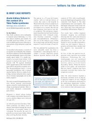
Hypertension<br />
systemic<br />
Generalized atherosclerosis<br />
Renal artery<br />
stenosis<br />
Renal tissue hypoxia<br />
local renin<br />
synthesis<br />
local Ang II<br />
production<br />
Intrarenal<br />
atheroembolism<br />
vascular<br />
rarefaction<br />
in<br />
interstitium<br />
Currently, in daily clinical practice it is thought that<br />
revascularisation in patients with ischemic nephropathy is<br />
not indicated in several clinical situation like: a) in patients<br />
with normotension or when normal blood pressure is<br />
obtained with antihypertensive therapy, b) when stenosed<br />
renal artery is supplying small cirrhotic kidney (longitudinal<br />
diameter less than 8.0cm in adult patient) c) in patients with<br />
high intrarenal resistance assessed by Doppler sonography<br />
(RI higher than 0.8) corresponding to the advanced kidney<br />
fibrosis due to chronic ischemic nephropathy.<br />
Table 1. Predictors of salvageability of ischemic<br />
nephropathy after revascularisation<br />
Glomerulosclerosis<br />
ROS<br />
PDGF-B<br />
TGF-β<br />
interstitial<br />
fibrosis<br />
GFR<br />
- Recent increase in serum creatinine concentration<br />
- Decrease in GFR during ACEI or ARB treatment<br />
- Absence of glomerular or interstitial fibrosis on kidney<br />
biopsy<br />
- Kidney longitudinal diameter >_8.0 cm<br />
- Intrarenal resistance index _70%<br />
- Presence of relative hypoxia in BOLD-MRI examination<br />
Figure 1. Pathogenesis of ischemic nephropathy.<br />
Ang II: angiotensin II; GFR: glomerular filtration rate; PDGF-B:<br />
platelet-derived growth factor-B; ROS: reactive oxygen species;<br />
TGF-β: transforming growth factor β.<br />
436<br />
ACEI: angiotensin converting enzyme inhibitor; ARB:<br />
angiotensin receptor blocker; BOLD-MRI: blood oxygen leveldependent<br />
magnetic resonance imaging; GFR: glomerular<br />
filtration rate.<br />
Nefrologia 2012;32(4):432-38