BBL MGIT - BD
BBL MGIT - BD
BBL MGIT - BD

You also want an ePaper? Increase the reach of your titles
YUMPU automatically turns print PDFs into web optimized ePapers that Google loves.
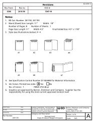
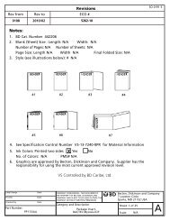
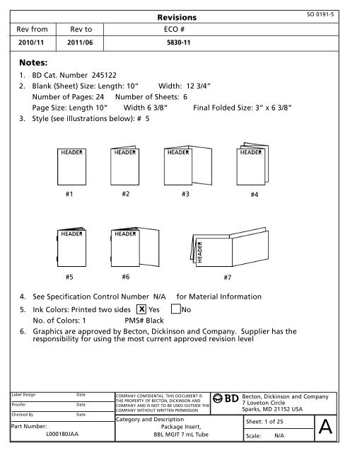
Revisions<br />
Rev from Rev to ECO #<br />
2010/11 2011/06 5830-11<br />
SO 0191-5<br />
Notes:<br />
1. <strong>BD</strong> Cat. Number 245122<br />
2. Blank (Sheet) Size: Length: 10” Width: 12 3/4”<br />
Number of Pages: 24 Number of Sheets: 6<br />
Page Size: Length 10” Width 6 3/8” Final Folded Size: 3” x 6 3/8”<br />
3. Style (see illustrations below): # 5<br />
#1 #2 #3 #4<br />
#5 #6 #7<br />
4. See Specification Control Number N/A for Material Information<br />
x<br />
5. Ink Colors: Printed two sides Yes No<br />
No. of Colors: 1<br />
PMS# Black<br />
6. Graphics are approved by Becton, Dickinson and Company. Supplier has the<br />
responsibility for using the most current approved revision level<br />
Label Design<br />
Date<br />
Proofer<br />
Date<br />
Checked By<br />
Date<br />
Part Number:<br />
L000180JAA<br />
COMPANY CONFIDENTAL. THIS DOCUMENT IS<br />
THE PROPERTY OF BECTON, DICKINSON AND<br />
COMPANY AND IS NOT TO BE USED OUTSIDE THE<br />
COMPANY WITHOUT WRITTEN PERMISSION<br />
Category and Description<br />
Package Insert,<br />
<strong>BBL</strong> <strong>MGIT</strong> 7 mL Tube<br />
Becton, Dickinson and Company<br />
7 Loveton Circle<br />
Sparks, MD 21152 USA<br />
Sheet: 1 of 25<br />
Scale:<br />
N/A<br />
A
<strong>BBL</strong> <strong>MGIT</strong><br />
Mycobacteria Growth Indicator Tube 7 mL<br />
With BACTEC <strong>MGIT</strong> 960 Supplement Kit<br />
English: pages 1 – 4 Italiano: pagine 11 – 15 L000180JAA<br />
Français : pages 4 – 8 Português: páginas 15 – 18 2011/06<br />
Deutsch: Seiten 8 – 11 Español: páginas 18 – 21<br />
<br />
INTENDED USE<br />
The <strong>BBL</strong> <strong>MGIT</strong> Mycobacteria Growth Indicator Tube supplemented with BACTEC <strong>MGIT</strong> Growth Supplement and <strong>BBL</strong> <strong>MGIT</strong><br />
PANTA antibiotic mixture is intended for the detection and recovery of mycobacteria using the BACTEC <strong>MGIT</strong> 960 and BACTEC <strong>MGIT</strong><br />
320 Systems. Acceptable specimen types are digested and decontaminated clinical specimens (except urine), and sterile body fluids (except<br />
blood).<br />
SUMMARY AND EXPLANATION<br />
From 1985 to 1992, the number of reported cases of infection with Mycobacterium tuberculosis (MTB) increased 18%. Tuberculosis still<br />
kills an estimated 3 million persons a year worldwide, making it the leading infectious disease cause of death. 1 Between 1981 and 1987,<br />
AIDS case surveillances indicated that 5.5% of the patients with AIDS had disseminated nontuberculous mycobacterial infections; e.g.,<br />
MAC. By 1990, the increased cases of disseminated nontuberculous mycobacterial infections had resulted in a cumulative incidence of<br />
7.6%. 2 In addition to the resurgence of MTB, multidrug-resistant MTB (MDR-TB) has become an increasing concern. Laboratory delays in the<br />
growth, identification and reporting of these MDR-TB cases contributed at least in part to the spread of the disease.3<br />
The U.S. Centers for Disease Control and Prevention (CDC) have recommended that every effort must be made for laboratories to use the<br />
most rapid methods available for diagnostic mycobacteria testing. These recommendations include the use of both a liquid and a solid<br />
medium for mycobacterial culture.3,4<br />
The <strong>MGIT</strong> Mycobacteria Growth Indicator Tube contains 7 mL of modified Middlebrook 7H9 Broth base. 5,6 The complete medium, with OADC<br />
enrichment and PANTA antibiotic mixture, is one of the most commonly used liquid media for the cultivation of mycobacteria.<br />
All types of clinical specimens, pulmonary as well as extrapulmonary (except blood and urine) can be processed for primary isolation in<br />
the <strong>MGIT</strong> tube using conventional methods. 4 The processed specimen is inoculated into a <strong>MGIT</strong> tube, placed into the BACTEC <strong>MGIT</strong><br />
instrument for continuous monitoring until positive or the end of the testing protocol.<br />
PRINCIPLES OF THE PROCEDURE<br />
A fluorescent compound is embedded in silicone on the bottom of 16 x 100 mm round bottom tubes. The fluorescent compound is<br />
sensitive to the presence of oxygen dissolved in the broth. Initially, the large amount of dissolved oxygen quenches emissions from the<br />
compound and little fluorescence can be detected. Later, actively respiring microorganisms consume the oxygen and allow the<br />
fluorescence to be detected.<br />
Tubes entered into the BACTEC <strong>MGIT</strong> instrument are continuously incubated at 37°C and monitored every 60 min for increasing fluorescence.<br />
Analysis of the fluorescence is used to determine if the tube is instrument positive; i.e., the test sample contains viable organisms. An<br />
instrument positive tube contains approximately 10 5 to 10 6 colony forming units per milliliter (CFU/mL). Culture vials which remain negative for<br />
a minimum of 42 days (up to 56 days) and which show no visible signs of positivity are removed from the instrument as negatives and<br />
sterilized prior to discarding.<br />
The BACTEC <strong>MGIT</strong> Growth Supplement is added to each <strong>MGIT</strong> tube to provide substances essential for the rapid growth of mycobacteria.<br />
Oleic acid is utilized by tubercle bacteria and plays an important role in the metabolism of mycobacteria. Albumin acts as a protective<br />
agent by binding free fatty acids which may be toxic to Mycobacterium species, thereby enhancing their recovery. Dextrose is an energy<br />
source. Catalase destroys toxic peroxides that may be present in the medium.<br />
Contamination is reduced when supplementing the <strong>BBL</strong> <strong>MGIT</strong> broth base with BACTEC <strong>MGIT</strong> Growth Supplement/<strong>BBL</strong> <strong>MGIT</strong> PANTA<br />
antibiotic mixture prior to inoculation with a clinical specimen.<br />
REAGENTS<br />
The <strong>BBL</strong> <strong>MGIT</strong> Mycobacteria Growth Indicator Tube contains: 110 µL of fluorescent indicator and 7 mL of broth. The indicator contains<br />
Tris 4, 7-diphenyl-1, 10-phenanthroline ruthenium chloride pentahydrate in a silicone rubber base. The tubes are flushed with 10% CO 2<br />
and capped with polypropylene caps.<br />
Approximate Formula* Per L of Purified Water:<br />
Modified Middlebrook 7H9 Broth base .......... 5.9 g<br />
Casein peptone.................................................. 1.25 g<br />
BACTEC <strong>MGIT</strong> Growth Supplement contains 15 mL Middlebrook OADC enrichment.<br />
Approximate Formula* Per L of Purified Water:<br />
Bovine albumin................................................ 50.0 g Catalase .............................................................. 0.03 g<br />
Dextrose .......................................................... 20.0 g Oleic acid ............................................................ 0.1 g<br />
Polyoxyethylene stearate (POES)...................... 1.1 g<br />
The <strong>BBL</strong> <strong>MGIT</strong> PANTA vial contains a lyophilized mixture of antimicrobial agents.<br />
Approximate Formula* Per Vial Lyophilized PANTA:<br />
Polymyxin B ................................................ 6,000 units Trimethoprim ................................................ 600 µg<br />
Amphotericin B.............................................. 600 µg Azlocillin ........................................................ 600 µg<br />
Nalidixic acid .............................................. 2,400 µg<br />
* Adjusted and/or supplemented as required to meet performance criteria.<br />
Storage of Reagents: <strong>BBL</strong> <strong>MGIT</strong> Mycobacteria Growth Indicator Tubes – On receipt, store at 2 – 25°C. DO NOT FREEZE. Minimize exposure<br />
to light. Broth should appear clear and colorless. Do not use if turbid. <strong>MGIT</strong> tubes stored as labeled prior to use may be inoculated up to<br />
the expiration date and incubated for up to eight weeks.
BACTEC <strong>MGIT</strong> Growth Supplement – On receipt, store in the dark at 2 – 8°C. Avoid freezing or overheating. Do not open until ready to use.<br />
Minimize exposure to light.<br />
<strong>BBL</strong> <strong>MGIT</strong> PANTA Antibiotic Mixture – On receipt, store lyophilized vials at 2 – 8°C. Once reconstituted, the PANTA mixture must be<br />
stored at 2 – 8°C and used within 5 days.<br />
WARNINGS AND PRECAUTIONS:<br />
For in vitro Diagnostic Use.<br />
This Product Contains Dry Natural Rubber.<br />
Pathogenic microorganisms, including hepatitis viruses and Human Immunodeficiency Virus, may be present in clinical specimens. “Standard<br />
Precautions” 7-10 and institutional guidelines should be followed in handling all items contaminated with blood and other body fluids.<br />
Working with Mycobacterium tuberculosis grown in culture requires Biosafety Level 3 practices, containment equipment and facilities. 4<br />
Prior to use, each <strong>MGIT</strong> tube should be examined for evidence of contamination or damage. Discard any tubes if they appear unsuitable.<br />
Dropped tubes should be examined carefully. If damage is seen, the tube should be discarded.<br />
In the event of tube breakage: 1) Close the instrument drawers; 2) Turn off the instrument; 3) Vacate the area immediately; 4) Consult<br />
your facility/CDC guidelines. An inoculated leaking or broken vial may produce an aerosol of mycobacteria; appropriate handling should<br />
be observed.<br />
Autoclave all inoculated <strong>MGIT</strong> tubes prior to disposal.<br />
SPECIMEN COLLECTION AND HANDLING<br />
All specimens should be collected and transported as recommended by the CDC, the Clinical Microbiology Procedures Handbook or your<br />
laboratory procedure manual. 11<br />
DIGESTION, DECONTAMINATION AND CONCENTRATION<br />
Specimens from different body sites should be processed for inoculation of <strong>MGIT</strong> tubes as follows:<br />
SPUTUM: Specimens should be processed using the NALC-NaOH method as recommended by the CDC’s Public Health Mycobacteriology:<br />
A Guide for the Level III Laboratory. 4 Alternatively, use the <strong>BBL</strong> MycoPrep kit for processing mycobacterial specimens (see<br />
“Availability”).<br />
GASTRIC ASPIRATES: Specimens should be decontaminated as for sputum. If the volume of the specimen is more than 10 mL, concentrate<br />
by centrifugation. Resuspend the sediment in about 5 mL of sterile water and then decontaminate. Add a small amount of NALC powder<br />
(50 to 100 mg) if the specimen is thick or mucoid. After decontamination, concentrate again prior to inoculation into <strong>MGIT</strong> tube.<br />
BODY FLUIDS: (CSF, synovial fluid, pleural fluid, etc.): Specimens which are collected aseptically and are expected to contain no other bacteria<br />
can be inoculated without decontamination. If the specimen volume is more than 10 mL, concentrate by centrifugation at 3,000 x g for<br />
15 min. Pour off supernatant fluid. Inoculate <strong>MGIT</strong> tube with sediment. Specimens that are expected to contain other bacteria must be<br />
decontaminated.<br />
TISSUE: Tissue specimens should be processed as recommended by the CDC’s Public Health Mycobacteriology: A Guide for the Level III<br />
Laboratory. 4<br />
The routine inoculation of solid media is especially important for optimal recovery of mycobacteria from tissue specimens as these specimen<br />
types are particularly susceptible to sporadic organism recovery.<br />
STOOL: Suspend 1 g of feces in 5 mL of Middlebrook Broth. Agitate the suspension on a vortex mixer for 5 s. Proceed to the NALC-NaOH<br />
procedure as recommended by the CDC’s Public Health Mycobacteriology: A Guide for the Level III Laboratory. 4<br />
NOTE: For all specimen processing methods, a phosphate buffer solution (pH 6.8) should be used to QS the sample decontaminant mixture<br />
to 50 mL prior to centrifugation. Resuspension of pellet must also be done using a fresh preparation of phosphate buffer solution<br />
(pH 6.8).<br />
PROCEDURE<br />
Materials Provided: <strong>BBL</strong> <strong>MGIT</strong> Mycobacteria Growth Indicator Tubes and BACTEC <strong>MGIT</strong> 960 Supplement Kit, containing BACTEC <strong>MGIT</strong><br />
Growth Supplement and <strong>BBL</strong> <strong>MGIT</strong> PANTA Antibiotic Mixture (see “Availability”).<br />
Materials Required But Not Provided: Falcon brand 50 mL centrifuge tubes, 4% sodium hydroxide, 2.9% sodium citrate solution,<br />
N-acetyl-L-cysteine powder, phosphate buffer pH 6.8, vortex mixer, 37°C incubator, 1 mL sterile pipettes, sterile transfer pipettes,<br />
<strong>BBL</strong> Middlebrook and Cohn 7H10 Agar, <strong>BBL</strong> MycoPrep Specimen Digestion / Decontamination Kit, <strong>BBL</strong> Middlebrook 7H9 Broth (see<br />
“Availability”) or other mycobacterial agars or egg-based media. Tissue homogenizer or sterile swab, <strong>BBL</strong> Normal Saline (see<br />
“Availability”), microscope and materials for staining slides, adjustable 1000 µL pipetter, corresponding sterile pipette tips, 5% sheep<br />
blood agar plates and tuberculocidal disinfectant.<br />
INOCULATION OF <strong>MGIT</strong> TUBES:<br />
<strong>BBL</strong> <strong>MGIT</strong> 7 mL Tubes must be used with a BACTEC <strong>MGIT</strong> instrument.<br />
1. Reconstitute a lyophilized vial of <strong>BBL</strong> <strong>MGIT</strong> PANTA Antibiotic Mixture with 15 mL of BACTEC <strong>MGIT</strong> Growth Supplement.<br />
2. Label the <strong>MGIT</strong> tube with the specimen number.<br />
3. Unscrew the cap and aseptically add 0.8 mL of Growth Supplement/<strong>MGIT</strong> PANTA Antibiotic Mixture. For best results, the addition of<br />
Growth Supplement/<strong>MGIT</strong> PANTA Antibiotic Mixture should be made just prior to specimen inoculation.<br />
4. Add 0.5 mL of the concentrated specimen suspension prepared above. Also add a drop (0.1 mL) of specimen to a 7H10 agar plate or<br />
other mycobacterial solid agar or egg-based medium.<br />
5. Tightly recap the tube and mix well.<br />
6. Tubes entered into the instrument will be automatically tested for the duration of the recommended 42 day testing protocol.<br />
For specimens in which mycobacteria with different incubation requirements are suspected, a duplicate <strong>MGIT</strong> tube can be set up and<br />
incubated at the appropriate temperature; e.g., 30 or 42°C. Inoculate and incubate at the required temperature. These tubes must be<br />
manually read (refer to the BACTEC <strong>MGIT</strong> Instrument User’s Manual).<br />
For specimens suspected of containing Mycobacterium haemophilum, a source of hemin must be introduced into the tube at the time of<br />
inoculation and the tube incubated at 30°C. Aseptically place one strip of <strong>BBL</strong> Taxo X factor strip into each <strong>MGIT</strong> tube requiring the<br />
addition of hemin prior to inoculation of specimen (see “Availability”). These tubes must be manually read (refer to the BACTEC <strong>MGIT</strong><br />
Instrument User’s Manual).<br />
7. Positive tubes, identified by the BACTEC <strong>MGIT</strong> instrument should be subcultured and an acid-fast smear prepared (see “Results”).<br />
All quality control testing, reprocessing, smear preparations, sub-culturing, etc., of presumptive positive tubes must be performed using biosafety<br />
level (BSL) III practices and containment facilities.<br />
Processing a Positive <strong>MGIT</strong> Tube: NOTE – All steps should be performed in a biological safety cabinet.<br />
1. Remove the <strong>MGIT</strong> tube from the instrument and transport to an area using BSL III practices and containment facilities.<br />
2 Using a sterile transfer pipet, remove an aliquot from the bottom of the tube (approx. 0.1 mL) for stain preparations (AFB and Gram<br />
stains).<br />
3. Inspect smear and preparations. Report preliminary results only after acid-fast smear evaluation.<br />
2
At the end of six weeks incubation, perform a visual check of all instrument negative tubes. If the tube appears visually positive (i.e., nonhomogenous<br />
turbidity, small grains or clumps) it should be subcultured, acid-fast stained and treated as a presumptive positive, provided the<br />
acid-fast smear result is positive. If the tube shows no signs of positivity, it should be sterilized prior to discarding.<br />
Reprocessing Contaminated <strong>MGIT</strong> tubes: Contaminated <strong>MGIT</strong> tubes may be re-decontaminated and re-concentrated using the procedure in<br />
Appendix E - Supplemental Procedures of the BACTEC <strong>MGIT</strong> Instrument User’s Manual.<br />
User Quality Control: Quality control requirements must be performed in accordance with applicable local, state and/or federal<br />
regulations or accreditation requirements and your laboratory's standard Quality Control procedures. It is recommended that the user<br />
refer to pertinent CLSI guidance and CLIA regulations for appropriate Quality Control practices.<br />
Quality Control Certificates are provided on the <strong>BD</strong> website. Quality Control Certificates list test organisms, including ATCC cultures<br />
specified in the CLSI Approved Standard M22-A3, Quality Control for Commercially Prepared Microbiological Culture Media. 12<br />
NOTE: Middlebrook 7H9 Broth (supplemented) is exempt from User QC testing according to CLSI M22-A3. 12<br />
RESULTS<br />
An instrument-positive sample is determined by the BACTEC <strong>MGIT</strong> instrument and confirmed by an acid-fast smear.<br />
REPORTING OF RESULTS<br />
An instrument positive tube must be confirmed by acid-fast smear. A positive AFB smear result indicates the presence of mycobacteria.<br />
If AFB smear positive, subculture to solid media and report as: Instrument positive, AFB smear positive, ID pending.<br />
If microorganisms other than AFB are present report as: Instrument positive, AFB smear negative. Contaminated.<br />
If no microorganisms are present: Reenter the tube into the instrument as an ongoing negative tube within 5 h of removal. Allow tube<br />
to complete test protocol. No reportable result.<br />
Perform subculture from the <strong>BBL</strong> <strong>MGIT</strong> tube for identification and drug susceptibility testing.<br />
LIMITATIONS OF THE PROCEDURE<br />
Recovery of mycobacteria in the <strong>MGIT</strong> tube is dependent on the number of organisms present in the specimen, specimen collection<br />
methods, patient factors such as presence of symptoms, prior treatment and the methods of processing.<br />
Decontamination with the N-acetyl-L-cysteine Sodium hydroxide (NALC-NaOH) method is recommended. Other decontamination methods<br />
have not been tested in conjunction with the <strong>BBL</strong> <strong>MGIT</strong> medium. Digestant/decontaminant solutions may have harmful effects on<br />
mycobacteria.<br />
Colony morphology and pigmentation can only be determined on solid media. Mycobacteria may vary in acid-fastness depending on<br />
strain, age of culture and other variables. The consistency of microscopic morphology in <strong>BBL</strong> <strong>MGIT</strong> medium has not been established.<br />
An AFB smear-positive <strong>MGIT</strong> tube can be subcultured, to both selective and nonselective mycobacterial media, for isolation to perform<br />
identification and susceptibility testing.<br />
<strong>MGIT</strong> tubes which are instrument-positive may contain other non-mycobacterial species. Non-mycobacterial species may overgrow<br />
mycobacteria present. Such <strong>MGIT</strong> tubes should be re-decontaminated and re-cultured (refer to the BACTEC <strong>MGIT</strong> Instrument User’s<br />
Manual). Reprocessing is strongly recommended if the original specimen source cannot be easily recollected; e.g. tissue specimen.<br />
<strong>MGIT</strong> tubes which are instrument-positive may contain one or more species of mycobacteria. Faster growing mycobacteria may be<br />
detected prior to slower growing mycobacteria; therefore, it is important to subculture positive <strong>MGIT</strong> tubes to ensure proper<br />
identification of all mycobacteria present in the sample.<br />
Due to the richness of the <strong>MGIT</strong> broth and to the non-selective nature of the <strong>MGIT</strong> indicator, it is important to follow the stated<br />
digestion/decontamination procedure to reduce the possibility of contamination. Adherence to procedural instructions, which includes use of<br />
recommended inoculum volume (0.5 mL), is critical for optimum recovery of mycobacteria.<br />
The use of PANTA antibiotic mixture, although necessary for all non-sterile specimens, may have inhibitory effects on some mycobacteria.<br />
Seeded culture studies were performed with twenty-four species (ATCC and wild strains) of mycobacteria using inoculum levels ranging<br />
from 10 1 to 10 2 CFU/mL. The following species were detected as positive in the BACTEC <strong>MGIT</strong> 960 System:<br />
M. avium* M. gordonae* M. nonchromogenicum M. terrae<br />
M. abscessus M. haemophilum† M. phlei M. trivale<br />
M. bovis M. intracellulare M. simiae* M. tuberculosis*<br />
M. celatum M. kansasii* M. scrofulaceum M. xenopi*<br />
M. fortuitum* M. malmoense M. smegmatis<br />
M. gastri M. marinum M. szulgai*<br />
* Species recovered during clinical evaluation of the BACTEC <strong>MGIT</strong> 960 System. In addition, M. mucogenicum was recovered at one of the<br />
clinical sites.<br />
† The M. haemophilum was recovered using the addition of a source of hemin to the <strong>MGIT</strong> tube prior to inoculation.<br />
Clinical studies have demonstrated recovery of mycobacteria from respiratory specimens, gastric aspirates, tissue, stool and sterile body<br />
fluids except blood; recovery of mycobacteria from other body fluids has not been established for this product.<br />
EXPECTED VALUES<br />
Figure 1 –- Frequency distribution of recovery times for clinical trial specimens positive in the BACTEC <strong>MGIT</strong> 960 System<br />
PERFORMANCE CHARACTERISTICS<br />
The BACTEC <strong>MGIT</strong> 960 System was evaluated at six clinical sites including one non-US site, which represented public health laboratories as<br />
well as large acute care hospitals in geographically diverse areas. The site population included patients infected with HIV,<br />
immunocompromised patients and transplant patients. The BACTEC <strong>MGIT</strong> 960 System was compared to the BACTEC 460TB radiometric<br />
system and conventional solid growth media for the detection and recovery of mycobacteria from clinical specimens, except blood. A<br />
total of 3330 specimens were tested during the study. A total of 353 specimens were positive which represented 362 isolates recovered<br />
during the study. The distribution of positives by specimen type is: respiratory (90%), tissue (7%), body fluids (1%), stool (0.85%) and<br />
3
one marrow (0.65%). Of the 362 isolates, 289 (80%) were recovered by the BACTEC <strong>MGIT</strong> 960 System, 271 (75%) were recovered by the<br />
BACTEC 460TB System and 250 (69%) were recovered by conventional solid media. Of the 3330 specimens tested in the clinical study, 27<br />
(0.8%) <strong>MGIT</strong> 960 tubes were determined to be false positive (instrument-positive, smear and/or subculture-negative). Of the 313 <strong>MGIT</strong><br />
960 instrument positive tubes, 27 (8.6%) were determined to be false positive. The false negative rate (instrument-negative, smear and/or<br />
subculture-positive) was determined to be 0.5% based on terminal subcultures of 15% of instrument negative vials. The average<br />
breakthrough contamination rate for the BACTEC <strong>MGIT</strong> 960 System was 8.1% with a range of 1.8 – 14.6%.<br />
Table 2: Detection of Mycobacteria Positive Isolates in Clinical Evaluations<br />
Isolates Total Total <strong>MGIT</strong> Total BACTEC BACTEC Total CONV<br />
isolates <strong>MGIT</strong> 960 Only 460TB 460TB Only CONV Only<br />
MTB 132 102 4 119 11 105 3<br />
MAC 172 147 36 123 12 106 3<br />
M. asiaticum 1 0 0 0 0 1 1<br />
M. fortuitum/chelonae 22 18 6 13 1 15 1<br />
M. genavense 1 0 0 1 0 1 0<br />
M. kansasii 5 5 1 4 0 4 0<br />
M. malmoense 1 0 0 1 0 1 0<br />
M. marinum 1 0 0 0 0 1 1<br />
M. mucogenicum 1 1 1 0 0 0 0<br />
M. simiae 1 1 0 1 0 1 0<br />
M. szulgai 2 2 0 2 0 2 0<br />
M. xenopi 2 2 1 1 0 0 0<br />
MOTT 2 1 1 1 1 0 0<br />
Mycobacteria spp. 2 2 1 1 0 1 0<br />
M. gordonae 11 6 3 3 2 6 3<br />
M. nonchromogenicum 6 2 0 1 0 6 4<br />
All MYCO 362 289 54 271 27 250 16<br />
AVAILABILITY<br />
Cat. No. Description<br />
245122 <strong>BBL</strong> <strong>MGIT</strong> Mycobacteria Growth Indicator Tubes,<br />
7 mL, carton of 100 tubes.<br />
245124 BACTEC <strong>MGIT</strong> 960 Supplement Kit, 6 vials, 15 mL,<br />
BACTEC <strong>MGIT</strong> Growth Supplement and 6 vials,<br />
lyophilized, <strong>BBL</strong> <strong>MGIT</strong> PANTA Antibiotic Mixture.<br />
Each Growth Supplement/PANTA vial sufficient for<br />
15 – 18 <strong>MGIT</strong> tubes.<br />
220908 <strong>BBL</strong> Lowenstein-Jensen Medium Slants, package of 10<br />
(20 x 148 mm tubes with cap).<br />
220909 <strong>BBL</strong> Lowenstein-Jensen Medium Slants, carton of 100<br />
(20 x 148 mm tubes with cap).<br />
240862 <strong>BBL</strong> MycoPrep Specimen Digestion/Decontamination<br />
Kit, ten 75 mL bottles of NALC-NaOH solution and<br />
5 packages of phosphate buffer.<br />
Cat. No. Description<br />
240863 <strong>BBL</strong> MycoPrep Specimen Digestion/Decontamination<br />
Kit, ten 150 mL bottles of NALC-NaOH solution and<br />
10 packages of phosphate buffer.<br />
221174 <strong>BBL</strong> Middlebrook and Cohn 7H10 Agar, package of 20.<br />
295939 <strong>BBL</strong> Middlebrook 7H9 Broth, 8 mL, package of 10 tubes.<br />
221818 <strong>BBL</strong> Normal Saline, 5 mL, package of 10.<br />
221819 <strong>BBL</strong> Normal Saline, 5 mL, carton of 100.<br />
231106 <strong>BBL</strong> Taxo X Factor Strips, 1 vial, 50 strips.<br />
REFERENCES<br />
1. Bloom, B.R., and C.J.L. Murray. 1992. Tuberculosis: commentary on a reemergent killer. Science 257:1055-1064.<br />
2. Horsburg, C.R., Jr., 1991. Mycobacterium avium complex infection in the acquired immunodefieciency syndrome. N. Engl. J. .Med. 324:1332-1338.<br />
3. Tenover, F.C., et al, 1993. The resurgence of tuberculosis: is your laboratory ready? J. Clin. Microbiol. 31:767-770.<br />
4. Kent, P.T., and G.P. Kubica. 1985. Public health mycobacteriology: a guide for the level III laboratory. USDHHS, Centers for Disease Control, Atlanta.<br />
5. Cohn, M.L., R.F. Waggoner and J.K. McClatchy. 1968. The 7H11 medium for the cultivation of mycobacteria. Am. Rev. Respir. Dis. 98:295-296.<br />
6. Youmans, G.P. 1979. Cultivation of mycobacteria, the morphology and metabolism of mycobacteria, p. 25-35. Tuberculosis. W.B. Saunders Co.,<br />
Philadelphia.<br />
7. Clinical and Laboratory Standards Institute. 2005. Approved Guideline M29-A3. Protection of laboratory workers from occupationally acquired<br />
infections, 3rd ed. CLSI, Wayne, Pa.<br />
8. Garner, J.S. 1996. Hospital Infection Control Practices Advisory Committee, U.S. Department of Health and Human Services, Centers for Disease<br />
Control and Prevention. Guideline for isolation precautions in hospitals. Infect. Control Hospital Epidemiol. 17:53-80.<br />
9. U.S. Department of Health and Human Services. 2007. Biosafety in microbiological and biomedical laboratories, HHS Publication (CDC), 5th ed.<br />
U.S. Government Printing Office, Washington, D.C.<br />
10. Directive 2000/54/EC of the European Parliament and of the Council of 18 September 2000 on the protection of workers from risks related to<br />
exposure to biological agents at work (seventh individual directive within the meaning of Article 16(1) of Directive 89/391/EEC). Offical Journal L262,<br />
17/10/2000, p. 0021-0045.<br />
11. Isenberg, Henry D. (ed.) 1992. Clinical microbiology procedures handbook. vol. 1. American Society for Microbiology, Washington, D.C.<br />
12. Clinical and Laboratory Standards Institute. 2004. Approved Standard M22-A3. Quality control for commercially prepared microbiological culture<br />
media, 3rd ed., CLSI, Wayne, Pa.<br />
<strong>BBL</strong> <strong>MGIT</strong><br />
Tube avec indicateur de croissance mycobactérienne 7 mL<br />
Avec le coffret du supplément BACTEC <strong>MGIT</strong> 960<br />
Français<br />
APPLICATION<br />
Le tube avec indicateur de croissance mycobactérienne <strong>BBL</strong> <strong>MGIT</strong> additionné du supplément de croissance BACTEC <strong>MGIT</strong> et du complexe d’antibiotiques<br />
<strong>BBL</strong> <strong>MGIT</strong> PANTA est destiné à la détection et l’isolement de mycobactéries au moyen aux systèmes BACTEC <strong>MGIT</strong> 960 et BACTEC <strong>MGIT</strong> 320. Les types<br />
d’échantillons acceptables sont des échantillons cliniques digérés et décontaminés (à l’exception de l’urine), et des liquides biologiques stériles (à<br />
l’exception du sang).<br />
RESUME ET EXPLICATION<br />
De 1985 à 1992, le nombre des cas confirmés d’infection avec Mycobacterium tuberculosis (MTB) a augmenté de 18 %. La tuberculose tue encore un<br />
nombre estimé à environ 3 millions de personnes par an à l’échelle mondiale, en faisant ainsi la principale maladie infectieuse pour cause de mortalité. 1<br />
Entre 1981 et 1987, le suivi des cas de SIDA indiquait que 5,5 % des malades du SIDA avaient contracté des infections mycobactériennes non<br />
tuberculeuses ; par exemple MAC. Dès 1990 l’augmentation des cas de dissémination des infections mycobactériennes non tuberculeuses se traduisait<br />
par une incidence cumulée de 7,6 %. 2 En plus de la recrudescence de la tuberculose, les souches de TB résistantes aux antibiotiques (MDR-TB)<br />
4
deviennent un souci croissant. Les délais pris par les la-boratoires au niveau de la culture, l’identification et la publication de ces cas de résistance aux<br />
antibiotiques ont au moins en partie favorisé la dissémination de cette maladie. 3<br />
Les U.S. Centers for Disease Control and Prevention (CDC) recommandent que les laboratoires fassent tous les efforts possibles pour appliquer les<br />
méthodes les plus rapides actuellement disponibles pour le diagnostic des mycobactéries. Ces recommandations font état de l’utilisation conjointe<br />
d’un milieu liquide et d’un milieu solide pour la culture des mycobactéries. 3,4<br />
Le tube avec indicateur de croissance mycobactérienne <strong>MGIT</strong> contient 7 mL d’un Bouillon de base Middlebrook 7H9 modifié. 5,6 Le milieu complet,<br />
additionné de supplément d’enrichissement OADC et de complexe d’antibiotiques PANTA, est un des milieux liquides les plus communément utilisés<br />
pour la culture des mycobactéries.<br />
Les méthodes traditionnelles peuvent être appliquées à tous les types d’échantillons cliniques, pulmonaires ou non (à l’exception du sang et de<br />
l’urine) pour réaliser un isolement primaire dans le tube <strong>MGIT</strong>. 4 L’échantillon traité est inoculé dans un tube <strong>MGIT</strong> et placé dans l’instrument<br />
BACTEC <strong>MGIT</strong> pour un suivi continu jusqu’à l’obtention d’un résultat positif ou la fin du protocole d’analyse.<br />
PRINCIPES DE LA METHODE<br />
Un composé fluorescent est incorporé à de la silicone au fond de tubes de 16 x 100 mm à fond rond. Le composé fluorescent est sensible à la<br />
présence de l’oxygène dissous dans le bouillon. Initialement, la grande quantité d’oxygène dissous inhibe les émissions du composé et une faible<br />
fluorescence peut être détectée. Subséquemment, les microorganismes, en respirant, consomment l’oxygène du milieu et permettent la détection de<br />
la fluorescence.<br />
Les tubes analysés avec l’instrument BACTEC <strong>MGIT</strong> sont incubés sans interruption à 37 ºC et contrôlés toutes les 60 minutes à la recherche d’une<br />
augmentation de la fluorescence. L’analyse de la fluorescence sert à déterminer si le tube est positif selon l’appareil ; c’est-à-dire si l’échantillon<br />
contient des orga-nismes vivants. Un tube positif selon l’appareil contient approximativement 10 5 à10 6 d’unités formant colonies par millilitre<br />
(UFC/mL). Les flacons de culture qui restent négatifs pendant au moins 42 jours (jusqu’à 56 jours) et qui ne montrent aucun signe visible de positivité<br />
sont retirés de l’appareil en tant que négatifs et stérilisés avant d’être jetés.<br />
Le supplément de croissance BACTEC <strong>MGIT</strong> est ajouté à chaque tube <strong>MGIT</strong> de façon à apporter les éléments essentiels à la croissance rapide des<br />
mycobactéries. L’acide oléique est utilisé par le bacille tuberculeux et joue un rôle important dans le métabolisme des mycobactéries. L’albumine agit<br />
comme agent protecteur en liant les acides gras libres qui peuvent être toxiques pour des espèces de Mycobacterium, augmentant ainsi leur<br />
récupération. Le dextrose est une source d’énergie. La catalase détruit les péroxydes toxiques qui peuvent être présents dans le milieu.<br />
La contamination peut être réduite par l’addition au bouillon de base <strong>BBL</strong> <strong>MGIT</strong> du supplément de croissance BACTEC <strong>MGIT</strong>/complexe<br />
d’antibiotiques <strong>MGIT</strong> PANTA avant l’inoculation avec un échantillon clinique.<br />
REACTIFS<br />
Le tube avec indicateur de croissance mycobactérienne <strong>BBL</strong> <strong>MGIT</strong> contient : 110 µL d’un indicateur fluorescent et 7 mL de bouillon. L’indicateur<br />
contient du chlorure de Tris 4, 7-diphényl-1, 10-phénanthroline ruthénium pentahydraté dans une base de caoutchouc à silicone. Les tubes sont<br />
gazés avec 10 % de CO 2 et fermés avec des capuchons en polypropylène.<br />
Formule approximative* par L d’eau purifiée :<br />
Bouillon de base Middlebrook 7H9 modifié.......... 5,9 g<br />
Peptone de caséine.................................................. 1,25 g<br />
Le supplément de croissance BACTEC <strong>MGIT</strong> contient 15 mL de supplément d'enrichissement Middlebrook OADC.<br />
Formule approximative* par L d'eau purifiée :<br />
Albumine bovine.................................................... 50,0 g Catalase .................................................................... 0,03 g<br />
Dextrose .................................................................. 20,0 g Acide oléique ............................................................ 0,1 g<br />
Stéarate de polyoxyéthylène (POES) ...................... 1,1, g<br />
L’ampoule de <strong>BBL</strong> <strong>MGIT</strong> PANTA contient un mélange lyophilisé d’agents antimicrobiens.<br />
Formule approximative* par ampoule lyophilisée PANTA :<br />
Polymixine B ...................................................... 6.000 unités Triméthoprime ...................................................... 600 µg<br />
Amphothéricine B ................................................ 600. µg Azlocilline .............................................................. 600 µg<br />
Acide nalidixique .............................................. 2.400, µg<br />
* Ajustée et/ou supplémentée en fonction des critères de performance imposés.<br />
Conservation des réactifs : tubes avec indicateur de croissance mycobactérienne <strong>BBL</strong> <strong>MGIT</strong> – Dès réception, conserver entre 2 – 25 ºC. NE PAS CONGELER.<br />
Minimiser l’exposition à la lumière. Le bouillon doit être clair et incolore. Ne pas l'utiliser s'il est turbide. Les tubes <strong>MGIT</strong> conservés dans les conditions<br />
décrites sur l'étiquette jusqu'au moment de l'utilisation peuvent être inoculés jusqu'à la date de péremption et incubés jusqu'à huit semaines.<br />
Supplément de croissance BACTEC <strong>MGIT</strong> – Dès réception, conserver à l’obscurité entre 2 – 8 °C. Eviter la congélation ou une surchauffe. Ne pas<br />
l’ouvrir avant d'être prêt à l'utiliser. Minimiser l’exposition à la lumière.<br />
Complexe d’antibiotiques <strong>BBL</strong> <strong>MGIT</strong> PANTA – Dès réception, conserver les ampoules lyophilisées entre 2 – 8 °C. Une fois reconstitué, le mélange<br />
PANTA doit être conservé entre 2 – 8 °C et utilisé dans les 5 jours.<br />
AVERTISSEMENTS ET PRECAUTIONS<br />
Pour diagnostic in vitro.<br />
Ce produit contient du caoutchouc naturel sec.<br />
Des microorganismes pathogènes, notamment les virus de l'hépatite et de l'immunodéficience humaine, sont susceptibles d'être présents dans les<br />
échantillons cliniques. Respecter les " Précautions standard " 7-10 et les consignes en vigueur dans l'établissement pour manipuler tout objet<br />
contaminé avec du sang ou d'autres liquides organiques. Stériliser à l'autoclave les récipients contenant les échantillons et d'autres matériaux<br />
contaminés avant de les éliminer.<br />
Travailler avec des cultures de Mycobacterium tuberculosis nécessite d'observer des procédures de protection contre les dangers biologiques de<br />
niveau 3 et l'usage d'équipement et de matériel de confinement. 4<br />
Avant d'être utilisé, chaque tube <strong>MGIT</strong> doit être inspecté pour vérifier l'absence de contamination ou de dommage. Tout tube qui paraît ne pas<br />
convenir doit être jeté.<br />
Les tubes qui sont tombés doivent être soigneusement ezaminés. S’ils sont endommagés d’une mannière quelconque, ils doivent être jetés.<br />
Dans le cas de bris du tube : 1) Eteindre l’appareil ; 2) Fermer les tiroirs de l’appareil ; 3) Evacuer la zone immédiatement ; 4) Se reporter aux<br />
directives de votre laboratoire/du CDC. Un flacon inoculé fêlé ou qui fuit peut produire un aérosol de bactéries ; une manipulation appropriée doit<br />
donc être respectée.<br />
Tous les tubes <strong>MGIT</strong> inoculés devront être autoclavés avant d'être jetés.<br />
PRELEVEMENT ET MANIPULATION DES ECHANTILLONS<br />
Tous les échantillons doivent être recueillis et transportés selon les recommandations des CDC, du Clinical Microbiology Procedures Handbook ou les<br />
directives de votre laboratoire. 11<br />
DIGESTION, DECONTAMINATION ET CONCENTRATION<br />
Avant de servir à l'inoculation des tubes <strong>MGIT</strong>, les échantillons provenant de différents sites anatomiques devraient être traités comme suit :<br />
CRACHATS : les échantillons doivent être traités selon la méthode utilisant NALC-NaOH comme recommandé par les CDC dans Public Health<br />
Mycobacteriology: A Guide for the Level III Laboratory. 4 Alternativement, utiliser la trousse <strong>BBL</strong> MycoPrep pour analyser les échantillons mycobactériens<br />
(voir “Matériel disponible”).<br />
ASPIRATIONS GASTRIQUES : les échantillons doivent être décontaminés comme des crachats. Si le volume de l'échantillon est supérieur à 10 mL,<br />
concentrer par centrifugation. Remettre en suspension le sédiment dans environ 5 mL d'eau stérile et décontaminer. Ajouter une petite quantité de<br />
poudre de NALC (50 – 100 mg) si l'échantillon est visqueux ou mucoïde. Après décontamination, concentrer de nouveau avant d'inoculer un tube <strong>MGIT</strong>.<br />
LIQUIDES BIOLOGIQUES : (LCR, liquide synovial, liquide pleural etc.) : les échantillons prélevés de manière aseptique et présumés exempts de bactéries<br />
autres que des mycobactéries peuvent être inoculés sans décontamination. Si le volume de l'échantillon est plus grand que 10 mL, concentrer par<br />
5
centrifugation à 3.000 x g pendant 15 min. Jeter le surnageant. Inoculer le tube <strong>MGIT</strong> avec le sédiment. Les échantillons présumés contaminés par<br />
d'autres bactéries doivent être décontaminés.<br />
TISSUS : les échantillons tissulaires doivent être analysés comme recommandé par les CDC dans Public Health Mycobacteriology: A Guide for the Level III<br />
Laboratory. 4<br />
Pour une mise en évidence optimale des mycobactéries, il est essentiel que les milieux solides soient inoculés systématiquement, parce que ces types<br />
d’échantillon livrent des résultats très aléatoires.<br />
SELLE : suspendre 1 g de matières fécales dans 5 mL de Bouillon Middlebrook. Agiter la suspension avec un agitateur vortex pendant 5 sec. Suivre la<br />
méthode utilisant NALC-NaOH comme recommandé par les CDC dans Public Health Mycobacteriology: A Guide for the Level III Laboratory. 4<br />
NOTA : pour toutes les méthodes de préparation des échantillons, une solution tampon au phosphate (pH 6,8) devrait être utilisée pour ramener le<br />
mélange de décontamination de l’échantillon à 50 mL avant centrifugation. La remise en suspension du sédiment doit aussi être effectuée à<br />
l’aide d’une solution fraîche de tampon au phosphate (pH 6,8).<br />
MODE OPERATOIRE<br />
Matériel fourni : tubes avec indicateur de croissance mycobactérienne <strong>BBL</strong> <strong>MGIT</strong> et coffret de supplément BACTEC <strong>MGIT</strong> 960 contenant le<br />
supplément de croissance BACTEC <strong>MGIT</strong> et le complexe d'antibiotiques <strong>BBL</strong> <strong>MGIT</strong> PANTA (voir “Matériel disponible”).<br />
Matériaux requis mais non fournis : tubes à centrifuger de 50 mL de marque Falcon, hydroxyde de sodium à 4 %, solution de citrate de sodium à 2,9 %,<br />
poudre de N-acétyle-L-cystéine, tampon phosphate pH 6,8, agitateur vortex, incubateur à 37 °C, pipettes stériles de 1 mL, pipettes de transfert stériles,<br />
Gélose <strong>BBL</strong> Middlebrook et Cohn 7H10, trousse de digestion/décontamination d’échantillon <strong>BBL</strong> MycoPrep, Bouillon <strong>BBL</strong> Middlebrook 7H9 (voir<br />
“Matériel disponible”), ou autres géloses ou milieux à base d’oeufs pour mycobactéries. Homogénéiseur pour tissus ou écouvillons stériles, sérum<br />
physiologique normal <strong>BBL</strong> (voir “Matériel disponible”), microscope et matériel nécessaire pour colorer les lames, multipette réglable de 1000 µL,<br />
embouts correspondants, boîtes de pétri de gélose à 5 % de sang de mouton et désinfectant tuberculocide.<br />
INOCULATION DES TUBES <strong>MGIT</strong><br />
Les tubes <strong>BBL</strong> <strong>MGIT</strong> 7 mL doivent être utilisés avec un appareil BACTEC <strong>MGIT</strong>.<br />
1. Reconstituer une ampoule lyophilisée de complexe d’antibiotiques <strong>BBL</strong> <strong>MGIT</strong> PANTA avec 15 mL de supplément de croissance BACTEC <strong>MGIT</strong>.<br />
2. Etiqueter le tube <strong>MGIT</strong> avec le numéro de l’échantillon.<br />
3. Dévisser le capuchon et ajouter de manière aseptique 0,8 mL de supplément de croissance/complexe d’antibiotiques <strong>MGIT</strong> PANTA. Pour de<br />
meilleurs résultats, l’ajout du supplément de croissance/complexe d’antibiotiiques <strong>MGIT</strong> PANTA devrait être fait juste avant l’inoculation de<br />
l’échantillon.<br />
4. Ajouter 0,5 mL de la suspension concentrée d’échantillon préparée comme ci-dessus. Déposer aussi une goutte (0,1 mL) d’échantillon sur une<br />
gélose 7H10 ou tout autre milieu solide pour mycobactéries à base d’agar ou d’oeufs.<br />
5. Refermer le tube hermétiquement et bien mélanger.<br />
6. Les tubes placés dans l’appareil seront automatiquement analysés jusqu’à la fin du protocole d’analyse (42 jours).<br />
Pour les échantillons suspectés de contenir des mycobactéries demandant des conditions d’incubation différentes, un double du tube <strong>MGIT</strong> peut<br />
être préparé et incubé à la température appropriée ; soit 30 ou 42 °C. Inoculer et incuber à la température demandée. Ces tubes doivent être lus<br />
manuellement (se reporter au Manuel d'utilisation de l'instrument BACTEC <strong>MGIT</strong>).<br />
Pour les échantillons suspectés de contenir Mycobacterium haemophilum, une source d’hémine peut être incorporée au tube au moment où<br />
l’inoculation est faite, et le tube incubé à 30 °C. Introduire aseptiquement une bandelette <strong>BBL</strong> Taxo avec facteur X dans chaque tube <strong>MGIT</strong><br />
demandant l’addition d’hémine avant d’inoculer l’échantillon (voir. “Matériel disponible”). Ces tubes doivent être lus manuellement (se reporter au<br />
Manuel d'utilisation de l'instrument BACTEC <strong>MGIT</strong>).<br />
7. Les tubes positifs, identifiés par l’appareil BACTEC <strong>MGIT</strong> doivent être repiqués et des frottis pour tester l’acido-résistance doivent être<br />
préparés (voir “Résultats”).<br />
Toutes les analyses de contrôle de la qualité, les traitements, les préparations de frottis, les repiquages, etc., de tubes considérés positifs doivent être<br />
effectués selon les pratiques de biosécurité de niveau III (BSL) et dans des installations de confinement.<br />
Traitement d’un tube <strong>MGIT</strong> positif : NOTA – toute la procédure doit être effectuée dans une hotte de sécurité biologique.<br />
1. Retirer le tube <strong>MGIT</strong> de l’appareil et le transporter dans une zone ayant des installations de confinement et appliquant les pratiques de<br />
biosécurité de niveau III.<br />
2 A l’aide d’une pipette stérile, prélever une fraction aliquote dans le fond du tube (environ 0,1 mL) pour les colorations (acido-résistant et de Gram).<br />
3. Inspecter le frottis et les préparations. Noter les résultats préliminaires seulement après avoir évalué la coloration acido-résistant.<br />
A la fin des six semaines d’incubation, effectuez un contrôle visuel de tous les tubes jugés négatifs par l’instrument. Si le tube apparaît positif (c’est-à-dire,<br />
si une turbidité non-homogène, des petits grains ou des granules sont visibles), il doit être repiqué, soumis à une coloration acido-résistant et traité<br />
comme un positif présumé dans la mesure où la coloration acido-résistant est positive. Si le tube ne montre aucun signe de positivité, il doit être<br />
stérilisé avant d’être jeté.<br />
Traitement des tubes <strong>MGIT</strong> contaminés : les tubes <strong>MGIT</strong> contaminés peuvent être décontaminés et re-concentrés au moyen de la procédure utilisée<br />
dans l’annexe E - Procédures supplémentaires du Manuel d'utilisation de l'instrument BACTEC <strong>MGIT</strong>.<br />
Contrôle de qualité réalisé par l’utilisateur : Effectuer les contrôles de qualité conformément à la réglementation nationale et/ou internationale, aux<br />
exigences des organismes d'homologation concernés et aux procédures de contrôle de qualité en vigueur dans l'établissement. Il est recommandé à<br />
l'utilisateur de consulter les directives CLSI et la réglementation CLIA concernées pour plus d'informations sur les modalités de contrôle de qualité.<br />
Des certificats de contrôle de qualité se trouvent dans le site web de <strong>BD</strong>. Les certificats de contrôle de qualité dressent la liste des microorganismes<br />
de test, y compris les cultures ATCC spécifiées dans la norme M22-A3 approuvée par le CLSI, Quality Control for Commercially Prepared<br />
Microbiological Culture Media. 12<br />
REMARQUE : Le bouillon Middlebrook 7H9 (supplémenté) n'est pas soumis aux tests de Contrôle Qualité par l'utilisateur selon CLSI M22-A3. 12<br />
RESULTATS<br />
Un échantillon positif selon l’instrument est identifié par l’appareil BACTEC <strong>MGIT</strong> et ce résultat est confirmé par une coloration acido-résistant (AFB).<br />
RAPPORT DES RESULTATS<br />
Un tube positif selon l’instrument doit être confirmé par frottis pour la coloration acido-résistant. Un frottis positif pour la coloration AFB indique la<br />
présence de mycobactéries.<br />
Si le frottis est positif pour la coloration AFB, repiquer sur milieux solides et noter : positif selon l’appareil, frottis positif pour AFB, identification en cours.<br />
Si des microorganismes autres que des AFB sont présents, noter : positif selon l’instrument, frottis négatif pour AFB. Contaminé.<br />
Si aucun microorganisme n’est présent : dans un délai maximal de 5 h après l’avoir retiré, remettre le tube dans l’instrument en tant que négatif en<br />
cours. Soumettre le tube à la totalité du protocole d’analyse. Aucun résultat ne peut être noté.<br />
Effectuez un repiquage du tube <strong>BBL</strong> <strong>MGIT</strong> pour l’identification et l’analyse de la sensibilité aux drogues.<br />
LIMITES DE LA METHODE<br />
L’obtention de mycobactéries dans un tube <strong>MGIT</strong> dépend du nombre de microorganismes présents dans l’échantillon, des méthodes de prélèvement<br />
de l’échantillon, de facteurs propres au patient tels que la présence de symptômes, des traitements antérieurs et des méthodes d’analyse.<br />
Une décontamination avec la N-acétyle-L-cystéine combinée à de l’hydroxyde de sodium (NALC-NaOH) est recommandée. D’autres méthodes de<br />
décontamination n’ont pas été expérimentées avec le milieu <strong>BBL</strong> <strong>MGIT</strong>. Les solutions digestives de décontamination peuvent avoir des effets néfastes sur<br />
les mycobactéries.<br />
La morphologie et la pigmentation des colonies ne peuvent être déterminées que sur des milieux solides. Les mycobactéries peuvent montrer des<br />
différences au niveau de la coloration acido-résistante, en fonction de la souche, de l’âge de la culture ou d’autre paramètres. La constance de la<br />
morphologie microscopique dans le milieu <strong>BBL</strong> <strong>MGIT</strong> n’a pas été établie.<br />
Un tube <strong>MGIT</strong> ayant donné un frottis positif pour AFB peut être repiqué sur des milieux sélectifs et des milieux non-sélectifs pour donner des isolats<br />
sur lesquels peuvent être effectuées des identifications et des analyses de la sensibilité.<br />
6
Les tubes <strong>MGIT</strong> jugés positifs selon l’instrument peuvent contenir des espèces autres que des mycobactéries. La croissance des espèces nonmycobactériennes<br />
peut excéder celle des mycobactéries présentes. De tels tubes <strong>MGIT</strong> doivent être décontaminés et repiqués (se reporter au Manuel<br />
d'utilisation de l'instrument BACTEC <strong>MGIT</strong>). Le traitement est fortement recommandé si la source d’échantillons initiale ne peut être facilement récupérée ;<br />
par exemple les échantillons de tissus.<br />
Les tubes <strong>MGIT</strong> jugés positifs selon l’instrument peuvent contenir une ou plusieurs espèces de mycobactéries. Les mycobactéries à croissance plus<br />
rapide peuvent être décelées avant les mycobactéries à croissance plus lente ; c’est pourquoi il est important de repiquer les tubes <strong>MGIT</strong> positifs afin<br />
d’assurer une identification correcte de toutes les mycobactéries présentes dans l’échantillon.<br />
Du fait de la richesse du Bouillon <strong>MGIT</strong> et de la nature non sélective de l’indicateur <strong>MGIT</strong>, il est important de suivre la procédure décrite de<br />
digestion/décontamination pour réduire le risque de contamination. Une stricte observation des instructions de procédure, y compris l’utilisation du volume<br />
recommandé d’inoculum (0,5 mL) est essentielle pour une récupération optimale des mycobactéries.<br />
L’utilisation du complexe d’antibiotiques PANTA, quoique nécessaire pour tous les échantillons non stériles, peut avoir des effets inhibiteurs sur certaines<br />
mycobactéries.<br />
Des études de cultures ensemencées ont été réalisées sur vingt-quatre espèces de mycobactéries (ATCC et sauvages) avec des inoculums<br />
comptant 10 1 – 10 2 UFC/mL. Les espèces suivantes ont été considérées comme positives dans le système BACTEC <strong>MGIT</strong> 960 :<br />
M. avium* M. gordonae* M. nonchromogenicum M. terrae<br />
M. abscessus M. haemophilum† M. phlei M. trivale<br />
M. bovis M. intracellulare M. simiae* M. tuberculosis*<br />
M. celatum M. kansasii* M. scrofulaceum M. xenopi*<br />
M. fortuitum* M. malmoense M. smegmatis<br />
M. gastri M. marinum M. szulgai*<br />
*Espèces obtenues lors de l’évaluation clinique du système BACTEC <strong>MGIT</strong> 960. De plus, M. mucogenicum a été collecté à un des sites cliniques.<br />
† Le M. haemophilum a été collecté grace à l’ajout d’une source d’hémine au tube <strong>MGIT</strong> avant inoculation.<br />
Les études cliniques ont démontré que des mycobactéries ont pu être collectées à partir d’échantillons respiratoires, d’aspirâts gastriques,<br />
d’échantillons tissulaires, de selles et de liquides biologiques stériles à l’exception du sang ; la croissance de mycobactéries à partir d’autres liquides<br />
biologiques n’a pas été réalisée avec ce produit.<br />
VALEURS ESCOMPTEES<br />
Figure 1 –- Histogramme de la fréquence des délais de récupération pour les échantillons<br />
des essais cliniques positifs dans le système BACTEC <strong>MGIT</strong> 960<br />
CARACTERISTIQUES DE PERFORMANCE<br />
Le système BACTEC <strong>MGIT</strong> 960 a été évalué dans six sites cliniques, dont un ne se trouvant pas aux Etats-Unis, comprenant aussi bien des laboratoires<br />
publics que de grands hôpitaux de soins intensifs situés dans diverses régions géographiques. La population de chaque site comprenait des patients<br />
infectés par VIH, des patients avec des déficiences immunologiques et des patients ayant reçu une greffe. Le système BACTEC <strong>MGIT</strong> 960 a été comparé<br />
au système de radiométrie BACTEC 460TB et aux milieux solides traditionnels pour la détection et l’obtention de mycobactéries dans des échantillons<br />
cliniques (à l’exception du sang). Un total de 3330 échantillons ont été analysés lors de cette étude. Un total de 353 échantillons étaient positifs, ce qui<br />
correspondait aux 362 isolats obtenus lors de cette étude. Le classement des échantillons analysés en fonction de leur source d’origine était :<br />
respiratoire (90 %), tissulaire (7,0 %), liquide biologique (1,0 %), selle (0,85 %) et moelle osseuse (0,65 %). De ces 362 isolats, 289 (80 %) ont été<br />
récupérés par le système BACTEC <strong>MGIT</strong> 960, 271 (75 %) ont été récupérés par le BACTEC 460TB et 250 (69 %) ont été obtenus avec les milieux solides<br />
traditionnels. Des 3330 échantillons analysés dans cette étude, 27 (0,8 %) tubes <strong>MGIT</strong> 960 ont donné un taux de faux positifs (positif selon<br />
l’instrument, négatif selon le frottis et/ou le repiquage). Des 313 tubes <strong>MGIT</strong> 960 positifs selon l’instrument, 27 (8,6 %) ont donné un taux de faux<br />
positifs. Le taux de faux négatifs (négatif selon l’instrument, positif selon le frottis et/ou le repiquage) a été évalué à 0,5 % basé sur les repiquages<br />
finaux de ~ 15 % de flacons négatifs selon l’instrument. Le taux de contamination moyen pour le système BACTEC <strong>MGIT</strong> 960 était de 8,1 % allant de<br />
1,8 % à 14,6 %.<br />
TABLEAU 2: Détection des isolats positifs de mycobactéries dans les évaluations cliniques<br />
Isolat Total Total <strong>MGIT</strong> Total BACTEC BACTEC Total CONV<br />
des isolates <strong>MGIT</strong> 960 seulement 460TB 460TB seulement CONV seulement<br />
MTB 132 102 4 119 11 105 3<br />
MAC 172 147 36 123 12 106 3<br />
M. asiaticum 1 0 0 0 0 1 1<br />
M. fortuitum/chelonae 22 18 6 13 1 15 1<br />
M. genavense 1 0 0 1 0 1 0<br />
M. kansasii 5 5 1 4 0 4 0<br />
M. malmoense 1 0 0 1 0 1 0<br />
M. marinum 1 0 0 0 0 1 1<br />
M. mucogenicum 1 1 1 0 0 0 0<br />
M. simiae 1 1 0 1 0 1 0<br />
M. szulgai 2 2 0 2 0 2 0<br />
M. xenopi 2 2 1 1 0 0 0<br />
MOTT 2 1 1 1 1 0 0<br />
Mycobacteria spp. 2 2 1 1 0 1 0<br />
M. gordonae 11 6 3 3 2 6 3<br />
M. nonchromogenicum 6 2 0 1 0 6 4<br />
MYCO tous 362 289 54 271 27 250 16<br />
7
MATERIEL DISPONIBLE<br />
No réf. Description<br />
245122 Tubes avec indicateur de croissance mycobactérienne<br />
<strong>BBL</strong> <strong>MGIT</strong>, 7 mL, carton de 100 tubes.<br />
245124 Coffret de supplément BACTEC <strong>MGIT</strong> 960, 6 flacons, 15 mL,<br />
supplément de croissance BACTEC <strong>MGIT</strong> et 6 flacons de<br />
complexe d’antibiotiques <strong>BBL</strong> <strong>MGIT</strong> PANTA, lyophilisé.<br />
Chaque ampoule de supplément de croissance/PANTA<br />
est suffisante pour 15 – 18 tubes <strong>MGIT</strong>.<br />
220908 Géloses inclinées <strong>BBL</strong> Lowenstein-Jensen, coffret de 10<br />
(tubes de 20 x 148 mm avec capuchon).<br />
220909 Géloses inclinées <strong>BBL</strong> Lowenstein-Jensen, coffret de 100<br />
(tubes de 20 x 148 mm avec capuchon).<br />
No réf. Description<br />
240862 Trousse <strong>BBL</strong> MycoPrep de digestion et de décontamination<br />
d’échantillons, comprenant dix flacons de 75 mL de<br />
solution de NALC-NaOH et 5 sachets de tampon<br />
phosphate.<br />
240863 Trousse <strong>BBL</strong> MycoPrep de digestion et de décontamination<br />
d’échantillons, comprenant dix flacons de 150 mL de<br />
solution de NALC-NaOH et 10 sachets de tampon<br />
phosphate.<br />
221174 Gélose <strong>BBL</strong> Middlebrook et Cohn 7H10, coffret de 20.<br />
295939 Bouillon <strong>BBL</strong> Middlebrook 7H9, 8 mL, coffret de 10 tubes.<br />
221818 Sérum physiologique normal <strong>BBL</strong>, 5 mL, coffret de 10.<br />
221819 Sérum physiologique normal <strong>BBL</strong>, 5 mL, coffret de 100.<br />
231106 Bandelettes <strong>BBL</strong> Taxo avec facteur X, un flacon,<br />
50 bandelettes.<br />
BIBLIOGRAPHIE : voir la rubrique “References” du texte anglais.<br />
<strong>BBL</strong> <strong>MGIT</strong><br />
Indikatorröhrchen für Mykobakterienwachstum 7 mL<br />
Mit BACTEC <strong>MGIT</strong> 960-Supplement-Kit<br />
Deutsch<br />
VERWENDUNGSZWECK<br />
Das <strong>BBL</strong> <strong>MGIT</strong>-Indikatorröhrchen für Mykobakterienwachstum, das mit dem BACTEC <strong>MGIT</strong>-Wachstumssupplement und dem <strong>BBL</strong> <strong>MGIT</strong> PANTA-<br />
Antibiotischen Gemisch angereichert werden kann, dient zum Nachweis und zur Isolierung von Mykobakterien unter Verwendung des BACTEC <strong>MGIT</strong> 960-<br />
und BACTEC <strong>MGIT</strong> 320-Geräts. Geeignete Proben sind digestierte und dekontaminierte klinische Proben (außer Urin) und sterile Körperflüssigkeiten<br />
(außer Blut).<br />
ZUSAMMENFASSUNG UND ERKLÄRUNG<br />
Zwischen 1985 und 1992 stieg die Zahl der gemeldeten Infektionen mit Mycobacterium tuberculosis (MTB) um 18 %. Tuberkulose ist die führende<br />
infektionsbedingte Todesursache, da weltweit immer noch etwa 3 Millionen Menschen jährlich an dieser Erkrankung sterben. 1 Die zwischen 1981<br />
und 1987 durchgeführten Untersuchungen von AIDS-Fällen zeigten, daß 5,5 % der Patienten mit AIDS disseminierte, nichttuberkulöse<br />
mykobakterielle Infektionen aufwiesen, wie z.B. MAC. Im Jahre 1990 war das kumulative Vorkommen durch die gestiegene Zahl der Fälle<br />
disseminierter nichttuberkulöser Infektionen mit Mykobakterien bereits auf 7,6 % angestiegen. 2 Sowohl der Wiederanstieg von MTB als auch<br />
mehrfachresistente MTB (MDR-TB) stellen ein zunehmendes Problem dar. Laborseitige Verzögerungen bei der Kultivierung, Identifizierung und<br />
Meldung dieser MDR-TB-Fälle trugen zumindest teilweise zur Ausbreitung der Krankheit bei. 3<br />
Die U.S. Centers for Disease Control and Prevention (CDC) empfehlen, daß Labors alle Anstrengungen unternehmen, um die schnellsten verfügbaren<br />
Methoden zum diagnostischen Testen auf Mykobakterien einzusetzen. Diese Empfehlungen umfassen die Verwendung sowohl eines flüssigen als<br />
auch eines festen Mediums zur Kultivierung von Mykobakterien. 3,4<br />
Das <strong>MGIT</strong>-Indikatorröhrchen für Mykobakterienwachstum enthält 7 mL modifizierte Middlebrook 7H9-Bouillonbasis. 5,6 Das komplette, mit OADC-<br />
Anreicherung und PANTA-Antibiotischem Gemisch angereicherte Medium ist eines der am häufigsten verwendeten flüssigen Medien zur Kultivierung von<br />
Mykobakterien.<br />
Alle klinischen Probentypen, pulmonale sowie extra-pulmonale Proben (außer Blut und Urin) können mit Hilfe konventioneller Methoden für die<br />
Primärisolierung im <strong>MGIT</strong>-Röhrchen vorbereitet werden. 4 Die vorbereitete Probe wird in ein <strong>MGIT</strong>-Röhrchen inokuliert, dann in ein BACTEC <strong>MGIT</strong>-<br />
Gerät zur kontinuierlichen Beobachtung gesetzt bis es positiv oder das Ende des Testprotokolls erreicht ist.<br />
VERFAHRENSPRINZIP<br />
Eingebettet in Silikon am Boden von 16 x 100-mm-Röhrchen mit rundem Boden befindet sich eine fluoreszierende Verbindung. Die fluoreszierende<br />
Verbindung spricht auf das Vorliegen von in der Bouillon aufgelöstem Sauerstoff an. Anfänglich ist nur wenig Fluoreszenz nachweisbar, da die große<br />
Menge aufgelösten Sauerstoffs die Emissionen der Verbindung absorbiert. Später nehmen die aktiv respirierenden Mikroorganismen den Sauerstoff<br />
auf, und die Fluoreszenz kann nachgewiesen werden.<br />
Die in das BACTEC <strong>MGIT</strong>-Gerät gegebenen Röhrchen werden fortwährend bei 37 °C inkubiert und alle 60 Minuten auf ansteigende Fluoreszenz<br />
überprüft. Durch Analyse der Fluoreszenz ist feststellbar, ob das Fläschchen gerätepositiv ist, d.h. ob die Probe lebensfähige Organismen enthält. Ein<br />
geräte–positives Röhrchen enthält etwa 10 5 bis 10 6 koloniebildende Einheiten pro Milliliter (KBE/mL). Kulturfläschchen, die nach mindestens 42<br />
Tagen (bis zu 56 Tagen) negativ bleiben und die keine sichtbare Zeichen von Positivität aufweisen, sind als negativ zu behandeln und werden von<br />
dem Gerät entfernt und vor dem Entsorgen sterilisiert.<br />
Das BACTEC <strong>MGIT</strong>-Wachstumssupplement wird jedem <strong>MGIT</strong>-Röhrchen hinzugefügt, um Substanzen, die wesentlich zum schnellen Wachstum von<br />
Mykobakterien beitragen, zur Verfügung zu stellen. Ölsäure wird von Tuberkelbakterien verwertet und spielt beim Stoffwechsel von Mykobakterien<br />
eine wichtige Rolle. Albumin agiert als Schutzmittel und verbessert die Isolierung von Mycobacterium-Spezies, indem es freie Fettsäuren bindet, die<br />
für diese toxisch sein können. Dextrose ist eine Energiequelle. Katalase zerstört eventuell im Medium vorkommende toxische Peroxide.<br />
Durch Supplementierung der <strong>BBL</strong> <strong>MGIT</strong>-Bouillonbasis mit BACTEC <strong>MGIT</strong>-Wachstumssupplement/<strong>MGIT</strong> PANTA-Antibiotischem Gemisch vor der<br />
Inokulation mit den klinischen Proben wird die Kontamination reduziert.<br />
REAGENZIEN<br />
Das <strong>BBL</strong> <strong>MGIT</strong>-Indikatorröhrchen für Mykobakterienwachstum enthält: 110 µL Fluoreszenzindikator und 7 mL Bouillon. Der Indikator enthält Tris-4,7-<br />
Diphenyl-1,10-Phenanthrolin-Rutheniumchlorid-Pentahydrat in Silikonkautschuk-Basis. Die Röhrchen sind mit 10%igem CO 2 ausgespült und mit<br />
Polypropylenkappen verschlossen.<br />
Ungefähre Zusammensetzung* pro L destilliertem Wasser:<br />
Modifizierte Middlebrook 7H9 Bouillonbasis ........ 5,9 g<br />
Casein-Pepton .......................................................... 1,25 g<br />
BACTEC <strong>MGIT</strong>-Wachstumssupplement enthält 15 mL Middlebrook OADC-Anreicherung.<br />
Ungefähre Zusammensetzung* pro L destilliertem Wasser:<br />
Rinderalbumin ........................................................ 50,0 g Katalase .............................................................. 0,03 g<br />
Dextrose .................................................................. 20,0 g Ölsäure ................................................................ 0,1 g<br />
Polyoxyethylen-Stearat (POES) ................................1,1 g<br />
Das <strong>BBL</strong> <strong>MGIT</strong> PANTA-Fläschchen enthält eine lyophilisierte Mischung von antimikrobiellen Agenzien.<br />
Ungefähre Zusammensetzung* pro Fläschchen lyophilisiertem PANTA:<br />
Polymixin B ........................................................ 6.000 Einheiten Trimethoprim ..................................................600 µg<br />
Amphotericin B .................................................... 600 µg Azlocillin.......................................................... 600 µg<br />
Nalidixinsäure .................................................... 2.400 µg<br />
*Abgestimmt und/oder supplementiert auf die geforderten Testkriterien.<br />
Aufbewahrung der Reagenzien: <strong>BBL</strong> <strong>MGIT</strong>-Indikatorröhrchen für Mykobakterienwachstum – Nach Erhalt bei 2 – 25 °C lagern. NICHT EINFRIEREN.<br />
Lichteinfall auf ein Minimum beschränken. Die Bouillon sollte klar und farblos sein. Bei Trübung nicht verwenden. <strong>MGIT</strong>-Röhrchen, die vor Gebrauch<br />
gemäß der Anleitung auf dem Etikett gelagert werden, können bis zum Verfallsdatum inokuliert und bis zu acht Wochen lang inkubiert werden.<br />
8
BACTEC <strong>MGIT</strong>-Wachstumssupplement – Nach Erhalt im Dunkeln bei 2 – 8 °C lagern. Einfrieren und Überhitzen vermeiden. Erst unmittelbar vor Gebrauch<br />
öffnen. Vor hellem Licht schützen.<br />
<strong>BBL</strong> <strong>MGIT</strong> PANTA-Antibiotisches Gemisch – Lyophiliserte Fläschchen nach Erhalt bei 2 – 8 °C lagern. Nach Rekonstituierung muß das PANTA-Gemisch<br />
bei 2 – 8 °C gelagert und innerhalb von 5 Tagen verwendet werden.<br />
WARNUNGEN UND VORSICHTSMAßNAHMEN<br />
Zur in vitro-Diagnostik.<br />
Dieses Produkt enthält trockenen Naturkautschuk.<br />
Klinische Proben können pathogene Mikroorganismen, wie z.B. Hepatitis-Viren und HIV, enthalten. Beim Umgang mit allen mit Blut oder anderen<br />
Körperflüssigkeiten kontaminierten Artikeln sind die "Allgemeinen Vorsichtsmaßnahmen" 7-10 sowie die einschlägigen Institutionsrichtlinien zu<br />
beachten. Nach Gebrauch Probenbehälter und andere kontaminierte Materialien im Autoklaven sterilisieren und erst dann entsorgen.<br />
Verfahren, die in Kultur gezüchtetes Mycobacterium tuberculosis beinhalten, müssen nach den Richtlinien und unter Anwendung der<br />
Sicherheitsvorrichtungen der Biologischen Sicherheitsstufe 3 durchgeführt werden. 4<br />
Vor Gebrauch muß jedes <strong>MGIT</strong>-Röhrchen auf Anzeichen von Kontamination oder Beschädigung untersucht werden. Fläschchen, die unbrauchbar<br />
erscheinen, sind zu entsorgen.<br />
Röhrchen, die auf den Boden gefallen sind, sollten sorgfältig auf Beschädigung untersucht werden. Bei Beschädigungen Röhrchen verwerfen.<br />
Bei Beschädigung des Fläschchens: 1) Gerätefächer zumachen 2) Gerät abschalten 3) Bereich sofort verlassen 4) CDC oder laborinterne Richtlinien zu<br />
Rate ziehen. Ein inokuliertes undichtes oder zerbrochenes Fläschchen kann ein Mykobakterien-Aerosol erzeugen; deshalb sollten angemessene<br />
Maßnahmen verwendet werden.<br />
Alle inokulierten <strong>MGIT</strong>-Röhrchen vor dem Entsorgen autoklavieren.<br />
PROBENENTNAHME UND -HANDHABUNG<br />
Alle Proben sollten gemäß den Empfehlungen der CDC, dem Clinical Microbiology Procedures Handbook oder den Verfahrensvorschriften des<br />
jeweiligen Labors entnommen und transportiert werden. 11<br />
DIGESTION, DEKONTAMINATION UND KONZENTRATION<br />
Proben aus verschiedenen Körperstellen sollten wie folgt zur Inokulation von <strong>MGIT</strong>-Röhrchen verarbeitet werden:<br />
SPUTUM: Proben sollten unter Anwendung des NALC-NaOH-Verfahrens gemäß den in Public Health Mycobacteriology: A Guide for the Level III<br />
Laboratory 4 gegebenen Empfehlungen der CDC verarbeitet werden. Als Alternative kann der <strong>BBL</strong> MycoPrep-Kit zur Verarbeitung von<br />
Mykobakterienproben verwendet werden (s. “Lieferbare Produkte”).<br />
MAGENASPIRATE: Die Proben sollten auf die gleiche Art und Weise wie Sputum dekontaminiert werden. Wenn das Probenvolumen mehr als 10 mL<br />
beträgt, durch Zentrifugieren konzentrieren. Das Sediment in ca. 5 mL sterilem Wasser resuspendieren und dann dekontaminieren. Eine kleine Menge<br />
NALC-Pulver (50 – 100 mg) hinzufügen, falls die Probe dickflüssig oder schleimähnlich ist. Die Probe nach der Dekontamination und vor der Inokulierung<br />
in das <strong>MGIT</strong>-Röhrchen erneut konzentrieren.<br />
KÖRPERFLÜSSIGKEITEN: (Liquor, Synovialflüssigkeit, Pleuralflüssigkeit, etc.): Proben, die aseptisch entnommen wurden und bei denen kein Verdacht<br />
auf das Vorkommen von anderen Bakterien vorliegt, können ohne Dekontamination inokuliert werden. Wenn das Probenvolumen mehr als 10 mL<br />
beträgt, durch 15 min Zentrifugieren bei 3.000 x g konzentrieren. Die Überstandsflüssigkeit abgießen. Das <strong>MGIT</strong>-Röhrchen mit dem Sediment<br />
inokulieren. Proben, die wahrscheinlich andere Bakterien enthalten, müssen dekontaminiert werden.<br />
GEWEBE: Gewebeproben sollten gemäß den in Public Health Mycobacteriology: A Guide for the Level III Laboratory 4 gegebenen Empfehlungen der<br />
CDC verarbeitet werden.<br />
Regelmäßige Inokulation von festen Medien ist zur optimalen Gewinnung von Mykobakterien aus Gewebeproben besonders wichtig, da Organismen<br />
aus diesen Probentypen besonders schwer zu isolieren sind.<br />
STUHL: 1 g Fäzes in 5 mL Middlebrook-Bouillon suspendieren. Suspension 5 Sek. im Vortex-Mixer mischen. Die weitere Verarbeitung sollte unter<br />
Anwendung des NALC-NaOH-Verfahrens gemäß den in Public Health Mycobacteriology: A Guide for the Level III Laboratory 4 gegebenen<br />
Empfehlungen der CDC erfolgen.<br />
HINWEIS: Bei allen Probenverarbeitungsmethoden muß eine Phosphatpufferlösung (pH 6,8) verwendet werden, um damit vor der Zentrifugation eine<br />
Qualitätskontrolle von bis zu 50 mL des Probendekontaminierungsgemisches durchzuführen. Eine Resuspendierung des Pellets muß ebenfalls<br />
unter Verwendung einer frischen Zubereitung von Phosphatpufferlösung (pH 6,8) durchgeführt werden.<br />
VERFAHREN<br />
Mitgeliefertes Arbeitsmaterial: <strong>BBL</strong> <strong>MGIT</strong>-Indikatorröhrchen für Mykobakterienwachstum und BACTEC <strong>MGIT</strong> 960-Supplementskit mit BACTEC <strong>MGIT</strong><br />
Wachstumssupplement und <strong>BBL</strong> <strong>MGIT</strong> PANTA-Antibiotischem Gemisch (s. “Lieferbare Produkte”).<br />
Benötigtes, jedoch nicht mitgeliefertes Arbeitsmaterial: Falcon 50-mL-Zentrifugenröhrchen, 4%iges Natriumhydroxid, 2,9%ige Natriumcitratlösung,<br />
N-Acetyl-L-Cystein-Pulver, Phosphatpuffer mit pH 6,8, Vortex-Mixer, Inkubator (37 °C), sterile 1-mL-Pipetten, sterile Transferpipetten,<br />
<strong>BBL</strong> Middlebrook und Cohn 7H10 Agar, <strong>BBL</strong> MycoPrep Aufschluß-/Dekontaminationskit für Proben, <strong>BBL</strong> Middlebrook 7H9-Bouillon (s. “Lieferbare<br />
Produkte”) oder andere Agar- oder Eiermedien für Mykobakterien. Gewebehomogenisator oder steriler Tupfer, <strong>BBL</strong> Normale Kochsalzlösung<br />
(s. “Lieferbare Produkte”), Mikroskop und Materialien zum Färben der Objektträger, verstellbare 1000-µL-Pipette, dazupassende sterile<br />
Pipettenspitzen, 5%ige Schafblut-Agarplatten und Tuberkelbazillen-Desinfektionsmittel.<br />
INOKULIERUNG DER <strong>MGIT</strong>-RÖHRCHEN<br />
<strong>BBL</strong> <strong>MGIT</strong> 7mL-Röhrchen müssen mit einem BACTEC <strong>MGIT</strong>-Gerät verwendet werden.<br />
1. Ein lyophilisiertes Fläschchen mit <strong>BBL</strong> <strong>MGIT</strong> PANTA-Antibiotischem Gemisch mit 15 mL BACTEC <strong>MGIT</strong> Wachstumssupplement rekonstituieren.<br />
2. Das <strong>MGIT</strong>-Röhrchen mit der Probennummer beschriften.<br />
3. Die Kappe entfernen und aseptisch 0,8 mL Wachstumssupplement/<strong>MGIT</strong> PANTA-Antibiotisches Gemisch hinzufügen. Um beste Ergebnisse zu<br />
erzielen, sollten Wachstumssupplement/<strong>MGIT</strong> PANTA-Antibiotisches Gemisch erst unmittelbar vor der Inokulierung hinzugefügt werden.<br />
4. 0,5 mL der vorbereiteten konzentrierten Probensuspension hinzufügen. Außerdem einen Tropfen (0,1 mL) der Probe auf eine 7H10-Agarplatte<br />
oder einen anderen festen Agar- oder Eiernährboden für Mykobakterien geben.<br />
5. Das Röhrchen wieder fest verschließen und gut durchmischen.<br />
6. Im Gerät befindliche Röhrchen werden für die Gesamtdauer der Testprotokollaufnahme (42 Tage) automatisch getestet.<br />
Wenn Mykobakterien mit unterschiedlichem Inkubationsbedarf in einer Probe vermutet werden, kann ein zweites <strong>MGIT</strong>-Röhrchen vorbereitet<br />
und bei der entsprechenden Temperatur inkubiert werden, (z.B. 30 oder 42 °C). Die Röhrchen inokulieren und bei der erforderlichen Temperatur<br />
inkubieren. Diese Röhrchen müssen manuell abgelesen werden (s. BACTEC <strong>MGIT</strong>-Gerät Benutzerhandbuch).<br />
Bei Proben mit Verdacht auf Mycobacterium haemophilum muß bei der Inokulation eine Häminquelle in das Röhrchen zugeführt werden und das<br />
Röhrchen bei 30 °C inkubiert werden. Einen <strong>BBL</strong> Taxo X-Faktorstreifen aseptisch in jedes <strong>MGIT</strong>-Röhrchen legen, in das Hämin vor der<br />
Probeninokulation zugefügt werden muß (s. “Lieferbare Produkte”). Diese Röhrchen müssen manuell abgelesen werden (s. BACTEC <strong>MGIT</strong>-Gerät<br />
Benutzerhandbuch).<br />
7. Von positiven Röhrchen, die vom BACTEC <strong>MGIT</strong>-Gerät identifiziert wurden, sollte eine Subkultur angelegt und ein Ausstrich für eine Säurefestigkeitsfärbung<br />
angefertigt werden (s. “Ergebnisse”).<br />
Alle Testverfahren zur Qualitätskontrolle, erneute Verarbeitung, Ausstrich-Anfertigung und Anlegen einer Subkultur von präsumtiv positiven Röhrchen<br />
müssen gemäß Biosicherheitsstufe III und unter Verwendung von Eindämmungseinrichtungen gehandhabt werden.<br />
Verarbeitung eines positiven <strong>MGIT</strong>-Röhrchens: HINWEIS – Alle Schritte sollten in einer biologischen Sicherheitswerkbank ausgeführt werden.<br />
1. <strong>MGIT</strong>-Röhrchen aus dem Gerät nehmen und Transport gemäß Biosicherheitsstufe III und unter Verwendung von Eindämmungseinrichtungen<br />
durchführen.<br />
2 Mit Hilfe einer sterilen Transferpipette ein Aliquot (etwa 0,1 mL) vom Boden des Röhrchens zur Anfertigung von Färbungen entnehmen<br />
(Säurefestigkeitsfärbung und Gram-Färbung).<br />
9
3. Ausstrich und Färbung prüfen. Vorläufige Ergebnisse erst nach Beurteilung des Säurefestigkeitsausstriches dokumentieren.<br />
Am Ende einer sechswöchigen Inkubation eine visuelle Überprüfung aller gerätenegativen Röhrchen durchführen. Erscheint das Röhrchen visuell positiv<br />
(d.h. inhomogene Trübung, kleine Körner oder Klümpchen), sollte eine Subkultur angelegt und eine Säurefestigkeitsfärbung angefertigt werden und das<br />
Röhrchen als positiv behandelt werden, vorausgesetzt das Ergebnis des Säurefestigkeitsausstriches ist positiv. Weist das Röhrchen keine sichtbaren Zeichen<br />
von Positivität auf, sollte es vor dem Entsorgen sterilisiert werden.<br />
Erneute Verarbeitung kontaminierter <strong>MGIT</strong>-Röhrchen: Kontaminierte <strong>MGIT</strong>-Röhrchen können durch Verwendung der in Anhang E - Zusätzliche Verfahren<br />
des Benutzerhandbuches für das BACTEC <strong>MGIT</strong>-Gerät wieder dekontaminiert und konzentriert werden.<br />
Qualitätskontrolle durch den Anwender: Es sind die geltenden gesetzlichen und behördlichen und in den Akkreditierungsbedingungen festgelegten<br />
Vorschriften zur Qualitätskontrolle sowie die laborinternen Standardvorgaben zur Qualitätskontrolle zu beachten. Benutzer sollten die vom CLSI<br />
relevanten Dokumente und CLIA-Vorschriften über geeignete Testverfahren zur Qualitätskontrolle einsehen.<br />
Qualitätskontrollzertifikate sin auf der <strong>BD</strong>-Website verfügbar. In den Qualitätskontrollzertifikaten sind Testorganismen, einschließlich der im CLSI<br />
zugelassenen Standard M22-A3, Quality Control for Commercially Prepared Microbiological Culture Media, spezifizierten ATCC-Kulturen,<br />
aufgeführt. 12<br />
HINWEIS: Middlebrook 7H9-Bouillon (ergänzt) ist gemäß CLSI M22-A3 von Qualitätskontrolltests durch den Anwender ausgenommen. 12<br />
ERGEBNISSE<br />
Eine gerätepositive Probe wird durch das BACTEC <strong>MGIT</strong>-Gerät bestimmt und durch einen Ausstrich für eine Säurefestigkeitsfärbung bestätigt.<br />
BERICHTEN VON ERGEBNISSEN<br />
Ein gerätepositives Röhrchen muß durch eine Säurefestigkeitsfärbung bestätigt werden. Eine positive Säurefestigkeitsfärbung indiziert das Vorhandensein<br />
von Mikroorganismen im Röhrchen.<br />
Bei positiver Säurefestigkeitsfärbung eine Subkultur auf festem Medium anlegen und wie folgt dokumentieren: Gerätepositiv, Säurefestigkeitsfärbung<br />
positiv, Identifizierung ausstehend.<br />
Wenn andere Mikroorganismen als säurefeste Bakterien vorhanden sind, wie folgt dokumentieren: Gerätepositiv, Säurefestigkeitsfärbung negativ.<br />
Kontaminiert.<br />
Wenn keine Mikroorganismen vorhanden sind: Das Röhrchen innerhalb von 5 h nach dem Herausnehmen als ständige negative Kontrolle wieder in das<br />
Gerät stellen. Röhrchen bis zur Vollendung des Testprotokolls mitführen. Kein Ergebnis zu dokumentieren.<br />
Subkultur vom <strong>BBL</strong> <strong>MGIT</strong>-Röhrchen zur Identifikation und zur Arzneimittelempfindlichkeitsprüfung anlegen.<br />
VERFAHRENSBESCHRÄNKUNGEN<br />
Die Isolierung von Mykobakterien im <strong>MGIT</strong>-Röhrchen hängt von der Anzahl der in der Probe vorhandenen Organismen, der Probenentnahmemethoden,<br />
patientenbedingten Faktoren (z.B. das Vorhandensein von Symptomen oder vorhergehende Behandlungen) und der Verarbeitungsmethoden ab.<br />
Zur Dekontamination wird die NALC-NaOH-Methode (N-Acetyl-L-Cystein mit Natriumhydroxid) empfohlen. Andere Dekontaminationsverfahren<br />
wurden mit dem <strong>BBL</strong> <strong>MGIT</strong>-Medium bisher noch nicht getestet. Digestions-/Dekontaminationsmittel können das Mykobakterienwachstum<br />
beeinträchtigen.<br />
Koloniemorphologie und Färbeverhalten können nur auf festen Medien ermittelt werden. Die Säurefestigkeit von Mykobakterien kann abhängig vom<br />
Stamm, dem Alter der Kultur und anderen Variablen unterschiedlich sein. Die Konsistenz mikroskopischer Morphologie in <strong>BBL</strong> <strong>MGIT</strong>-Medium wurde bisher<br />
noch nicht ermittelt.<br />
Ein in der Säurefestigkeitsfärbung positiver Ausstrich eines <strong>MGIT</strong>-Röhrchens kann zur Isolierung für Identifizierungs- und Empfindlichkeitstests<br />
sowohl auf selektiven als auch nicht-selektiven Medien für Mykobakterien subkultiviert werden.<br />
<strong>MGIT</strong>-Röhrchen, die gerätepositiv sind, können neben Mykobakterien Begleitkeime enthalten. Begleitkeime können vorhandene Mykobakterien<br />
überwachsen. Solche <strong>MGIT</strong>-Röhrchen sollten erneut dekontaminiert und kultiviert werden (s. BACTEC <strong>MGIT</strong>-Gerät Benutzerhandbuch). Erneute<br />
Verarbeitung wird nachdrücklich empfohlen, wenn die Originalprobe nur schwer zu erhalten ist, z.B. Gewebeproben.<br />
<strong>MGIT</strong>-Röhrchen, die gerätepositiv sind, können mehrere Spezies von Mykobakterien enthalten. Schneller wachsende Mykobakterien können vor<br />
langsamer wachsenden Mykobakterien nachgewiesen werden; daher ist es wichtig, eine Subkultur positiver <strong>MGIT</strong>-Röhrchen anzulegen, um alle in der<br />
Probe vorhandenen Mykobakterien korrekt zu identifizieren.<br />
Da die <strong>MGIT</strong>-Bouillon sehr wachstumsfördernd und der <strong>MGIT</strong>-Indikator nicht selektiv ist, müssen die angegebenen Verfahren zur<br />
Digestion/Dekontamination unbedingt eingehalten werden, um die Möglichkeit einer Kontamination zu verringern. Die Beachtung der<br />
Verfahrensanleitungen, die die Verwendung des empfohlenen Inokulumvolumens (0,5 mL) einschließt, ist für eine optimale Isolierung von<br />
Mykobakterien äußerst wichtig.<br />
Die Verwendung des PANTA-Antibiotischen Gemisches ist zwar für alle nicht-sterilen Proben unerläßlich, kann jedoch das Wachstum einiger Mykobakterien<br />
hemmen.<br />
Studien mit beimpften Kulturen wurden unter Verwendung von vierundzwanzig Mykobakterienspezies (mit ATCC-Stämmen und Wildstämmen)<br />
durchgeführt. Dabei wurden Inokulumkonzentrationen von 10 1 bis 10 2 KBE/mL verwendet. Die folgenden Spezies wurden im BACTEC <strong>MGIT</strong> 960-System<br />
als positiv nachgewiesen:<br />
M. avium* M. gordonae* M. nonchromogenicum M. terrae<br />
M. abscessus M. haemophilum† M. phlei M. trivale<br />
M. bovis M. intracellulare M. simiae* M. tuberculosis*<br />
M. celatum M. kansasii* M. scrofulaceum M. xenopi*<br />
M. fortuitum* M. malmoense M. smegmatis<br />
M. gastri M. marinum M. szulgai*<br />
*Bei der klinischen Beurteilung des BACTEC <strong>MGIT</strong> 960-Systems isolierte Spezies. Darüber hinaus wurde M. mucogenicum an einem der klinischen Labors<br />
nachgewiesen.<br />
† M. haemophilum wurde isoliert, indem eine zusätzliche Häminquelle dem <strong>MGIT</strong>-Röhrchen vor der Inokulation zugegeben wurde.<br />
Klinische Studien belegten die Isolierung von Mykobakterien aus Proben der Atemwege, aus Magenaspiraten, Gewebe, Stuhl und sterilen<br />
Körperflüssigkeiten (außer Blut); die Isolierung von Mykobakterien aus anderen Körperflüssigkeiten wurde mit diesem Produkt noch nicht<br />
nachgewiesen.<br />
10
ERWARTETE WERTE<br />
Abbildung 1 – Häufigkeitsverteilung der Isolierungszeit für im BACTEC <strong>MGIT</strong> 960-System positive Studienproben<br />
LEISTUNGSMERKMALE<br />
Das BACTEC <strong>MGIT</strong> 960-System wurde an sechs klinischen Zentren (darunter ein außeramerikanisches Zentrum), die öffentliche Gesundheitslabors<br />
und große Notfallkrankenhäuser an geographisch unterschiedlichen Standorten darstellten, beurteilt. Die Patientenpopulation der Zentren<br />
umfaßte HIV-infizierte und immungeschwächte Patienten und Transplantatempfänger. Das BACTEC <strong>MGIT</strong> 960-System wurde mit dem<br />
radiometrischen BACTEC 460TB-System und konventionellen festen Wachstumsmedien zum Nachweis und zur Isolierung von Mykobakterien aus<br />
klinischen Proben (außer Blut) verglichen. Im Verlauf der Studie wurden insgesamt 3330 Proben getestet. Insgesamt waren 353 Proben positiv, die<br />
362 während der Studie gewonnene Isolate repräsentierten. Die Anteile der positiven Proben nach Entnahmestelle waren: Atemwege (90 %),<br />
Gewebe (7 %), Körperflüssigkeiten (1 %), Stuhl (0,85 %) und Knochenmark (0,65 %). Von diesen 362 Isolaten wurden 289 (80 %) mit Hilfe des<br />
BACTEC <strong>MGIT</strong> 960-Systems, 271 (75 %) mit dem BACTEC 460TB-System und 250 (69 %) mit konventionellen festen Medien isoliert. Von den 3330 in<br />
der klinischen Studie getesteten Proben, 27 (0,8 %) <strong>MGIT</strong> 960-Röhrchen wiesen falschpositive Ergebnisse auf (gerätepositiv, Ausstrich und/oder<br />
Subkultur negativ). Von den 313 <strong>MGIT</strong> 960 gerätepositiven Röhrchen wiesen 27 (8,6 %) falschpositive Ergebnisse auf. Anhand von Subkulturen von<br />
etwa 15 % gerätenegativen Fläschchen konnte das Ergebnis der falschpositiven Rate (gerätenegativ, Ausstrich und/oder Subkultur positiv) auf 0,5 %<br />
festgelegt werden. Die durchschnittliche Durchbruch-Kontaminationsrate bei dem BACTEC <strong>MGIT</strong> 960-System betrug 8,1 %, wobei der Bereich von<br />
1,8 % – 14,6 % schwankte.<br />
Tabelle 2: Nachweis von Mykobakterien-positiven Isolaten bei klinischen Beurteilungene<br />
Isolate Total <strong>MGIT</strong> 960 Nur BACTEC Nur CONV Nur<br />
gesamt gesamt <strong>MGIT</strong> 460TB gesamt BACTEC 460TB gesamt CONV<br />
MTB 132 102 4 119 11 105 3<br />
MAC 172 147 36 123 12 106 3<br />
M. asiaticum 1 0 0 0 0 1 1<br />
M. fortuitum/chelonae 22 18 6 13 1 15 1<br />
M. genavense 1 0 0 1 0 1 0<br />
M. kansasii 5 5 1 4 0 4 0<br />
M. malmoense 1 0 0 1 0 1 0<br />
M. marinum 1 0 0 0 0 1 1<br />
M. mucogenicum 1 1 1 0 0 0 0<br />
M. simiae 1 1 0 1 0 1 0<br />
M. szulgai 2 2 0 2 0 2 0<br />
M. xenopi 2 2 1 1 0 0 0<br />
MOTT 2 1 1 1 1 0 0<br />
Mycobacteria spp. 2 2 1 1 0 1 0<br />
M. gordonae 11 6 3 3 2 6 3<br />
M. nonchromogenicum 6 2 0 1 0 6 4<br />
MYCO gesamt 362 289 54 271 27 250 16<br />
LIEFERBARE PRODUKTE<br />
Best.- Nr. Beschreibung<br />
245122 <strong>BBL</strong> <strong>MGIT</strong>-Indikatorröhrchen für Mykobakterienwachstum,<br />
7 mL, Karton mit 100 Röhrchen.<br />
245124 BACTEC <strong>MGIT</strong> 960-Supplement-kit, mit 6 15-mL-Fläschchen<br />
BACTEC <strong>MGIT</strong> Wachstumssupplement und 6 Fläschchen<br />
lyophilisiertem <strong>BBL</strong> <strong>MGIT</strong> PANTA-Antibiotischem Gemisch.<br />
Jedes Wachstums-supplement/PANTA-Fläschchen reicht für<br />
15 – 18 <strong>MGIT</strong>-Röhrchen aus.<br />
220908 <strong>BBL</strong> Löwenstein-Jensen-Schrägagar, Packung mit 10 Stück<br />
(20 x 148-mm-Röhrchen mit Kappe).<br />
220909 <strong>BBL</strong> Löwenstein-Jensen-Schrägagar, Karton mit 100 Stück<br />
(20 x 148-mm-Röhrchen mit Kappe).<br />
Best.- Nr. Beschreibung<br />
240862 <strong>BBL</strong> MycoPrep Aufschluß-/Dekontaminationskit für Proben,<br />
zehn 75-mL-Fläschchen mit NALC-NaOH-Lösung und 5<br />
Packungen mit Phosphatpuffer.<br />
240863 <strong>BBL</strong> MycoPrep Aufschluß-/Dekontaminationskit für Proben,<br />
zehn 150-mL-Fläschchen mit NALC-NaOH-Lösung und 10<br />
Packungen mit Phosphatpuffer.<br />
221174 <strong>BBL</strong> Middlebrook und Cohn 7H10 Agar, Packung mit 20.<br />
295939 <strong>BBL</strong> Middlebrook 7H9-Bouillon, 8 mL, Packung mit 10<br />
Röhrchen.<br />
221818 Normale Kochsalzlösung, 5 mL, Packung mit 10.<br />
221819 <strong>BBL</strong> Normale Kochsalzlösung, 5 mL, Karton mit 100.<br />
231106 <strong>BBL</strong> Taxo X-Faktorstreifen, 1 Fläschchen, 50 Teststreifen.<br />
LITERATURNACHWEIS: S. “References” im englischen Text.<br />
<strong>BBL</strong> <strong>MGIT</strong><br />
Provetta per l’indicazione di crescita dei micobatteri 7 mL<br />
Con kit di supplemento BACTEC <strong>MGIT</strong> 960<br />
Italiano<br />
USO PREVISTO<br />
La provetta <strong>BBL</strong> <strong>MGIT</strong> per l’indicazione di crescita dei micobatteri, arricchita con il supplemento di crescita BACTEC <strong>MGIT</strong> e con la miscela antibiotica<br />
<strong>BBL</strong> <strong>MGIT</strong> PANTA, è indicata per la ricerca e l’isolamento dei micobatteri mediante i sistemi BACTEC <strong>MGIT</strong> 960 e BACTEC <strong>MGIT</strong> 320. I tipi di campioni che<br />
si prestano al test sono i campioni clinici digeriti e decontaminati (ad eccezione delle urine) e i liquidi biologici sterili (ad eccezione del sangue).<br />
11
SOMMARIO E SPIEGAZIONE<br />
Tra il 1985 e il 1992 il numero dei casi riportati di infezione da Mycobacterium tuberculosis (MTB) è aumentato del 18%. Si ritiene che la tubercolosi sia<br />
tuttora causa di morte, nel mondo, per circa 3 milioni di persone all’anno, cifra che la pone al primo posto tra le malattie infettive letali. 1 I controlli<br />
effettuati sui casi di AIDS tra il 1981 e il 1987 attestarono che il 5,5% dei pazienti affetti da AIDS contreva infezioni micobatteriche diverse dalla<br />
tubercolosi, es. il MAC. Entro il 1990 i casi riportati di infezioni micobatteriche diverse da tubercolosi erano aumentati fino ad avere un’incidenza globale<br />
del 7,6%. 2 Preoccupazione crescente è pure derivata, oltre che dalla ripresa dell’MTB, dall’apparizione dell’MTB resistente a molteplici farmaci (MDR-TB). I<br />
ritardi di laboratorio nell’identificare e diagnosticare questi casi di MDR-TB hanno contribuito almeno in parte alla diffusione della malattia. 3<br />
Gli U.S. Centers for Disease Control and Prevention (CDC) hanno esteso ai laboratori la raccomandazione di impiegare ogni sforzo per adottare i<br />
metodi più rapidi a disposizione per l’analisi diagnostica dei Micobatteri. Tali raccomandazioni comprendono l’utilizzo di un terreno liquido e di uno<br />
solido per la coltura di micobatteri. 3,4<br />
La provetta <strong>MGIT</strong> per l’indicazione di crescita dei micobatteri contiene 7 mL di Brodo di base Middlebrook 7H9 modificato. 5,6 Il terreno completo, con<br />
arricchimento OADC e miscela antibiotica PANTA, è uno dei brodi più comunemente usati per la coltivazione dei micobatteri.<br />
Qualsiasi tipo di campione clinico, polmonare, extra-polmonare (ad eccezione del sangue ed urina), può essere sottoposto a trattamento per<br />
l’isolamento primario nella provetta <strong>MGIT</strong>, mediante i metodi tradizionali. 4 Il campione trattato viene inoculato in una provetta <strong>MGIT</strong>, inserito nello<br />
strumento BACTEC <strong>MGIT</strong> e monitorato in continuazione finché non risulta positivo o fino a completamento del protocollo di analisi.<br />
PRINCIPI DELLA PROCEDURA<br />
Un composto fluorescente è incapsulato nel silicone posto sul fondo delle provette circolari da 16 x 100 mm. Il composto fluorescente è sensibile alla<br />
presenza dell’ossigeno sciolto nel brodo. All’inizio, la grande quantità di ossigeno contenuto nel terreno riduce le emissioni del composto e si nota solo<br />
una debole fluorescenza. In un secondo tempo, i microrganismi che respirano attivamente consumano l’ossigeno e permettono di osservare la<br />
fluorescenza.<br />
Le provette inserite nello strumento BACTEC <strong>MGIT</strong> vengono incubate a 37 ºC per un periodo ininterrotto e monitorate ogni 60 minuti per rilevarne<br />
l’aumento di fluorescenza. L’analisi della fluorescenza viene usata per determinare se la provetta è positiva per lo strumento, vale a dire se il campione<br />
contiene organismi vivi. Una provetta positiva per lo strumento contiene tra 10 5 e 10 6 circa di Unità Formanti Colonie per millilitro (UFC/mL). I flaconi di<br />
coltura che rimangono negativi per un minimo di 42 giorni (fino a 56 giorni) e che non mostrano alcun segno di positività vengono tolti dallo strumento<br />
in quanto negativi, sterilizzati e poi eliminati.<br />
Ad ogni provetta <strong>MGIT</strong> viene aggiunto il supplemento di crescita BACTEC <strong>MGIT</strong> che fornisce sostanze essenziali alla crescita rapida dei micobatteri.<br />
L’acido oleico viene utilizzato dai bacilli della tubercolosi e riveste un ruolo importante nel metabolismo dei micobatteri. L’albumina agisce quale<br />
agente protettivo legando gli acidi grassi liberi, tossici per le specie Mycobacterium, favorendo così il loro isolamento. Il destrosio è fonte di energia.<br />
La catalasi distrugge i perossidi tossici che potrebbero essere presenti nel terreno di coltura.<br />
La contaminazione può essere ridotta arricchendo il brodo di base <strong>BBL</strong> <strong>MGIT</strong> con il supplemento di crescita BACTEC <strong>MGIT</strong>/la miscela antibiotica<br />
<strong>BBL</strong> <strong>MGIT</strong> PANTA, prima dell’inoculo del campione clinico.<br />
REAGENTI<br />
La provetta <strong>BBL</strong> <strong>MGIT</strong> per l’indicazione di crescita dei micobatteri contiene: 110 µL di indicatore fluorescente e 7 mL di brodo di coltura. L’indicatore<br />
contiene Tris 4, 7-difenil-1, 10-fenantrolina cloruro di rutenio pentaidrato, in una base di gomma al silicone. Le provette contengono CO 2 al 10% e<br />
sono chiuse con tappi di polipropilene.<br />
Formula approssimata* per L di acqua purificata:<br />
Brodo di base Middlebrook 7H9 modificato ........ 5,9 g<br />
Peptone di caseina .................................................. 1,25 g<br />
Il supplemento di crescita BACTEC <strong>MGIT</strong> contiene 15 mL di arricchimento OADC Middlebrook.<br />
Formula approssimata* per L di acqua purificata:<br />
Albumina bovina .................................................. 50,0 g Catalasi ...................................................................... 0,03 g<br />
Destrosio ................................................................ 20,0 g Acido oleico .............................................................. 0,1 g<br />
Poliossietilene stearato (POES) ................................ 1,1 g<br />
La fiala <strong>BBL</strong> <strong>MGIT</strong> PANTA contiene una miscela liofilizzata di agenti antimicrobici.<br />
Formula approssimata* per fiala di PANTA liofilizzato:<br />
Polimixina B ...................................................... 6.000 unità Trimethoprim ........................................................ 600 µg<br />
Anfotericina B ...................................................... 600 µg Azlocillina .............................................................. 600 µg<br />
Acido nalidixico ................................................ 2.400 µg<br />
* Controllata e/o corretta per soddisfare i criteri di rendimento.<br />
Conservazione dei reagenti: Provette <strong>BBL</strong> <strong>MGIT</strong>, indicatori della crescita di Micobatteri – Al ricevimento, riporre a 2 – 25º C. NON CONGELARE.<br />
Minim izzare l’esposizione alla luce. Il brodo di coltura deve essere limpido e incolore. Non usare se torbido. Le provette <strong>MGIT</strong> conservate secondo le<br />
indicazioni fino al momento dell’uso possono essere inoculate fino alla data di scadenza e incubate per un periodo massimo di otto settimane.<br />
Supplemento di crescita BACTEC <strong>MGIT</strong> – Al ricevimento, riporre al buio a 2 – 8º C. Evitare il congelamento o il surriscaldamento. Non aprire fino al<br />
momento dell’uso. Minimizzare l’esposizione alla luce.<br />
Miscela antibiotica <strong>BBL</strong> <strong>MGIT</strong> PANTA – Al ricevimento, riporre le fiale liofilizzate a 2 – 8º C. Una volta ricostituita, la miscela PANTA deve essere<br />
conservata a 2 – 8º C e utilizzata entro 5 giorni.<br />
AVVERTENZE E PRECAUZIONI<br />
Per uso diagnostico in vitro.<br />
Prodotto contenente gomma naturale allo stato secco.<br />
I campioni clinici possono contenere microrganismi patogeni, inclusi i virus dell'epatite e i virus dell'immunodeficienza umana. Manipolare tutti i materiali<br />
e gli articoli contaminati con sangue e altri fluidi biologici in conformità alle linee guide dell'istituto e alle "Precauzioni standard". 7-10 Prima dello<br />
smaltimento, sterilizzare in autoclave i contenitori dei campioni e gli altri materiali contaminati.<br />
Quando si lavora con una coltura di Mycobacterium tuberculosis, si richiedono le pratiche di sicurezza biologica di livello 3 e tutte le apparecchiature e<br />
attrezzature adatte al caso. 4<br />
Prima dell’uso, esaminare ogni provetta <strong>MGIT</strong> per assicurarsi che non vi sia traccia di contaminazione o danneggiamento. Eliminare tutte le provette<br />
non idonee.<br />
Esaminare bene ogni provetta caduta inavvertitamente ed eliminarla in caso abbia subito qualche danno.<br />
In caso di rottura di una provetta: 1) chiudere i cassetti dello strumento; 2) spegnere lo strumento; 3) evacuare la zona immediatamente; 4) consultare le<br />
norme del laboratorio/le linee guida del CDC. Una fiala inoculata rotta o con perdite può produrre un aerosol di micobatteri, perciò bisogna manipolarla<br />
in modo adeguato.<br />
Sterilizzare in autoclave tutte le provette <strong>MGIT</strong> inoculate prima di eliminarle.<br />
RACCOLTA E TRATTAMENTO DEI CAMPIONI<br />
Tutti i campioni devono essere prelevati e trasportati secondo le modalità stabilite dal CDC, dal Clinical Microbiology Procedures Handbook o dal<br />
regolamento in vigore presso il laboratorio locale. 11<br />
DIGESTIONE, DECONTAMINAZIONE E CONCENTRAZIONE<br />
I campioni provenienti dalle diverse parti del corpo devono essere trattati per l’inoculo con il Sistema <strong>MGIT</strong> nel modo seguente:<br />
ESPETTORATO: I campioni devono venir trattati con il procedimento che usa NALC-NaOH, come raccomandato dal CDC nel Public Health<br />
Mycobacteriology: A Guide for the Level III Laboratory. 4 Come alternativa, usare il kit <strong>BBL</strong> MycoPrep per il trattamento di campioni micobatterici<br />
(vedere “Disponibilità”).<br />
ASPIRATO GASTRICO: I campioni devono essere decontaminati, come fatto per l’espettorato. Se il volume del campione è superiore a 10 mL,<br />
concentrarlo in centrifuga. Sospendere di nuovo il sedimento in circa 5 mL d’acqua sterile e poi decontaminare. Se il campione è denso o mucoide,<br />
aggiungere una piccola quantità di NALC in polvere (50 – 100 mg). Dopo la decontaminazione, procedere ancora alla concentrazione prima<br />
dell’inoculo nella provetta <strong>MGIT</strong>.<br />
12
LIQUIDI BIOLOGICI (LCS, liquido sinoviale, liquido pleurico, ecc.) I campioni raccolti sterilmente e che si ritiene non contengano altri batteri, possono<br />
essere inoculati senza previa decontaminazione. Se il volume del campione supera i 10 mL, concentrare in centrifuga a 3.000 x g per 15 min. Eliminare il<br />
supernatante. Inoculare il sedimento nella provetta <strong>MGIT</strong>. I campioni che si prevede contengano altri batteri devono essere decontaminati.<br />
TESSUTO: I campioni di tessuto devono essere trattati secondo la procedura raccomandata dal CDC nel Public Health Mycobacteriology: A Guide for<br />
the Level III Laboratory. 4<br />
L’inoculo routinario di terreni solidi è molto importante per il recupero ottimale di Micobatteri dai campioni di tessuto, poiché l’isolamento di<br />
organismi da questo tipo di campioni può essere soggetto ad un’alta variabilità.<br />
CAMPIONI FECALI: Sospendere 1 g di feci in 5 mL di Brodo Middlebrook. Centrifugare la sospensione in un vortex per 5 sec. Procedere usando NALC-<br />
NaOH come raccomandato dal CDC nel Public Health Mycobacteriology: A Guide for the Level III Laboratory. 4<br />
NOTA: Per tutti i metodi di trattamento dei campioni si deve usare una soluzione di tampone fosfato (pH 6,8), quanto basta per ottenere 50 mL di<br />
miscela di decontaminazione prima della centrifugazione. La risospensione del sedimento deve essere effettuata usando una soluzione di<br />
tampone fosfato (pH 6,8) preparata di fresco.<br />
PROCEDURA<br />
Materiali forniti: Provette <strong>BBL</strong> <strong>MGIT</strong> per l’indicazione di crescita dei micobatteri e kit per supplemento BACTEC <strong>MGIT</strong> 960, contenente il supplemento<br />
di crescita BACTEC <strong>MGIT</strong> e la miscela antibiotica <strong>BBL</strong> <strong>MGIT</strong> PANTA (vedere “Disponibilità”).<br />
Materiali non forniti: Provette per centrifuga da 50 mL Falcon, idrossido di sodio al 4%, soluzione di citrato di sodio al 2,9%, polvere di N-acetil-Lcisteina,<br />
tampone fosfato pH 6,8, vortex, incubatore a 37 ºC, pipette sterili da 1 mL, pipette sterili da trasporto, Agar <strong>BBL</strong> Middlebrook e Cohn 7H10,<br />
<strong>BBL</strong> MycoPrep, kit per digestione/decontaminazione di campioni, Brodo <strong>BBL</strong> Middlebrook 7H9 (vedere “Disponibilità”) o altri agar o terreni a base<br />
d’uovo per Micobatteri; omogeneizzatore di tessuti o tampone sterile, soluzione salina normale <strong>BBL</strong> (vedere “Disponibilità”), microscopio e materiali<br />
per la colorazione di vetrini, pipettatore regolabile da 1000 µL e relativi puntali sterili, piastre agar con sangue di montone al 5% e disinfettante<br />
tubercolicida.<br />
INOCULO DELLE PROVETTE <strong>MGIT</strong>:<br />
Le provette <strong>BBL</strong> <strong>MGIT</strong> da 7 mL devono essere usate con lo strumento BACTEC <strong>MGIT</strong>.<br />
1. Ricostituire una fiala liofilizzata di miscela antibiotica <strong>BBL</strong> <strong>MGIT</strong> PANTA con 15 mL di supplemento di crescita BACTEC <strong>MGIT</strong>.<br />
2. Contrassegnare la provetta <strong>MGIT</strong> col numero del campione.<br />
3. Svitare il tappo e aggiungere sterilmente 0,8 mL di supplemento di crescita/miscela antibiotica <strong>MGIT</strong> PANTA. Per ottenere risultati ottimali,<br />
l’aggiunta di supplemento di crescita/miscela antibiotica <strong>MGIT</strong> PANTA deve avvenire immediatamente prima dell’inoculo del campione.<br />
4. Aggiungere 0,5 mL di sospensione concentrata del campione preparata in precedenza. Aggiungere anche una goccia di campione (0,1 mL) su una<br />
piastra agar 7H10 o altro agar solido o terreno a base d’uovo per Micobatteri.<br />
5. Riavvitare bene il tappo e miscelare accuratamente.<br />
6. Le provette inserite nello strumento saranno analizzate automaticamente per tutta la durata raccomandata del protocollo di test, cioè 42 giorni.<br />
Per i campioni in cui si sospetta la presenza di Micobatteri con esigenze di incubazione diverse, preparare un duplicato della provetta <strong>MGIT</strong> e<br />
incubarlo alla temperatura idonea, es. 30 ºC o 42 ºC. Inoculare ed incubare alla temperatura richiesta. Queste provette devono esser lette<br />
manualmente (consultare il Manuale d’uso dello strumento BACTEC <strong>MGIT</strong>).<br />
Se si sospetta che un campione possa contenere Mycobacterium haemophilum, si deve introdurre nella provetta una sostanza contenente emina al<br />
momento dell’inoculo e poi incubare la provetta a 30 ºC. Usando una tecnica sterile, porre una striscia <strong>BBL</strong> Taxo a fattore X in ogni provetta <strong>MGIT</strong><br />
che richiede l’aggiunta di emina, prima di inoculare il campione (vedere “Disponibilità”). Queste provette devono esser lette manualmente<br />
(consultare il Manuale d’uso dello strumento BACTEC <strong>MGIT</strong>).<br />
7. Le provette identificate come positive dallo strumento BACTEC <strong>MGIT</strong> devono essere sottoposte a coltura secondaria e si deve allestire un vetrino<br />
per l’acido-resistenza (vedere “Risultati”).<br />
Ogni prova per il controllo di qualità, ogni ripetizione di trattamento, preparazione di vetrini, subcoltura, ecc. di provette ritenute positive deve essere<br />
effettuata con attrezzature e metodi conformi alle norme di sicurezza biologica (BSL) di livello III.<br />
Trattamento delle provette <strong>MGIT</strong> positive: NOTA – Tutte le procedure devono essere eseguite in una cella di sicurezza biologica.<br />
1. Togliere la provetta <strong>MGIT</strong> dallo strumento e trasportarla in altro luogo rispettando le norme sulle attrezzature e sulla sicurezza biologica di livello III.<br />
2 Usando una pipetta sterile da trasporto, prelevare un’aliquota dal fondo della provetta (circa 0,1 mL) per la preparazione di colorazioni (per l’acidoresistenza<br />
e colorazione di Gram).<br />
3. Ispezionare il vetrino e le colorazioni. Dare il referto preliminare solo dopo una valutazione del vetrino per l’acido-resistenza.<br />
Dopo sei settimane d’incubazione, esaminare a occhio nudo tutte le provette negative per lo strumento. Se una provetta appare positiva (es.<br />
torbidità non omogenea, piccoli coaguli o grumi), si deve eseguire una subcoltura, la colorazione per l’acido-resistenza e, se lo striscio per l’acidoresistenza<br />
è positivo, la provetta va considerata positiva. In mancanza di qualsiasi segno di positività, la provetta va sterilizzata ed eliminata.<br />
Ripetizione del trattamento sulle provette <strong>MGIT</strong> contaminate: È possibile decontaminare e concentrare nuovamente le provette <strong>MGIT</strong> contaminate<br />
utilizzando la procedura illustrata all’Appendice E - Procedure supplementari, del Manuale d’uso dello strumento BACTEC <strong>MGIT</strong>.<br />
Controllo di qualità per l’utilizzatore: Le procedure prescritte per il controllo di qualità devono essere effettuate in conformità alle norme vigenti o<br />
ai requisiti di accreditazione e alla prassi di controllo di qualità del laboratorio specifico. Per una guida alla prassi di controllo di qualità appropriata,<br />
si consiglia di consultare le norme CLIA e la documentazione CLSI in merito.<br />
Nel sito web di <strong>BD</strong> sono disponibili dei certificati di controllo qualità. I certificati di controllo qualità riportano i microrganismi di controllo, incluse le<br />
colture ATCC specificate nella norma M22-A3 approvata dal CLSI Quality Control for Commercially Prepared Microbiological Culture Media. 12<br />
NOTA: Il Brodo Middlebrook 7H9 (supplementato) è esente dai test di controllo qualità a cura dell'utilizzatore ai sensi della norma CLSI M22-A3. 12<br />
RISULTATI<br />
Un campione positivo per lo strumento viene identificato come tale dallo strumento BACTEC <strong>MGIT</strong> e confermato mediante un vetrino per l’acido-resistenza.<br />
RAPPORTO DEI RISULTATI<br />
La positività di una provetta, riscontrata dallo strumento, deve essere confermata dal vetrino per l’acido-resistenza. Il risultato positivo di tale vetrino<br />
sta ad indicare la presenza di Micobatteri.<br />
Se il vetrino per l’acido-resistenza è positivo, eseguire una subcoltura su terreno solido e segnalare come: Positivo per lo strumento, Positivo al vetrino<br />
per l’acido-resistenza, In corso di identificazione.<br />
Se sono presenti microrganismi diversi dai bacilli acido-resistenti, segnalare come: Positivo per lo strumento, Negativo al vetrino per l’acidoresistenza,<br />
Contaminato.<br />
Se non sono presenti microrganismi: Rimettere la provetta nello strumento in quanto ancora negativa e lasciarla dentro fino al completamento delle 5 h<br />
di durata del protocollo di prova. Non va dato alcun risultato.<br />
Eseguire una subcoltura, a partire dalla provetta <strong>BBL</strong> <strong>MGIT</strong>, per le prove d’identificazione e sensibilità ai farmaci.<br />
LIMITAZIONI DELLA PROCEDURA<br />
Il grado di recupero dei Micobatteri in una provetta <strong>MGIT</strong> dipende dal numero degli organismi presenti nel campione, dai metodi di prelievo, da fattori<br />
dipendenti dal paziente, come la presenza di sintomi, la terapia somministrata in precedenza e dal metodo di trattamento del campione.<br />
Si raccomanda la decontaminazione col procedimento a base di N-acetil-L-cisteina idrossido di sodio (NALC-NaOH). Il terreno <strong>BBL</strong> <strong>MGIT</strong> non è stato<br />
testato con nessun altro metodo di decontaminazione. Altre soluzioni di digestione-decontaminazione possono avere effetti dannosi sui Micobatteri.<br />
La morfologia e la pigmentazione delle colonie possono essere determinate solo su terreni solidi. L’acido-resistenza dei Micobatteri può variare a seconda<br />
del ceppo, dell’età della coltura e di altre variabili. Non è stata stabilita alcuna uniformità della morfologia microscopica nel terreno <strong>BBL</strong> <strong>MGIT</strong>.<br />
Le provette <strong>MGIT</strong> positive al vetrino per l’acido-resistenza possono essere subcolturate su terreni per Micobatteri sia selettivi che non selettivi, allo<br />
scopo di isolamento, con successiva identificazione, e per effettuare prove di sensibilità.<br />
È possibile che provette <strong>MGIT</strong> positive per lo strumento contengano specie non appartenenti ai Micobatteri. Tali specie non micobatteriche possono<br />
crescere più dei Micobatteri presenti. Queste provette <strong>MGIT</strong> devono essere decontaminate di nuovo e rimesse in coltura (consultare il Manuale d’uso<br />
13
dello strumento BACTEC <strong>MGIT</strong>). La ripetizione del trattamento è vivamente raccomandata nel caso di difficoltà di nuovo prelievo del campione<br />
originario, come nel caso dei campioni di tessuto.<br />
Le provette <strong>MGIT</strong> che risultano positive per lo strumento possono contenere una o più specie di Micobatteri. I Micobatteri a crescita rapida possono<br />
essere rilevati prima di quelli a crescita lenta; perciò è importante eseguire una coltura secondaria delle provette <strong>MGIT</strong> positive per assicurare la<br />
corretta identificazione di tutti i Micobatteri presenti nel campione.<br />
Poiché il brodo <strong>MGIT</strong> è un brodo arricchito e poiché l’indicatore <strong>MGIT</strong> è per natura non selettivo, è importante seguire la procedura indicata per la<br />
digestione/decontaminazione, al fine di ridurre il rischio di contaminazione. Per un recupero ottimale dei Micobatteri, è essenziale seguire<br />
fedelmente le istruzioni relative alla procedura, compreso il volume d’inoculo raccomandato (0,5 mL).<br />
L’uso della miscela antibiotica PANTA, anche se necessario per tutti i campioni non sterili, può avere effetti inibitori su alcuni Micobatteri.<br />
Sono stati compiuti studi su colture per semina con ventiquattro specie di Micobatteri (ceppi ATCC e selvaggi), usando concentrazioni d’inoculo tra 10 1 e 10 2<br />
UFC/mL. Le specie seguenti si sono rivelate positive con il sistema BACTEC <strong>MGIT</strong> 960:<br />
M. avium* M. gordonae* M. nonchromogenicum M. terrae<br />
M. abscessus M. haemophilum† M. phlei M. trivale<br />
M. bovis M. intracellulare M. simiae* M. tuberculosis*<br />
M. celatum M. kansasii* M. scrofulaceum M. xenopi*<br />
M. fortuitum* M. malmoense M. smegmatis<br />
M. gastri M. marinum M. szulgai*<br />
*Specie isolate nel corso della valutazione clinica del sistema BACTEC <strong>MGIT</strong> 960. In una delle sedi cliniche è stato isolato anche M. mucogenicum.<br />
† M. haemophilum è stato isolato grazie all’aggiunta di una sorgente di emina alla provetta <strong>MGIT</strong> prima dell’inoculo.<br />
Gli studi clinici hanno dato prova dell’isolamento di Micobatteri da campioni respiratori, aspirato gastrico, tessuto, feci e liquidi biologici sterili ad<br />
eccezione del sangue. Non è stato determinato, per questo prodotto, il recupero di Micobatteri da altri liquidi biologici.<br />
VALORI PREVISTI<br />
Figura 1 – Distribuzione della frequenza dei tempi di rilevazione nei campioni sottoposti<br />
alle prove cliniche e risultati positivi con il sistema BACTEC <strong>MGIT</strong> 960<br />
CARATTERISTICHE DI PERFORMANCE<br />
Il sistema BACTEC <strong>MGIT</strong> 960 è stato sottoposto a valutazione in sei sedi cliniche, tra cui una fuori degli Stati Uniti, comprendenti laboratori di igiene<br />
pubblica e grossi ospedali di cura intensiva situati in località geografiche diverse. La popolazione esaminata comprendeva pazienti infetti da HIV,<br />
pazienti immunocompromessi o che avevano subìto trapianti. Il sistema BACTEC <strong>MGIT</strong> 960 è stato messo a confronto con il sistema radiometrico BACTEC<br />
460TB e con terreni di crescita convenzionali per il rilevamento e il recupero dei Micobatteri da campioni clinici (ad eccezione del sangue). I campioni<br />
analizzati durante lo studio sono stati in totale 3330. I campioni positivi sono stati 353, su un totale di 362 isolati recuperati durante lo studio. La<br />
distribuzione dei campioni risultati positivi era: campioni respiratori (90%), tessuto (7%), liquidi biologici (1%), feci (0,85%) e midollo (0,65%). Sul totale<br />
di 362 isolati, 289 (80%) sono stati rilevati col sistema BACTEC <strong>MGIT</strong> 960, 271 (75%) col sistema BACTEC 460TB e 250 (69%) mediante terreni solidi<br />
convenzionali. Dei 3330 campioni analizzati durante le prove cliniche, 27 provette <strong>MGIT</strong> 960 (0,8%) si sono rivelate falso positive (positive per lo<br />
strumento, negative a striscio e/o subcoltura). Delle 313 provette positive per lo strumento <strong>MGIT</strong> 960, 27 (8,6%) sono state riscontrate falso positive. Il<br />
tasso di falso negatività (negativo per lo strumento, positivo a striscio e/o subcoltura) è stato dello 0,5%, in base alle subcolture terminali di ~ 15% delle<br />
provette negative per lo strumento. Il tasso medio di contaminazione per il sistema BACTEC <strong>MGIT</strong> 960 è stato dell’8,1%, con un range di 1,8 – 14,6%.<br />
TABELLA 2 Isolati positivi alla presenza di Micobatteri nelle valutazioni cliniche<br />
Isolato Totale Totale <strong>MGIT</strong> Totale BACTEC 460TB Totale CONV<br />
Isolati <strong>MGIT</strong> 960 solamente BACTEC 460TB solamente CONV solamente<br />
MTB 132 102 4 119 11 105 3<br />
MAC 172 147 36 123 12 106 3<br />
M. asiaticum 1 0 0 0 0 1 1<br />
M. fortuitum/chelonae 22 18 6 13 1 15 1<br />
M. genavense 1 0 0 1 0 1 0<br />
M. kansasii 5 5 1 4 0 4 0<br />
M. malmoense 1 0 0 1 0 1 0<br />
M. marinum 1 0 0 0 0 1 1<br />
M. mucogenicum 1 1 1 0 0 0 0<br />
M. simiae 1 1 0 1 0 1 0<br />
M. szulgai 2 2 0 2 0 2 0<br />
M. xenopi 2 2 1 1 0 0 0<br />
MOTT 2 1 1 1 1 0 0<br />
Mycobacteria spp. 2 2 1 1 0 1 0<br />
M. gordonae 11 6 3 3 2 6 3<br />
M. nonchromogenicum 6 2 0 1 0 6 4<br />
Tutti i MICO 362 289 54 271 27 250 16<br />
14
DISPONIBILITÀ<br />
N. di cat. Descrizione<br />
245122 Provette <strong>BBL</strong> <strong>MGIT</strong> per l’indicazione di crescita dei<br />
micobatteri, 7 mL, confezione da 100 provette.<br />
245124 Kit per supplemento BACTEC <strong>MGIT</strong> 960, contenente 6 fiale<br />
da 15 mL di supplemento di crescita BACTEC <strong>MGIT</strong> e 6 fiale<br />
di miscela antibiotica liofilizzata <strong>BBL</strong> <strong>MGIT</strong> PANTA. Ogni fiala<br />
di supplemento di crescita/PANTA è sufficiente per 15 – 18<br />
provette <strong>MGIT</strong>.<br />
220908 Becchi di clarino <strong>BBL</strong> per terreno Lowenstein-Jensen,<br />
confezione da 10 (provette da 20 x 148 mm, con tappo).<br />
220909 Becchi di clarino <strong>BBL</strong> per terreno Lowenstein-Jensen,<br />
confezione da 100 (provette da 20 x 148 mm, con tappo).<br />
N. di cat. Descrizione<br />
240862 Kit <strong>BBL</strong> MycoPrep per digestione/decontaminazione di<br />
campioni, dieci flaconi da 75 mL di soluzione NALC-NaOH<br />
e 5 confezioni di tampone fosfato.<br />
240863 Kit <strong>BBL</strong> MycoPrep per digestione/decontaminazione di<br />
campioni, dieci flaconi da 150 mL di soluzione NALC-NaOH<br />
e 10 confezioni di tampone fosfato.<br />
221174 Agar <strong>BBL</strong> Middlebrook e Cohn 7H10, confezione da 20.<br />
295939 Brodo <strong>BBL</strong> Middlebrook 7H9, 8 mL, confezione da 10 provette.<br />
221818 Soluzione salina normale <strong>BBL</strong>, 5 mL, confezione da 10.<br />
221819 Soluzione salina normale <strong>BBL</strong>, 5 mL, confezione da 100.<br />
231106 Strisce <strong>BBL</strong> Taxo a fattore X, 1 flacone, 50 strisce.<br />
BIBLIOGRAFIA: Vedere “References” nel testo inglese.<br />
<strong>BBL</strong> <strong>MGIT</strong><br />
Mycobacteria Growth Indicator Tube 7 mL<br />
Com BACTEC <strong>MGIT</strong> 960 Supplement Kit<br />
Português<br />
USO A QUE SE DESTINA<br />
O <strong>BBL</strong> <strong>MGIT</strong> Mycobacteria Growth Indicator Tube (Tubo Indicador de Crescimento de Micobactérias 7 mL) suplementado com BACTEC <strong>MGIT</strong> Growth<br />
Supplement e <strong>BBL</strong> <strong>MGIT</strong> PANTA antibiotic mixture destina-se à detecção e isolamento de micobactérias utilizando os sistemas BACTEC <strong>MGIT</strong> 960 e<br />
BACTEC <strong>MGIT</strong> 320. Os tipos de amostras aceitáveis são amostras clínicas digeridas e descontaminadas (excepto urina) e líquidos corporais estéreis<br />
(excepto sangue).<br />
RESUMO E EXPLICAÇÃO<br />
De 1985 para 1992, o número de casos notificados de infecção pelo Mycobacterium tuberculosis (MTB) aumentou 18%. A tuberculose ainda mata<br />
cerca de 3 milhões de pessoas por ano em todo o mundo, o que a torna a principal causa de morte entre as doenças infecciosas. 1 Entre 1981 e 1987,<br />
a vigilância dos casos de SIDA indicou que 5,5% dos doentes com SIDA apresentavam infecções micobacterianas não tuberculosas disseminadas, por<br />
exemplo, o MAC. Em 1990, o aumento dos casos de infecções micobacterianas não tuberculosas disseminadas resultou numa incidência cumulativa<br />
de 7,6%. 2 Além do ressurgimento do MTB, o MTB resistente a múltiplos fármacos (MDR-TB) tem constituído uma preocupação crescente. Os atrasos<br />
laboratoriais no crescimento, identificação e notificação destes casos de MDR-TB contribuiu, pelo menos em parte, para a disseminação da doença. 3<br />
O Centers for Disease Control and Prevention (Centro de Controlo e Prevenção de Doenças) (CDC) dos E.U. recomendou aos laboratórios que<br />
efectuassem todos os esforços no sentido de utilizarem os métodos mais rápidos disponíveis para o teste de diagnóstico de micobactérias. Estas<br />
recomendações incluem a utilização de um meio líquido e de um meio sólido para a cultura de micobactérias. 3,4<br />
O <strong>MGIT</strong> Mycobacteria Growth Indicator Tube contém 7 mL de base de Meio Líquido Middlebrook 7H9 modificado. 5,6 O meio completo, enriquecido<br />
com OADC e com a PANTA antibiotic mixture, é um dos meios líquidos mais frequentemente utilizados para a cultura de micobactérias.<br />
Todos os tipos de amostras clínicas, pulmonares e extrapulmonares (excepto sangue e urina) podem ser processadas para o isolamento primário no<br />
tubo <strong>MGIT</strong> utilizando os métodos convencionais. 4 A amostra processada é inoculada dentro de um tubo <strong>MGIT</strong>, colocada dentro do instrumento<br />
BACTEC <strong>MGIT</strong>, sendo efectuada a monitorização contínua até a amostra ser positiva ou até ao fim do protocolo do teste.<br />
PRINCÍPIO DO PROCEDIMENTO<br />
No fundo dos tubos de 16 x 100 mm com fundo redondo existe um composto fluorescente envolvido em silicone. O composto fluorescente é sensível<br />
à presença de oxigénio dissolvido no caldo. Inicialmente, a grande quantidade de oxigénio dissolvido anula as emissões provenientes do composto<br />
sendo detectada apenas uma pequena quantidade de fluorescência. Posteriormente, a respiração activa dos microrganismos consome o oxigénio e<br />
permite que a fluorescência seja detectada.<br />
Os tubos introduzidos dentro do instrumento BACTEC <strong>MGIT</strong> são incubados, de forma contínua, a 37ºC e monitorizados a cada 60 min relativamente<br />
ao aumento da fluorescência. A análise da fluorescência é utilizada para determinar se o tubo apresenta um resultado positivo no instrumento; ou<br />
seja, se a amostra testada contém organismos viáveis. Um tubo com resultado positivo no instrumento contém cerca de 10 5 a 10 6 unidades<br />
formadoras de colónia por mililitro (UFC/mL). Os frascos de cultura que se mantenham negativos durante um período mínimo de 42 dias (até 56<br />
dias) e que não apresentem sinais visíveis de positividade são retirados do instrumento como sendo negativos e são esterilizados antes de serem<br />
eliminados.<br />
O BACTEC <strong>MGIT</strong> Growth Supplement é adicionado a cada tubo <strong>MGIT</strong> para fornecer substâncias essenciais para o crescimento rápido de<br />
micobactérias. O ácido oleico é utilizado pelas bactérias da tuberculose e desempenha um papel importante no metabolismo das micobactérias. A<br />
albumina actua como agente protector, ligando-se aos ácidos gordos livres que podem ser tóxicos para as espécies de Mycobacterium, melhorando<br />
assim o seu isolamento. A dextrose é uma fonte de energia. A catalase destrói os peróxidos tóxicos que possam estar presentes no meio.<br />
A contaminação é reduzida quando, antes da inoculação com uma amostra clínica, a base de meio líquido <strong>BBL</strong> <strong>MGIT</strong> é suplementada com o<br />
BACTEC <strong>MGIT</strong> Growth Supplement/<strong>BBL</strong> <strong>MGIT</strong> PANTA antibiotic mixture.<br />
REAGENTES<br />
O <strong>BBL</strong> <strong>MGIT</strong> Mycobacteria Growth Indicator Tube contém: 110 µL de indicador fluorescente e 7 mL de meio líquido. O indicador contém Tris 4,<br />
7-difenil-1, pentahidrato de cloreto de ruténio de 10-fenantrolina numa base de borracha de silicone. Os tubos são purgados com CO 2 a 10% e<br />
fechados com tampas de polipropileno.<br />
Fórmula Aproximada* Por Litro de Água Purificada<br />
Base de Meio Líquido Middlebrook 7H9 modificado ........................5,9 g<br />
Peptona de caseína.............................................................................. 1,25 g<br />
O BACTEC <strong>MGIT</strong> Growth Supplement é enriquecido com 15 mL de Middlebrook OADC.<br />
Fórmula Aproximada* Por Litro de Água Purificada<br />
Albumina bovina...................................................... 50,0 g<br />
Catalase .......................................................... 0,03 g<br />
Dextrose.................................................................... 20,0 g<br />
Ácido oleico.................................................... 0,1 g<br />
Estearato de polioxietileno (POES) .......................... 1,1 g<br />
O frasco do <strong>BBL</strong> <strong>MGIT</strong> PANTA contém uma mistura liofilizada de agentes antimicrobianos.<br />
Fórmula* Aproximada Por Frasco Liofilizado de PANTA:<br />
Polimixina B .......................................................................... 6.000 unidades Trimetroprim .............................................. 600 µg<br />
Anfotericina B .......................................................................... 600 µg Azlocilina .................................................... 600 µg<br />
Ácido nalidíxico .................................................................... 2.400 µg<br />
*Ajustado e/ou suplementado conforme necessário para cumprir os critérios de desempenho.<br />
Armazenamento dos reagentes: <strong>BBL</strong> <strong>MGIT</strong> Mycobacteria Growth Indicator Tubes - Após a recepção, armazene-os entre 2 e 25ºC. NÃO CONGELE.<br />
Minimizar a exposição à luz. O caldo de carne deverá ter um aspecto transparente e incolor. Não usar se estiver turvo. Os tubos <strong>MGIT</strong> que forem<br />
armazenados conforme é indicado no rótulo, antes de serem utilizados, podem ser inoculados até ao fim do prazo de validade e incubados durante<br />
um período de até oito semanas.<br />
15
BACTEC <strong>MGIT</strong> Growth Supplement - Após a recepção, armazene entre 2 e 8ºC, ao abrigo da luz. Evite congelar ou aquecer excessivamente. Abrir<br />
apenas quando estiver pronto a utilizar. Minimizar a exposição à luz.<br />
<strong>BBL</strong> <strong>MGIT</strong> PANTA Antibiotic Mixture - Após a recepção, armazene os frascos liofilizados entre 2 e 8ºC. Logo que seja reconstituída, a mistura PANTA<br />
deve ser armazenada entre 2 e 8°C e utilizada num período de 5 dias.<br />
AVISOS E PRECAUÇÕES<br />
Para uso em Diagnóstico in-vitro.<br />
Este produto contém borracha natural desidratada.<br />
Nas amostras podem existir microorganismos patogénicos, incluindo os vírus das hepatites e o vírus da imunodeficiência humana. No manuseamento<br />
de todos os itens contaminados com sangue e outros fluidos corporais, devem ser seguidas as "Precauções padrão" 7-10 e as linhas de orientação da<br />
instituição.<br />
Para trabalhar com o Mycobacterium tuberculosis cultivado em meio de cultura são exigidas práticas, equipamentos de contenção e instalações de<br />
Nível 3 de Biosegurança. 4<br />
Antes de ser utilizado, cada tubo <strong>MGIT</strong> deve ser examinado relativamente a contaminação ou danos. Elimine todos os tubos que pareçam alterados.<br />
Tubos que caiam deverão ser analisados cuidadosamente. Se forem observados danos, o tubo deverá ser descartado.<br />
Em caso de quebra do tubo: 1) Fechar as gavetas do instrumento; 2) Desligar o instrumento; 3) Desocupar a área de imediato; 4) Consultar as normas<br />
da instituição/CDC. Um frasco inoculado que apresente fugas ou se encontre partido pode produzir aerossóis de micobactérias; o frasco deverá ser<br />
manipulado de forma apropriada.<br />
Submeter todos os tubos <strong>MGIT</strong> inoculados ao autoclave antes de proceder ao seu descarte.<br />
RECOLHA E MANUSEAMENTO DAS AMOSTRAS<br />
A colheita e o transporte de todas as amostras devem ser efectuados de acordo com as recomendações do CDC, do Clinical Microbiology Procedures<br />
Handbook (Manual de Procedimentos de Microbiologia Clínica) ou do manual de procedimentos do seu laboratório. 11<br />
DIGESTÃO, DESCONTAMINAÇÃO E CONCENTRAÇÃO<br />
Para a inoculação dos tubos <strong>MGIT</strong>, as amostras de diferentes locais do corpo deverão ser processadas da seguinte forma:<br />
SALIVA: As amostras devem ser processadas utilizando o método de NALC-NaOH, tal como é recomendado pela publicação do CDC, Public Health<br />
Mycobacteriology: A Guide for the Level III Laboratory (Micobacteriologia de Saúde Pública: Um Guia para o Laboratório de Nível III) 4 Em alternativa,<br />
utilize o <strong>BBL</strong> MycoPrep kit para o processamento de amostras de micobactérias (consulte a secção "Disponibilidade").<br />
ASPIRADO GÁSTRICO: As amostras devem ser descontaminadas de forma idêntica à das amostras de saliva. Se o volume da amostra for superior a<br />
10 mL, concentre-a por centrifugação. Volte a suspender o sedimento em cerca de 5 mL de água estéril e, em seguida, descontamine. Se a amostra<br />
for espessa ou mucóide, adicione uma pequena quantidade de pó NALC (50 a 100 mg). Após a descontaminação, concentre novamente a amostra<br />
antes de a inocular dentro do tubo <strong>MGIT</strong>.<br />
LÍQUIDOS CORPORAIS: (LCR, líquido sinovial, líquido pleural, etc.): As amostras cuja colheita foi efectuada de forma asséptica e que se prevê não<br />
conterem outras bactérias, podem ser inoculadas sem serem descontaminadas. Se o volume da amostra for superior a 10 mL, concentre-a por<br />
centrifugação a 3.000 x g durante 15 min. Elimine o líquido sobrenadante. Inocule o tubo <strong>MGIT</strong> com o sedimento. As amostras que se espera<br />
poderem conter outras bactérias devem ser descontaminadas.<br />
TECIDOS: As amostras de tecidos devem ser processadas de acordo com as recomendações da publicação do CDC, Public Health Mycobacteriology: A<br />
Guide for the Level III Laboratory. 4<br />
A inoculação de rotina de meios sólidos é especialmente importante para o isolamento óptimo de micobactérias a partir de amostras de tecidos, uma<br />
vez que estes tipos de amostras são particularmente susceptíveis ao isolamento esporádico de organismos.<br />
FEZES: Suspenda 1 g de fezes em 5 mL de Meio Líquido Middlebrook. Agite a suspensão num misturador vortex durante 5 s. Prossiga com o<br />
procedimento NALC-NaOH, tal como é recomendado pela publicação do CDC, Public Health Mycobacteriology: A Guide for the Level III Laboratory. 4<br />
NOTA: Para todos os métodos de processamento das amostras, deve ser adicionada uma solução de tampão fosfato (pH 6,8) à mistura<br />
descontaminante q.b.p. 50 mL, antes da centrifugação. A resuspensão do concentrado também deve ser efectuada utilizando uma preparação<br />
fresca de solução de tampão fosfato (pH 6,8).<br />
PROCEDIMENTO<br />
Material Fornecido: <strong>BBL</strong> <strong>MGIT</strong> Mycobacteria Growth Indicator Tubes e BACTEC <strong>MGIT</strong> 960 Supplement Kit contendo o BACTEC <strong>MGIT</strong> Growth<br />
Supplement e a <strong>BBL</strong> <strong>MGIT</strong> PANTA Antibiotic Mixture (consulte a secção "Disponibilidade").<br />
Material Necessário mas Não Fornecido: Tubos de centrífuga de 50 mL da marca Falcon, hidróxido de sódio a 4%, solução de citrato de sódio a 2,9%,<br />
N-acetil-L-cisteína em pó, tampão fosfato, pH 6,8, misturador vortex, incubadora a 37°C, pipetas estéreis de 1 mL, pipetas de distribuição estéreis,<br />
Agar <strong>BBL</strong> Middlebrook e Cohn 7H10, <strong>BBL</strong> MycoPrep Specimen Digestion / Decontamination Kit (Kit de Digestão / Descontaminação das Amostras<br />
MycoPrep <strong>BBL</strong>), <strong>BBL</strong> Middlebrook 7H9 Broth (Meio Líquido Middlebrook 7H9 <strong>BBL</strong>) (consulte a secção "Disponibilidade") ou outros agares ou meios à<br />
base de ovo para micobactérias. Homogeneizador de tecidos ou esfregaço estéril, <strong>BBL</strong> Normal Saline (Solução Salina Normal <strong>BBL</strong>) (consulte a secção<br />
"Disponibilidade"), microscópio e materiais para a coloração de lâminas, pipetador de 1000 µL regulável, pontas de pipetas estéreis correspondentes,<br />
placas de agar com sangue de ovino a 5% e desinfectante tuberculocida.<br />
INOCULAÇÃO DOS TUBOS <strong>MGIT</strong><br />
Os tubos <strong>BBL</strong> <strong>MGIT</strong> 7 mL devem ser utilizados com um instrumento BACTEC <strong>MGIT</strong>.<br />
1. Reconstitua um frasco liofilizado da <strong>BBL</strong> <strong>MGIT</strong> PANTA Antibiotic Mixture com 15 mL de BACTEC <strong>MGIT</strong> Growth Supplement.<br />
2. Identifique o tubo <strong>MGIT</strong> com o número da amostra.<br />
3. Desenrosque a tampa e adicione, de forma asséptica, 0,8 mL de Growth Supplement/<strong>MGIT</strong> PANTA Antibiotic Mixture. Para obter melhores<br />
resultados, a adição do Growth Supplement/<strong>MGIT</strong> PANTA Antibiotic Mixture deverá ser efectuada momentos antes da inoculação da amostra.<br />
4. Adicione 0,5 mL da suspensão de amostra concentrada preparada conforme é indicado em cima. Adicione também uma gota (0,1 mL) de<br />
amostra a uma placa de agar 7H10 ou a outro agar sólido ou meio à base de ovo para micobactérias.<br />
5. Volte a colocar a tampa no tubo, aperte e misture bem.<br />
6. Os tubos introduzidos dentro do instrumento serão automaticamente testados durante o protocolo de teste com 42 dias de duração<br />
recomendado.<br />
Para as amostras nas quais se suspeita da existência de micobactérias com diferentes necessidades de incubação, pode ser preparado outro tubo<br />
<strong>MGIT</strong>, o qual será incubado à temperatura apropriada; por exemplo, 30 ou 42ºC. Inocule e incube à temperatura exigida. Estes tubos devem ser<br />
lidos manualmente (consulte o Manual do Utilizador do instrumento BACTEC <strong>MGIT</strong>).<br />
Para as amostras nas quais se suspeita da existência de Mycobacterium haemophilum, deverá ser introduzida dentro do tubo, no momento da<br />
inoculação, uma fonte de hemina; o tubo deverá ser incubado a 30ºC. Antes de inocular a amostra, coloque, de forma asséptica, uma tira de<br />
<strong>BBL</strong> Taxo X factor strip (tiras do factor X Taxo <strong>BBL</strong>) dentro de cada um dos tubos <strong>MGIT</strong> em que é necessária a adição de hemina (consulte a<br />
secção "Disponibilidade"). Estes tubos devem ser lidos manualmente (consulte o Manual do Utilizador do instrumento BACTEC <strong>MGIT</strong>).<br />
7. A partir dos tubos identificados como positivos pelo instrumento BACTEC <strong>MGIT</strong>, deverá ser efectuada uma repicagem e deverá ser preparada<br />
uma coloração ácida rápida (consulte a secção "Resultados").<br />
Todos os testes de controlo da qualidade, reprocessamento, preparação de esfregaços, repicagens, etc., de tubos presumivelmente positivos devem<br />
ser efectuados utilizando práticas de biossegurança de nível (BSL) III e instalações de isolamento.<br />
Processamento de um Tubo <strong>MGIT</strong> Positivo: NOTA - Todos os passos devem ser executados numa câmara de segurança biológica.<br />
1. Retire o tubo <strong>MGIT</strong> do instrumento e transporte-o para um área utilizando práticas e instalações de contenção BSL III (Nível de Biosegurança III).<br />
2 Utilizando uma pipeta de distribuição estéril, retire uma alíquota do fundo do tubo (aproximadamente 0,1 mL) para a realização das<br />
preparações coradas (colorações AFB e Gram).<br />
3. Inspeccione o esfregaço e as preparações. Efectue o relatório dos resultados preliminares apenas após a avaliação da coloração ácida rápida.<br />
No fim do período de seis semanas de incubação, inspeccione visualmente todos os tubos negativos existentes no instrumento. Se, o tubo apresentar<br />
sinais de positividade (ou seja, turvação não homogénea, pequenos grumos ou colónias), deverá ser efectuada uma repicagem, uma coloração ácida<br />
16
ápida e o tubo deverá ser tratado como sendo presuntivamente positivo, desde que o resultado do esfregaço com coloração ácida rápida seja<br />
positivo. Se o tubo não apresentar sinais de positividade, deverá ser esterilizado antes de ser eliminado.<br />
Reprocessamento dos tubos <strong>MGIT</strong> Contaminados: Os tubos <strong>MGIT</strong> contaminados podem voltar a ser descontaminados e concentrados utilizando o<br />
procedimento no Anexo E - Procedimentos Suplementares do Manual do Utilizador do instrumento BACTEC <strong>MGIT</strong>).<br />
Controlo de Qualidade do Utilizador: Os requisitos do controlo de qualidade devem ser efectuados de acordo com os regulamentos ou requisitos de<br />
acreditação europeus e/ou nacionais aplicáveis e com os procedimentos padrão de controlo de qualidade do seu laboratório. O utilizador deve<br />
consultar as orientações CLSI e os regulamentos CLIA relevantes sobre as práticas de Controlo da Qualidade adequadas.<br />
O site da <strong>BD</strong> na internet fornece Certificados do Controlo de Qualidade. Os Certificados do Controlo de Qualidade contêm uma lista dos organismos<br />
para teste, incluindo as culturas ATCC especificadas na norma M22-A3 aprovada CLSI, Quality Control for Commercially Prepared Microbiological<br />
Culture Media. 12<br />
NOTA: O caldo Middlebrook 7H9 (suplementado) está isento de testes de controlo de qualidade do utilizador, em conformidade com a norma CLSI<br />
M22-A3. 12<br />
RESULTADOS<br />
A determinação da positividade de uma amostra no instrumento é efectuada pelo instrumento BACTEC <strong>MGIT</strong> e é confirmada por um esfregaço com<br />
coloração ácida rápida.<br />
PARTICIPAÇÃO DOS RESULTADOS<br />
Um tubo positivo no instrumento deve ser confirmado por um esfregaço com coloração ácida rápida. Um esfregaço com coloração AFB com um<br />
resultado positivo indica a presença de micobactérias.<br />
Se o esfregaço for AFB positivo, efectue uma repicagem para um meio sólido e descreva como: Positivo no instrumento, esfregaço AFB positivo, ID<br />
pendente.<br />
Se existirem outros microorganismos além de AFB, descreva como: Positivo no instrumento, esfregaço AFB negativo. Contaminado.<br />
Se não existirem microorganismos: Volte a introduzir o tubo dentro do instrumento, como um tubo negativo pendente, no máximo até 5 h após a<br />
remoção. Permita a conclusão do protocolo do teste do tubo. Sem resultado a relatar.<br />
Efectue uma repicagem a partir do tubo <strong>BBL</strong> <strong>MGIT</strong> para a identificação e teste da susceptibilidade a fármacos.<br />
LIMITAÇÕES DO PROCEDIMENTO<br />
O isolamento de micobactérias no tubo <strong>MGIT</strong> depende do número de organismos presentes na amostra, dos métodos de colheita da amostra, de<br />
factores relacionados com o doente, tais como a presença de sintomas, tratamentos anteriores e métodos de processamento.<br />
É recomendada a descontaminação com o método da N-acetil-L-cisteína Hidróxido de sódio (NALC-NaOH). Não foram testados outros métodos de<br />
descontaminação em conjunto com o meio <strong>BBL</strong> <strong>MGIT</strong>. As soluções utilizadas para a digestão/descontaminação podem ter efeitos prejudiciais sobre as<br />
micobactérias.<br />
A morfologia e a pigmentação das colónias apenas podem ser determinadas em meios sólidos. As micobactérias podem variar na rapidez com que<br />
adquirem a coloração ácida, dependendo da estirpe, da idade da cultura e de outras variáveis. A consistência da morfologia microscópica no<br />
<strong>BBL</strong> <strong>MGIT</strong> medium ainda não foi estabelecida.<br />
Poderá ser efectuada uma repicagem de um tubo <strong>MGIT</strong> com esfregaço AFB positivo em meios selectivos e não selectivos para micobactérias, para o<br />
isolamento e identificação e para testes de susceptibilidade.<br />
Os tubos <strong>MGIT</strong> que são positivos no instrumento podem conter outras espécies além de micobactérias. O crescimento de outras espécies pode<br />
sobrepor-se ao das micobactérias presentes. Deve ser efectuada uma nova descontaminação e nova cultura destes tubos <strong>MGIT</strong> (consulte o Manual do<br />
Utilizador do instrumento BACTEC <strong>MGIT</strong>). Recomenda-se veemente a repetição do processamento caso não possa ser facilmente efectuada uma nova<br />
colheita da fonte original da amostra; por exemplo, amostra de tecidos.<br />
Os tubos <strong>MGIT</strong> que são positivos no instrumento podem conter uma ou mais espécies de micobactérias. As micobactérias com crescimento mais<br />
rápido podem ser detectadas antes das micobactérias com crescimento lento; portanto, é importante efectuar uma repicagem dos tubos <strong>MGIT</strong><br />
positivos para garantir uma identificação correcta de todas as micobactérias presentes na amostra.<br />
Devido à riqueza do meio líquido <strong>MGIT</strong> e à natureza não selectiva do indicador <strong>MGIT</strong>, deverá cumprir o procedimento de digestão/descontaminação<br />
descrito para diminuir a possibilidade de contaminação. O cumprimento das instruções do procedimento, as quais incluem a utilização de um volume<br />
de inóculo recomendado (0,5 mL) é crucial para a optimização do isolamento de micobactérias.<br />
A utilização da PANTA antibiotic mixture, apesar de ser necessária para todas as amostras não estéreis, pode exercer efeitos inibidores sobre algumas<br />
micobactérias.<br />
Os estudos das culturas semeadas foram efectuados com vinte e quatro espécies (ATCC e estirpes selvagens) de micobactérias utilizando níveis de inóculo<br />
que variaram entre 10 1 e 10 2 UFC/mL. As espécies seguintes foram detectadas como positivas no Sistema BACTEC <strong>MGIT</strong> 960:<br />
M. avium* M. gordonae* M. nonchromogenicum M. terrae<br />
M. abscessus M. haemophilum† M. phlei M. trivale<br />
M. bovis M. intracellulare M. simiae* M. tuberculosis*<br />
M. celatum M. kansasii* M. scrofulaceum M. xenopi*<br />
M. fortuitum* M. malmoense M. smegmatis<br />
M. gastri M. marinum M. szulgai*<br />
*Espécies isoladas durante a avaliação clínica do Sistema BACTEC <strong>MGIT</strong> 960. Além disso, o M. mucogenicum foi isolado em um dos locais clínicos.<br />
† O M. haemophilum foi isolado através da adição de uma fonte de hemina ao tubo <strong>MGIT</strong>, antes da inoculação.<br />
Os estudos clínicos efectuados demonstraram o isolamento de micobactérias a partir de amostras do aparelho respiratório, tecidos, fezes e líquidos<br />
corporais estéreis excepto sangue; o isolamento de micobactérias a partir de outros líquidos corporais ainda não foi estabelecido para este produto.<br />
VALORES ESPERADOS<br />
Figura 1 – Distribuição da frequência dos tempos de recuperação para as amostras do ensaio clínico positivas no Sistema BACTEC <strong>MGIT</strong> 960<br />
Número Recuperado<br />
Dias até à Detecção<br />
CARACTERÍSTICAS DE DESEMPENHO<br />
O Sistema BACTEC <strong>MGIT</strong> 960 foi avaliado em seis locais clínicos, incluindo um local fora dos EU, representando laboratórios de saúde pública, assim<br />
como hospitais de grandes dimensões em diversas áreas geográficas. A população local incluiu doentes infectados com o VIH, doentes<br />
imunodeprimidos e doentes transplantados. O Sistema BACTEC <strong>MGIT</strong> 960 foi comparado com o sistema radiométrico BACTEC 460TB, e com meios de<br />
17
crescimento sólidos convencionais para a detecção e isolamento de micobactérias em amostras clínicas, excepto sangue. Durante o estudo, foram<br />
testadas um total de 3330 amostras. Durante o estudo, um total de 353 amostras foram positivas, o que representou a detecção de 362 isolados. A<br />
distribuição dos resultados positivos de acordo com o tipo de amostra é: aparelho respiratório (90%), tecidos (7%), líquidos corporais (1%), fezes<br />
(0,85%) e medula óssea (0,65%). Dos 362 isolados, 289 (80%) foram isolados pelo Sistema BACTEC <strong>MGIT</strong> 960, 271 (75%) foram isolados pelo Sistema<br />
BACTEC 460TB e 250 (69%) foram isolados pelos meios sólidos convencionais. Das 3330 amostras testadas no estudo clínico, 27 (0,8%) tubos <strong>MGIT</strong><br />
960 eram falsos positivos (positivo no instrumento, negativo no esfregaço e/ou na repicagem). Dos 313 tubos positivos no instrumento <strong>MGIT</strong> 960, 27<br />
(8,6%) eram falsos positivos. A taxa de falsos negativos (negativos no instrumento, positivos no esfregaço e/ou na repicagem) foi de 0,5% baseada<br />
nas repicagens terminais de 15% de frascos negativos no instrumento. A taxa média de contaminação por sobreposição para o Sistema BACTEC <strong>MGIT</strong><br />
960 foi de 8,1% com uma variação entre 1,8 e 14,6%.<br />
Quadro 2: Detecção de Isolados Positivos de Microbactérias nas Avaliações Clínicas<br />
Isolados Total de Total Apenas Total Apenas Total Apenas<br />
isolados <strong>MGIT</strong> 960 <strong>MGIT</strong> BACTEC 460TB BACTEC 460TB CONV CONV<br />
MTB 132 102 4 119 11 105 3<br />
MAC 172 147 36 123 12 106 3<br />
M. asiaticum 1 0 0 0 0 1 1<br />
M. fortuitum/chelonae 22 18 6 13 1 15 1<br />
M. genavense 1 0 0 1 0 1 0<br />
M. kansasii 5 5 1 4 0 4 0<br />
M. malmoense 1 0 0 1 0 1 0<br />
M. marinum 1 0 0 0 0 1 1<br />
M. mucogenicum 1 1 1 0 0 0 0<br />
M. simiae 1 1 0 1 0 1 0<br />
M. szulgai 2 2 0 2 0 2 0<br />
M. xenopi 2 2 1 1 0 0 0<br />
MOTT 2 1 1 1 1 0 0<br />
Micobactérias spp. 2 2 1 1 0 1 0<br />
M. gordonae 11 6 3 3 2 6 3<br />
M. nonchromogenicum 6 2 0 1 0 6 4<br />
Todas MICO 362 289 54 271 27 250 16<br />
DISPONIBILIDADE<br />
No. de cat. Descrição<br />
245122 <strong>BBL</strong> <strong>MGIT</strong> Mycobacteria Growth Indicator Tubes, 7 mL, caixa de 100 tubos<br />
245124 BACTEC <strong>MGIT</strong> 960 Supplement Kit, 6 frascos, 15 mL, BACTEC <strong>MGIT</strong> Growth Supplement e 6 frascos, liofilizados, de <strong>BBL</strong> <strong>MGIT</strong> PANTA<br />
Antibiotic Mixture. Cada frasco de Suplemento de Crescimento/PANTA é suficiente para 15 - 18 <strong>MGIT</strong> tubes.<br />
220908 <strong>BBL</strong> Lowenstein-Jensen Medium Slants, embalagem de 10 (tubos de 20 x 148 mm com tampa).<br />
220909 <strong>BBL</strong> Lowenstein-Jensen Medium Slants, caixa de 100 (tubos de 20 x 148 mm com tampa).<br />
240862 <strong>BBL</strong> MycoPrep Specimen Digestion/Decontamination Kit (Kit de Digestão/Descontaminação de Amostras <strong>BBL</strong> MycoPrep), dez frascos de<br />
75 mL de Solução NALC-NaOH e 5 embalagens de tampão de fosfato.<br />
240863 <strong>BBL</strong> MycoPrep Specimen Digestion/Decontamination Kit, dez frascos de 150 mL de Solução NALC-NaOH e 10 embalagens de tampão de<br />
fosfato.<br />
221174 <strong>BBL</strong> Middlebrook and Cohn 7H10 Agar, embalagem de 20.<br />
295939 <strong>BBL</strong> Middlebrook 7H9 Broth, 8 mL, embalagem de 10 tubos.<br />
221818 <strong>BBL</strong> Normal Saline, 5 mL, embalagem de 10.<br />
221819 <strong>BBL</strong> Normal Saline, 5 mL, caixa de 100.<br />
231106 <strong>BBL</strong> Taxo X Factor Strips, 1 Frasco de 50 Tiras.<br />
REFERÊNCIAS: Consulte "References" no texto em Inglês.<br />
Somente para uso diagnóstico in vitro<br />
Registrado no Brasil por:<br />
Becton Dickinson Indústrias Cirúrgicas Ltda<br />
Av. Presidente Juscelino Kubitschek, 273 Juiz de Fora - MG - Brasil<br />
CNPJ 21.551.379/0001-06<br />
Registro ANVISA nº 10033430437 (Tubo Indicador de Crescimento de Micobactérias, 7 mL)<br />
Registro ANVISA nº 10033430418 (BACTEC <strong>MGIT</strong> 960 Supplement Kit)<br />
Centro de Relacionamento com o Cliente: 0800 0555654<br />
<strong>BBL</strong> <strong>MGIT</strong><br />
Tubo indicador de crecimiento micobacteriano 7 mL<br />
Con kit de suplemento BACTEC <strong>MGIT</strong> 960<br />
Español<br />
USO PREVISTO<br />
El tubo <strong>BBL</strong> <strong>MGIT</strong> indicador de crecimiento micobacteriano enriquecido con el suplemento de crecimiento BACTEC <strong>MGIT</strong> y la mezcla antibiótica<br />
<strong>BBL</strong> <strong>MGIT</strong> PANTA está destinado para la detección y recuperación de micobacterias utilizando los sistemas BACTEC <strong>MGIT</strong> 960 y BACTEC <strong>MGIT</strong> 320. Los<br />
tipos de muestras aceptables son muestras clínicas digeridas y descontaminadas (excepto orina) y fluidos corporales estériles (excepto sangre).<br />
RESUMEN Y EXPLICACION<br />
Entre 1985 y 1992, el número de casos comunicados de infecciones causadas por Mycobacterium tuberculosis (MTB) aumentó un 18%. La tuberculosis<br />
todavía produce una mortalidad anual aproximada de 3 millones de personas en todo el mundo, la cual la convierte en la principal causa infecciosa de<br />
muerte. 1 Entre 1981 y 1987, la vigilancia de los casos de SIDA encontró un 5,5% de pacientes con SIDA con infecciones micobacterianas no tuberculosas<br />
diseminadas, por ejemplo, MAC. En 1990, el aumento en el número de casos de infecciones micobacterianas no tuberculosas diseminadas había<br />
producido una incidencia cumulativa del 7,6%. 2 Por encima del resurgimiento de MTB, el MTB con polifármaco resistencia (MDR-TB) genera cada vez más<br />
preocupación. La lentitud de los laboratorios en el crecimiento, identificación y comunicación de estos casos de MDR-TB ha contribuido, al menos en<br />
parte, a la diseminación de la enfermedad. 3<br />
Los U.S. Centers for Disease Control and Prevention (CDC) han recomendado que los laboratorios se esfuercen todo lo posible en utilizar los métodos<br />
más rápidos disponibles para el análisis diagnóstico de micobacterias. Estas recomendaciones incluyen la utilización de medios de cultivo tanto líquidos<br />
como sólidos para el cultivo de micobacterias. 3,4<br />
El tubo <strong>MGIT</strong> indicador de crecimiento micobacteriano contiene 7 mL de base de Caldo Middlebrook 7H9 modificado. 5,6 El medio de cultivo completo, con<br />
caldo de enriquecimiento OADC y mezcla antibiótica PANTA, es uno de los medios líquidos usados más frecuentemente para el cultivo de micobacterias.<br />
18
Todos los tipos de muestras clínicas, tanto pulmonares como extrapulmonares (excepto sangre y orina), pueden ser procesadas para el aislamiento<br />
primario en el tubo <strong>MGIT</strong> utilizando los métodos convencionales. 4 La muestra procesada se inocula en un tubo <strong>MGIT</strong> y se coloca en el instrumento<br />
BACTEC <strong>MGIT</strong> para el control continuo hasta obtener una lectura positiva o hasta el final del procedimiento de análisis.<br />
PRINCIPIOS DEL PROCEDIMIENTO<br />
Un compuesto fluorescente está incluido en silicona en la parte inferior de los tubos de 16 x 100 mm de fondo redondo. El compuesto fluorescente es<br />
sensible a la presencia de oxígeno disuelto en el caldo. Inicialmente, la gran cantidad de oxígeno disuelto apaga las emisiones procedentes del compuesto<br />
y se puede detectar muy poca fluorescencia. Más tarde, los microorganismos que respiran activamente consumen el oxígeno y permiten que se detecte la<br />
fluorescencia.<br />
Los tubos que se introducen en el instrumento BACTEC <strong>MGIT</strong> son incubados continuamente a 37 ºC y éste controla los tubos cada 60 minutos para<br />
detectar un aumento en la fluorescencia. El análisis de la fluorescencia se utiliza para determinar si el instrumento detecta el tubo como positivo, es<br />
decir, que la muestra analizada contiene organismos viables. Cada tubo que el instrumento detecta como positivo contiene aproximadamente 10 5 to 10 6<br />
unidades formadoras de colonias por mililitro (UFC/mL). Los frascos de cultivo que permanecen negativos durante un mínimo de 42 días (hasta 56 días) y<br />
que no muestran indicios de ser positivos se retiran del instrumento como negativos y se esterilizan antes de ser desechados.<br />
El suplemento de crecimiento BACTEC <strong>MGIT</strong> se añade a cada tubo <strong>MGIT</strong> para proporcionar substancias esenciales para el crecimiento rápido de<br />
micobacterias. El ácido oleico es usado por bacterias tuberculosas y desempeña un papel importante en el metabolismo micobacteriano. La albúmina<br />
actúa como agente protector al ligar ácidos grasos libres, los cuales pueden ser tóxicos para las especies Mycobacterium y, por lo tanto, ayuda en su<br />
recuperación. La dextrosa es una fuente de energía. La catalasa destruye las peroxidasas tóxicas que pueden estar presentes en el medio.<br />
La contaminación se reduce al suplementar el caldo de base <strong>BBL</strong> <strong>MGIT</strong> con el suplemento de crecimiento BACTEC <strong>MGIT</strong>/mezcla antibiótica <strong>BBL</strong> <strong>MGIT</strong><br />
PANTA antes de la inoculación con una muestra clínica.<br />
REACTIVOS<br />
El tubo indicador de crecimiento micobacteriano <strong>BBL</strong> <strong>MGIT</strong> contiene: 110 µL de indicador fluorescente y 7 mL de caldo. El indicador contiene cloruro<br />
pentahidratado de Tris-4, 7-difenil-1, 10-fenantrolina rutenio en una base de silicona. Los tubos se limpian con un chorro de CO 2 al 10% y se cierran<br />
con tapones de polipropileno.<br />
Fórmula aproximada* por L de agua purificada:<br />
Base de caldo Middlebrook 7H9 modificado ........ 5,9 g<br />
Peptona de caseína.................................................. 1,25 g<br />
El suplemento de crecimiento BACTEC <strong>MGIT</strong> contiene 15 mL de Caldo de enriquecimiento Middlebrook OADC.<br />
Fórmula aproximada* por L de agua purificada:<br />
Albúmina bovina .................................................. 50,0 g Catalasa .............................................................. 0,03 g<br />
Dextrosa ................................................................ 20,0 g Ácido oléico ........................................................ 0,1 g<br />
Estearato de polioxietileno (POES) ........................ 1,1 g<br />
El frasco <strong>BBL</strong> <strong>MGIT</strong> PANTA contiene una mezcla liofilizada de agentes antimicrobianos.<br />
Fórmula aproximada* por frasco liofilizado de PANTA:<br />
Polimixina B ...................................................... 6.000 unidades Trimetoprima .................................................. 600 µg<br />
Anfotericina B ...................................................... 600 µg Azlocilina ........................................................ 600 µg<br />
Acido nalidíxico ................................................ 2.400 µg<br />
*Ajustada y/o suplementada para satisfacer los criterios de rendimiento.<br />
Almacenamiento de reactivos: Tubos <strong>BBL</strong> <strong>MGIT</strong> indicadores de crecimiento micobacteriano – En cuanto los reciba, almacénelos entre 2 – 25º C. NO LOS<br />
CONGELE. Reduzca al mínimo la exposición a la luz. El caldo debe estar transparente e incoloro. No lo use si está turbio. Los tubos <strong>MGIT</strong> que han sido<br />
almacenados antes del uso como lo indiquen sus etiquetas pueden ser inoculados hasta la fecha de caducidad e incubados hasta ocho semanas.<br />
Suplemento de crecimiento BACTEC <strong>MGIT</strong> – En cuanto lo reciba, guárdelo en un sitio oscuro entre 2 – 8º C. Evite la congelación o el<br />
sobrecalentamiento. No los abra hasta el momento de utilizarlos. Reduzca al mínimo la exposición a la luz.<br />
Mezcla antibiótica <strong>BBL</strong> <strong>MGIT</strong> PANTA – En cuanto los reciba, almacene los frascos liofilizados entre 2 – 8º C. Una vez que se reconstituya, la mezcla<br />
PANTA debe ser almacenada entre 2 – 8º C y utilizada dentro de 5 días.<br />
ADVERTENCIAS Y PRECAUCIONES<br />
Para diagnóstico in vitro.<br />
Este producto contiene goma seca natural.<br />
En las muestras clínicas puede haber microorganismos patógenos, como los virus de la hepatitis y el virus de la inmunodeficiencia humana. Para la<br />
manipulación de todos los elementos contaminados con sangre u otros líquidos corporales deben seguirse las "Precauciones estándar" 7-10 y las<br />
directrices del centro. Esterilizar en autoclave los recipientes para muestras y cualquier otro material contaminado antes de desecharlos.<br />
El trabajo con Mycobacterium tuberculosis en cultivo precisa la utilización de prácticas del Nivel de bioseguridad 3 y equipo e instalaciones para<br />
contención. 4<br />
Antes de utilizarlos, inspeccione todos los tubos <strong>MGIT</strong> en busca de indicios de contaminación o deterioro. Deseche todos los tubos que no reúnan las<br />
condiciones adecuadas.<br />
Si algún tubo se cae, debe ser examinado cuidadosamente. Si nota cualquier daño, deseche el tubo.<br />
Si se rompe un tubo: 1) Cierre los cajones del instrumento;2) Apague el instrumento; 3) Evacúe el área inmediatamente; 4) Consulte las normas de su<br />
establecimiento/CDC. Un frasco inoculado roto o que gotea puede producir un aerosol de micobacterias, por lo que se debe emplear una manipulación<br />
adecuada.<br />
Todos los tubos <strong>MGIT</strong> inoculados deben ser esterilizados en autoclave antes de desecharse.<br />
RECOGIDA Y MANIPULACION DE LAS MUESTRAS<br />
Todas las muestras deben ser recogidas y transportadas siguiendo las recomendaciones de los CDC, del Clinical Microbiology Procedures Handbook o<br />
del manual de procedimientos de su laboratorio. 11<br />
DIGESTION, DESCONTAMINACION Y CONCENTRACION<br />
Las muestras procedentes de distintos sitios del cuerpo deben ser procesadas para la inoculación en los tubos <strong>MGIT</strong> en la forma siguiente:<br />
ESPUTO: Las muestras deben ser procesadas utilizando el método NALC-NaOH de acuerdo con las recomendaciones de los CDC en el Public Health<br />
Mycobacteriology: A Guide for the Level III Laboratory. 4 Alternativamente, utilice el equipo <strong>BBL</strong> MycoPrep para el procesamiento de muestras<br />
micobacterianas (vea “Disponibilidad”).<br />
ASPIRADOS GASTRICOS: Las muestras deben ser descontaminadas como para el esputo. Si el volumen de la muestra es más de 10 mL, concéntrela por<br />
centrifugación. Suspenda de nuevo el sedimento en aproximadamente 5 mL de agua estéril y después descontamine. Añada una pequeña cantidad de<br />
NALC en polvo (entre 50 – 100 mg) si la muestra es espesa o mucoidea. Después de la descontaminación, concéntrela de nuevo antes de la inoculación en<br />
el tubo <strong>MGIT</strong>.<br />
FLUIDOS CORPORALES: (LCR, líquido sinovial, líquido pleural, etc.): Las muestras que son recogidas asépticamente y que no se espera que contengan<br />
otras bacterias pueden ser inoculadas sin descontaminar. Si el volumen de la muestra es más de 10 mL, concéntrela por centrifugación a 3.000 x g<br />
durante 15 min. Elimine el líquido sobrenadante. Inocule el tubo <strong>MGIT</strong> con el sedimento. Las muestras que se espera que tengan otras bacterias deben<br />
ser descontaminadas.<br />
TEJIDO: Las muestras de tejido deben ser procesadas de acuerdo con las recomendaciones de los CDC en el Public Health Mycobacteriology: A Guide<br />
for the Level III Laboratory. 4<br />
Es de especial importancia la inoculación de rutina de medios sólidos para la recuperación óptima de micobacterias a partir de muestras de tejido, puesto<br />
que estos tipos de muestras son especialmente susceptibles a la recuperación esporádica de organismos.<br />
MATERIA FECAL: Suspenda 1 g de heces en 5 mL de caldo Middlebrook. Agite la suspensión en un agitador vórtex durante 5 seg. Continúe con el<br />
procedimiento de NALC-NaOH siguiendo las recomendaciones de los CDC en el Public Health Mycobacteriology: A Guide for the Level III Laboratory. 4<br />
NOTA: Para todos los métodos de procesamiento de muestras, debe emplearse una solución tampón de fosfato (pH 6,8) para ajustar la cantidad<br />
suficiente de la mezcla descontaminante de la muestra a 50 mL antes de centrifugar. La nueva suspensión del sedimento también debe<br />
realizarse utilizando una preparación fresca de solución tampón de fosfato (pH 6,8).<br />
19
PROCEDIMIENTO<br />
Materiales suministrados: Tubos <strong>BBL</strong> <strong>MGIT</strong> indicadores de crecimiento micobacteriano y kit de suplemento BACTEC <strong>MGIT</strong> 960 que contiene suplemento<br />
de crecimiento BACTEC <strong>MGIT</strong> y mezcla antibiótica <strong>BBL</strong> <strong>MGIT</strong> PANTA (vea “Disponibilidad”).<br />
Materiales necesarios pero no suministrados: Tubos de centrifugación de 50 mL marca Falcon, hidróxido de sodio al 4%, solución de citrato de sodio<br />
al 2,9%, N-acetil-L-cisteína en polvo, solución tampón de fosfato pH 6,8, agitador vórtex, incubadora para 37 ºC, pipetas estériles de 1 mL, pipetas<br />
estériles de transferencia, Agar <strong>BBL</strong> Middlebrook y Cohn 7H10, kit para digestión/descontaminación de muestras <strong>BBL</strong> MycoPrep, Caldo<br />
<strong>BBL</strong> Middlebrook 7H9 (vea “Disponibilidad”) u otros medios de cultivo de micobacterias a base de agar o de huevo. Homogenizador de tejidos o<br />
torunda estéril, solución salina normal <strong>BBL</strong> (vea “Disponibilidad”), microscopio y materiales para teñir preparaciones en portaobjetos, pipeta<br />
ajustable de 1000 µL, puntas estériles de pipetas correspondientes, placas de agar con 5% de sangre de carnero y desinfectante tuberculocida.<br />
INOCULACIÓN DE LOS TUBOS <strong>MGIT</strong><br />
Los tubos de 7 mL <strong>BBL</strong> <strong>MGIT</strong> deben utilizarse con un instrumento BACTEC <strong>MGIT</strong>.<br />
1. Reconstituya un tubo liofilizado de mezcla antibiótica <strong>BBL</strong> <strong>MGIT</strong> PANTA con 15 mL de suplemento de crecimiento BACTEC <strong>MGIT</strong>.<br />
2. Rotule el tubo <strong>MGIT</strong> con el número de la muestra.<br />
3. Desenrosque el tapón y añada asépticamente 0,8 mL de suplemento de crecimiento/mezcla antibiótica <strong>MGIT</strong> PANTA. Para obtener los mejores<br />
resultados, la adición de suplemento de crecimiento/mezcla antibiótica <strong>MGIT</strong> PANTA debe hacerse justo antes de la inoculación de la muestra.<br />
4. Añada 0,5 mL de la suspensión de muestra concentrada preparada anteriormente. También añada una gota (0,1 mL) de la muestra a una placa<br />
de agar 7H10 o a otro medio micobacteriano de agar sólido o a base de huevo.<br />
5. Vuelva a tapar el tubo firmemente y mezcle bien.<br />
6. Los tubos que se ponen en el instrumento serán analizados automáticamente durante el procedimiento del análisis recomendado de 42 días.<br />
Para aquellas muestras en que se sospeche la presencia de micobacterias que tienen necesidades de incubación diferentes, se puede preparar e<br />
incubar un segundo tubo <strong>MGIT</strong> a la temperatura apropiada, por ejemplo, 30 o 42 ºC. Inocule e incube a la temperatura necesaria. Estos tubos deben<br />
leerse manualmente (consulte el Manual del usuario del instrumento BACTEC <strong>MGIT</strong>).<br />
Para aquellas muestras en que se sospeche la presencia de Mycobacterium haemophilum, hay que introducir en el tubo una substancia que<br />
contiene hemina en el momento de la inoculación e incubar el tubo a 30 ºC. Coloque asépticamente una tira <strong>BBL</strong> Taxo con factor X en cada tubo<br />
<strong>MGIT</strong> que requiere la adición de hemina antes de inocular la muestra (vea “Disponibilidad”). Estos tubos deben leerse manualmente (consulte el<br />
Manual del usuario del instrumento BACTEC <strong>MGIT</strong>).<br />
7. Se deben hacer subcultivos y frotis ácido resistentes a partir de los tubos positivos identificados por el instrumento BACTEC <strong>MGIT</strong> (vea<br />
“Resultados”).<br />
Todos los análisis de control de calidad, reprocesamiento, preparación de frotis, subcultivos, etc. de tubos presuntamente positivos deben ser realizados<br />
utilizando medios de contención y a un nivel 3 de bioseguridad.<br />
Procesamiento de un tubo <strong>MGIT</strong> positivo: NOTA – Todos los pasos deben ser efectuados en un gabinete de seguridad biológica.<br />
1. Saque el tubo <strong>MGIT</strong> del instrumento y transporte a un área utilizando medios de contención y prácticas a un nivel 3 de bioseguridad.<br />
2 Utilizando una pipeta de transferencia estéril, saque una alícuota del fondo del tubo (aprox. 0,1 mL) para hacer preparaciones teñidas (tinciones<br />
ácidos resistentes y de Gram).<br />
3. Inspeccione el frotis y las preparaciones. Sólo deben comunicarse los resultados preliminares después de evaluar el frotis ácido resistente.<br />
Después de seis semanas de incubación, inspeccione visualmente todos los tubos que el instrumento determina negativos. Si el tubo parece positivo a<br />
simple vista (es decir, turbidez no homogénea, gránulos pequeños o flóculos) debe efectuar un subcultivo, frotis ácido resistente y considerarlo<br />
presuntamente positivo, siempre que el resultado del frotis ácido resistente sea positivo. Si el tubo no muestra ningún indicio de ser positivo, debe<br />
ser esterilizado antes de ser desechado.<br />
Reprocesamiento de tubos <strong>MGIT</strong> contaminados: Los tubos <strong>MGIT</strong> contaminados pueden ser descontaminados y concentrados de nuevo utilizando el<br />
procedimiento descrito en el Anexo E - Procedimientos adicionales del Manual del usuario del instrumento BACTEC <strong>MGIT</strong>.<br />
Control de calidad por parte del usuario: El control de calidad debe llevarse a cabo conforme a la normativa local y/o nacional, a los requisitos de los<br />
organismos de acreditación y a los procedimientos estándar de control de calidad del laboratorio. Se recomienda consultar las instrucciones de CLSI y<br />
normativas de CLIA correspondientes para obtener información acerca de las prácticas adecuadas de control de calidad.<br />
El sitio web de <strong>BD</strong> suministra certificados de control de calidad. En los certificados de control de calidad aparecen los organismos de prueba, incluidos los<br />
cultivos ATCC especificados en el estándar aprobado del CLSI M22-A3, Quality Control for Commercially Prepared Microbiological Culture Media. 12<br />
NOTA: Según el CLSI M22-A3, no es necesario que el usuario realice un control de la calidad del caldo Middlebrook 7H9 (suplementado). 12<br />
RESULTADOS<br />
Una muestra positiva por el instrumento se determina utilizando el instrumento BACTEC <strong>MGIT</strong> y se confirma mediante un frotis ácido resistente.<br />
INFORME DE RESULTADOS<br />
Un tubo que el instrumento detecta como positivo debe ser confirmado mediante un frotis ácido resistente. El resultado positivo de un frotis ácido<br />
resistente indica la presencia de micobacterias.<br />
Si la tinción es positiva para bacilos ácido resistentes, haga un subcultivo en medios sólidos y comunique: Positivo en el instrumento, tinción de bacilos<br />
ácido resistentes positiva, identificación pendiente.<br />
Si están presentes otros microorganismos además de bacilos ácido resistentes, comunique: Positivo en el instrumento, tinción de bacilos ácido<br />
resistentes negativa, contaminado.<br />
Si no están presentes otros microorganismos: Vuelva a poner el tubo en el instrumento como tubo constantemente negativo dentro de 5 h de la<br />
extracción. Permita que el tubo complete el procedimiento de análisis. No hay resultado comunicable.<br />
Realice un subcultivo del tubo <strong>BBL</strong> <strong>MGIT</strong> para hacer la identificación y pruebas de susceptibilidad.<br />
LIMITACIONES DEL PROCEDIMIENTO<br />
La recuperación de micobacterias en el tubo <strong>MGIT</strong> depende del número de organismos presentes en la muestra, los métodos de recogida de la<br />
muestra, factores del paciente, tales como la presencia de síntomas y el tratamiento previo, y los métodos de procesamiento.<br />
Se recomienda la descontaminación utilizando el método de N-acetil-L-cisteína de hidróxido de sodio (NALC-NaOH). Otros métodos de<br />
descontaminación no han sido estudiados conjuntamente con el medio <strong>BBL</strong> <strong>MGIT</strong>. Las soluciones digestivo-descontaminantes pueden dañar las<br />
micobacterias.<br />
La morfología y pigmentación de las colonias sólo pueden determinarse en medios sólidos. La ácido resistencia de las micobacterias puede variar<br />
dependiendo de la cepa, la edad del cultivo y otras variables. La uniformidad de la morfología microscópica en el medio <strong>BBL</strong> <strong>MGIT</strong> no ha sido establecida.<br />
Se pueden hacer subcultivos a partir de un tubo <strong>MGIT</strong> que tiene el frotis positivo para bacilos ácido resistentes en medios micobacterianos selectivos<br />
y no selectivos con el fin de obtener aislados para hacer la identificación y pruebas de susceptibilidad.<br />
Los tubos <strong>MGIT</strong> que el instrumento detecta como positivos pueden contener especies no micobacterianas. Las especies no micobacterianas pueden<br />
crecer más que las micobacterias presentes. Estos tubos <strong>MGIT</strong> deben se descontaminados y cultivados de nuevo (consulte el Manual del usuario del<br />
instrumento BACTEC <strong>MGIT</strong>). Se recomienda firmemente realizar el reprocesamiento si la fuente de la muestra original no puede volver a extraerse<br />
fácilmente, por ejemplo, muestra de tejido.<br />
Los tubos <strong>MGIT</strong> que el instrumento detecta como positivos pueden contener una o más especies de micobacterias. Las micobacterias de crecimiento más<br />
rápido pueden ser detectadas antes que las micobacterias de crecimiento más lento; por lo tanto, es importante hacer subcultivos de los tubos <strong>MGIT</strong><br />
positivos para asegurar la identificación adecuada de todas las micobacterias presentes en la muestra.<br />
Debido a la riqueza del caldo <strong>MGIT</strong> y la naturaleza no selectiva del indicador <strong>MGIT</strong>, es importante seguir el procedimiento de<br />
digestión/descontaminación descrito para reducir la posibilidad de contaminación. Es importante seguir las instrucciones que acompañan el<br />
procedimiento, que incluyen el uso de volumen recomendado de inóculo (0,5 mL), para asegurar una recuperación óptima de micobacterias.<br />
La utilización de la mezcla antibiótica PANTA, aunque es necesaria para todas las muestras no estériles, puede ejercer un efecto inhibidor sobre<br />
algunas micobacterias.<br />
20
Se hicieron estudios de cultivos sembrados con veinticuatro especies (ATCC y cepas salvajes) de micobacterias utilizando niveles de inóculo desde<br />
10 1 a 10 2 UFC/mL. Las siguientes especies dieron resultados positivos en el sistema BACTEC <strong>MGIT</strong> 960:<br />
M. avium* M. gordonae* M. nonchromogenicum M. terrae<br />
M. abscessus M. haemophilum† M. phlei M. trivale<br />
M. bovis M. intracellulare M. simiae* M. tuberculosis*<br />
M. celatum M. kansasii* M. scrofulaceum M. xenopi*<br />
M. fortuitum* M. malmoense M. smegmatis<br />
M. gastri M. marinum M. szulgai*<br />
*Especies recuperadas durante la evaluación clínica del sistema BACTEC <strong>MGIT</strong> 960. Además, se recuperó M. mucogenicum en uno de los centros clínicos.<br />
† M. haemophilum se recuperó al añadir una fuente de hemina al tubo <strong>MGIT</strong> antes de la inoculación.<br />
Los estudios clínicos han demostrado la recuperación de micobacterias a partir de muestras del aparato respiratorio, aspirados gástricos, tejidos,<br />
heces y fluidos corporales estériles con la excepción de sangre; la recuperación de micobacterias de otros fluidos corporales no ha sido establecida<br />
para este producto.<br />
VALORES ESPERADOS<br />
Figura 1 – Distribución de las frecuencias de los tiempos de recuperación para muestras<br />
positivas en ensayo clinico del sistema BACTEC <strong>MGIT</strong> 960<br />
CARACTERISTICAS DE RENDIMIENTO<br />
El sistema BACTEC <strong>MGIT</strong> 960 fue evaluado en seis centros clínicos que incluyeron un centro extranjero, que representaron laboratorios de salud<br />
pública y grandes hospitales de asistencia a pacientes agudos, situados en zonas geográficas muy diferentes. La población de los centros incluyó<br />
pacientes con infección por VIH, pacientes inmunocomprometidos y pacientes de trasplante. El sistema BACTEC <strong>MGIT</strong> 960 fue comparado con el<br />
sistema radiométrico BACTEC 460TB y medios de cultivo sólidos convencionales para la detección y recuperación de micobacterias de muestras<br />
clínicas, excepto sangre. Un total de 3330 muestras fue analizado durante el estudio. Un total de 353 muestras fue positivo, que representó 362 de<br />
los aislados recuperados durante el estudio. La distribución de las muestras positivas según el tipo de muestra es: respiratorias (90%), tejido (7%),<br />
fluidos corporales (1%), fecales (0,85%) y médula ósea (0,65%). De estos 362 aislados, 289 (80%) fueron recuperados por el sistema BACTEC <strong>MGIT</strong><br />
960, 271 (75%) fueron recuperados por el sistema BACTEC 460TB y 250 (69%) fueron recuperados por medios sólidos convencionales. De las 3330<br />
muestras analizadas durante el estudio clínico, se determinó que 27 (0,8%) tubos <strong>MGIT</strong> 960 eran falsos positivos (positivos en el instrumento,<br />
negativos en frotis y/o subcultivo). De los 313 tubos <strong>MGIT</strong> que el instrumento detectó como positivos, 27 (8,6%) fueron falsos positivos. Se<br />
determinó que el porcentaje de falsos negativos (negativos en el instrumento, positivos en frotis y/o subcultivo) fue 0,5% según subcultivos<br />
terminales de ~ 15% de frascos que el instrumento detectó como negativos. La tasa media de contaminación hallada en el sistema BACTEC <strong>MGIT</strong><br />
960 fue 8,1% con un margen de 1,8% – 14,6%.<br />
TABLA 2: Detección de aislados positivos para micobacterias en evaluaciones clínicas<br />
Aislado Total Total Sólo Total Sólo Total Sólo<br />
aislados <strong>MGIT</strong> 960 <strong>MGIT</strong> BACTEC 460TB BACTEC 460TB CONV CONV<br />
MTB 132 102 4 119 11 105 3<br />
MAC 172 147 36 123 12 106 3<br />
M. asiaticum 1 0 0 0 0 1 1<br />
M. fortuitum/chelonae 22 18 6 13 1 15 1<br />
M. genavense 1 0 0 1 0 1 0<br />
M. kansasii 5 5 1 4 0 4 0<br />
M. malmoense 1 0 0 1 0 1 0<br />
M. marinum 1 0 0 0 0 1 1<br />
M. mucogenicum 1 1 1 0 0 0 0<br />
M. simiae 1 1 0 1 0 1 0<br />
M. szulgai 2 2 0 2 0 2 0<br />
M. xenopi 2 2 1 1 0 0 0<br />
MOTT 2 1 1 1 1 0 0<br />
Mycobacteria spp. 2 2 1 1 0 1 0<br />
M. gordonae 11 6 3 3 2 6 3<br />
M. nonchromogenicum 6 2 0 1 0 6 4<br />
Total MICO 362 289 54 271 27 250 16<br />
DISPONIBILIDAD<br />
N.º ref. Descripción<br />
245122 Tubos <strong>BBL</strong> <strong>MGIT</strong> indicadores de crecimiento micobacteriano<br />
de 7 mL, caja de 100 tubos.<br />
245124 Kit de suplemento BACTEC <strong>MGIT</strong> 960, caja de 6 frascos, 15 mL,<br />
suplemento de crecimiento BACTEC <strong>MGIT</strong> y 6 frascos<br />
liofilizados, mezcla antibiótica <strong>BBL</strong> <strong>MGIT</strong> PANTA. Cada frasco<br />
de suplemento de crecimiento/PANTA es suficiente para<br />
15 – 18 tubos <strong>MGIT</strong>.<br />
220908 Medios inclinados <strong>BBL</strong> Lowenstein-Jensen, paquete de 10<br />
(tubos de 20 x 148 mm con tapa).<br />
220909 Medios inclinados <strong>BBL</strong> Lowenstein-Jensen, caja de 100 (tubos<br />
de 20 x 148 mm con tapa).<br />
N.º ref. Descripción<br />
240862 Kit de digestión/descontaminación de muestras <strong>BBL</strong> MycoPrep,<br />
diez frascos de 75 mL de solución NALC-NaOH y 5 paquetes<br />
con tampón fosfato.<br />
240863 Kit de digestión/descontaminación de muestras <strong>BBL</strong> MycoPrep,<br />
diez frascos de 150 mL de solución NALC-NaOH y 10 paquetes<br />
con tampón fosfato.<br />
221174 Agar <strong>BBL</strong> Middlebrook y Cohn 7H10, paquete de 20.<br />
295939 Caldo <strong>BBL</strong> Middlebrook 7H9 de 8 mL, paquete de 10 tubos.<br />
221818 Solución salina normal <strong>BBL</strong> de 5 mL, paquete de 10.<br />
221819 Solución salina normal <strong>BBL</strong> de 5 mL, caja de 100.<br />
231106 Tiras <strong>BBL</strong> Taxo con factor X, 1 frasco, 50 tiras.<br />
BIBLIOGRAFIA: Ver References en el texto en inglés.<br />
21
Becton, Dickinson and Company<br />
7 Loveton Circle<br />
Sparks, MD 21152 USA<br />
800-638-8663<br />
www.bd.com/ds<br />
Benex Limited<br />
Rineanna House<br />
Shannon Free Zone<br />
Shannon, County Clare, Ireland<br />
ATCC is a trademark of the American Type Culture Collection.<br />
<strong>BD</strong>, <strong>BD</strong> Logo, BACTEC, <strong>BBL</strong>, Falcon, <strong>MGIT</strong>, MycoPrep, PANTA and Taxo are trademarks of Becton, Dickinson and Company. © 2011 <strong>BD</strong>