Info - Leica Biosystems
Info - Leica Biosystems
Info - Leica Biosystems
You also want an ePaper? Increase the reach of your titles
YUMPU automatically turns print PDFs into web optimized ePapers that Google loves.
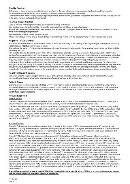
Quality Control<br />
Differences in tissue processing and technical procedures in the user’s laboratory may produce significant variability in results,<br />
necessitating regular performance of in-house controls in addition to the following procedures.<br />
Controls should be fresh autopsy/biopsy/surgical specimens, formalin-fixed, processed and paraffin wax-embedded as soon as possible<br />
in the same manner as the patient sample(s).<br />
Positive Tissue Control<br />
Used to indicate correctly prepared tissues and proper staining techniques.<br />
One positive tissue control should be included for each set of test conditions in each staining run.<br />
A tissue with weak positive staining is more suitable than a tissue with strong positive staining for optimal quality control and to detect<br />
minor levels of reagent degradation. 2<br />
Recommended positive control tissue is thyroid.<br />
If the positive tissue control fails to demonstrate positive staining, results with the test specimens should be considered invalid.<br />
Negative Tissue Control<br />
Should be examined after the positive tissue control to verify the specificity of the labeling of the target antigen by the primary antibody.<br />
Recommended negative control tissue is tonsil.<br />
Alternatively, the variety of different cell types present in most tissue sections frequently offers negative control sites, but this should be<br />
verified by the user.<br />
Non-specific staining, if present, usually has a diffuse appearance. Sporadic staining of connective tissue may also be observed in<br />
sections from excessively formalin-fixed tissues. Use intact cells for interpretation of staining results. Necrotic or degenerated cells often<br />
stain non-specifically. 3 False-positive results may be seen due to non-immunological binding of proteins or substrate reaction products.<br />
They may also be caused by endogenous enzymes such as pseudoperoxidase (erythrocytes), endogenous peroxidase<br />
(cytochrome C), or endogenous biotin (eg. liver, breast, brain, kidney) depending on the type of immunostain used. To differentiate<br />
endogenous enzyme activity or non-specific binding of enzymes from specific immunoreactivity, additional patient tissues may be stained<br />
exclusively with substrate chromogen or enzyme complexes (avidin-biotin, streptavidin, labeled polymer) and substrate-chromogen,<br />
respectively. If specific staining occurs in the negative tissue control, results with the patient specimens should be considered invalid.<br />
Negative Reagent Control<br />
Use a non-specific negative reagent control in place of the primary antibody with a section of each patient specimen to evaluate<br />
non-specific staining and allow better interpretation of specific staining at the antigen site.<br />
Patient Tissue<br />
Examine patient specimens stained with NCL-L-TTF-1 last. Positive staining intensity should be assessed within the context of any<br />
non-specific background staining of the negative reagent control. As with any immunohistochemical test, a negative result means that<br />
the antigen was not detected, not that the antigen was absent in the cells/tissue assayed. If necessary, use a panel of antibodies to<br />
identify false-negative reactions.<br />
Results Expected<br />
Normal Tissues<br />
Clone SPT24 detected the thyroid transcription factor-1 protein in the nucleus of follicular epithelial cells of the thyroid (13/13), type II<br />
pneumocytes and Clara cells of the lung (9/9). Some reactivity was also noted in glial cells in cerebrum (3/3).<br />
Clone SPT24 did not stain TTF-1 in a variety of other normal tissues (n=140) including colon (0/12), brain (0/8), placenta (0/4), kidney<br />
(0/6), prostate (0/4), testis (0/4), heart (0/4), spleen (0/4), cervix (0/5), lymph node (0/2), thymus (0/13), tongue (0/1), endometrium (0/2),<br />
skeletal muscle (0/4), tonsil (0/4), salivary gland (0/4), myometrium (0/1), umbilical cord (0/1), ureter (0/1), bronchus (0/1), ovary (0/4),<br />
fallopian tube (0/1), liver (0/4), breast (0/4), esophagus (0/4), stomach (0/4), spinal cord (0/1), eye (0/1), pancreas (0/4), ileum (0/4),<br />
cecum (0/1), rectum (0/1), adrenal (0/4), mesothelium (0/1), parathyroid (0/1), peripheral nerve (0/3), pituitary (0/3), bone marrow (0/3),<br />
uterus (0/3) and skin (0/4).<br />
Abnormal Tissues<br />
Clone SPT24 detected the thyroid transcription factor-1 protein in the nucleus of 26/32 lung adenocarcinoma, 4/7 lung small cell<br />
carcinoma, 2/3 lung bronchioalveolar carcinoma, 6/6 thyroid papillary carcinoma, 4/4 thyroid medullary carcinoma, 3/3 thyroid follicular<br />
carcinoma, 1/1 thyroid follicular adenoma, 1/1 Hashimoto’s thyroiditis, 19/56 thymoma, 1/1 desmoplastic small round cell tumor, 3/38<br />
moderately differentiated colon adenocarcinoma and 1/1 rectal carcinoma.<br />
Clone SPT24 did not stain lung squamous cell carcinoma (0/18), lung large cell carcinoma (0/5), poorly differentiated colon<br />
adenocarcinoma (0/4), well differentiated colon adenocarcinoma (0/3), ungraded colon adenocarcinoma (0/3), mesothelioma (0/5),<br />
small bowel carcinoid (0/4), thymus atypical carcinoid (1/5), metastatic thymic tumor (0/1), breast tumor (0/6), liver tumor (0/8), kidney<br />
renal cell carcinoma (0/5), kidney transitional cell carcinoma (0/1), ovary thecoma (0/1), ovary granulosa cell tumor (0/1), ovary juvenile<br />
granulosa (0/1), ovary serous carcinoma (0/3), ovary mucinous carcinoma (0/2), ovary germ cell tumor (0/1), ovary clear cell carcinoma<br />
(0/1), stomach adenocarcinoma (0/4), pancreas adenocarcinoma (0/2), pancreas papillary mucinous carcinoma (0/1), pancreas islet<br />
cell tumor (0/1), pancreas glucagonoma (0/1), testis seminoma (0/3), testis mixed germ cell tumor (0/1), testis embryonal carcinoma<br />
(0/1), brain astrocytoma (0/1), brain choroid plexus papilloma (0/1), melanoma (0/3), skin basal cell carcinoma (0/1), skin squamous<br />
cell carcinoma (0/2), penis squamous cell carcinoma (0/2), esophagus squamous cell carcinoma (0/2), larynx squamous cell carcinoma<br />
(0/1), tongue squamous cell carcinoma (0/2), cervix squamous carcinoma (0/1), small bowel carcinoma (0/1), GIST (0/1), synovial<br />
sarcoma (0/1), leiomyosarcoma (0/1), Ewing’s sarcoma (0/1), spindle cell rhabdomyosarcoma (0/1), omental fibrous tumor (0/1), bladder<br />
transitional cell carcinoma (0/2), bladder small cell carcinoma (0/1), large B-cell lymphoma (0/1), adrenal oncocytoma (0/2), adrenal<br />
adenoma (0/1), ganglioneuroma (0/1), prostate adenocarcinoma (0/1), benign prostatic hyperplasia (0/1), endometrial stromal sarcoma<br />
(0/1), endometrial adenocarcinoma (0/1), endometrial clear cell carcinoma (0/1), pheochromocytoma (0/1), paraganglioma (0/1) and<br />
cervix tumor (0/2).<br />
Thyroid Transcription Factor-1 (clone SPT24) is recommended for the identification of TTF-1 in normal and neoplastic tissues<br />
and for use in diagnosis as part of a panel of antibodies.<br />
TTF-1-L-CE<br />
Page 3


