Mycotrim® RS - Irvine Scientific
Mycotrim® RS - Irvine Scientific
Mycotrim® RS - Irvine Scientific

You also want an ePaper? Increase the reach of your titles
YUMPU automatically turns print PDFs into web optimized ePapers that Google loves.
M. pneumoniae colony appearance:<br />
M. pneumoniae colonies are 60 to 200 microns in diameter,<br />
about 1/10 as large as a small bacterial colony, 1 to 5 times<br />
larger than a single buccal epithelial cell, and 10 to 15 times<br />
larger than a single white blood cell. Mycoplasma colonies<br />
have a characteristic “fried egg” or “sand patty” (grainy)<br />
appearance. Epithelial cells are clearly distinguished from<br />
mycoplasma colonies by their sharp, angular margins and<br />
clear cytoplasm (often folded over upon itself).<br />
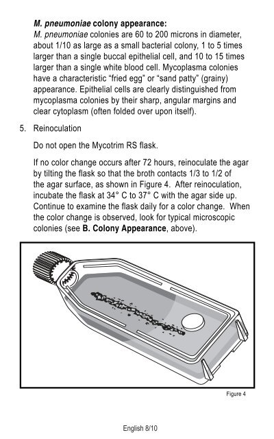
5. Reinoculation<br />
Do not open the Mycotrim <strong>RS</strong> flask.<br />
If no color change occurs after 72 hours, reinoculate the agar<br />
by tilting the flask so that the broth contacts 1/3 to 1/2 of<br />
the agar surface, as shown in Figure 4. After reinoculation,<br />
incubate the flask at 34° C to 37° C with the agar side up.<br />
Continue to examine the flask daily for a color change. When<br />
the color change is observed, look for typical microscopic<br />
colonies (see B. Colony Appearance, above).<br />
English 8/10<br />
Figure 4