Nature Immunology
Nature Immunology
Nature Immunology

You also want an ePaper? Increase the reach of your titles
YUMPU automatically turns print PDFs into web optimized ePapers that Google loves.
© 2009 <strong>Nature</strong> America, Inc. All rights reserved.<br />
www.nature.com/natureimmunology<br />
EDITORIAL OFFICE immunology@natureny.com<br />
75 Varick Street, Fl 9, New York, NY 10013-1917<br />
Tel: (212) 726 9207, Fax: (212) 696 9752<br />
Editor: Jamie D. K. Wilson<br />
Senior Editors: Christine Borowski, Laurie A. Dempsey<br />
Copy Editor: Jennifer Fosmire<br />
Production Editors: Marina Corral, Matt Hansen<br />
Senior Illustrator: Katie Vicari<br />
Illustrator: Kimberly Caesar<br />
Senior Editorial Assistant: Adam Lipkin<br />
MANAGEMENT OFFICES<br />
NPG New York<br />
75 Varick Street, Fl 9, New York, NY 10013-1917<br />
Tel: (212) 726 9200, Fax: (212) 696 9006<br />
Executive Editor: Linda Miller<br />
Chief Technology Officer: Howard Ratner<br />
Head of <strong>Nature</strong> Research & Reviews Marketing: Sara Girard<br />
Marketing Manager: Leah Rodriguez<br />
Assistant Production Coordinator: Karen Wilson<br />
Head of Web Services: Anthony Barrera<br />
Web Production Editor: Colleen Diez<br />
NPG London<br />
The Macmillan Building, 4 Crinan Street, London N1 9XW<br />
Tel: 44 207 833 4000, Fax: 44 207 843 4996<br />
Managing Director: Steven Inchcoombe<br />
Publishing Director: Alison Mitchell<br />
Publisher: Stephanie Diment<br />
Editor-in-Chief, <strong>Nature</strong> Publications: Philip Campbell<br />
Marketing Director: Della Sar<br />
Director of Web Publishing: Timo Hannay<br />
NPG <strong>Nature</strong> Asia-Pacific<br />
Chiyoda Building, 2-37 Ichigayatamachi, Shinjuku-ku, Tokyo 162-0843<br />
Tel: 81 3 3267 8751, Fax: 81 3 3267 8746<br />
Publishing Director — Asia-Pacific: David Swinbanks<br />
Associate Director: Antoine E. Bocquet<br />
Manager: Koichi Nakamura<br />
Senior Marketing Manager: Peter Yoshihara<br />
Asia-Pacific Sales Director: Kate Yoneyama<br />
Asia-Pacific Sales Manager: Ken Mikami<br />
DISPLAY ADVERTISING display@natureny.com (US/Canada)<br />
display@nature.com (Europe)<br />
nature@natureasia.com (Asia)<br />
Global Head of Display Advertising Sales: John Michael, Tel: 44 207 843 4960, Fax: 44 207<br />
843 4996<br />
Asia-Pacific Sales Manager: Ken Mikami, Tel: 81 3 3267 8765, Fax: 81 3 3267 8746<br />
Display Account Managers:<br />
New England: Sheila Reardon, Tel: (617) 399 4098, Fax: (617) 426 3717<br />
New York/Mid-Atlantic/Southeast: Jim Breault, Tel: (212) 726 9334, Fax: (212) 696 9481<br />
Midwest: Mike Rossi, Tel: (212) 726 9255, Fax: (212) 696 9481<br />
West Coast South: George Lui, Tel: (415) 781 3804, Fax: (415) 781 3805<br />
West Coast North: Bruce Shaver, Tel: (415) 781 6422, Fax: (415) 781 3805<br />
Germany/Switzerland/Austria: Sabine Hugi-Fürst, Tel: 41 52761 3386, Fax: 41 52761 3419<br />
United Kingdom/Ireland/France/Belgium/Eastern Europe: Jeremy Betts, Tel: 44 207 843 4968,<br />
Fax: 44 207 843 4749<br />
Scandinavia/The Netherlands/Italy/Spain/Portugal/Israel/Iceland: Graham Combe,<br />
Tel: 44 207 843 4914, Fax: 44 207 843 4749<br />
Greater China/Singapore: Gloria To, Tel: 852 2811 7191, Fax: 852 2811 0743<br />
NATUREJOBS naturejobs@natureny.com (US/Canada)<br />
naturejobs@nature.com (Europe)<br />
nature@natureasia.com (Asia)<br />
US Sales Manager: Peter Bless, Tel: (212) 726 9248, Fax: (212) 696 9482<br />
European Sales Manager: Andrew Douglas, Tel: 44 207 843 4975, Fax: 44 207 843 4996<br />
Asia-Pacific Sales Manager: Ayako Watanabe, Tel: 81 3 3267 8765, Fax: 81 3 3267 8746<br />
SITE LICENSE BUSINESS UNIT<br />
Americas: Tel: (888) 331 6288 institutions@natureny.com<br />
Asia/Pacific: Tel: 81 3 3267 8751 institutions@natureasia.com<br />
Australia/New Zealand: Tel: 61 3 9825 1160 nature@macmillan.com.au<br />
India: Tel: 91 124 2881054/55 npgindia@nature.com<br />
ROW: Tel: 44 207 843 4759 institutions@nature.com<br />
CUSTOMER SERVICE www.nature.com/help<br />
Senior Global Customer Service Manager: Gerald Coppin<br />
For all print and online assistance, please visit www.nature.com/help<br />
Purchase subscriptions:<br />
Americas: <strong>Nature</strong> <strong>Immunology</strong>, Subscription Dept., 342 Broadway, PMB 301, New York, NY<br />
10013-3910. Tel: (866) 363 7860, Fax: (212) 689 9108<br />
Europe/ROW: <strong>Nature</strong> <strong>Immunology</strong>, Subscription Dept., Macmillan Magazines Ltd., Brunel<br />
Road, Houndmills, Basingstoke, RG21 6XS, United Kingdom. Tel: 44 1256 329 242, Fax: 44<br />
1256 812 358<br />
Japan: <strong>Nature</strong> <strong>Immunology</strong>, NPG <strong>Nature</strong> Asia-Pacific, Chiyoda Building, 2-37<br />
Ichigayatamachi, Shinjuku-ku, Tokyo 162-0843. Tel: 81 3 3267 8751, Fax: 81 3 3267 8746<br />
India: <strong>Nature</strong> <strong>Immunology</strong>, NPG India, 3A, 4th Floor, DLF Corporate Park, Gurgaon 122002,<br />
India. Tel: 91 124 2881054/55, Fax: 91 124 2881052<br />
REPRINTS reprint@natureny.com<br />
<strong>Nature</strong> <strong>Immunology</strong>, Reprint Department, <strong>Nature</strong> Publishing Group, 75 Varick Street, Fl 9,<br />
New York, NY 10013-1917, USA.<br />
For commercial reprint orders of 600 or more, please contact:<br />
UK Reprints: Tel: 44 1256 302 923, Fax: 44 1256 321 531<br />
US Reprints: Tel: (212) 726 9278, Fax: (212) 679 0843
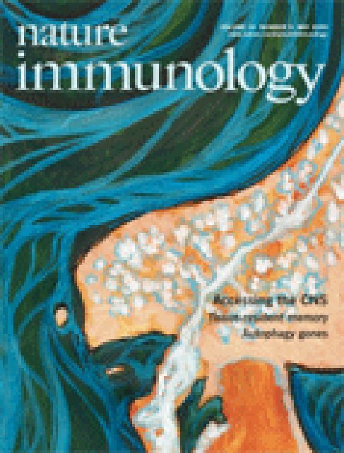
experimental autoimmune<br />
encephalomyelitis requires the entry of<br />
disease-inducing T cells into the brain.<br />
reboldi and colleagues (p 514; see also<br />
news and views by Steinman,<br />
p 453) find that T H -17 cells initiate<br />
this disease by entering the brain<br />
through the choroid plexus. The original<br />
image shows human brain tissue in<br />
which choroid plexus epithelial cells<br />
are stained with antibody to CCl20<br />
(fuchsia) and astrocytes are ‘decorated’<br />
by antibody to glial fibrillary acidic<br />
protein (brown). original image by<br />
andrew elston (lifeSpan bioSciences).<br />
artwork by lewis long.<br />
brahms and inteferon (p 447)<br />
Editorial<br />
445 The final push?<br />
Essay<br />
447 Aimez-vous Brahms? A story capriccioso from the discovery of a cytokine family<br />
and its regulators<br />
tadatsugu taniguchi<br />
nEws and viEws<br />
451 Local advantage: skin DCs prime; skin memory T cells protect<br />
akiko iwasaki see also pp 488 & 524<br />
453 Gaining entry to an uninflamed brain<br />
robert C axtell & lawrence steinman see also p 514<br />
455 Crohn’s disease–associated Nod2 mutants reduce IL10 transcription<br />
dana J Philpott & stephen E Girardin see also p 471<br />
457 The Foxo and the hound: chasing the in vivo regulation of T cell populations<br />
during infection<br />
Elia d tait & Christopher a Hunter see also p 504<br />
459 rEsEarCH HiGHliGHts<br />
rEviEw<br />
461 Autophagy genes in immunity<br />
Herbert w virgin & Beth levine<br />
artiClEs<br />
volume 10 number 5 may 2009<br />
471 A Crohn’s disease–associated NOD2 mutation suppresses transcription of human<br />
IL10 by inhibiting activity of the nuclear ribonucleoprotein hnRNP-A1<br />
Eiichiro noguchi, yoichiro Homma, Xiaoyan Kang, Mihai G netea & Xiaojing Ma<br />
see also p 455<br />
480 Autophagy enhances the presentation of endogenous viral antigens on MHC class I<br />
molecules during HSV-1 infection<br />
luc English, Magali Chemali, Johanne duron, Christiane rondeau, annie laplante,<br />
diane Gingras, diane alexander, david leib, Christopher norbury, roger lippé &<br />
Michel desjardins<br />
<strong>Nature</strong> <strong>Immunology</strong> (issn 1529-2908) is published monthly by nature Publishing Group, a trading name of nature america inc. located at 75 varick<br />
street, Fl 9, new york, ny 10013-1917. Periodicals postage paid at new york, ny and additional mailing post offices. Editorial Office: 75 varick street,<br />
Fl 9, new york, ny 10013-1917. tel: (212) 726 9207, Fax: (212) 696 9752. Annual subscription rates: Usa/Canada: Us$225 (personal), Us$3,060<br />
(institution). Canada add 7% Gst #104911595rt001; Euro-zone: €287 (personal), €2,430 (institution); rest of world (excluding China, Japan, Korea):<br />
£185 (personal), £1,570 (institution); Japan: Contact nPG nature asia-Pacific, Chiyoda Building, 2-37 ichigayatamachi, shinjuku-ku, tokyo 162-0843.<br />
tel: 81 (03) 3267 8751, Fax: 81 (03) 3267 8746. POSTMASTER: send address changes to <strong>Nature</strong> <strong>Immunology</strong>, subscriptions department, 342 Broadway,<br />
PMB 301, new york, ny 10013-3910. Authorization to photocopy material for internal or personal use, or internal or personal use of specific clients,<br />
is granted by nature Publishing Group to libraries and others registered with the Copyright Clearance Center (CCC) transactional reporting service,<br />
provided the relevant copyright fee is paid direct to CCC, 222 rosewood drive, danvers, Ma 01923, Usa. identification code for <strong>Nature</strong> <strong>Immunology</strong>:<br />
1529-2908/04. Back issues: Us$45, Canada add 7% for Gst. CPC PUB aGrEEMEnt #40032744. Printed on acid-free paper by dartmouth Journal<br />
services, Hanover, nH, Usa. Copyright © 2009 nature Publishing Group. Printed in Usa.<br />
i
nature immunology<br />
Getting aID<br />
(p 540)<br />
The Foxo factor<br />
(pp 457 and 504)<br />
Skin dendritic cells<br />
(pp 451 and 488)<br />
488 Cross-presentation of viral and self antigens by skin-derived CD103 + dendritic<br />
cells<br />
sammy Bedoui, Paul G whitney, Jason waithman, liv Eidsmo, linda wakim,<br />
irina Caminschi, rhys s allan, Magdalena wojtasiak, Ken shortman,<br />
Francis r Carbone, andrew G Brooks & william r Heath see also pp 451 & 524<br />
496 Divergent functions for airway epithelial matrix metalloproteinase 7 and retinoic<br />
acid in experimental asthma<br />
sangeeta Goswami, Pornpimon angkasekwinai, Ming shan, Kendra J Greenlee,<br />
wade t Barranco, sumanth Polikepahad, alexander seryshev, li-zhen song,<br />
david redding, Bhupinder singh, sanjiv sur, Prescott woodruff, Chen dong,<br />
david B Corry & Farrah Kheradmand<br />
504 Transcription factor Foxo3 controls the magnitude of T cell immune responses by<br />
modulating the function of dendritic cells<br />
anne s dejean, daniel r Beisner, irene l Ch’en, yann M Kerdiles, anna Babour,<br />
Karen C arden, diego H Castrillon, ronald a dePinho & stephen M Hedrick<br />
see also p 457<br />
514 C-C chemokine receptor 6–regulated entry of T H-17 cells into the CNS through<br />
the choroid plexus is required for the initiation of EAE<br />
andrea reboldi, Caroline Coisne, dirk Baumjohann, Federica Benvenuto,<br />
denise Bottinelli, sergio lira, antonio Uccelli, antonio lanzavecchia,<br />
Britta Engelhardt & Federica sallusto see also p 453<br />
524 Memory T cells in nonlymphoid tissue that provide enhanced local immunity<br />
during infection with herpes simplex virus<br />
thomas Gebhardt, linda M wakim, liv Eidsmo, Patrick C reading, william r Heath &<br />
Francis r Carbone see also pp 451 & 488<br />
531 T cell antigen receptor signaling and immunological synapse stability require<br />
myosin IIA<br />
tal ilani, Gaia vasiliver-shamis, santosh vardhana, anthony Bretscher &<br />
Michael l dustin<br />
540 HoxC4 binds to the promoter of the cytidine deaminase AID gene to induce AID<br />
expression, class-switch DNA recombination and somatic hypermutation<br />
seok-rae Park, Hong Zan, Zsuzsanna Pal, Jinsong Zhang, ahmed al-Qahtani,<br />
Egest J Pone, Zhenming Xu, thach Mai & Paolo Casali<br />
551 CorriGEnda<br />
natUrE iMMUnoloGy ClassiFiEd<br />
See back pages.<br />
volume 10 number 5 may 2009<br />
iii
© 2009 <strong>Nature</strong> America, Inc. All rights reserved.<br />
The final push?<br />
Over 20 years ago, the Global Polio Eradication Initiative was launched. Today, polio is still endemic in four countries.<br />
With great fanfare, the World Health Assembly launched the<br />
Global Polio Eradication Initiative (GPEI) just over 20 years<br />
ago. In what was described as a “magnificent gift from the 20th<br />
century to future generations of children,” public health officials and<br />
volunteers committed themselves to ridding the planet of polio by the<br />
year 2000. Unfortunately, there are still new cases of infection in 2009.<br />
Although worldwide cases of polio have fallen from 350,000 in 1988 to<br />
under 2,000 in 2008, some experts question whether eradication will<br />
ever be possible. Many elements are conspiring against the efforts to<br />
eliminate disease, including the present political and economic crisis.<br />
By 2001, it almost seemed that the battle against polio had been won—<br />
worldwide cases fell to an all-time low of 791. Poliomyelitis type II had<br />
been completely eradicated by 1999. However, since 2003 the initiative<br />
has suffered several setbacks. In Nigeria, rumors began circulating that<br />
the polio vaccine contained the AIDS virus and was part of a Western plot<br />
to sterilize Muslim girls. The vaccine was described as “tainted by evildoers<br />
from America” and was “America’s revenge for September 11th.”<br />
As a result, the Nigerian government halted the vaccination campaign<br />
until the vaccine could be proven safe. As a consequence, 18 formerly<br />
polio-free countries suffered outbreaks traceable to Nigeria. A few countries,<br />
such as Sudan, are still struggling to regain their polio-free status.<br />
At present, polio is endemic in four countries: Nigeria, Afghanistan,<br />
Pakistan and India. In 2007, an intensified eradication effort was<br />
launched by the GPEI to circumvent the remaining technical, financial<br />
and operational barriers that were preventing polio eradication in<br />
these countries (http://www.polioeradication.org/content/publications/<br />
PolioStrategicPlan09-13_Framework.pdf). By mid-2008, two independent<br />
World Health Organization (WHO) advisory bodies concluded<br />
that the intensified efforts could overcome these challenges, and consequently<br />
a new Strategic Plan 2009–2013 has been endorsed. This 5-year<br />
plan combines proven eradication strategies with recently developed<br />
tools and tactics, including improving the efficacy of the vaccine. But<br />
even with a new strategic plan in place, is it realistic to expect a poliofree<br />
world by 2013?<br />
The GPEI will need to overcome many obstacles, not least the political<br />
instability of many regions in the polio-endemic countries. Afghanistan<br />
and Pakistan will probably pose the greatest challenge. Because of<br />
successful vaccine drives, no cases of polio have been reported in the<br />
relatively peaceful northern provinces of Afghanistan, but the conflict–<br />
ridden south and southeastern provinces still suffer recurrent outbreaks.<br />
Access of aid workers to vulnerable communities in these areas has been<br />
increasingly limited. Many aid workers have been killed, abducted or<br />
threatened by criminal gangs and Taliban insurgents in recent years,<br />
according to the Afghanistan NGO Safety Office. Only a few months<br />
ago, a day of tranquility organized by the United Nations Assistance<br />
Mission in Afghanistan to enable immunizations in remote areas was<br />
ediTorial<br />
cancelled by the WHO after two Afghan doctors were killed by a suicide<br />
car bombing caused by the Taliban.<br />
Pakistan has also become increasingly unstable. The Taliban and<br />
Al-Qaeda resurgence in the North-West Frontier Province of Pakistan<br />
is allowing the virus to cross borders between the two countries unimpeded.<br />
Cases of polio have exploded in Pakistan, causing outbreaks<br />
in previously polio-free areas. In 2007, the cleric Mufti Khalid Shah<br />
declared a fatwa on employees of the United Nations, WHO and all<br />
other foreign organizations in 2007, and aid workers in Bannu were<br />
sent a 500-rupee note with a letter stating they could either stop or buy<br />
their own coffin.<br />
Cultural differences also present problems for the vaccine program<br />
in these regions. Finding sufficient numbers of women to join the vaccination<br />
team has been difficult. However, the participation of women<br />
is essential, as by tradition only women can enter a Muslim household<br />
when the husband is away, and women with children are usually better<br />
at persuading other mothers to vaccinate. The GPEI must also persuade<br />
people that the vaccine is safe, improve poor sanitation that can interfere<br />
with uptake of the oral vaccine, and offset fatigue that can set in among<br />
volunteers, donors and the general populace. It only takes one unvaccinated<br />
child to trigger a new epidemic.<br />
Fortunately, fatigue has not affected the organizers of the eradication<br />
movement. In January 2009, the Bill & Melinda Gates Foundation,<br />
Rotary International and the governments of the UK and Germany<br />
pledged US$630 million over 5 years for a massive final push to rid<br />
humanity of this scourge. But this may not be enough. Heidemarie<br />
Wieczorek-Zeul, the German Federal Minister for Economic Cooperation<br />
and Development, estimated that even with the new injection of funds,<br />
the global initiative is still some US$340 million short of its budget for<br />
2009–2010. Given that funding agencies and charities, including the<br />
Gates Foundation, are having to prune grant growth because of the recession,<br />
these are uncertain times for the eradication program.<br />
Nevertheless, there are many reasons to be optimistic. The government<br />
in Nigeria is now firmly behind the GPEI, and Muslim clerics who<br />
initially shunned the vaccine program are now actively campaigning for<br />
acceptance of the polio vaccine. In March, President Barack Obama<br />
announced the mobilization of more troops in Afghanistan to “disrupt,<br />
dismantle and defeat” the terrorist Al-Qaeda network in Afghanistan<br />
and neighboring Pakistan. Although the final push for eradication will<br />
be costly and difficult, it certainly will not compare with the costs the<br />
world would face if polio were allowed to reemerge. Since 1988, more<br />
than 2 million children have been immunized, and it is estimated that<br />
as a result, 5 million fewer people have been paralyzed by polio. It is<br />
a testament to the success of the GPEI that worldwide cases of polio<br />
have fallen by over 99%. Now is not the time to give up, even when the<br />
going seems to be getting tough.<br />
nature immunology volume 10 number 5 may 2009 445
© 2009 <strong>Nature</strong> America, Inc. All rights reserved.<br />
Aimez-vous Brahms? A story capriccioso<br />
from the discovery of a cytokine family and<br />
its regulators<br />
Tadatsugu Taniguchi<br />
Do you delight in Brahms? Do you delight in immunology? Tada Taniguchi recounts the story of Type 1 interferon and<br />
its downstream regulators.<br />
“The Master said ‘Those who know it are not<br />
comparable to those who love it; those who love<br />
it are not compatible to those who delight in<br />
it.’” —Analects of Confucius<br />
Music aficionados may recognize the title of<br />
this article as a phrase originating from<br />
Françoise Sagan’s romantic novel of the same<br />
name (published in English as Goodbye Again).<br />
A typical laboratory scene on a Sunday afternoon<br />
in the 1970s was one in which I would<br />
whistle the first movement of the Brahms second<br />
piano concerto and then the next portion<br />
of the melody would be whistled back from<br />
the nearby office of my graduate school mentor<br />
Charles Weissmann. In this environment, I<br />
began the scientific achievements that would<br />
shape my career. To the credit of Massimo<br />
Libonati, my mentor from two prior years<br />
in Napoli, and my very rudimentary Italian,<br />
I had made my debut at a scientific meeting<br />
held in Roma in 1972 (ref. 1) before meeting<br />
Charles, my subsequent friend and mentor. I<br />
had left Tokyo for Naples without even a master’s<br />
degree, partly because I was appassionato<br />
about classic music. Indeed, it was through my<br />
desire to do science in Italy that I was able to<br />
derive tremendous enjoyment from my personal<br />
meetings with many artists, including the<br />
tenor Mario del Monaco and a retired German<br />
prima donna who lived in a friend’s villa in<br />
Tadatsugu Taniguchi is in the Department of<br />
<strong>Immunology</strong>, Graduate School of Medicine and<br />
Faculty of Medicine, at Tokyo University, Tokyo,<br />
Japan.<br />
e-mail: tada@m.u-tokyo.ac.jp<br />
Capri and recounted to me many episodes<br />
of her—sometimes romantic—experiences<br />
with the conductors Bruno Walter, Wilhelm<br />
Furtwängler, Herbert von Karajan and others.<br />
In Charles’ lab at the Institut für<br />
Molekularbiologie I der Universität Zürich,<br />
the project that was to become my PhD thesis<br />
involved site-directed mutagenesis of the RNA<br />
phage Qβ, a technique invented by Charles and<br />
one of my brothers in science, Richard Flavell 2 ,<br />
thereby spawning the concept of ‘reverse<br />
genetics’ that was only later applied to DNA.<br />
Although it was not very easy to catch up with<br />
the other members of the lab—who came to<br />
Zurich from top institutes in the world—in<br />
terms of either language or science, their<br />
exceptional kindness helped me to adjust in a<br />
relatively short time. I was fortunate enough<br />
to publish several papers on Qβ, including a<br />
description of the use of recombinant DNA<br />
technology to generate a plasmid encoding<br />
the genome of this virus as a means to produce<br />
an infectious RNA virus 3 . On weekends,<br />
I was invited to Charles’s home to write papers:<br />
as I recall well, writing was accompanied by<br />
music, often the chamber music of Brahms<br />
and Beethoven but sometimes an opera such<br />
as Berlioz’s Les Troyens which, as many readers<br />
may have experienced, was quite beneficial to<br />
scientific writing.<br />
It was in 1978 that the hitherto unheard-of<br />
word “interferon” (IFN) came to my ears<br />
at a lecture given by Peter Lengyel of Yale<br />
University, a good friend of Charles and a<br />
pioneer of IFN research. It was a stormy experience,<br />
and it made me very curious about the<br />
phenomenon by which this molecule with<br />
Silhouette of Brahms.<br />
ESSAy<br />
antiviral activity is produced in mammalian<br />
cells infected by viruses; in my mind, at the<br />
time, I envisioned IFN as a ‘Sleeping Beauty’<br />
gene(s) awakened upon ‘un bacio’ (a kiss) of<br />
a virus. By this point, I had finished my PhD,<br />
and I was about to return to Tokyo for a new<br />
position when Charles asked me to begin<br />
work on the initial isolation of human leukocyte<br />
IFN (afterward renamed IFN-α) mRNA<br />
and the development of strategies for its eventual<br />
gene cloning, in collaboration with Kari<br />
Cantell in Helsinki.<br />
My curiosity about the nature of IFN was<br />
strongly sostenuto after my return to the Cancer<br />
Institute in Tokyo at the end of 1978, and so I<br />
decided to work on the fibroblast IFN (afterward<br />
renamed IFN-β, or IFNB) gene, which<br />
was presumed to be distinct from leukocyte<br />
IFN 4 , mainly in order to avoid unnecessary<br />
overlap and competition with the project in<br />
Charles’s lab. As it turned out, this decision led<br />
to the identification of IFN-α and IFN-β by<br />
Charles’s and my group, respectively, resulting<br />
in the first recognition of a cytokine gene fam-<br />
nature immunology volume 10 number 5 may 2009 447
© 2009 <strong>Nature</strong> America, Inc. All rights reserved.<br />
eSSay<br />
Tada Taniguchi, his mentor Charles Weissmann<br />
and colleagues in Zurich (1978). Charles (second<br />
from right) is wearing a uniform because he had<br />
just returned from military service.<br />
ily. My initial inclination was to search around<br />
for someone working on fibroblast IFN; the<br />
gene was extremely ricercato by numerous<br />
pharmaceutical companies, and there were<br />
many aspirations for its clinical use, so I was<br />
not very optimistic about getting support<br />
from them because of their desire to protect<br />
trade secrets and potential patents. Although<br />
IFNs and other soluble mediators of immune<br />
responses, later collectively termed ‘cytokines’,<br />
had already garnered considerable attention<br />
in the biological sciences and medicine at that<br />
time, the structure and function of these molecules<br />
remained elusive, along with the underlying<br />
mechanisms of signal transmission and<br />
the regulation of their expression. Indeed,<br />
addressing these issues was hampered by the<br />
fact that these molecules have pleiotropic, or<br />
multiple, biological activities, are usually produced<br />
simultaneously and are expressed only<br />
weakly by a variety of different cell types, making<br />
it difficult to obtain a single cytokine in<br />
pure form.<br />
My initial efforts to obtain a consistent<br />
source of starting material were in vain until<br />
I read a book about IFN, written in Japanese<br />
with great scientific enthusiasm and dedication<br />
by Shigeyasu Kobayashi of Toray, Inc. I<br />
visited Dr. Kobayashi in Toray’s Basic Research<br />
Institute and explained my enthusiasm for<br />
elucidating the structure of fibroblast IFN and<br />
the prospect that such work could lead to the<br />
discovery of the gene-switching mechanism in<br />
mammalian cells, an idea that was beginning<br />
to be addressed worldwide. He listened to me<br />
attentively, and then promptly told me that he<br />
would be willing to provide large amounts of<br />
his fibroblast cell line for the gene cloning. I was<br />
honestly surprised not only by the speed with<br />
which he made his decision but also because he<br />
placed no conditions on my receiving his help. I<br />
then realized that this was a kind of gentlemen’s<br />
agreement, although nowadays such an agreement<br />
is difficult to imagine: perhaps it was our<br />
shared scientific enthusiasm for the discovery<br />
of IFN, which was almost totally unknown at<br />
that time, that helped forge our collaboration.<br />
After all, “Curiosity has its own reason for<br />
existence,” as Albert Einstein (a fellow Zurich<br />
alumnus) once wrote.<br />
There was another big obstacle to my work:<br />
human gene cloning had not yet been done in<br />
Japan and the regulation of recombinant DNA<br />
technology was so strict that all work had to<br />
be done in a P3 facility using only the safest<br />
bacteria, that is, those that are the most difficult<br />
to grow (for example, Escherichia coli strain<br />
χ1776). Fortunately for me, the Institute had<br />
such a facility almost ready to use, though interestingly<br />
I was required, before the start of my<br />
experiments, to spray Neurospora crassa spores<br />
into the cabinet and monitor their capture so<br />
as to demonstrate the facility’s suitability for<br />
P3-level experiments. Finally, with the strong<br />
support and leadership of the Institute’s magnanimous<br />
director Haruo Sugano, I was soon<br />
allowed to proceed with the gene cloning.<br />
I was aware at the time that the competition<br />
for this project worldwide was very strong.<br />
Moreover, because I had to work solo, I thought<br />
it necessary to devise a strategy that would minimize<br />
my time and effort as much as possible.<br />
The most notable difficulty at that time was<br />
to identify cDNA clones for mRNAs that were<br />
very weakly expressed, of which IFN cDNA<br />
was a typical example. As a result, I came upon<br />
a two-step approach by which I would first<br />
search for all suspected clones before proceeding<br />
to identify the desired clone among them.<br />
I first prepared ‘hot’ ( 32 P-labeled) cDNA from<br />
mRNA isolated from poly(I:C)-stimulated<br />
fibroblast cells, and then hybridized the cDNA<br />
with mRNA from unstimulated cells. After<br />
separation of unhybridized cDNA (probe A)<br />
from hybridized cDNA (probe B), I subjected<br />
the unhybridized cDNA to Grunstein-Hogness<br />
in situ colony hybridization with two identical<br />
sets of filters harboring ~3,600 χ1776 colonies.<br />
I then assayed the four clones that preferentially<br />
hybridized to probe A by a ‘hybridizationtranslation<br />
assay’ to search more rigorously for<br />
the cDNA clone that contained the IFN mRNA<br />
sequence. This strategy worked well for me,<br />
and by the end of the summer of 1979, I was<br />
done 5 . Although I can no longer recall this, my<br />
wife Yoko says that I seldom came back home<br />
before midnight, even on holidays.<br />
I sequenced the cDNA (again solo) by the<br />
Maxam-Gilbert method, though the information<br />
I could obtain from one piece of endlabeled<br />
DNA was very limited at that time.<br />
Finally, after a few months, I elucidated the<br />
coding sequence of fibroblast IFN; it was on 2<br />
February 1980, while I was listening to one of<br />
the Beethoven’s Rasumovsky quartets at home<br />
with my son, who celebrated his second birth-<br />
day that day. In the interim, Kathy Zoon and<br />
Ernest Knight, together with Mike Hunkapiller,<br />
Lee Hood and colleagues, had determined the<br />
N-terminal sequences of human IFN-α and<br />
IFN-β, respectively, and Shigekazu Nagata,<br />
another brother in science in Charles’s lab,<br />
and his colleagues had cloned and expressed<br />
a human IFN-α (IFNA) cDNA 6–8 .<br />
I then received two international phone calls,<br />
one from Charles and the other from Mark<br />
Ptashne, already one of the most renowned<br />
molecular biologists around, who was then<br />
working at Harvard; Mark is also a renowned<br />
violinist and so might perhaps be reclassified<br />
as a ‘molecular violinist’. Charles and I talked<br />
about the structures of IFN-α and IFN-β, and<br />
we eventually published papers back to back<br />
on their sequences 9,10 . Naturally, we were very<br />
interested in comparing their sequences. Mark,<br />
meanwhile, was very keen to exploit the new<br />
technique developed by Lenny Guarente and<br />
Tom Roberts of expressing cDNA in E. coli and<br />
so invited me to Harvard so that we could work<br />
together. At the end of February, Yoko and I<br />
left Tokyo for Zurich with a final destination of<br />
Boston. Charles and I, sitting together again in<br />
his room where we had written so many papers,<br />
began the comparison between our IFN-α and<br />
IFN-β sequences by hand. I remember that it<br />
was he who noticed the first portion of the<br />
sequence similarities, which got us very excited,<br />
though only later would we become aware that<br />
this was just the beginning of the discovery of<br />
many cytokine families.<br />
Charles and I then decided to write a paper<br />
together. He kindly said something to the effect<br />
of, “Well, Tada, you are still young and need further<br />
development.” So, after carefully discussing<br />
the issue further with his colleagues Marco<br />
Schwarzstein and Ned Mantei, who sequenced<br />
the IFN-α cDNA, Charles generously gave me<br />
the first authorship. Our paper was eventually<br />
published as an Article in <strong>Nature</strong> back to back<br />
with the paper by Rik Derynck, Walter Fiers<br />
and colleagues, who also cloned IFN-β 11,12 .<br />
This was for us a truly singular experience<br />
with <strong>Nature</strong> in that we originally submitted the<br />
manuscript as a Letter, which the editors then<br />
converted into the longer Article format. Yoko<br />
and I then continued on to Boston, where we<br />
received a warm welcome from Mark and his<br />
colleagues. Thanks to the kind help provided<br />
by Jan Vilcek (IFN assay), Alice Wong (virus),<br />
Vicki Sato (human cells) and many others, the<br />
work was done in a relatively short period of<br />
time 13 . During our sojourn, we were given<br />
many opportunities to interact socially with<br />
many scientists and artists, including Wally<br />
Gilbert, Lew Cantley, Hidde Ploegh, (the late)<br />
pianist Patricia Zander and cellist Yo-Yo Ma.<br />
Of the many concerts we enjoyed, perhaps the<br />
448 volume 10 number 5 may 2009 nature immunology
© 2009 <strong>Nature</strong> America, Inc. All rights reserved.<br />
most unforgettable was Sergey Rachmaninov’s<br />
second sonata for piano played by his friend<br />
Vladimir Horowitz at Boston Symphony Hall.<br />
Overall, my friendships and experiences from<br />
this time have endured ever since then and are<br />
a great treasure of my life.<br />
After returning to Tokyo, I started working<br />
on another class of cytokines, now known as<br />
the interleukins (ILs), and specifically on T<br />
cell growth factor 14 (since renamed IL-2), with<br />
Junji Hamuro, Hiroshi Matsui and colleagues<br />
at Ajinomoto Inc. IL-2 was also the subject<br />
of considerable hopes for its use in basic and<br />
clinical immunology, and we succeeded in<br />
elucidating the structure of human IL-2 and<br />
producing recombinant IL-2 in 1982 (ref. 15).<br />
It is remarkable that, since then, more than<br />
30 ILs have been cloned and characterized. In<br />
the interim, my colleague Shigeo Ohno joined<br />
my lab and discovered that the 5′ region of the<br />
human IFN-β gene confers virus infection–<br />
dependent activation of a reporter gene—the<br />
inception of the virus-inducible promoter<br />
analysis 16 . In parallel, both the IFN-α and<br />
IFN-β promoters were subsequently analyzed<br />
by many investigators, most notably by Charles<br />
and by Tom Maniatis and colleagues (reviewed<br />
in ref. 17). Indeed, the study of the IFN-β gene<br />
has led to the concept of the ‘enhanceosome’<br />
developed by Tom and his colleagues, which<br />
has become a model for understanding the<br />
mechanisms whereby genes are turned on and<br />
off in mammalian cells 18 .<br />
After I moved my lab and family to Osaka,<br />
another talented colleague, Takashi Fujita,<br />
joined my lab and identified a minimal DNA<br />
consensus sequence that functions as virusinducible<br />
enhancer 19 . Then, working together<br />
with Takashi, a master’s degree graduate student,<br />
Masaaki Miyamoto used an expression-<br />
cloning strategy to screen a cDNA library for<br />
gene products that would bind to Takashi’s<br />
consensus sequence. For several reasons, we<br />
originally proposed to name the newly discovered<br />
factor ‘cytokine regulatory factor-1’, but<br />
Ben Lewin, then editor of Cell, rejected our<br />
proposal. So the gene was instead named ‘interferon<br />
regulatory factor-1’ or IRF1 (ref. 20). Soon<br />
after, another master’s degree student, Hisashi<br />
Harada, discovered a related gene, termed IRF2,<br />
which officially signified the recognition of an<br />
IRF family. The family has since been extended<br />
to nine members in mammals by a number of<br />
other investigators (reviewed in ref. 21). In retrospect,<br />
the term ‘CRF’ gene family might indeed<br />
have been more accurate in the light of what we<br />
know now of this family’s role, but c’est la vie.<br />
The roles of IRFs have since been extensively<br />
studied by a number of groups worldwide.<br />
Indeed, interest in IRFs has only grown, particularly<br />
with the recent discovery of patternrecognition<br />
receptors whose activation evokes<br />
type I IFN and other innate immune responses,<br />
such as Toll-like receptors (pioneered by Jules<br />
Hoffman’s group working on Drosophila Toll<br />
and Ruslan Medzhitov, Charles Janeway and<br />
Bruce Beutler on TLR4 in mammals) and cytosolic<br />
receptors for nucleic acids (by Mitsutoshi<br />
Yoneyama and Takashi Fujita on RIG-I and<br />
MDA5, by my laboratory on DAI/DLM-1/ZBP1<br />
and recently by several groups on AIM2). We<br />
now know that IRF1, IRF3 and IRF7 (and possibly<br />
also IRF5) play critical roles in the regulation<br />
of type I IFN (IFN-α and IFN-β) genes,<br />
wherein the contribution of each depends<br />
on the nature of the stimuli and the cell type<br />
(reviewed in ref. 22). The role of IRFs in other<br />
biological responses, for example, in the regulation<br />
of oncogenesis, has also received much<br />
attention: accordingly, the numbers of articles<br />
describing IRFs are rapidly increasing. I am particularly<br />
grateful to Tak Mak, Shigeru Noguchi<br />
and Nobuaki Yoshida, who so kindly collaborated<br />
with us on the generation and analyses of<br />
a number of IRF knockout mice.<br />
Upon reflection, I find it quite remarkable<br />
that, since its independent discovery by Isaacs<br />
and Lindenman and by Nagano and Kojima<br />
more than 50 years ago, the type I IFN family<br />
of cytokines has become the prototype for<br />
cytokine research over the past three decades.<br />
Indeed, IFN research has led directly to the<br />
discovery of Janus-family (JAK) kinases,<br />
signal transducers and activators of transcription<br />
(STATs), the enhanceosome, IRFs<br />
and IFN-inducible genes; and, as key regulators<br />
of pathological and protective immune<br />
responses, IFNs are likely to lead to new<br />
research discoveries in the years to come.<br />
ACKNOWLEDGMENTS<br />
I thank J. Vilcek and D. Savitsky for their kind<br />
reading of this essay. My particular appreciation<br />
goes to my mentor C. Weissmann, from whom I<br />
learned so much about science and music (and<br />
good jokes), and to the colleagues who spent time<br />
in my laboratory through the many facets of the<br />
studies described above. I also thank my wife Yoko<br />
and my friends, either mentioned herein or not, for<br />
their continuous support. Our studies were mostly<br />
supported by Kakenhi, Grants-in-Aid for Scientific<br />
Research from the Ministry of Education, Culture,<br />
Sports, Science and Technology, Japan. Finally, I<br />
also thank baseball’s Hanshin Tigers for their longstanding<br />
roller coaster of hope and disappointment,<br />
which in addition to music has significantly<br />
enriched my life.<br />
eSSay<br />
1. Taniguchi, T., Libonati, M. & Leone, e. azione della<br />
subtilisina sulla ribonucleasi BS-1. Boll. Soc. Ital. Biol.<br />
Sper. XLVIII, 1115–1119 (1972).<br />
2. Flavell, R.a., Sabo, D.L., Bandle, e.F. & Weissmann,<br />
C. Site-directed mutagenesis: generation of an extracistronic<br />
mutation in bacteriophage Qβ RNa. J. Mol. Biol.<br />
89, 255–272 (1974).<br />
3. Taniguchi, T., Palmieri, M. & Weissmann, C.Q. β DNa–<br />
containing hybrid plasmids giving rise to Qβ phage<br />
formation in the bacterial host. <strong>Nature</strong> 274, 223–228<br />
(1978).<br />
4. Cavalieri, R.L., Havell, e.a., Vilcek, J. & Pestka, S.<br />
Synthesis of human interferon by Xenopus laevis oocytes:<br />
two structural genes for interferons in human cells. Proc.<br />
Natl. Acad. Sci. USA 74, 3287–3291 (1977).<br />
5. Taniguchi, T. et al. Construction and identification of a<br />
bacterial plasmid containing the human fibroblast interferon<br />
gene sequence. Proc. Jpn. Acad. 55B, 464–469<br />
(1979).<br />
6. Knight, e., Jr, Hunkapiller, M.W., Korant, B.D., Hardy,<br />
R.W. & Hood, L.e. Human fibroblast interferon: amino<br />
acid analysis and amino terminal amino acid sequence.<br />
Science 207, 525–526 (1980).<br />
7. Nagata, S. et al. Synthesis in E. coli of a polypeptide<br />
with human leukocyte interferon activity. <strong>Nature</strong> 284,<br />
316–320 (1980).<br />
8. Zoon, K.C. et al. amino terminal sequence of the major<br />
component of human lymphoblastoid interferon. Science<br />
207, 527–528 (1980).<br />
9. Mantei, N. et al. The nucleotide sequence of a cloned<br />
human leukocyte interferon cDNa. Gene 10, 1–10<br />
(1980).<br />
10. Taniguchi, T., Ohno, S., Fujii-Kuriyama, y. & Muramatsu,<br />
M. The nucleotide sequence of human fibroblast interferon<br />
cDNa. Gene 10, 11–15 (1980).<br />
11. Derynck, R. et al. Isolation and structure of a human<br />
fibroblast interferon gene. <strong>Nature</strong> 285, 542–547<br />
(1980).<br />
12. Taniguchi, T. et al. Human leukocyte and fibroblast interferons<br />
are structurally related. <strong>Nature</strong> 285, 547–549<br />
(1980).<br />
13. Taniguchi, T. et al. expression of the human fibroblast<br />
interferon gene in Escherichia coli. Proc. Natl. Acad. Sci.<br />
USA 77, 5230–5233 (1980).<br />
14. Morgan, D.a., Ruscetti, F.W. & Gallo, R. Selective in<br />
vitro growth of T lymphocytes from normal human bone<br />
marrows. Science 193, 1007–1008 (1976).<br />
15. Taniguchi, T. et al. Structure and expression of a cloned<br />
cDNa for human interleukin-2. <strong>Nature</strong> 302, 305–310<br />
(1983).<br />
16. Ohno, S. & Taniguchi, T. Inducer-responsive expression<br />
of the cloned human interferon β1 gene introduced into<br />
cultured mouse cells. Nucleic Acids Res. 10, 967–977<br />
(1982).<br />
17. Honda, K., Takaoka, a. & Taniguchi, T. Type I interferon<br />
gene induction by the interferon regulatory factor family<br />
of transcription factors. Immunity 25, 349–360<br />
(2006).<br />
18. Kim, T.K. & Maniatis, T. The mechanism of transcriptional<br />
synergy of an in vitro assembled interferon-β<br />
enhanceosome. Mol. Cell 1, 119–129 (1997).<br />
19. Fujita, T., Shibuya, H., Hotta, H., yamanishi, K. &<br />
Taniguchi, T. Interferon-β gene regulation: tandemly<br />
repeated sequences of a synthetic 6 bp oligomer function<br />
as a virus-inducible enhancer. Cell 49, 357–367<br />
(1987).<br />
20. Miyamoto, M. et al. Regulated expression of a gene<br />
encoding a nuclear factor, IRF-1, that specifically binds<br />
to IFN-β gene regulatory elements. pCell 54, 903–913<br />
(1988).<br />
21. Tamura, T., yanai, H., Savitsky, D. & Taniguchi, T. The<br />
IRF family transcription factors in immunity and oncogenesis.<br />
Annu. Rev. Immunol. 26, 535–584 (2008).<br />
22. Honda, K. & Taniguchi, T. IRFs: master regulators of<br />
signalling by Toll-like receptors and cytosolic patternrecognition<br />
receptors. Nat. Rev. Immunol. 6, 644–658<br />
(2006).<br />
nature immunology volume 10 number 5 may 2009 449
© 2009 <strong>Nature</strong> America, Inc. All rights reserved.<br />
Local advantage: skin DCs prime; skin memory T cells<br />
protect<br />
Akiko Iwasaki<br />
How the immune system responds to local infection and establishes protective immunity in susceptible tissues<br />
remains unclear. Two new studies show that local tissue-resident dendritic cells prime cytotoxic T lymphocyte<br />
responses and that memory cytotoxic T lymphocytes remain in the tissue to provide antiviral immunity.<br />
Cytotoxic T lymphocytes (CTLs) are key<br />
effector cells that provide protection from<br />
viral infection. CD8 + T cells bearing T cell antigen<br />
receptors specific for a given viral antigen<br />
are primed in the secondary lymphoid organs<br />
by dendritic cells (DCs). Exactly which subset<br />
of DCs primes CTLs in response to viral infection<br />
has been an area of intense debate. Many<br />
pathogens enter the host via a specific niche,<br />
most commonly the mucosal surfaces, where<br />
they establish local acute and chronic infection.<br />
Herpes virus family members represent<br />
a good example of such a pathogen. In particular,<br />
herpes simplex virus type 1 (HSV-1) enters<br />
the human host through the oral mucosa and<br />
establishes latency in the trigeminal ganglion.<br />
Reactivation of latent HSV-1 in the ganglion<br />
leads to anterograde transport of virus back<br />
to the skin, causing ‘cold sore’ lesions around<br />
the mouth. CTLs are important both in controlling<br />
HSV-1 reactivation1 and in limiting<br />
viral replication in the peripheral site of replication2<br />
. Given the restricted nature of HSV-1<br />
replication in the epithelial cells and latency<br />
in the innervating ganglia, which cells prime<br />
CD8 + T cells in the draining lymph node and<br />
how protection is afforded by memory T cells<br />
are important unresolved issues. In this issue<br />
of <strong>Nature</strong> <strong>Immunology</strong>, two papers show that<br />
local tissue-resident cells do both: Heath and<br />
colleagues demonstrate that CTL priming is<br />
accomplished mainly by langerin-positive<br />
CD103 + dermal DCs3 , whereas Carbone<br />
and colleagues report that memory CTLs in<br />
the skin remain in the tissue and maximize<br />
Akiko Iwasaki is in the Department of<br />
Immunobiology, Yale University School of Medicine,<br />
New Haven, Connecticut, USA.<br />
e-mail: akiko.iwasaki@yale.edu<br />
NewS AND vIewS<br />
protective immunity to subsequent challenge<br />
with HSV-1 (ref. 4).<br />
There are three DC subsets in the skin:<br />
Langerhans cells in the epidermis, and two<br />
subsets of dermal DCs, consisting of langerinnegative<br />
dermal DCs and the newly described<br />
langerin-positive CD103 + dermal DCs 5–7<br />
(Fig. 1). Traditionally, it has been thought<br />
that local tissue-resident DCs ingest microbial<br />
antigens by phagocytosis and migrate to the<br />
draining lymph node to prime naive T cells.<br />
However, that paradigm has been challenged<br />
by many studies showing that lymph noderesident<br />
DCs are the only cells that present<br />
antigens to T cells. As for the DCs involved in<br />
CTL priming, the importance of the lymph<br />
node–resident CD8α + DCs has become well<br />
accepted 8 . CD8α + DCs are the main antigenpresenting<br />
cells for CTLs that are generated<br />
after infection by HSV-1, influenza virus,<br />
vaccinia virus, lymphocytic choriomeningitis<br />
virus and Listeria monocytogenes. CD8α +<br />
DCs can exclusively prime CTL responses<br />
whether HSV-1 is injected by needle into the<br />
footpad, by the intravenous route or by dermal<br />
abrasion 9 (Fig. 1a). In their study presented<br />
here, Bedoui et al. make the intriguing<br />
observation that reactivating HSV-1 causes a<br />
second phase of infection of the entire skin<br />
dermatome innervated by the infected ganglion<br />
3 (Fig. 1b), which results in a second<br />
wave of antigen presentation in the lymph<br />
nodes draining the new site of viral replication.<br />
Reactivating HSV-1 replicates in the<br />
epithelium, notably in the absence of any<br />
artificial manipulation of the skin, allowing<br />
the authors to study the course of natural<br />
infection. Taking advantage of this system,<br />
they assess DC subsets for their ability to<br />
stimulate CTLs. They find that whereas cross-<br />
presentation of viral antigen during primary<br />
infection after scarification is mediated by the<br />
lymph node–resident CD8α + DCs 3,9 , antigen<br />
presentation after natural infection with HSV-1<br />
in the skin during recrudescence is handled<br />
almost exclusively by the CD103 + dermal DCs<br />
(Fig. 1b). These results are consistent with the<br />
fact that dermal DCs are the main antigen-<br />
presenting cells after natural infection of<br />
vaginal mucosa with HSV-2 (ref. 10) and are also<br />
supported by a study showing differences in the<br />
participation of migrant versus lymph node–<br />
resident DCs in CTL priming after natural<br />
mucosal infection versus skin abrasion with<br />
HSV-1, respectively 11 .<br />
Several intriguing questions arise from this<br />
study 3 . First, why are different DCs involved<br />
in CTL priming during the primary and secondary<br />
infection? Is it possible that scarification<br />
allows HSV-1 to be carried by the lymph,<br />
circumventing the requirement for presentation<br />
by migrant DCs? This is unlikely, as<br />
migrant DCs are still needed for the CD8α +<br />
DCs to prime CTL immunity after scarification<br />
9 , which indicates that even if direct entry<br />
of the virus into the lymph node does occur,<br />
it is insufficient for priming by CD8α + DCs.<br />
Because scarification causes considerable tissue<br />
damage, it is conceivable that the CD103 +<br />
dermal DC functions may be suppressed by<br />
tissue-derived factors, rendering them unable<br />
to prime CD8 + T cells. Langerin-positive dermal<br />
DCs are shown to be responsible for crosspresenting<br />
epidermally expressed self antigen 3<br />
and are required for contact-hypersensitivity<br />
responses 7 , which suggests that these cells are<br />
able to present a diverse set of antigens. In<br />
contrast, langerin-negative dermal DCs are<br />
the main antigen-presenting cell for CD4 +<br />
T cells after scarification-induced HSV-1<br />
nature immunology volume 10 number 5 may 2009 451
© 2009 <strong>Nature</strong> America, Inc. All rights reserved.<br />
nEwS AnD ViEwS<br />
a Primary infection<br />
b Secondary infection<br />
c<br />
Scarification<br />
Basement<br />
membrane<br />
Spinal column<br />
Dorsal root<br />
ganglion<br />
HSV-1<br />
8<br />
4<br />
Skin<br />
Epidermis<br />
Dermis<br />
Draining lymph<br />
node<br />
infection and after natural reactivation<br />
(Fig. 1a,b). Thus, it will also be important to<br />
understand the cellular and molecular mechanisms<br />
by which CD103 + dermal DCs versus<br />
CD103 – dermal DCs prime CD8 + T and CD4 +<br />
T cells. Second, which wave of T cell priming<br />
results in the establishment of protective<br />
immunity? Are the effector and memory CTLs<br />
induced by the first and second waves of HSV-1<br />
infection quantitatively and qualitatively similar?<br />
The answers to these questions will not<br />
only be important for development of vaccines<br />
against HSV-1 but also provide key clues to the<br />
findings reported by Carbone et al. 4 .<br />
Once primed by DCs, the virus-specific<br />
CD8 + T cells differentiate into at least two<br />
types of memory cells: central memory T cells<br />
home to lymphoid organs and have limited<br />
effector functions, whereas effector memory<br />
T cells home to peripheral tissues and rapidly<br />
secrete cytokines 12 . In their study presented<br />
here, Carbone and colleagues propose the<br />
existence of another type of memory T cells:<br />
tissue-resident memory T cells 4 . The authors<br />
carry out an elegant set of transplantation<br />
studies to demonstrate that these cells reside<br />
both near the latently infected ganglia and<br />
in the skin near the primary site of HSV-1<br />
infection (Fig. 1c). Unlike central and effec-<br />
4<br />
Langerhans cells CD103 4<br />
– dDC Langerin-positive CD103 + dDC CD8α + lymph node DC CD4 + T cell 8 CD8 + T cell<br />
tor memory T cells, tissue-resident memory<br />
T cells do not readily enter circulation once<br />
they establish residency in a given tissue and<br />
can proliferate locally after secondary viral<br />
challenge. Notably, tissue-resident memory T<br />
cells enter and remain in the tissue even in the<br />
absence of virus and, presumably, viral antigens.<br />
This is demonstrated by the finding that<br />
scarification alone induces the recruitment<br />
of these cells to damaged tissue and that skin<br />
containing these cells, when transplanted into<br />
a naive recipient, retains these cells for over 3<br />
weeks even though it is separated from the<br />
latently infected ganglia. Most importantly,<br />
when secondary HSV-1 challenge is applied to<br />
previously infected flank (containing tissueresident<br />
memory T cells) or to the opposite<br />
flank (able to recruit only effector memory<br />
T cells), the tissue containing tissue-resident<br />
memory T cells has 1% as much virus as is<br />
present in the site in which only the effector<br />
memory T cells are newly recruited. However,<br />
effector memory T cells still provide some<br />
protection relative to that afforded by unimmunized<br />
control by diminishing viral load to<br />
1% as much as naive control. These data show<br />
that complete protection from a viral challenge<br />
requires not only systemic CTL memory<br />
but also that tissue-resident memory T cells<br />
be mobilized to the site of potential viral<br />
encounter before infection.<br />
Tissue-resident memory T cells could<br />
represent a distinct lineage of memory cells<br />
that arise from the effector CTL pool, or they<br />
may differentiate from effector memory T<br />
cells once they arrive in the infected and/or<br />
damaged tissue. Tissue-resident memory T<br />
cells have high expression of integrin α 1 β 1<br />
(VLA-1) and CD69 but are CD62L lo and<br />
CD122 lo . However, this expression pattern<br />
is also shared by effector memory T cells.<br />
If tissue-resident memory T cells are a distinct<br />
lineage of memory cells, what factors<br />
influence their development? Or if effector<br />
memory T cells do differentiate into<br />
tissue-resident memory T cells, this would<br />
indicate that the chemokines that recruit<br />
effector memory T cells are responsible for<br />
the eventual existence of these cells in a given<br />
tissue. The cues that are responsible for the<br />
conversion of effector memory T cells into<br />
tissue-resident memory T cells and how long<br />
this takes will be important areas of research.<br />
Another question that needs an answer is<br />
how tissue-resident memory T cells are<br />
retained in a particular tissue, given that viral<br />
antigen is not required. This is particularly<br />
relevant for autoimmune diseases in which<br />
452 volume 10 number 5 may 2009 nature immunology<br />
8<br />
4<br />
4<br />
T RM<br />
T RM<br />
T RM<br />
T RM<br />
Memory<br />
T RM T RM<br />
T EM<br />
T RM<br />
T CM<br />
Blood vessel<br />
Figure 1 The induction and execution of immune responses to HSV-1 are coordinated by local DCs and memory T cells. (a) Primary infection by HSV-1<br />
is initiated by mechanical scarification of the superficial layer of the epidermis, which allows the virus to enter and replicate locally. HSV-1 infects the<br />
innervating ganglion by retrograde transport from the nerve endings in the skin and establishes latency. Virus introduced by this route is taken up by local<br />
skin-resident DCs, which, after migrating, present antigens to CD4 + T cells. However, cross-priming of CD8 + T cells is done uniquely by the lymph node–<br />
resident CD8α + DCs. (b) Reactivation of latent virus in the ganglion results in anterograde migration of infectious virions to the skin and infection of epithelial<br />
cells throughout the dermatome innervated by the ganglion. After this natural route of reinfection, the viral antigens are cross-presented to CD8 + T cells by<br />
langerin-positive CD103 + dermal DCs, not CD8α + DCs. (c) CTLs induced by DCs differentiate into three kinds of memory cells: central memory T cells (T CM ),<br />
effector memory T cells (T EM ) and tissue-resident memory T cells (T RM ). Only tissue-resident memory T cells take up residency in the skin (at the previous<br />
site of virus infection) and near the latently infected ganglion. After tertiary infection by HSV-1, tissue-resident memory T cells provide bulk of the protection,<br />
although effector memory T cells can also be recruited to the site from systemic circulation.<br />
Kim Caesar
© 2009 <strong>Nature</strong> America, Inc. All rights reserved.<br />
tissue-resident memory T cells may cause<br />
chronic tissue destruction.<br />
Those questions aside, the findings<br />
reported by Gebhardt et al. 4 have important<br />
implications for vaccine development.<br />
Most pathogens gain a foothold in the host<br />
through specific portals of entry. For example,<br />
HIV-1 enters through the genital and<br />
rectal mucosa, whereas Mycobacterium tuberculosis<br />
gains access through the respiratory<br />
mucosa. Thus, provision of effective protection<br />
requires mobilization of tissue-resident<br />
memory T cells to the appropriate mucosal<br />
surfaces. Once again, the mechanism of the<br />
recruitment and retention of these cells<br />
in a given tissue needs to be investigated.<br />
One unconventional proposal would be to<br />
‘scratch and save (a life)’: creating minor<br />
scarification to recruit these cells to the<br />
potential site of pathogen encounter after<br />
conventional parenteral immunization. Of<br />
course, not all surfaces are conducive to this<br />
type of manipulation. A more universal and<br />
powerful approach would be to immunize at<br />
the potential site of pathogen encounter. The<br />
development of safe mucosal vaccines able to<br />
establish tissue-resident memory T cells at<br />
the site of pathogen entry might be the way<br />
to prevent the transmission of deadly virus<br />
infection in humans.<br />
In conclusion, these studies by the groups<br />
of Heath 3 and Carbone 4 provide important<br />
insight into the priming and execution of<br />
antiviral immunity to a local viral infection<br />
and open new avenues of investigation<br />
and, possibly, therapeutic approaches. With<br />
the development of a new in vivo approach<br />
for temporally and selectively depleting the<br />
skin of langerin-positive dermal DCs 7 , future<br />
studies should identify the function of these<br />
cells in immune responses to a variety of<br />
antigens. In addition, elucidating the biol-<br />
Gaining entry to an uninflamed brain<br />
Robert C Axtell & Lawrence Steinman<br />
ogy of tissue-resident memory T cells will<br />
provide clues about the generation of tissuespecific<br />
memory that is needed for vaccine<br />
development and for immune intervention<br />
in autoimmune diseases.<br />
1. Divito, S., Cherpes, T.L. & Hendricks, R.L. Immunol.<br />
Res. 36, 119–126 (2006).<br />
2. Zhu, J. et al. J. Exp. Med. 204, 595–603 (2007).<br />
3. Bedoui, S. et al. Nat. Immunol. 10, 488–495<br />
(2009).<br />
4. Gebhardt, T. et al. Nat. Immunol. 10, 524–530<br />
(2009).<br />
5. Poulin, L.F. et al. J. Exp. Med. 204, 3119–3131<br />
(2007).<br />
6. Ginhoux, F. et al. J. Exp. Med. 204, 3133–3146<br />
(2007).<br />
7. Bursch, L.S. et al. J. Exp. Med. 204, 3147–3156<br />
(2007).<br />
8. Heath, w.R. et al. Immunol. Rev. 199, 9–26<br />
(2004).<br />
9. Allan, R.S. et al. Immunity 25, 153–162 (2006).<br />
10. Zhao, X. et al. J. Exp. Med. 197, 153–162 (2003).<br />
11. Lee, H.K. et al. J. Exp. Med. 206, 359–370<br />
(2009).<br />
12. Lanzavecchia, A. & Sallusto, F. Curr. Opin. Immunol.<br />
17, 326–332 (2005).<br />
Little is known about how pathogenic T cells gain access to the uninflamed brain in multiple sclerosis and<br />
experimental autoimmune encephalomyelitis. A new study reports that interleukin 17–producing T helper cells enter<br />
the uninflamed central nervous system through the choroid plexus by a CCR6-CCL20–dependent mechanism.<br />
The blood-brain barrier is a protective<br />
barricade that limits the entry of large<br />
molecules and cells such as erythrocytes,<br />
platelets and lymphoid cells into the central<br />
nervous system (CNS) in normal conditions.<br />
Although there are well known ‘windows’<br />
in this barrier, particularly adjacent to the<br />
hypothalamus, which allow cytokines such as<br />
interleukin 1 (IL-1), IL-6 and tumor necrosis<br />
factor to elicit fever1 , in general, the brain is<br />
very selective about allowing large molecules<br />
and cells to gain entry. Yet, how autoreactive<br />
T cells gain access to the uninflamed brain<br />
to initiate disease has remained unknown.<br />
In this issue of <strong>Nature</strong> <strong>Immunology</strong>, Reboldi<br />
and colleagues2 demonstrate that autoreactive<br />
IL-17-producing T helper cells (TH-17 cells) enter the CNS through a ‘chink in<br />
the armor’ of the blood-brain barrier. In a<br />
special location called the ‘choroid plexus’,<br />
these TH-17 cells initiate the inflammatory<br />
cascade, causing demyelination through an<br />
Robert C. Axtell and Lawrence Steinman are in the<br />
Department of Neurological Sciences and Neurology,<br />
Stanford University, Stanford, California, USA.<br />
e-mail: steinman@stanford.edu<br />
interaction between the chemokine CCL20<br />
and its receptor, CCR6.<br />
For years, the brain has been considered a<br />
site of ‘immune privilege’, a place that tends to<br />
exclude members, both cells and molecules, of<br />
the immune community. ‘Privilege’ has its price,<br />
and of course, the immune system is any case<br />
skilled at outwitting protective ‘covenants’. In<br />
multiple sclerosis, the blood-brain barrier is<br />
breached by a variety of immune cells, including<br />
macrophages, dendritic cells, B cells and autoreactive<br />
T cells 3 . T cells are thought to be the<br />
earliest sentinels that penetrate the blood-brain<br />
barrier, but data to support this idea are scant. So<br />
far, much has been reported on the cellular and<br />
molecular processes involved in the migration of<br />
lymphocytes to the brain through inflamed vessels.<br />
Research on homing through the inflamed<br />
blood-brain barrier has shown that the integrin<br />
α 4 β 1 is critical for this 3 . Such studies of the<br />
blood-brain barrier in inflammation have led<br />
to the development of the most potent drug so<br />
far approved for treatment relapsing remitting<br />
multiple sclerosis, natalizumab, a humanized<br />
monoclonal antibody specific for α 4 β 1 .<br />
The migration of T H-17 cells to the CNS has<br />
been linked to the induction of inflammation<br />
nEwS AnD ViEwS<br />
in multiple sclerosis and in the animal model<br />
of experimental autoimmune encephalomyelitis<br />
(EAE) 4,5 . It has been shown in humans<br />
that T H -17 cells ‘preferentially’ express CCR6<br />
(ref. 6). Reboldi and colleagues now confirm<br />
that mice have a similar expression pattern<br />
in which CCR6 expression is restricted to<br />
T H -17 cells and is not found on the surface<br />
of T helper type 1 (T H 1) or T H 2 cells 2 . Given<br />
those data, the authors next explore the contribution<br />
of this chemokine receptor to EAE.<br />
The authors find that Ccr6 –/– mice are completely<br />
resistant to EAE 2 . This effect is not due<br />
to a block in the differentiation of T H -17 or<br />
T H 1 cells in the lymph nodes after induction<br />
of EAE but is associated with a lower capacity<br />
of T H 1 and T H -17 cells to migrate to the CNS.<br />
EAE disease signs are restored in the Ccr6 –/–<br />
mice by the transfer of myelin-specific T cells<br />
from Ccr6 +/+ 2D2 mice. Notably, in this transfer<br />
system, most of the T cells in the spinal<br />
cords and brain at the peak of disease are not<br />
of the Ccr6 +/+ donor origin but are almost all<br />
from the Ccr6 –/– recipient. This finding indicates<br />
that the initial trigger of inflammation is<br />
caused by CCR6-dependent infiltration of the<br />
uninflamed CNS by autoreactive T H-17 cells<br />
nature immunology volume 10 number 5 may 2009 453
© 2009 <strong>Nature</strong> America, Inc. All rights reserved.<br />
nEwS AnD ViEwS<br />
Subarachnoid<br />
space<br />
Dendritic<br />
cell<br />
CCR6 + T H -17<br />
cell<br />
that then causes a ‘second wave’ of infiltration<br />
by a CCR6-independent mechanism.<br />
The choroid plexus is a highly vascularized<br />
region in the brain where the cerebrospinal fluid<br />
is formed. It is also a chief site for the entry of<br />
lymphocytes into the CNS, where they perform<br />
their normal immune surveillance of the fluid<br />
that surrounds the brain in the subarachnoid<br />
space 7 . Furthermore, there is speculation that<br />
the choroid plexus is an area in which autoreactive<br />
lymphocytes gain access to the CNS to cause<br />
multiple sclerosis 8 . Notably, the authors establish<br />
that CCL20, one of the ligands for CCR6, has<br />
higher expression in the choroid plexus than in<br />
other regions of the brain in both healthy mice<br />
and mice with EAE 2 . Additionally, they find that<br />
Ccr6 –/– cells of the immune system are unable to<br />
pass through the epithelial barrier of the choroid<br />
plexus to gain access to the CNS during EAE.<br />
These elegant mouse experiments are successfully<br />
‘translated’ to the human arena.<br />
The investigators find that this mechanism<br />
may actually occur during the early stages of<br />
multiple sclerosis. In patients experiencing<br />
their first neurological episode, memory T<br />
cells from the cerebrospinal fluid have much<br />
higher expression of CCR6 on their surface<br />
than do cells from peripheral blood 2 . In<br />
addition, CCL20 protein is present in greater<br />
abundance in the choroid plexus in brains of<br />
both healthy subjects and patients with multiple<br />
sclerosis than in the parenchyma of the<br />
brain. Such ‘translation’ of experiments in the<br />
EAE model to the human disease of multiple<br />
Choroid plexus<br />
Venule<br />
CCL20<br />
Choroid plexus<br />
epithelial cell<br />
Ventricular<br />
epithelial cell<br />
Figure 1 CCR6-dependent entry into the CnS. Epithelial cells of the uninflamed choroid plexus<br />
constitutively express CCL20, which attracts T H -17 cells and facilitates the crossing of T cells into<br />
the subarachnoid space of the CnS. in the subarachnoid space, the autoreactive T cells engage their<br />
cognate peptide–major histocompatibility complex expressed on dendritic cells to initiate inflammation.<br />
sclerosis is to be applauded and represents an<br />
important message for immunologists: findings<br />
in mice beg for confirmation in man.<br />
When results can be compared and found to<br />
be concordant from mouse to man, it is reassuring,<br />
and the importance of any such study<br />
is elevated above those far more numerous<br />
investigations restricted to the mouse alone.<br />
‘Translation’ to man is critical; the most<br />
potent therapy so far for the treatment of<br />
relapsing-remitting multiple sclerosis, natalizumab,<br />
came from initial experiments on the<br />
adhesion of human lymphocytes to inflamed<br />
brains of rodents with EAE 8 .<br />
These data are compelling and the evidence<br />
suggests that the earliest event of inflammation<br />
of the CNS in multiple sclerosis occurs<br />
through the interaction of CCR6 + T H -17 cells<br />
with CCL20 expressed by the epithelium of<br />
the choroid plexus 2 (Fig. 1). After this initial<br />
insult, the vasculature in the deep white<br />
matter becomes inflamed and recruits other<br />
immune cells to the CNS by the classic integrin<br />
VLA-4 (α 4 β 1 )–VCAM adhesion molecule–<br />
dependent mechanism 3,8,9 .<br />
There has been an active debate about<br />
which subset of helper T cells is most critical<br />
for the pathogenesis of multiple sclerosis and<br />
EAE. Originally, multiple sclerosis and EAE<br />
were regarded as T H 1 diseases. Interferon-γ<br />
(IFN-γ) is found in considerable abundance<br />
in the cerebrospinal fluid of patients with<br />
multiple sclerosis and mice with EAE 10 . Mice<br />
deficient in the ‘master’ transcription factor<br />
critical for T H 1 differentiation, T-bet, are<br />
resistant to the EAE 11 . Moreover, an early<br />
clinical trial reported that IFN-γ treatment<br />
exacerbated symptoms in multiple sclerosis<br />
patients 12 . However, subsequent studies<br />
have shown that the T H 1 cytokines IFN-γ<br />
and IL-12 have anti-inflammatory effects in<br />
EAE 13,14 . Mice deficient in these cytokines<br />
develop an aggressive atypical form of EAE<br />
that is indicative of greater infiltration of cells<br />
of the immune system into the brain. Those<br />
data catalyzed the discovery of T H -17 cells,<br />
and now, this population has gained notoriety<br />
as the pathogenic population in EAE and<br />
multiple sclerosis. IL-17 is also found in the<br />
inflamed CNS, and altering T H -17 differentiation<br />
can inhibit signs of EAE in mice 4,5 .<br />
But to complicate the issue, two provocative<br />
papers have provided data that question<br />
the pathogenic potential of T H -17 cells in<br />
neuroinflammation: first, mice with conditional<br />
deletion of IL-17 in T cells develop EAE<br />
normally 15 ; second, experimental uveitis is<br />
cured in rats by treatment with recombinant<br />
IL-17 (ref. 16). Assessing the data reported<br />
by Reboldi and colleagues 2 in the context<br />
of those other observations illuminates the<br />
problems of placing functional importance<br />
on a particular cytokine that might define a<br />
particular subset of autoreactive effector T<br />
cells. With this in mind, the implications of<br />
the data from Reboldi et al. 2 suggest that the<br />
important autoreactive effector T cells are not<br />
those that express IL-17 or IFN-γ but actually<br />
those T cells with an autoreactive brain<br />
tissue–specific T cell antigen receptor that also<br />
concomitantly express CCR6. In other words,<br />
the pathogenic function of these cells is due<br />
to their ability to infiltrate the target tissue<br />
and engage major histocompatibility complex<br />
to generate an immune response against the<br />
nervous system. This feature is independent<br />
of whether the pathogenic T cells are T H 1,<br />
T H -17, T H 2 or ‘T H -9’. These studies, illuminating<br />
how pathogenic T cells gain entry into<br />
the normal brain, are marvelous both for their<br />
implications for understanding immune surveillance<br />
of the nervous system and for the<br />
elegant ‘translation’ of experimental work all<br />
the way from mice to man.<br />
1. Conti, B., Tabarean, i., Andrei, C. & Bartfai, T. Front.<br />
Biosci. 9, 1433–1449 (2004).<br />
2. Reboldi, A. et al. Nat. Immunol. 10, 514–523<br />
(2009).<br />
3. Steinman, L. Nat. Rev. Drug Discov. 4, 510–519<br />
(2005).<br />
4. Tzartos, J.S. et al. Am. J. Pathol. 172, 146–155<br />
(2008).<br />
5. Langrish, C.L. et al. J. Exp. Med. 201, 233–240<br />
(2005).<br />
6. Acosta-Rodriguez, E.V. et al. Nat. Immunol. 8, 639–<br />
646 (2007).<br />
7. Ransohoff, R.M., Kivisakk, P. & Kidd, G. Nat. Rev.<br />
454 volume 10 number 5 may 2009 nature immunology<br />
Kim Caesar
© 2009 <strong>Nature</strong> America, Inc. All rights reserved.<br />
Immunol. 3, 569–581 (2003).<br />
8. Yednock, T. et al. <strong>Nature</strong> 356, 63–66 (1992).<br />
9. Engelhardt, B., wolburg-Buchholz, K. & wolburg, H.<br />
Microsc. Res. Tech. 52, 112–129 (2001).<br />
10. Sospedra, M. & Martin, R. Annu. Rev. Immunol. 23,<br />
683–747 (2005).<br />
11. Bettelli, E. et al. J. Exp. Med. 200, 79–87 (2004).<br />
12. Panitch, H.S., Hirsch, R.L., Haley, A.S. & Johnson, K.P.<br />
Lancet 1, 893–895 (1987).<br />
13. willenborg, D.O., Fordham, S., Bernard, C.C., Cowden,<br />
w.B. & Ramshaw, i.A. J. Immunol. 157, 3223–3227<br />
(1996).<br />
14. Gran, B. et al. J. Immunol. 169, 7104–7110 (2002).<br />
15. Haak, S. et al. J. Clin. Invest. 119, 61–69 (2009).<br />
16. Ke, Y. et al. J. Immunol. 182, 3183–3190 (2009).<br />
Crohn’s disease-associated Nod2 mutants reduce IL10<br />
transcription<br />
Dana J Philpott & Stephen E Girardin<br />
The 3020insC mutation in Nod2 is associated with Crohn’s disease, but how it influences disease pathogenesis is<br />
unknown. A new study shows that the 3020insC mutant protein fails to activate a key transcription factor that drives<br />
interleukin 10 expression, resulting in reduced production of this anti-inflammatory cytokine.<br />
Crohn’s disease, a chronic inflammatory<br />
disorder that can affect the entire gastrointestinal<br />
tract1 , is generally believed to<br />
result from an inappropriate immune reaction<br />
to the normal commensal flora of the<br />
intestine. The most persuasive argument<br />
for this conclusion is that in many mouse<br />
models of inflammatory bowel disease,<br />
regardless of the underlying cause, intestinal<br />
inflammation does not develop if the animals<br />
are housed under germ-free conditions.<br />
Moreover, there is some improvement in<br />
patients who take antibiotics or have intestinal<br />
shunts, which divert intestinal contents<br />
away from the inflamed areas. The molecular<br />
basis for this overexuberant response<br />
to commensal flora is not understood, but<br />
the gene encoding nucleotide oligomerization<br />
domain 2 (Nod2) is present within a<br />
previously described Crohn’s ‘hot-spot’ on<br />
chromosome 16 called IBD1, and three Nod2<br />
coding region polymorphisms are associated<br />
with increased susceptibility to disease2,3 . In<br />
this issue of <strong>Nature</strong> <strong>Immunology</strong>, Noguchi<br />
et al. find that some of these mutations—<br />
specifically, those in the leucine-rich repeat<br />
(LRR) domain of Nod2—result in suppression<br />
of the production of the immunosuppressive<br />
cytokine interleukin 10 (IL-10).<br />
One of these polymorphisms is a frameshift<br />
mutation resulting from an insertion<br />
of a cytosine at nucleotide position 3020 in<br />
Nod2 (3020insC). The nucleotide insertion<br />
generates a premature stop codon, resulting<br />
in a truncated protein lacking part of the last<br />
Dana J. Philpott is in the Department of<br />
<strong>Immunology</strong> and Stephen e. Girardin is in<br />
the Department of Laboratory Medicine and<br />
Pathobiology at the University of Toronto, Toronto,<br />
Ontario, Canada.<br />
e-mail: dana.philpott@utoronto.ca<br />
LRR near the C terminus of the protein. The<br />
relative risk for developing Crohn’s disease<br />
in individuals bearing a homozygous frameshift<br />
mutation in Nod2 has been calculated<br />
to be approximately 30 times that of normal<br />
individuals. Moreover, these individuals<br />
show a more severe disease phenotype, with<br />
increased risk of ileal stenosis and the need<br />
for surgical intervention 4 .<br />
Since the discovery of the link between<br />
Nod2 and Crohn’s disease, many studies<br />
have focused on trying to understand how<br />
mutations in Nod2 influence Crohn’s disease<br />
development. Nod2 is an Apaf-like<br />
molecule with a distinct domain organization<br />
comprising two N-terminal caspase-activating<br />
and recruitment domains<br />
(CARDs), a central nucleotide binding site<br />
(NBS) and a C-terminal series of LRRs 5 .<br />
The domain architecture of Nod2 is similar<br />
to plant R (for resistance) proteins, which<br />
mediate defense against invading pathogens.<br />
Indeed, Nod2, a member of a family<br />
of cytosolic innate immune molecules called<br />
NBS-LRR receptors (NLRs), is also involved<br />
in host defense. Comprising 22 members<br />
so far, the NLR family includes proteins<br />
that are involved in sensing both microbe-<br />
associated and danger-associated molecular<br />
patterns within cells and commencing innate<br />
immune responses 6 .<br />
Nod2 responds to a motif called muramyl<br />
dipeptide (MDP), which is found in the cell<br />
wall of Gram-positive and Gram-negative<br />
bacteria 7,8 and is a component of peptidoglycan.<br />
It is not yet known how Nod2 senses<br />
MDP; indeed, no study so far has been able<br />
to show whether MDP binds directly to<br />
Nod2. On the basis of studies in the Tolllike<br />
receptor field and plant R proteins, it has<br />
been postulated that the LRR region is likely<br />
to represent the site at which ligand sens-<br />
nEwS AnD ViEwS<br />
ing occurs, either directly or through a coreceptor.<br />
MDP sensing is thought to unfold<br />
Nod2, leading to oligomerization through<br />
the NBS domain. This process exposes the<br />
CARDs, which then provide a platform for<br />
the recruitment of Rip2, a CARD-containing<br />
signaling kinase that triggers the NF-κB<br />
pathway 5 . Rip2 also activates the p38 and Erk<br />
MAP kinases upon MDP stimulation 9 . These<br />
inflammatory pathways lead to the transcription<br />
and expression of several genes, many of<br />
which are proinflammatory or provide some<br />
defensive function.<br />
In relation to Crohn’s disease, the 3020insC<br />
mutation in Nod2 results in a protein product<br />
that, when expressed in vitro, no longer<br />
reacts to MDP to drive NF-κB signaling 7,8 .<br />
These findings are supported by studies<br />
showing that when cells are isolated from the<br />
blood of humans with Crohn’s disease who<br />
are homozygous for the 3020insC mutation<br />
and treated in vitro with MDP, they do not<br />
produce certain cytokines in response to this<br />
bacterial product 10 . A key point, however, is<br />
that no study so far has proven that a mutated<br />
Nod2 protein is actually present in tissues<br />
from subjects with Crohn’s disease. It is possible<br />
that this mutation results in an unstable<br />
protein product. If this is the case, then the<br />
Nod2-deficient mouse is an acceptable model<br />
for the disease. However, if the protein is<br />
present, it might have ligand-independent<br />
functions. Indeed, the paper by Noguchi et<br />
al. supports the latter possibility 11 .<br />
Over the years, much research has gone<br />
into trying to unravel the apparent paradox:<br />
the 3020insC mutation in Nod2 results in a<br />
protein product incapable of responding to<br />
a bacterial ligand, yet Crohn’s disease results<br />
in overt inflammation that probably is triggered<br />
by bacterial products. One explanation<br />
that might help resolve this paradox<br />
nature immunology volume 10 number 5 may 2009 455
© 2009 <strong>Nature</strong> America, Inc. All rights reserved.<br />
nEwS AnD ViEwS<br />
MDP stimulated<br />
MDP<br />
IκB-α<br />
Nod2<br />
Rip2<br />
IKK<br />
Nod2<br />
NF-κB<br />
Proinflammatory<br />
gene transcription<br />
P<br />
Healthy human:<br />
normal Nod2<br />
Basal Basal MDP stimulated<br />
P<br />
p38<br />
Nod2<br />
P<br />
Nod2<br />
hnRNP-A1 hnRNP-A1<br />
P<br />
p38<br />
P P<br />
IL10<br />
transcription<br />
relies on the observation that cells or tissues<br />
isolated from Crohn’s disease patients show<br />
depressed IL-10 production 12 . IL-10 is produced<br />
by various hematopoietic cells, including<br />
macrophages, dendritic cells and T cells,<br />
and acts to contain and suppress inflammatory<br />
responses and thereby to downmodulate<br />
adaptive immune responses and minimize<br />
tissue damage in response to microbes. It<br />
seems that enhancing IL-10 production,<br />
which can occur after treatment with certain<br />
probiotic bacteria, helps to calm inflammation<br />
in Crohn’s disease 12 . Moreover, reduced<br />
intestinal colonization with an IL-10promoting<br />
member of the commensal flora,<br />
Fecalibacterium prausnitzii, is associated with<br />
enhanced risk of recurrence of Crohn’s disease<br />
after surgical resection 13 . Noguchi et al.<br />
shed light on why IL-10 expression is affected<br />
in Crohn’s disease.<br />
Noguchi et al. begin with the observation<br />
that bacterially induced IL-10 production<br />
is dampened in primary human monocytes<br />
from subjects with Crohn’s disease having<br />
the 3020insC Nod2 mutation. Although<br />
?<br />
?<br />
Mouse:<br />
normal Nod2<br />
P<br />
MDP<br />
3020insC<br />
Nod2<br />
No<br />
signaling<br />
P<br />
p38<br />
Crohn’s disease:<br />
mutant Nod2<br />
Basal Basal<br />
hnRNP-A1<br />
No basal Il10<br />
transcription<br />
IL10<br />
transcription<br />
actual suppression of IL-10 production is<br />
not clear from their studies with these cells,<br />
synergy between Toll-like receptors (TLRs)<br />
and Nod2 is clearly lost. Consistent with this<br />
finding, several previous studies have shown<br />
a synergistic production of cytokines upon<br />
coactivation of TLR2 and Nod2, which is lost<br />
in the 3020insC mutants. Transduction of<br />
human monocytes with a cDNA encoding the<br />
3020insC mutant Nod2 protein blocks basal<br />
transcription and expression of IL-10, but<br />
leaves other cytokines, such as IL-1β, unaffected.<br />
Expression of the 3020insC form of<br />
Nod2 blocks IL10 promoter activity as measured<br />
from a luciferase reporter. Notably, two<br />
other Crohn’s disease–associated Nod2 variants,<br />
R702W and G908R, similarly inhibit<br />
IL10 promoter activity. The 3020insC Nod2<br />
mutant has no affect on the activity of mouse<br />
Il10 reporter constructs and, similarly, the<br />
equivalent mutation made in the mouse<br />
Nod2 gene, referred to as 2939insC, does<br />
not affect human IL10 transcription. Taken<br />
together, these findings demonstrate that the<br />
human 3020insC mutant of Nod2 acts spe-<br />
cifically to block the human IL10 promoter<br />
(Fig. 1). Moreover, it may explain why the<br />
2939insC mouse mutation, when knocked<br />
into the mouse Nod2 locus, does not behave<br />
like the human 3020insC mutant with regard<br />
to proinflammatory cytokine production 14 .<br />
Noguchi et al. show that heterogeneous<br />
nuclear ribonucleoprotein A1 (hnRNP A1)<br />
binds constitutively to the IL10 promoter.<br />
When 3020insC is expressed in cells, the<br />
binding of hnRNP A1 to IL10 is reduced.<br />
Exogenously expressed hnRNP A1 stimulates<br />
IL10 promoter activity, and knocking down<br />
hnRNP A1 expression in primary human<br />
monocytes reduces lipopolysaccharide-<br />
stimulated production of IL-10. Convincingly,<br />
through chromatin immunoprecipitation<br />
analysis, the authors show reduced binding<br />
of hnRNP A1 to the IL10 promoter region in<br />
humans with the 3020insC Nod2 genotype,<br />
who are highly deficient in IL-10 production.<br />
But how does the presence of the 3020insC<br />
mutation in Nod2 result in reduced transcription<br />
factor binding at the IL10 promoter?<br />
Noguchi et al. found that wild-type<br />
Nod2 forms a trimeric complex in the cytoplasm<br />
with hnRNP A1 as well as active,<br />
phosphorylated p38, and it appears that p38<br />
is able to serine-phosphorylate hnRNP A1.<br />
This phosphorylation event is required for<br />
cleavage of hnRNP A1 into its active form,<br />
which can then translocate to the nucleus to<br />
act on the IL10 promoter (Fig. 1). However,<br />
the 3020insC Nod2 mutant protein interacts<br />
with p38 but not with p38hnRNP A1. The<br />
lack of this interaction reduces the presence<br />
of phosphorylated hnRNP A1 in the nucleus,<br />
thereby affecting basal transcription at the<br />
IL10 promoter. Interestingly, the mouse<br />
2939insC Nod2 mutant protein resembles<br />
wild-type mouse Nod2 in its influence on<br />
the interaction between phospho-p38 and<br />
hnRNP A1; this observation provides mechanistic<br />
insight into why the mouse mutant<br />
does not affect IL10 transcription.<br />
Taken together, the findings of Noguchi et<br />
al. stress an important point with regard to<br />
the 3020insC mutation in Nod2 associated<br />
with Crohn’s disease: this mutation might be<br />
a loss of function or gain of function, depending<br />
on the response examined. Indeed, it certainly<br />
seems that the mutant protein is unable<br />
to sense MDP. One might argue that the phenotype<br />
caused by the 3020insC mutation in<br />
Nod2 described by Noguchi et al. also represents<br />
a loss of function. Indeed, the 3020insC<br />
mutant Nod2 protein is unable to form a trimolecular<br />
complex that leads to the activation<br />
of hnRNP A1 and thereby affects the ability<br />
of hnRNP A1 to bind to the IL10 promoter.<br />
However, the authors’ point is well taken that<br />
456 volume 10 number 5 may 2009 nature immunology<br />
p38<br />
2939insC<br />
Nod2<br />
P<br />
?<br />
?<br />
Mouse:<br />
mutant Nod2<br />
P<br />
hnRNP-A1<br />
Figure 1 Reduced production of iL-10 in cells expressing Crohn’s disease–associated mutations in<br />
Nod2. in cells from humans and mice expressing wild-type Nod2, muramyl dipeptide (MDP) triggers<br />
nod2 unfolding, allowing its interaction with Rip2 and the subsequent activation of the nF-κB pathway,<br />
which drives proinflammatory cytokine gene transcription. noguchi et al. show that wild-type nod2<br />
forms a trimolecular complex with active, phosphorylated p38 and the transcription factor hnRnP A1.<br />
This association drives IL10 transcription during the steady-state and after bacterial stimulation (left<br />
panel). The Crohn’s disease–associated 3020insC mutant nod2 (right panel) fails to detect MDP and<br />
activate the nF-κB pathway, and does not efficiently interact with hnRnP A1; as a result, hnRnP A1<br />
activation and subsequent transcriptional control of the IL10 promoter is impaired. Reduced iL-10<br />
production is therefore a consequence of this mutation and may contribute to the hyperinflammatory<br />
response in the intestine that is characteristic of Crohn’s disease. The equivalent mutation translated<br />
to mouse nod2 results in a protein that retains its ability to activate Il10 transcription. These findings<br />
provide a possible explanation of why cells isolated from a knock-in mouse with this equivalent<br />
mutation do not behave like their human counterparts.<br />
P
© 2009 <strong>Nature</strong> America, Inc. All rights reserved.<br />
if the mutant protein is indeed expressed, it<br />
might have acquired a new, proinflammatory<br />
“gain of function” that is distinct from<br />
the activity of the wild-type version of Nod2.<br />
Finally, these studies bring ideas for possible<br />
treatments for Crohn’s disease: targeting<br />
hnRNP A1 directly to influence its ability to<br />
drive IL10 transcription might bypass the<br />
block erected by mutant Nod2.<br />
1. Xavier, R.J. & Podolsky, D.K. <strong>Nature</strong> 448, 427–434<br />
(2007).<br />
2. Hugot, J.P. et al. <strong>Nature</strong> 411, 599–603 (2001).<br />
3. Ogura, Y. et al. <strong>Nature</strong> 411, 603–606 (2001).<br />
4. Seiderer, J. et al. Inflamm. Bowel Dis. 12, 1114–1121<br />
(2006).<br />
5. Ogura, Y. et al. J. Biol. Chem. 276, 4812–4818<br />
(2001).<br />
6. Franchi, L., warner, n., Viani, K. & nunez, G. Immunol.<br />
Rev. 227, 106–128 (2009).<br />
7. Girardin, S.E. et al. J. Biol. Chem. 278, 8869–8872<br />
(2003).<br />
8. inohara, n. et al. J. Biol. Chem. 278, 5509–5512<br />
(2003).<br />
9. Park, J.H. et al. J. Immunol. 178, 2380–2386 (2007).<br />
10. netea, M.G. et al. J. Biol. Chem. 280, 35859–35867<br />
(2005).<br />
11. noguchi, E. et al. Nat. Immunol. 10, 471–479<br />
(2009).<br />
12. Baumgart, D.C. & Sandborn, w.J. Lancet 369, 1641–<br />
1657 (2007).<br />
13. Sokol, H. et al. Proc. Natl. Acad. Sci. USA 105, 16731–<br />
16736 (2008).<br />
14. Maeda, S. et al. Science 307, 734–738 (2005).<br />
The Foxo and the hound: chasing the in vivo regulation<br />
of T cell populations during infection<br />
Elia D Tait & Christopher A Hunter<br />
T cell expansion and contraction during the immune response to pathogens are regulated by a wide variety of cellintrinsic<br />
and cell-extrinsic factors. A new study identifies a role for CTLA-4 signaling and activation of the Foxo3<br />
transcription factor in modulating T cell populations.<br />
In this issue of <strong>Nature</strong> <strong>Immunology</strong>, Dejean<br />
et al. provide new insight into the role of the<br />
forkhead box (Fox) transcription factor Foxo3<br />
in limiting the magnitude of T cell responses1 .<br />
The Fox family of transcription factors participate<br />
in a multitude of biological processes,<br />
including the cell cycle, metabolism and the<br />
stress response. The activity of proteins in the<br />
Foxo subfamily is predominantly regulated by<br />
phosphorylation status and subcellular localization,<br />
but these transcription factors are also<br />
subject to a number of post-translational modifications,<br />
including acetylation and ubiquitination.<br />
Growth factors mobilize numerous kinases,<br />
including Akt, that phosphorylate Foxo proteins,<br />
leading to their export from the nucleus and<br />
inactivation. In contrast, stress stimuli mobilize<br />
a different set of kinases, notably Jun N-terminal<br />
kinase (Jnk), that promote nuclear import of<br />
Foxo and the initiation of Foxo-dependent<br />
gene regulation programs2 . These transcription<br />
factors can modulate gene expression by binding<br />
promoters at Foxo-binding sites to recruit<br />
transcriptional machinery, by competing for<br />
promoter binding sites with other factors, and<br />
by dynamically regulating interactions between<br />
cofactors and various elements of the transcriptional<br />
machinery3 .<br />
In the past decade, immunologists have come<br />
to appreciate the importance of the Fox family<br />
in coordinating immune responses, particularly<br />
elia D. Tait and Christopher A. Hunter are in the<br />
Department of Pathobiology, School of veterinary<br />
Medicine, University of Pennsylvania, Philadelphia,<br />
Pennsylvania, USA.<br />
e-mail: chunter@vet.upenn.edu<br />
in the context of Foxp3 expression by T regulatory<br />
(T reg ) cells. The Foxo subfamily members<br />
(in mammals, Foxo1, Foxo3, Foxo4 and Foxo6)<br />
have also been shown to influence T cell function.<br />
T cell receptor (TCR) and co-receptor<br />
ligation modulate the activity of Foxo proteins<br />
that upregulate prosurvival programs in the<br />
presence of growth factors and initiate apoptotic<br />
pathways in the absence of mitogen or<br />
cytokine 2 . In 2004, Pandiyan et al. demonstrated<br />
that signaling through the inhibitory receptor<br />
cytotoxic T lymphocyte–associated antigen-4<br />
(CTLA-4) protected T helper type 2 (T H 2)<br />
cells from apoptosis via Foxo3 inactivation and<br />
subsequent upregulation of the survival protein<br />
Bcl-2 (ref. 4). Consistent with a proapoptotic,<br />
regulatory role for Foxo3, Lin et al. reported<br />
that Foxo3a antagonizes signaling by the transcription<br />
factor NF-κB and that mice carrying a<br />
mutated Foxo3a allele underwent spontaneous<br />
T cell hyperproliferation and inflammation 5 . In<br />
2007, it was shown that Foxo3a participates in<br />
the maintenance of memory CD4 + T cells by<br />
coordinating signals from cytokine receptors<br />
and the TCR 6 . Thus, Foxo proteins appear to<br />
have an intrinsic role in T cell homeostasis and<br />
in limiting T cell activity.<br />
Dejean et al. examined how Foxo3 influences<br />
T cell population expansion and contraction<br />
during infection by lymphocytic choriomeningitis<br />
virus (LCMV). In this model, the absence<br />
of Foxo3 led to an exaggerated antigen-specific<br />
T cell accumulation, with largely normal contraction<br />
1 . Whereas a previous study had suggested<br />
that Foxo3 was “largely dispensable” 5 in<br />
modulating T cell apoptosis and was involved<br />
exclusively in spontaneous T cell population<br />
nEwS AnD ViEwS<br />
expansion, Dejean et al. showed that antigenspecific<br />
T cells in Foxo3-deficient mice had<br />
enhanced expression of the prosurvival factor<br />
Bcl-2 and decreased binding to the apoptosis<br />
marker annexin-5. Notably, the expansion<br />
of T cell populations observed in the Foxo3deficient<br />
mice was not T cell intrinsic and was<br />
instead dependent on increased production<br />
of the cytokine interleukin-6 (IL-6) by Foxo3deficient<br />
dendritic cells (DC) 1 . IL-6 has long<br />
been regarded as proinflammatory, and it also<br />
has a strong prosurvival effect on activated T<br />
cells 7 . Blockade of IL-6R α-chain restored the<br />
magnitude of the LCMV-driven T cell response<br />
in the Foxo3-deficient environment to a level<br />
comparable to that observed in wild-type mice.<br />
Finally, Dejean et al. showed that CTLA-4 ligation<br />
induced nuclear localization and activation<br />
of Foxo3 and inhibited the production of<br />
IL-6 in DCs in vitro 1 . These findings provide a<br />
previously unsuspected mechanism by which<br />
CTLA-4–expressing T cells can limit their own<br />
survival; by initiating Foxo3 activation and<br />
nuclear import in DCs, T cells modulate DC<br />
cytokine production and thereby influence their<br />
own viability (Fig. 1).<br />
These findings illustrate the importance<br />
of the interactions that take place between<br />
DCs and T cells in regulating the expansion<br />
and contraction of T cell populations during<br />
infection. In addition, this work identifies a<br />
new role for Foxo3 in modulating this process.<br />
Understanding how T cell responses are<br />
regulated and maintained during infection<br />
has obvious implications for vaccine development.<br />
To this end, multiple DC–T cell interactions<br />
that result in the activation of these two<br />
nature immunology volume 10 number 5 may 2009 457
© 2009 <strong>Nature</strong> America, Inc. All rights reserved.<br />
nEwS AnD ViEwS<br />
CTLA-4 signals:<br />
↓ IL-2 production<br />
↓ Activation<br />
↓ Cell cycle<br />
Lack of IL-6 signals:<br />
↓ Bcl-2<br />
↓ Viability<br />
IL-6R<br />
CTLA-4<br />
TCR<br />
CD4<br />
IL-6<br />
populations have been characterized. The<br />
CD40-CD40L interaction is one example of<br />
a mechanism through which T cells influence<br />
their own activation and accumulation<br />
by optimizing DC responses. In contrast,<br />
the interactions between T cells and DCs<br />
that govern the contraction and control of<br />
T cell numbers are less well defined 8 . DCs<br />
may imprint T cells during activation with<br />
apoptotic programs, and T cell population<br />
expansion is certainly limited by T cell–<br />
mediated DC killing 9 . Also, interactions<br />
of CTLA-4 and programmed death-1 with<br />
their respective ligands intriusically modulate<br />
T cell responses and are important in<br />
controlling autoimmunity 8 . Specifically,<br />
mice lacking CTLA-4 develop spontaneous<br />
lymphoproliferative disease (similar to<br />
that of the Foxo3-deficient mice described<br />
previously 5 ), indicating that this molecule<br />
is vital in maintaining T cell homeostasis 10 .<br />
Overall, however, the signals that orchestrate<br />
the fine balance between too much and too<br />
little T cell population expansion during an<br />
immune response are still unclear.<br />
The findings of Dejean et al. suggest a scenario<br />
in which activated T cells that express<br />
CTLA-4 (or T reg cells that constitutively<br />
express CTLA-4; ref. 11) can induce nuclear<br />
localization of Foxo3, and this active Foxo3<br />
then limits the production of IL-6 by DCs<br />
B7<br />
MHC II<br />
Jnk<br />
Foxo3<br />
P<br />
and thus controls T cell population expansion<br />
(Fig. 1) 1 . Certain aspects of this model<br />
need to be tested further, however. Some<br />
previous studies have shown that antibodymediated<br />
blockade of CTLA-4 can lead to<br />
enhanced T cell responses during infection 12<br />
and cancer 13 , and the model outlined in<br />
Figure 1 suggests that antagonizing CTLA-4<br />
in vivo during LCMV infection should<br />
lead to an increase in the expansion of T<br />
cell populations 1 . However, in agreement<br />
with other studies 14 , Dejean et al. found<br />
that this was not the case: LCMV-driven T<br />
cell responses were unaffected by CTLA-4<br />
blockade in vivo 1 . These negative data serve<br />
as a reminder that blocking CTLA-4 has<br />
different effects during different types of<br />
immune responses 1,4,8,12–14 . Thus, the effect<br />
of CTLA-4 signaling on the expansion and<br />
contraction of the T cell population overall<br />
might vary depending on context. The<br />
apparent contradiction between the model<br />
described in Figure 1 and the negative in vivo<br />
data of Dejean et al. 1 also indicates that there<br />
may be ligands other than CTLA-4 that lead<br />
to Foxo3 activation in vivo. The model proposed<br />
by Dejean et al. prompts other questions<br />
regarding the biology and function of<br />
Foxo3 in response to CTLA-4 signaling. The<br />
lack of Foxo3-binding sites in the Il6 promoter<br />
1 implies that Foxo3 may regulate Il6<br />
transcription through indirect means or by<br />
upregulating a broader anti-inflammatory<br />
gene expression program that includes<br />
downregulation of IL-6 production. Overall,<br />
the principles established by Dejean et al. are<br />
important in advancing our understanding<br />
of the role of DC–T cell interactions in controlling<br />
T cell populations, and they highlight<br />
some of the contradictions and future<br />
directions in this field.<br />
In total, the current literature suggests that<br />
CTLA-4 and Foxo3 have important roles in<br />
the control of spontaneous and antigeninduced<br />
T cell population expansion. In this<br />
issue, Dejean et al. make an important contribution<br />
by providing evidence linking these<br />
pathways 1 . In the context of treating human<br />
disease, particularly autoimmune disease,<br />
controlling the immune response using<br />
therapies that mimic natural cell-intrinsic<br />
immunoregulation is an appealing strategy.<br />
CTLA-4–Ig, a fusion between CTLA-4<br />
and IgG that mimics CTLA-4, was initially<br />
thought to act as an antagonist of B7-CD28–<br />
mediated signaling that would downregulate<br />
inappropriate T cell population expansion in<br />
the context of autoimmunity; we now appreciate<br />
that it has additional effects, including<br />
inducing the immunosuppressive indoleamine-2,3-dioxygenase<br />
pathway 8 . This fusion<br />
protein is now used for the management of<br />
rheumatoid arthritis, but the true biological<br />
mechanism behind its clinical success is<br />
not entirely clear. Whether Foxo3 signaling<br />
is also influenced during treatment with<br />
CTLA-4–Ig remains to be tested. Although<br />
CTLA-4 signaling and the in vivo biology<br />
of Foxo3 are still not fully understood, the<br />
work of Dejean et al. 1 prompts exciting new<br />
questions about the basic biology and clinical<br />
relevance of these factors.<br />
1. Dejean, A.S. et al. Nat. Immunol. 10, 504–513<br />
(2009).<br />
2. Peng, S.L. Oncogene 27, 2337–2344 (2008).<br />
3. Fu, Z. & Tindall, D.J. Oncogene 27, 2312–2319<br />
(2008).<br />
4. Pandiyan, P. et al. J. Exp. Med. 199, 831–842<br />
(2004).<br />
5. Lin, L., Hron, J.D. & Peng, S.L. Immunity 21, 203–<br />
213 (2004).<br />
6. Riou, C. et al. J. Exp. Med. 204, 79–91 (2007).<br />
7. Teague, T.K. et al. J. Exp. Med. 191, 915–926<br />
(2000).<br />
8. Fife, B.T. & Bluestone, J.A. Immunol. Rev. 224,<br />
166–182 (2008).<br />
9. Masson, F., Mount, A.M., wilson, n.S. & Belz, G.T.<br />
Immunol. Cell Biol. 86, 333–342 (2008).<br />
10. waterhouse, P. et al. Science 270, 985–988 (1995).<br />
11. wing, K. et al. Science 322, 271–275 (2008).<br />
12. Kaufmann, D.E. et al. Nat. Immunol. 8, 1246–1254<br />
(2007).<br />
13. Fong, L. & Small, E.J. J. Clin. Oncol. 26, 5275–5283<br />
(2008).<br />
14. Bachmann, M.F. et al. J. Immunol. 160, 95–100<br />
(1998).<br />
458 volume 10 number 5 may 2009 nature immunology<br />
Foxo3<br />
IL-6 IL-6<br />
Figure 1 T cells can limit their own accumulation by multiple mechanisms mediated by CTLA-4<br />
signaling and the Foxo3 transcription factor. During antigen presentation, B7 molecules on DCs engage<br />
CTLA-4 expressed on activated T cells or T reg cells. The resulting signals promote direct control of T cell<br />
activation and expansion through downregulation of iL-2 production, decreased T cell activation and<br />
decreased cell cycle progression. Also, the B7–CTLA-4 interaction initiates signaling in the DCs that<br />
mobilizes nuclear import and activation of Foxo3, which indirectly controls T cell population expansion<br />
by limiting DC production of the prosurvival cytokine iL-6. T cells with limited access to iL-6 are more<br />
susceptible to apoptosis and have lower expression of Bcl-2. MHC ii, major histocompatibility class ii.<br />
P<br />
Kim Caesar
© 2009 <strong>Nature</strong> America, Inc. All rights reserved.<br />
Dimers dampen T cell activity<br />
The transcription factor NFAT influences T cell function by<br />
acting together with other transcription factors, including<br />
AP-1, T-bet GATA-3 and Foxp3. In the Journal of Experimental<br />
Medicine, Macian and colleagues show that NFAT homodimers<br />
are needed for the transcription of some genes associated with<br />
T cell anergy. In T cells stimulated with ionomycin, mutant NFAT<br />
proteins that bind to AP-1 but not to other NFAT proteins induce<br />
activation of some (Tle4 and Dgka) but not other (Rnf128 and<br />
Casp3) anergy-associated genes. Tandem κB-like binding sites<br />
in the Rnf128 promoter—to which NFAT homodimers bind—are<br />
needed for ionomycin-induced transcription of this gene.<br />
T cells expressing the NFAT mutant that does not form dimers<br />
produce more interleukin 2 and have a lower anergy index score<br />
than do T cells transduced with a constitutively active form of<br />
NFAT. Whether NFAT proteins form dimers with other proteins<br />
to facilitate expression of anergy-related genes such as Tle4 and<br />
Dgka remains to be determined. CB<br />
J. Exp. Med. (23 March 2009) doi:10.1084/jem20082731<br />
Facilitating cross-presentation<br />
Although macrophages, neutrophils and dendritic cells (DCs) are known<br />
to have phagosomal pathways that differ in terms of oxidation, pH and<br />
degradation, it is not known whether phagocytic organization differs<br />
among DC subsets. In Immunity, Amigorena and colleagues compare the<br />
phagocytic pathways of splenic CD8 + and CD8 – DCs. The former subset<br />
is known to cross-present antigens, and cross-presentation requires a high<br />
pH in the phagosome. Consistent with that, CD8 + DCs restrict assembly<br />
of the NADPH oxidase complex to the phagosome, which results in the<br />
production of reactive oxygen species and local alkalization. CD8 – DCs,<br />
however, are unable to assemble this complex on the phagosomal membrane.<br />
The ability of CD8 + DCs to assemble the NADPH oxidase complex<br />
depends on the GTPas Rac2, and deficiency of Rac2 results in a lower<br />
phagosomal pH and cross-presentation efficacy. These data show that DC<br />
subpopulations have differences in their endophagocytic pathways that<br />
can explain their different abilities to cross-present antigen. JDKW<br />
Immunity (26 March 2009) doi:10.1016/j.immuni.2009.01.013<br />
Systemic suppression<br />
Retinoic acid (RA) induces regulatory T cells in the intestine,<br />
but whether it is involved in the systemic immune response<br />
is unclear. In <strong>Nature</strong> Medicine, Pulendran and colleagues<br />
now find that zymosan induces, through TLR2, expression<br />
of RA–metabolizing enzyme in splenic DCs. This Erk kinase–<br />
dependent pathway produces RA that functions in an autocrine<br />
way to upregulate SOCS3, a negative regulator of the kinase<br />
p38 and proinflammatory cytokines. Consistent with that,<br />
RA, in conjunction with IL-10, acts synergistically to induce<br />
regulatory T cells and suppress T helper type 1 (T H 1) and T H -17<br />
autoimmune responses in vivo. In the absence of TLR2, dectin-1,<br />
a C-type lectin receptor that also recognizes zymosan, induces a<br />
proinflammatory cytokine response in splenic DCs, which promotes<br />
T H1 and T H-17 responses. Identification of the physiological<br />
conditions in which zymosan triggers dectin-1 but not TLR2 in<br />
splenic DCs is needed. JDKW<br />
Nat. Med. (1 March 2009) doi:10.1038/nm.1925<br />
Written by Christine Borowski, Laurie A. Dempsey & Jamie D.K. Wilson<br />
Distinguishing danger<br />
How the body distinguishes sterile inflammation from pathogen-induced<br />
inflammatory responses has been unclear. In Science, Chen et al. show<br />
that the recognition of endogenous ‘danger-associated molecular pattern’<br />
(DAMP) proteins by CD24 and the signaling molecule Siglec-10 inhibits<br />
activation of the transcription factor NF-κB, thereby braking inflammation<br />
due to non–microbe-induced tissue damage. CD24, a glycosylphosphoinositol-anchored<br />
protein, in association with Siglec-10, binds DAMP<br />
proteins such as high-mobility group box 1 and heat-shock proteins 70<br />
and 90. Mice lacking either CD24 or Siglec-10 succumb to sublethal doses<br />
of acetaminophen, which is toxic to liver tissue and induces hepatocyte<br />
necrosis. Siglec-10 interacts with the SHP-1 tyrosine phosphatase to block<br />
activation and nuclear translocation of the NF-κB family member p65.<br />
Despite having more NF-κB activation in response to DAMP proteins,<br />
neither CD24- or Siglec-10-deficient dendritic cells show enhanced NF-κB<br />
activation in response to pathogen-associated molecular pattern motifs<br />
such as lipopolysaccharide. Thus, CD24–Siglec-10 regulation acts to limit<br />
tissue damage in response to non-pathogen threats. LAD<br />
Science 323, 1722–1725 (2009)<br />
Regulating stress<br />
ReseARch highlights<br />
Cellular stress triggered by an abundance of unfolded proteins<br />
in the endoplasmic reticulum activates the stress sensor<br />
IREα1. IREα1 contains an endoribonuclease whose splicing<br />
activity produces functional mRNA encoding the transcription<br />
factor XBP-1 (called ‘XBP-1s’), which, among other functions,<br />
is required for immunoglobulin secretion in plasma cells.<br />
In Molecular Cell, Glimcher and colleagues identify Bax<br />
inhibitor 1 (BI-1) as a negative regulator of this stress-induced<br />
activation of IREα1 and XBP-1s. Like IREα1, BI-1 localizes<br />
to the endoplasmic reticulum membrane and physiologically<br />
associates with IREα1 through its conserved carboxy-terminal<br />
cytoplasmic tail and inhibits the endoribonuclease activity of<br />
IREα1. After stress induction, cells that lack BI-1 have higher<br />
expression of XBP-1s and proteins encoded by its target genes.<br />
Lipopolysaccharide stimulation yields much higher titers of<br />
IgM in BI-1-deficient B cells than in wild-type B cells. How<br />
endoplasmic reticulum stress alters the BI-1–IREα1 interaction<br />
remains unknown. LAD<br />
Mol. Cell 33, 679–691 (2009)<br />
Degrading inflammatory mRNA<br />
If left unrestrained, TLR-driven immune responses induce unwanted tissue<br />
damage. In <strong>Nature</strong>, Akira and colleagues identify Zc3h12a, a CCCHtype<br />
zinc-finger protein that increases in abundance after TLR signaling, as<br />
an essential feedback inhibitor of TLR-induced inflammatory responses.<br />
Zc3h12a –/– mice show impaired survival and rampant inflammation<br />
characterized by excessive accumulation of plasma cells and activated T<br />
cells in several organs, granuloma formation in lymph nodes, and serum<br />
hyperimmunoglobulinemia. Macrophages from Zc3h12a –/– mice contain<br />
more Il6 and Il12p40 mRNA but similar amounts of Tnf and Cxcl1<br />
mRNA after TLR stimulation. Zc3h12a degrades—apparently through<br />
an endonuclease activity—Il6 and Il12p40 mRNA by a mechanism that<br />
depends on the 3′ untranslated regions of these molecules. Additional<br />
work is needed to determine if other ‘proinflammatory’ mRNA transcripts<br />
are degraded by Zc3h12a and contribute to the severely detrimental phenotype<br />
of Zc3h12a –/– mice. CB<br />
<strong>Nature</strong> (25 March 2009) doi: 10.1038/nature07924<br />
nature immunology volume 10 number 5 may 2009 459
© 2009 <strong>Nature</strong> America, Inc. All rights reserved.<br />
Autophagy genes in immunity<br />
Herbert W Virgin 1 & Beth Levine 2<br />
In its classical form, autophagy is a pathway by which cytoplasmic constituents, including intracellular pathogens, are<br />
sequestered in a double-membrane–bound autophagosome and delivered to the lysosome for degradation. This pathway has been<br />
linked to diverse aspects of innate and adaptive immunity, including pathogen resistance, production of type I interferon, antigen<br />
presentation, tolerance and lymphocyte development, as well as the negative regulation of cytokine signaling and inflammation.<br />
Most of these links have emerged from studies in which genes encoding molecules involved in autophagy are inactivated in<br />
immune effector cells. However, it is not yet known whether all of the critical functions of such genes in immunity represent<br />
‘classical autophagy’ or possible as-yet-undefined autophagolysosome-independent functions of these genes. This review<br />
summarizes phenotypes that result from the inactivation of autophagy genes in the immune system and discusses the pleiotropic<br />
functions of autophagy genes in immunity.<br />
Classical macroautophagy (called ‘autophagy’ here) involves the sequestration<br />
of cytoplasmic contents in a characteristic double-membraned<br />
vacuole, the autophagosome. Fusion of the outer autophagosomal<br />
membrane with the lysosome and the subsequent breakdown of the<br />
inner membrane results in the exposure of the sequestered cytoplasmic<br />
material to lysosomal hydrolases 1,2 (Fig. 1). A related term, ‘xenophagy’,<br />
refers to the use of the autophagy pathway to digest foreign rather than<br />
self constituents 3 . The autophagy pathway has two main physiological<br />
functions: it rids the cell of unwanted constituents, and it recycles cytoplasmic<br />
material so that cells can maintain macromolecular synthesis<br />
and energy homeostasis during stressful conditions. These functions<br />
probably underlie the well described roles of autophagy in cell survival<br />
and in the prevention of neurodegeneration, cancer and aging and, at<br />
least in part, the protective roles of autophagy in pathogen-infected<br />
cells 1,2 .<br />
The autophagy pathway involves the concerted action of evolutionarily<br />
conserved gene products involved in the initiation of autophagy,<br />
elongation and closure of the autophagosome, and lysosomal fusion<br />
(Fig. 1). Beclin 1, in complex with the class III phosphatidylinositol-<br />
3-OH kinase (PI(3)K) Vps34 and other proteins, is important for<br />
the nucleation of autophagosomes. This macromolecular complex is<br />
critical for integrating multiple signals that positively and negatively<br />
regulate autophagy. The autophagy (‘Atg’) protein Atg7 is required for<br />
two ubiquitin-like conjugation pathways that in concert result in the<br />
covalent conjugation of Atg5 to Atg12 and the conversion of LC3 (the<br />
mammalian ortholog of yeast Atg8) to its phosphatidylethanolamine-<br />
1 Department of Pathology and <strong>Immunology</strong>, Department of Molecular<br />
Microbiology and Department of Medicine, Washington University School<br />
of Medicine, St. Louis, Missouri, USA. 2Howard Hughes Medical Institute,<br />
Department of Internal Medicine and Department of Microbiology, University<br />
of Texas Southwestern Medical Center, Dallas, Texas, USA. Correspondence<br />
should be addressed to H.W.V. (virgin@wustl.edu) or B.L. (beth.levine@<br />
utsouthwestern.edu).<br />
Published online 20 April 2009; doi:10.1038/ni.1726<br />
reVIeW<br />
conjugated LC3-II form. The Atg5-Atg12 conjugate forms part of a large<br />
complex with Atg16L1 that is responsible for the membrane localization<br />
of the autophagic machinery and the generation of the autophagosome<br />
4 . At present, there are more than 20 recognized Atg proteins 1 , and<br />
it is likely that more will be identified. For example, FIP200, a protein<br />
reported to interact with the kinases FAK, Pyk2 and ASK1, the tumor<br />
suppressor TSC1, the tumor suppressor p53 and tumor necrosis factor<br />
receptor–associated factor 2, has been found to be critical for the initiation<br />
of autophagy through interaction with the kinases ULK1 and ULK2<br />
(mammalian orthologs of yeast Atg1) 5 . Furthermore, four independent<br />
groups have identified a candidate functional mammalian ortholog of<br />
yeast Atg14, known as either human Atg14 or Barkor, that interacts with<br />
Beclin 1 and the Vps34 class III PI(3)K complex to initiate autophagic<br />
vesicle nucleation 6–9 . Of the many proteins involved in autophagy, Beclin<br />
1, Atg5, Atg7, LC3, Atg12 and Atg16L1 have been studied in one or more<br />
aspects of immunity.<br />
The identification of this core molecular machinery has revolutionized<br />
the ability to detect and genetically manipulate the autophagy pathway.<br />
This has led to considerable advances in understanding the function<br />
of genes encoding molecules involved in autophagy (called ‘autophagy<br />
genes’ here) in immunity and other biological processes 1,2 . In terms of<br />
immunity, studies have shown that autophagy is regulated by pathways<br />
critical for the function and differentiation of cells of the immune system,<br />
including Toll-like receptors (TLRs), p47 GTPases, eIF2α kinases,<br />
and cytokines such as interferon-γ (IFN-γ) 10–13 . Furthermore, autophagy<br />
genes are important for thymic selection, lymphocyte development and<br />
survival, antigen presentation, killing of intracellular pathogens, and tissue<br />
homeostasis. There is also increasing evidence that autophagy genes<br />
may regulate innate immune signaling in a cell type–specific way.<br />
Although some of the functions described above, such as the targeting<br />
of microbes for lysosomal degradation (xenophagy), may unequivocally<br />
reflect the involvement of classical autophagy in immunity (Fig.<br />
2), it is not yet clear whether all functions of autophagy genes in immunity<br />
relate to the autophagy pathway itself (Fig. 3). This uncertainty<br />
arises from both the assays commonly used to detect autophagy and<br />
the lack of universal criteria for interpreting reverse-genetic studies.<br />
nature immunology volume 10 number 5 may 2009 461
© 2009 <strong>Nature</strong> America, Inc. All rights reserved.<br />
REVIEw<br />
For example, the most commonly used<br />
autophagy assays, biochemical detection of<br />
the lipidated form of LC3 (LC3-II) and detection<br />
of the localization of LC3-II to punctate<br />
dots by light microscopy, are subject to many<br />
interpretations. Most investigators now recognize<br />
that detection of increased LC3-II can<br />
indicate either more autophagosome formation<br />
or a block in autophagosomal maturation,<br />
which necessitates the use of ancillary<br />
approaches to distinguish between these<br />
possibilities 14 . Perhaps less well appreciated<br />
is the idea that LC3 dots may represent the<br />
targeting of LC3 to structures other than<br />
autophagosomes, such as phagosomes, double-membraned<br />
scaffolds for the replication<br />
complexes of positive-strand RNA viruses, or<br />
even protein aggregates 15–17 . Similarly, LC3<br />
may become lipidated, forming LC3-II, in<br />
the absence of autophagosome formation 18,19 .<br />
Thus, whereas LC3-II formation and localization<br />
of LC3 punctae are hallmark features of<br />
autophagosome formation (and sensitive<br />
parameters for the detection of autophagy),<br />
they lack complete specificity as markers of<br />
classical autophagy. The direct demonstration<br />
of a function for autophagosomes in a<br />
process is the gold standard for proving that<br />
autophagy is involved. An extensive discussion<br />
of the various assays used in autophagy<br />
research, as well as the criteria for their use and<br />
interpretation, has been provided in a consensus<br />
paper published by many of the authorities<br />
in the field 14 .<br />
The genetic knockout or knockdown of<br />
core autophagy genes is an effective way to<br />
turn off the autophagy pathway, but it is often<br />
Autophagy induction<br />
Starvation<br />
Growth factor deprivation<br />
Immune signals:<br />
IFN-γ<br />
TNF<br />
TLRs<br />
PKR-elF2α kinase<br />
Jnk<br />
FADD<br />
Immunity-related GTPases<br />
Autophagy suppression<br />
Nutrient abundance<br />
Insulin-Akt-TOR signaling<br />
Immune signals:<br />
IL-4<br />
IL-13<br />
FADD<br />
Immunity-related GTPases<br />
unclear whether resulting phenotypes are due to a deficiency of classical<br />
autophagy or autophagy-independent functions of the autophagy genes.<br />
As reviewed elsewhere, autophagy genes are known to have alternative<br />
functions, including those in other membrane-trafficking events, axonal<br />
elongation and cell death 20,21 . Furthermore, investigators often assume<br />
that phenotypes noted in autophagy gene–deficient cells or organisms<br />
are due to lack of classical autophagy without providing direct experimental<br />
proof. Notably, for many studies of the involvement of autophagy<br />
genes in immunity (Fig. 3), it is not clear how classical autophagy, involving<br />
the autophagolysosomal degradation of sequestered contents, could<br />
contribute to the described functions of autophagy genes. Thus, it is<br />
possible that autophagy genes act in other cellular processes that affect<br />
immune effector cell development and function. Here we will discuss<br />
advances related to the function of autophagy genes in the immune<br />
system.<br />
Autophagy genes in antimicrobial defense<br />
More than a decade ago, the first published paper on Beclin 1, a mammalian<br />
autophagy protein, demonstrated an antiviral function for<br />
exogenous neuronal expression of Beclin 1 in mice with alphavirus<br />
encephalitis 22 . Subsequently, endogenous autophagy genes were shown<br />
to be critical for the successful innate immune response to fungal, bacterial<br />
and viral pathogens in plants 23 , and viral evasion of Beclin 1 function<br />
by a herpes simplex virus neurovirulence factor was found to be essential<br />
Vesicle<br />
nucleation<br />
Isolation<br />
membrane<br />
Vesicle<br />
elongation<br />
Docking &<br />
fusion<br />
Autophagosome<br />
AUTOPHAGY<br />
Atg16L1 Atg12 Atg12<br />
Atg10 E2 Atg12<br />
Atg5 Atg5<br />
Atg5<br />
Atg5 Atg16L1 Atg16L1<br />
Atg12<br />
Atg16L1 Atg16L1<br />
Atg7 E1<br />
Atg5<br />
Atg12<br />
Atg5<br />
Atg12<br />
PE<br />
LC3 -Gly LC3<br />
Atg3 E2<br />
Atg4 LC3<br />
Lysosome<br />
Class III PI(3)K complex<br />
Other<br />
regulators<br />
Beclin 1 Atg14<br />
Bcl-2–Bcl-XL Vps34 Vps15<br />
Ubiquitin-like conjugation systems<br />
Vesicle breakdown<br />
& degradation<br />
Autolysosome<br />
Figure 1 The autophagy pathway and its regulation. The autophagy pathway proceeds through a series<br />
of stages, including nucleation of the autophagic vesicle, elongation and closure of the autophagosome<br />
membrane to envelop cytoplasmic constituents, docking of the autophagosome with the lysosome, and<br />
degradation of the cytoplasmic material inside the autophagosome. Vesicle nucleation depends on a<br />
class III PI(3)K complex that contains various proteins (in light yellow box at right), as well as additional<br />
proteins that regulate the activity of this complex (such as rubicon, UVRAG, Ambra-1 and Bif-1). Vesicle<br />
elongation and completion involves the activity of two ubiquitin-like conjugation systems (light blue<br />
box at right). The autophagy pathway is positively regulated (green box at left) and negatively regulated<br />
(red box at left) by diverse environmental and immunological signals. TNF, tumor necrosis factor; PE,<br />
phosphatidylethanolamine; Gly, glycine; E1 and E2, ligases for ubiquitin-like conjugation systems.<br />
for lethal encephalitis in mice 24 . These studies collectively suggested<br />
a likely function for autophagy genes in pathogen defense in vivo. In<br />
parallel, many studies demonstrated a function for autophagy in vitro<br />
in defense against invading pathogens, including group A Streptococcus,<br />
Shigella flexneri, Mycobacterium tuberculosis, Salmonella typhimurium<br />
and Toxoplasma gondii 10,11,13 .<br />
Two recent studies have further confirmed an antimicrobial function<br />
for autophagy genes in host defense in vivo against intracellular<br />
pathogens and have identified previously unknown relationships among<br />
innate immune signaling, autophagy genes and potential autophagyindependent<br />
functions of autophagy genes. An innate microbial sensor,<br />
the peptidoglycan-recognition protein PRGP-LE, which recognizes bacterial<br />
diaminopimelic acid–type peptidoglycan, is crucial for autophagic<br />
control of Listeria monocytogenes infection in fly hemocytes and for host<br />
survival 25 . PRGP-LE-mediated resistance is associated with autophagy<br />
induction, requires the autophagy gene Atg5 and presumably involves<br />
the xenophagic degradation of bacteria (as bacteria are visualized in<br />
autophagosomes in wild-type animals). In this case, xenophagy targets<br />
cytoplasmic bacteria that have escaped from the endosome for<br />
envelopment in an autophagosome and destruction. In other cases<br />
in which a pathogen inside a vesicular structure is targeted (such as<br />
mycobacteria inside phagosomes, or salmonella inside vacuoles), the<br />
membrane dynamics involved are incompletely defined. It may be that<br />
an autophagosome can envelop the entire vesicular structure containing<br />
462 volume 10 number 5 may 2009 nature immunology<br />
P<br />
LC3
© 2009 <strong>Nature</strong> America, Inc. All rights reserved.<br />
Antigen presentation<br />
Antigen<br />
Autophagosome<br />
Isolation<br />
membrane<br />
MIIC<br />
MHC class II<br />
antigen presentation<br />
Activation of<br />
interferon production<br />
in plasmacytoid<br />
dendritic cells<br />
Isolation<br />
membrane<br />
Viral<br />
nucleic acid<br />
the pathogen or that the autophagic machinery somehow enhances<br />
phagolyosomal maturation. An alternative mechanism is discussed<br />
below for T. gondii.<br />
Although TLR signaling has been linked to autophagy induction (discussed<br />
below), the consequences of this autophagy induction have not<br />
been studied so far; therefore, the finding of for a function for PRGP-LE<br />
in listeria infection in flies is the first to demonstrate involvement of a<br />
cytoplasmic pattern-recognition receptor in the delivery of microbes to<br />
autophagosomes for degradation and in autophagy-mediated pathogen<br />
defense. One speculation is that other pattern-recognition receptors<br />
function similarly in innate immunity to target diverse microbes<br />
or microbial products to the autophagosome. A related issue is whether<br />
autophagosomal targeting motifs, such as monoubiquitination or<br />
polyubiquitination, that can target cellular proteins and organelles to<br />
the autophagosome through the adaptor protein SQSTM1 (p62) 26 also<br />
target microbial products. It will be useful to determine how the selective<br />
delivery of microbes, through cytoplasmic recognition by innate sensors<br />
and/or ubiquitination, may result in the selective removal or killing of<br />
pathogens by xenophagy, the activation of innate immune signaling<br />
and/or the generation of antigenic peptides from these pathogens for<br />
presentation to T cells.<br />
In mice, phagocytic cell–specific deletion of Atg5 results in greater<br />
susceptibility to infection with two different types of intracellular pathogens,<br />
the bacterium L. monocytogenes and the protozoan T. gondii 27 .<br />
Such work has provided important confirmation that autophagy genes<br />
are involved in antimicrobial host defense in mammals. However, in<br />
contrast to studies in the fly, in which autophagy is believed to degrade<br />
bacteria that invade professional phagocytes called hemocytes, Atg5 is<br />
Virus<br />
Isolation<br />
membrane<br />
Autophagosome<br />
Endosome<br />
TLR7<br />
Type 1 IFN<br />
AutophagosomeLysosome<br />
Suppression of<br />
interferon production<br />
in fibroblasts<br />
and macrophages<br />
Damaged<br />
mitochondria<br />
Reactive<br />
oxygen<br />
species<br />
RIG-I-like<br />
receptor signaling<br />
Autolysosome<br />
Reactive<br />
oxygen<br />
species<br />
Autophagosome<br />
Cell<br />
survival and death<br />
‘decisions’<br />
Pathogen degradation<br />
Isolation<br />
membrane<br />
Autophagosome<br />
proposed to control T. gondii by a mechanism<br />
independent of autophagosome formation.<br />
Published work suggests that IFN-γ-mediated<br />
control of T. gondii involves the localization<br />
of immunity-related GTPases to the parasitophorous<br />
vacuole, vacuolar membrane<br />
stripping, and either the induction of green<br />
fluorescent protein–LC3–positive vesicular<br />
structures that localize together with GTPases 28<br />
or the formation of autophagic membranes<br />
near the damaged parasitophorous vacuoles 29 .<br />
In Atg5 –/– macrophages, the GTPase IIGP1<br />
(Irga6) fails to localize to the parasitophorous<br />
vacuole, which results in a lack of disruption<br />
of parasitophorous vacuole membranes. Thus,<br />
the autophagy machinery, or at least Atg5, may<br />
be critical in the destruction of a parasitecontaining<br />
vesicular structure through the<br />
recruitment of immunity-related GTPases to<br />
the parasitophorous vacuole.<br />
As for T. gondii–infected mouse astrocytes<br />
28 , autophagosomal membranes are not<br />
found surrounding the damaged parasitophorous<br />
vacuole in wild-type macrophages,<br />
which suggests that the GTPase-recruitment<br />
function of Atg5, rather than autophagic<br />
entrapment of T. gondii, may be the main<br />
mechanism by which Atg5 functions ‘downstream’<br />
of IFN-γ to mediate intracellular<br />
resistance to this parasite. Studies using<br />
real-time microscopy have documented that<br />
killing of T. gondii is not consistently associated<br />
with localization of LC3 to the parasi-<br />
tophorous vacuole 30 . These data collectively provide support for a<br />
model in which an autophagy gene, Atg5, is involved in host defense<br />
independently of autophagosome formation. An important question<br />
is whether recruitment of GTPase to the parasitophorous vacuole<br />
requires autophagy proteins other than Atg5 and how Atg5, and perhaps<br />
the autophagic machinery, mediates this trafficking event.<br />
Immunity-related GTPases and autophagy<br />
Several different immune signals positively regulate autophagy, including<br />
PKR, TLRs, tumor necrosis factor, the CD40–CD40 ligand interaction,<br />
IFN-γ and the immunity-related GTPases 10,13,27,31,32 , whereas T helper<br />
type 2 cytokines (IL-4 and 1L-13) negatively regulate autophagy 33 . One<br />
of the most rapidly evolving areas of research centers on the relationship<br />
between autophagy and immunity-related GTPases (IRGs or p47<br />
GTPases), a family of proteins crucial for IFN-γ-mediated resistance<br />
to intracellular pathogens 34 . Although only the expression of mouse<br />
p47 GTPases seems to be regulated by IFN-γ, both mouse (LRG47) and<br />
human (Irgm1) p47 GTPases are thought to be required for IFN-γinduced<br />
autophagy and antimycobacterial activity in macrophages 31,32 .<br />
Furthermore, delivery of T. gondii to autophagosomes in macrophages<br />
may occur in an Irgm3 (IGTP)-dependent way 29 . Thus, a consensus<br />
is emerging that p47 GTPases are important in the IFN-γ-dependent<br />
autophagic elimination of intracellular pathogens. The precise<br />
mechanisms by which p47 GTPases exert this effect, however, remain<br />
unknown. One theory is that their localization to pathogen-containing<br />
phagosomes or vacuoles somehow facilitates damage to the pathogencontaining<br />
compartment with subsequent targeting of pathogen to the<br />
autophagosome.<br />
nature immunology volume 10 number 5 may 2009 463<br />
Lysosome<br />
Lymphocyte<br />
development<br />
Figure 2 Immunological processes involving autophagy. The colored boxes present five immunological<br />
processes that, on the basis of data available at present, involve classical autophagy. MIIC, MHC class<br />
II loading compartment.<br />
REVIEw
© 2009 <strong>Nature</strong> America, Inc. All rights reserved.<br />
REVIEw<br />
Beyond the proposed involvement of p47 GTPases in the autophagic<br />
control of intracellular pathogens, evidence suggests potentially more<br />
complex crosstalk between this family of immune effectors and<br />
autophagy. As noted above, the autophagy protein Atg5 is needed<br />
for recruitment of the GTPase Irga6 to the T. gondii parasitophorous<br />
vacuole 27 . Thus, autophagy proteins can function ‘upstream’ of p47<br />
GTPases to regulate their trafficking to pathogen-containing compartments.<br />
As Irga6 that is associated with the parasitophorous vacuole<br />
is in an active GTP-bound state and Irga6 in uninfected cells is in an<br />
inactive GDP-bound state 35 , one obvious question is whether the<br />
effects of Atg5 on Irga6 localization occur through regulation of the<br />
GDP- or GTP-bound state of the GTPase. Another question is whether<br />
the Atg5-Atg12-Atg16L1 complex enhances the ability of Irga6, and<br />
perhaps other p47 GTPases, to form multimers on intracellular membranes.<br />
Furthermore, it is not yet known whether pathogen-containing<br />
phagosomes, in addition to parasitophorous vacuoles, may be sites of<br />
autophagy gene–dependent recruitment of p47 GTPase. Along these<br />
lines, it will be useful to determine whether Atg5 and other autophagy<br />
genes are required for the recruitment of p47 GTPases to mycobacteriacontaining<br />
phagosomes.<br />
A somewhat paradoxical function for Irgm1 in negatively regulating<br />
IFN-γ-induced autophagy has also been proposed. Mice deficient in<br />
Irgm1 have less proliferation of mature effector CD4 + T cell populations<br />
in the presence of IFN-γ 36 . This diminished expansion seems to be due<br />
to the induction of a caspase-independent cell death program that is<br />
blocked by PI(3)K inhibitors such as wortmanin or LY294002 or small<br />
interfering RNA specific for the autophagy gene encoding Beclin 1. Thus,<br />
Irgm1 may have dual interactions with the autophagy pathway that act<br />
in synergy to promote IFN-γ antimicrobial interactions; Irgm1 may signal<br />
autophagic sequestration of intracellular pathogens in macrophages<br />
while simultaneously limiting IFN-γ-induced autophagic death of effector<br />
T lymphocytes. That suggests the possibility of divergent functions<br />
for Irgm1 and potentially other p47 GTPases in autophagy regulation<br />
in different cell types. At present, it is unknown how Irgm1 might negatively<br />
regulate IFN-γ-induced autophagy to preserve activated CD4 +<br />
T cell populations. Furthermore, the possibility has not yet been formally<br />
excluded that the greater vacuolization in Irgm1 –/– lymphocytes<br />
is a function of aberrant membrane trafficking rather than enhanced<br />
autophagic flux. As reviewed elsewhere, whether autophagy is truly<br />
involved in mediating cell death also remains controversial 21 .<br />
TLRs and autophagy<br />
TLRs and the autophagy pathway intersect at many different levels 37 :<br />
TLRs can regulate autophagy induction 38,39 , the autophagy machinery<br />
can be used to deliver viral genetic material to endosomal TLRs for efficient<br />
induction of type I interferon 40 , and TLRs may act in the recruitment<br />
of autophagy proteins to phagosomal membranes 16 . Initially it<br />
was shown that bacterial lipopolysaccharide (LPS) induces autophagy<br />
through its cognate receptor TLR4 in macrophages by a mechanism<br />
that requires the TLR adaptor TRIF but not the adaptor MyD88 and<br />
requires the ‘downstream’ molecules receptor-interacting protein 1 and<br />
p38 mitogen-activated protein kinase 38 . Subsequently, another group<br />
confirmed induction of autophagy by LPS-TLR4 and also reported<br />
induction of autophagy by single-stranded RNA, poly(I:C) and imiquimod<br />
through TLR3 or TLR7 39 . TLR7-dependent induction of<br />
autophagy was found to be MyD88 dependent, and another study has<br />
proposed that in response to TLR activation, both TRIF and MyD88<br />
trigger autophagy through a direct interaction with Beclin 1 (ref. 41).<br />
However, it is not yet known how this interaction stimulates autophagy,<br />
and further studies are needed to more fully elucidate the molecular<br />
mechanism(s) by which TLR signaling activates autophagy.<br />
It is noteworthy that more robust TLR-dependent stimulation of<br />
autophagy in RAW264.7 mouse macrophages than in primary macrophages<br />
has been reported 39 and that autophagy induction has not<br />
been reported in primary fetal liver–derived macrophages treated with<br />
LPS or several other TLR stimuli 42 . These primary fetal liver–derived<br />
macrophages, however, are able to mount an autophagic response to<br />
starvation or invasive bacteria, which indicates that this lack of responsiveness<br />
to TLR stimuli does not reflect a deficiency in their general<br />
ability to undergo stress-induced autophagy. These data conflict with<br />
the data outlined above showing positive regulation of autophagy by<br />
TLR signaling. One possibility is that the function of TLR signaling in<br />
autophagy induction has been examined over different time frames in<br />
different studies. Alternatively, there may be TLR-independent mechanisms<br />
for the induction of autophagy in phagocytic cells, the regulation<br />
of autophagy by TLRs may be cell type specific or the function of TLRs<br />
may be tightly regulated by the differentiation state of the macrophage.<br />
The physiological importance of TLR regulation of autophagy requires<br />
further studies in primary cells, in cells other than macrophages and<br />
dendritic cells and, perhaps most importantly, in different in vivo models<br />
of microbial infection.<br />
Although the physiological function of TLR-mediated induction of<br />
autophagy is not yet known, it seems reasonable to speculate that it<br />
may function either in the autophagic control of intracellular pathogens<br />
and/or in other autophagy gene–dependent functions in immunity.<br />
As noted above, the autophagic control of L. monocytogenes in flies<br />
requires another type of pattern-recognition receptor, PGRP-LE 25 ; a<br />
critical question is whether such findings represent a general paradigm<br />
that applies to other families of pattern-recognition receptors in mammalian<br />
hosts, including TLRs and Nod-like receptors. At least in vitro,<br />
LPS stimulation of macrophages (which activates TLR4) or TLR7 activation<br />
results in greater localization of M. tuberculosis in autophagosomes<br />
and lower mycobacterial survival 38,39 , which raises the possibility that<br />
TLR induction of autophagy may similarly participate in autophagic<br />
control of intracellular pathogens in vivo. It is also possible that TLR<br />
induction of autophagy represents a type of feed-forward mechanism<br />
for priming the autophagy machinery to deliver viral nucleic acids to<br />
endosomal TLRs to activate innate immune signaling. Furthermore,<br />
another unexplored possibility is that induction of autophagy by TLRs<br />
or pattern-recognition receptors enhances autophagy-mediated presentation<br />
of endogenous antigens to major histocompatibility complex<br />
(MHC) class II loading compartments (Fig. 2).<br />
Studies also suggest that TLR signaling may not only induce classical<br />
autophagy but also recruit autophagy proteins to the phagosomal<br />
membrane and deliver phagocytosed material to the lysosome (Fig. 3).<br />
Engagement of TLRs during the phagocytosis of LPS-coated beads or<br />
zymosan particles induces the rapid recruitment of Beclin 1 and LC3<br />
to phagosomal membranes in an Atg5- and Atg7-dependent way 16 .<br />
Furthermore, proteomic analyses of latex bead–containing phagosomes<br />
from macrophages shows enhanced recruitment of LC3 to membranes<br />
in response to autophagy inducers 43 . The translocation of Beclin<br />
1 and LC3 to phagosomal membranes in macrophages treated with<br />
LPS-coated beads or zymosan particles is associated with enhanced<br />
phagosome fusion with lysosomes but not with detectable doublemembraned<br />
structures characteristic of autophagosomes 16 . Thus, it<br />
is postulated that TLR signaling, through recruitment of autophagy<br />
proteins to the phagosome, may constitute another mechanism for<br />
expediting lysosomal microbial elimination. Similar to the failure to<br />
detect autophagosomes surrounding T. gondii parasitophorous vacuoles<br />
in some studies 27,28,30 , it is not yet known whether the elimination<br />
of pathogen-containing phagosomes is truly a process that requires<br />
autophagy proteins but not the formation of autophagosomes or,<br />
464 volume 10 number 5 may 2009 nature immunology
© 2009 <strong>Nature</strong> America, Inc. All rights reserved.<br />
Figure 3 Immunological processes involving autophagy genes. The colored boxes present six<br />
immunologic processes that involve various autophagy genes but that are not yet proven to involve<br />
classical autophagy.<br />
alternatively, whether autophagosomes are sufficiently evanescent to<br />
have escaped detection in ultrastructural studies so far. Nevertheless,<br />
the findings obtained with T. gondii and with TLR signaling, phagocytosis<br />
and autophagy proteins do raise the possibility that components<br />
of the autophagy machinery may be involved in membrane-mediated<br />
events that do not involve the classical delivery of cytoplasmic constituents<br />
or pathogens to the lysosome (Fig. 3).<br />
Autophagy genes and cytokine secretion<br />
The first connection between autophagy and innate immune signaling<br />
emerged after the demonstration that Atg5 is essential for the production<br />
of type I interferon in plasmacytoid dendritic cells infected with vesicular<br />
stomatitis virus by a mechanism presumed to involve autophagymediated<br />
delivery of viral genetic material to endosomal TLRs 40 (Fig.<br />
2). Somewhat paradoxically, several studies have shown that absent or<br />
hypomorphic expression of autophagy genes in certain cell types can<br />
result in enhanced production of type I interferon or other cytokines,<br />
including proinflammatory molecules such as IL-1β and IL-18, or adipocytokines,<br />
such as leptin and adiponectin 42,44–46 (Fig. 3). As discussed<br />
below, enhanced secretion of such molecules may be involved in certain<br />
aspects of Crohn’s disease in mice deficient in the expression of the<br />
autophagy gene Atg16l1. Thus, the autophagic machinery may serve a<br />
dual function in innate immune signaling, acting not only to stimulate<br />
antiviral type I interferon responses in dendritic cells but also to ensure<br />
homeostatic balance by preventing excess innate immune activation in<br />
other cell types.<br />
Two distinct models have been proposed to explain the higher production<br />
of type I interferon in autophagy gene–deficient fibroblasts and<br />
primary macrophages (Fig. 3). Enhanced production of type I interferon<br />
in Atg5 –/– and Atg7 –/– mouse embryonic fibroblasts in response<br />
to infection with vesicular stomatitis virus or double-stranded RNA has<br />
REVIEw<br />
been reported 44 . In this model, the Atg5-Atg12<br />
conjugate can bind to cytoplasmic viral sensors,<br />
the retinoic acid–inducible helicases RIG-1 and<br />
Mda5 and their adaptor protein IPS-1, to suppress<br />
the activity of such helicases in stimulating<br />
the production of type I interferon. Thus, it<br />
is possible that the autophagic machinery may<br />
suppress innate immune signaling by direct<br />
inhibitory interactions with these helicases<br />
and their adaptor proteins. Of note, however, a<br />
completely different, but not mutually exclusive,<br />
mechanism for the higher production of type I<br />
interferon in Atg5-deficient mouse embryonic<br />
fibroblasts and primary macrophages has also<br />
been proposed 45 . Dysfunctional mitochondria<br />
have been shown to accumulate in the absence<br />
of Atg5, leading to higher concentrations of<br />
IPS-1 and a reactive oxygen species–dependent<br />
increase in retinoic acid–inducible helicase signaling<br />
and production of type I interferon in<br />
response to infection with vesicular stomatitis<br />
virus (Fig. 3). Therefore, it is also possible<br />
that the homeostatic functions of constitutive<br />
autophagy, especially mitochondrial turnover<br />
and quality control, indirectly regulate the production<br />
of type I interferon.<br />
It will be useful to determine whether this<br />
enhanced response to viral infection is present<br />
in cells deficient in autophagy genes that act<br />
in other steps of the autophagy pathway and<br />
whether the underlying mechanism reflects a deficiency of the cellular<br />
process of autophagy or alternative, as-yet-undefined functions of<br />
autophagy proteins in regulating interferon signaling. Because Atg5deficient<br />
plasmacytoid dendritic cells with impaired type I interferon<br />
responses are more sensitive to infection with vesicular stomatitis virus<br />
and Atg5-deficient mouse embryonic fibroblasts and primary macrophages<br />
with enhanced type I interferon responses are more resistant<br />
to such infection, another critical question is how potential cell type–<br />
specific differences in autophagy gene–dependent regulation of type I<br />
interferon signaling may interact in vivo to determine the fate of viral<br />
infection. It is not yet clear whether the autophagic machinery truly<br />
has a direct immunosuppressive function that increases susceptibility<br />
to viral infection by inhibiting cytokine release or if it merely functions<br />
as a negative feedback mechanism that provides a brake on unneeded<br />
innate antiviral signaling.<br />
Another study has shown an important function for the autophagy<br />
gene Atg16l1 in regulating the secretion of proinflammatory cytokines<br />
(Figs. 3,4). The Nod-like receptor protein cyropryrin (also called NALP<br />
or NLRP3) forms a complex known as the ‘inflammasome’, which contains<br />
the adaptor protein ASC (apoptosis-associated speck-like protein<br />
containing a caspase-activating and caspase-recruitment domain) and<br />
caspase 1 and is responsible for the processing of pro-IL-1β to its mature,<br />
secreted form 47,48 . Macrophages from mice lacking Atg16l1 produce<br />
more of the proinflammatory cytokines IL-1β and IL-18 after endotoxin<br />
stimulation of TLR4 (ref. 42). Similarly, mouse chimeras engrafted<br />
with Atg16l1 –/– fetal liver hematopoietic progenitors have higher serum<br />
concentrations of IL-1β and IL-18 after treatment with dextran sodium<br />
sulfate; this enhanced cytokine secretion probably contributes to pathology,<br />
as treatment with antibody to IL-1β and antibody to IL-18 results<br />
in lower susceptibility of Atg16l1 –/– chimeric mice to dextran sodium<br />
sulfate–induced colitis.<br />
nature immunology volume 10 number 5 may 2009 465
© 2009 <strong>Nature</strong> America, Inc. All rights reserved.<br />
REVIEw<br />
The precise mechanisms responsible for the enhanced secretion of<br />
IL-1β and IL-18 by Atg16l1-deficient cells are not yet clear, but TRIFdependent<br />
activation of caspase 1 and consequent enhanced processing<br />
of IL-1β in Atg16l1 –/– macrophages has been noted. In addition, similar<br />
to results obtained with virus-infected Atg5-deficient cells 45 , higher production<br />
of reactive oxygen species in LPS-stimulated Atg16l1 –/– macrophages<br />
has been found 42 . Thus, a gain of function in cytokine release<br />
may be obtained with deficiency in both Atg5 and Atg16l1, and in both<br />
cases it may result directly from effects of the autophagy machinery on<br />
signaling pathways that regulate cytokine production and/or indirectly<br />
from effects of autophagy on the cellular redox state.<br />
Another study has emphasized the involvement of Atg16L1 in regulating<br />
cytokine expression in a specialized intestinal epithelial cell that is<br />
important in innate immunity, the Paneth cell (Figs. 3,4). Substantially<br />
enhanced gene expression of acute-phase reactants, PPAR signaling molecules<br />
and adipocytokines such as leptin and adiponectin was observed<br />
after transcriptional profiling by microarray of Paneth cell RNA from<br />
mice with a hypomorphic Atg16l1 allele 46 . Similar alterations have not<br />
been found in thymocytes from these mice, which suggests that Atg16l1<br />
deficiency may upregulate cytokine production in a cell type–specific<br />
way. Furthermore, this study has emphasized the potential importance<br />
of the autophagic machinery in Paneth cells, as specialized cells of the<br />
innate immune system, in regulating the expression of molecules with a<br />
broad range of systemic pro- and anti-inflammatory effects. The mechanism<br />
by which deficiency in an autophagy gene results in these transcriptional<br />
changes in Paneth cells remains unknown. It is also unknown<br />
whether the polymorphism in human ATG16L1 that is associated with<br />
Crohn’s disease (ATG16L1 T300A) results in similar changes and, if so,<br />
whether such changes contribute to the pathophysiology of Crohn’s<br />
diseases and/or other types of inflammatory diseases.<br />
Autophagy, antigen presentation and thymic selection<br />
There is growing evidence that autophagy may be involved in the MHC<br />
class II presentation of certain endogenously synthesized peptides 10,49<br />
(Fig. 2). The involvement of autophagy in MHC class I–restricted antigen<br />
presentation remains more speculative 50 ; one paper has suggested<br />
involvement of Atg5 in MHC class I antigen presentation 51 . Although<br />
it is clear that autophagosomes can deliver peptides to MHC class II<br />
loading compartments for presentation to CD4 + T cells 52,53 , it is not yet<br />
known whether this represents a major pathway for antigen presentation<br />
during the generation of adaptive immune responses in physiological<br />
conditions in primary cells or in vivo. It will therefore be necessary to<br />
assess the involvement of autophagy in the function of professional<br />
antigen-presenting cells during immune responses in vivo. Regardless<br />
of the results, the specific targeting of antigens to autophagosomes by<br />
fusion with the LC3 autophagy protein may represent an effective vaccine<br />
strategy for enhancing CD4 + T cell responses; for example, a 20-fold<br />
enhancement of MHC class II presentation to CD4 + T cell clones has<br />
been noted when influenza virus matrix protein is targeted to autophagosomes<br />
by fusion with LC3 (ref. 53).<br />
One notable physiological context in which autophagy pathway–<br />
dependent endogenous loading of MHC class II may be important<br />
is in shaping the T cell repertoire during thymic selection. Thymic<br />
epithelial cells that function in positive and negative selection show<br />
constitutive autophagy, as demonstrated by the presence of Atg5dependent<br />
green fluorescent protein–LC3 dots 54,55 . Normal thymic T<br />
cell selection requires Atg5 expression in the stromal cells in thymic<br />
allografts 55 . Experiments with thymic transplantation have shown that<br />
the selection of certain MHC class II–dependent TCRs but not MHC<br />
class I–dependent TCRs requires Atg5. Furthermore, mice with Atg5 –/–<br />
thymic implants develop autoreactive CD4 + T cells, as measured both<br />
by the proportion of CD62L lo peripheral cells and the development of<br />
inflammatory infiltrates in many organs, including the intestine. This<br />
last observation supports speculation that abnormal negative selection<br />
might explain the association between a polymorphism in the autophagy<br />
protein Atg16L1 and susceptibility to Crohn’s disease (discussed below;<br />
Fig. 4). The development of autoreactive CD4 + T cells in mice with<br />
Atg5 –/– thymic implants is thought to reflect a function of Atg5 and,<br />
presumably, the autophagy pathway in controlling the repertoire of self<br />
peptides presented by thymic epithelial cells that are responsible for<br />
normal thymic selection and the generation of T cell tolerance. However,<br />
autophagy genes other than Atg5 have not yet been analyzed in similar<br />
studies, and thus the function of the cellular process of autophagy in<br />
thymic selection remains to be defined. It will be useful to determine<br />
whether the mechanism responsible for the function of Atg5 in thymic<br />
selection reflects the generation of peptides or a function for nutritional,<br />
survival or transcriptional responses regulated by Atg5 or other<br />
autophagy genes.<br />
Another important question is whether autophagy, in either the<br />
antigen-presenting cell or the cell donating antigen, is involved in efficient<br />
antigen cross-presentation. Indeed, one study has shown that small<br />
interfering RNA–mediated knockdown of either Beclin 1 or Atg12 in<br />
antigen-donor tumor cells results in less cross-presentation 56 . The<br />
authors postulate a mechanism involving direct involvement of the<br />
autophagosome as a carrier of protein antigens from tumor cells. More<br />
studies are needed to confirm this mechanism, as well as explore its<br />
potential clinical implications for cancer vaccines.<br />
An additional potential link between autophagy and cross-presentation<br />
has emerged from the observations that autophagy is required for<br />
the removal of apoptotic corpses in mouse embryoid body and chick<br />
retinal development 57,58 . In these settings, autophagy is required for<br />
dying cells to have sufficient energy to generate the engulfment signals<br />
necessary for the clearance of such corpses by phagocytes. Therefore, one<br />
interesting, as-yet-unexplored theory is that autophagy in dying cells<br />
may also be required for the generation of signals that induce efficient<br />
uptake of cell corpses for subsequent cross-presentation. This hypothesis<br />
is potentially important for understanding and enhancing the immunogenicity<br />
of cell-based tumor vaccines. The function of autophagy in<br />
the clearance of dead cells during normal tissue turnover might also<br />
contribute to peripheral tolerance.<br />
Autophagy genes and lymphocytes<br />
Lymphocytes undergo extensive rearrangement of cytoplasm and<br />
organelles during the selection, expansion and contraction of antigenspecific<br />
clones and frequently confront life-or-death decisions during<br />
their development and activation. Therefore, it is not unexpected that<br />
autophagy might have critical cell-intrinsic functions in lymphocyte<br />
biology. Indeed, several studies have shown that deletion of autophagy<br />
genes in T lymphocytes and B lymphocytes alters lymphocyte development,<br />
survival and/or function (Fig. 2).<br />
The thymus is a site of constitutive autophagy 54,55 , and the expression<br />
of Beclin 1 is regulated during T cell and B cell development and T cell<br />
activation 59 , which provides indirect evidence of potential links between<br />
autophagy and lymphocyte development. Of note, loss of Atg5 or Atg7<br />
impairs the survival and proliferation of mature T lymphocytes in<br />
vivo 60–62 , and Atg5 is required in B lymphocytes for the survival of developing<br />
pre–B cells in the bone marrow and of mature B-1a cells in the<br />
periphery 63 . On the basis of studies of lethally irradiated mice reconstituted<br />
with Atg5 –/– fetal liver progenitors, it seems that macrophages, plasmacytoid<br />
dendritic cells, neutrophils, erythrocytes and immature T cells<br />
develop and survive normally in the absence of Atg5. Therefore, a critical<br />
question is how deletion of Atg5 and Atg7 confers lymphocyte-specific<br />
466 volume 10 number 5 may 2009 nature immunology
© 2009 <strong>Nature</strong> America, Inc. All rights reserved.<br />
Abnormal secretion into<br />
intestinal lumen<br />
Abnormal<br />
granules<br />
mRNAs for adipocytokines<br />
and acute phase reactants<br />
Abnormal<br />
small<br />
vesicles<br />
Autophagy gene mutation<br />
(Atg5, Atg7, Atg16l1)<br />
in Paneth cells<br />
Degenerating<br />
mitochondria<br />
Intestinal inflammation<br />
Release of autoimmune T cells<br />
Abnormal<br />
thymus<br />
gland<br />
Abnormal<br />
negative<br />
selection<br />
Autophagy gene mutation<br />
(Atg5) in<br />
thymic epithelial cells<br />
and lymphocyte lineage–specific (for example, T cell and B-1a cell but<br />
not B2 B cell) effects on development and survival.<br />
It is not yet completely clear whether the effects of the deletion<br />
of Atg5 and Atg7 on lymphocyte development are mediated through<br />
autophagy (as proposed in Fig. 2) or other effects of these genes; studies<br />
of mice with mutations in genes whose products act at other stages in<br />
the autophagy pathway may be helpful in elucidating this. One mechanism<br />
postulated for the involvement of Atg7 and Atg5 in the survival of<br />
mature T cells involves a classical autophagy function in the clearance<br />
of mitochondria and consequent prevention of the accumulation of<br />
reactive oxygen species and imbalance of the expression of pro- and<br />
antiapoptotic proteins 60,62 . Notably, the developmental pattern of<br />
Beclin 1 in B lymphocytes, similar to that of the antiapoptotic protein<br />
Bcl-2, shows downregulation at the transition from pro–B cell to<br />
pre–B cell 58 . Thus, it will be useful to determine whether suppression<br />
of autophagy, in addition to apoptosis, is necessary for the physiological<br />
cell death that occurs at this stage in B lymphocyte development<br />
and whether null mutations in autophagy genes result in excessive cell<br />
death that is incompatible with efficient development of pre–B cells<br />
in the bone marrow.<br />
A controversial area is whether autophagy is exclusively a prosurvival<br />
pathway in lymphocytes or whether it also can be used as a death pathway.<br />
As noted before, deletion of Atg5 or Atg7 results in the impaired<br />
survival of certain populations of B cells or T cells 60–63 , which suggests a<br />
major function in lymphocyte survival. However, as discussed earlier, it<br />
has been postulated that IFN-γ may induce Irgm1-regulated autophagic<br />
cell death in CD4 + T cells 36 . In addition, knockdown of the gene encoding<br />
Beclin 1 or Atg7 results in less death of CD4 + T cells induced by<br />
growth-factor withdrawal 64 , and autophagy genes are required for the<br />
death of bystander CD4 + T cells triggered by the human immunodeficiency<br />
virus envelope protein 65 . Thus, a consensus is growing that<br />
autophagy may mediate activation-induced death of CD4 + T cells.<br />
Failure to control<br />
intracellular<br />
bacterial replication<br />
Increased ROS production<br />
Increased LPS-induced<br />
IL-1β and IL-18 secretion<br />
Autophagy gene mutation<br />
(Atg7, Atg16l1)<br />
in macrophages<br />
However, several caveats must be considered when determining<br />
whether autophagy is truly a cause of cell death (death by autophagy)<br />
rather than simply being associated with cell death (death with<br />
autophagy) 21 . An example of some of the complexities in determining<br />
the contribution of autophagy to T cell death is provided by a<br />
study examining the inter-relationships among signaling by the death<br />
domain protein FADD, autophagy and the death of primary CD8 + T<br />
cells 66 . Transgenic expression of dominant negative FADD in CD8 +<br />
T cells leads to enhanced autophagy and cell death in response to<br />
mitogenic signals, which can be prevented by either targeting of the<br />
autophagy pathway (as with pharmacological inhibitors, dominant<br />
negative Vps34 or short hairpin RNA specific for Atg7) or by use of the<br />
necropoptosis inhibitor Nec-1, which blocks the kinase RIPK1. Thus,<br />
FADD signaling may somehow dampen the autophagy response to<br />
prevent RIPK1-dependent necroptotic cell death, but it is as yet unclear<br />
whether autophagy is a true cell death pathway for proliferating CD8 +<br />
T cells or whether the autophagic machinery functions in signaling<br />
necroptosis. If autophagy does prove to be a cell death pathway for T<br />
cells, one possible explanation for the apparently conflicting observations<br />
is that basal autophagy is required for T cell survival, whereas<br />
unrestrained autophagy, which may occur in response to IFN-γ stimulation<br />
or compromise of FADD signaling, may be deleterious.<br />
The stress-activated signaling molecules Jnk1 and Jnk2 may also be<br />
involved in the dual integration of autophagy signaling with T cell differentiation,<br />
activation and function. Jnk1 and Jnk2 both serve important<br />
but nonredundant functions in CD4 + and CD8 + T cells 67–69 . Notably, several<br />
studies have indicated that Jnk kinases are important in autophagic<br />
signaling. Although one study has reported more green fluorescent<br />
protein–LC3 dots in T cells lacking Jnk1 or Jnk2 (ref. 64), another study<br />
has found that Jnk1 positively regulates autophagy by a mechanism that<br />
involves phosphorylation of Bcl-2 and disruption of the Bcl-2–Beclin 1<br />
complex, but Jnk2 does not 70 . The last study showed that Jnk2-deficient<br />
nature immunology volume 10 number 5 may 2009 467<br />
ROS<br />
IL-1β<br />
IL-18<br />
ROS<br />
Altered gut<br />
microbiota<br />
Natural<br />
antibody<br />
B1a<br />
B cell<br />
Atg5 mutation<br />
B cell<br />
precursor<br />
Altered<br />
intestinal<br />
immunity<br />
Altered T cell<br />
homeostasis<br />
Atg5, Atg7,<br />
Irgm1 mutations<br />
T cell<br />
Autophagy gene mutation<br />
(Atg5, Atg7, Irgm1)<br />
in lymphocytes<br />
REVIEw<br />
Figure 4 Possible mechanisms by which alterations in autophagy may be involved in the pathogenesis of human Crohn’s disease. The colored boxes present<br />
four potential mechanisms by which the mutation of an autophagy gene could contribute to Crohn’s disease, proposed on the basis of studies of model<br />
organisms in which various autophagy genes have been mutated in various cell types (boxes at bottom). So far, only ATG16L1 and IRGM1 have been linked to<br />
susceptibility to Crohn’s disease. ROS, reactive oxygen species.
© 2009 <strong>Nature</strong> America, Inc. All rights reserved.<br />
REVIEw<br />
mouse embryonic fibroblasts have enhanced Jnk1 signaling and hyperactive<br />
autophagy 70 . Therefore, it is plausible that the lack of autophagy<br />
in Jnk1-deficient mice (or perhaps even enhanced autophagy in Jnk2deficient<br />
mice) may contribute to alterations in T cell differentiation.<br />
Further assessment of the function of Jnk signaling in the regulation of<br />
autophagy in T cells and the effects of Jnk signaling–mediated autophagy<br />
regulation in T cell differentiation is clearly needed.<br />
Autophagy genes and Crohn’s disease<br />
One of the most exciting recent developments in the field of autophagy<br />
genes and immunity was catalyzed by genome-wide association studies<br />
identifying strong links between polymorphisms in two human<br />
autophagy genes, IRGM (called ‘IRGM1’ here) and ATG16L1, and susceptibility<br />
to inflammatory bowel disease 71,72 (Fig. 4). A polymorphism<br />
in IRGM1 is associated with both Crohn’s disease and ulcerative colitis;<br />
this association is due to the absence of specific single-nucleotide<br />
polymorphisms associated with a 2.7-kilobase deletion upstream of the<br />
IRGM1 transcriptional start site 71,73,74 . This raises the possibility that<br />
Irgm1-mediated regulation of autophagy may contribute to susceptibility<br />
to inflammatory bowel disease; however, so far there are no studies<br />
that mechanistically link Irgm1 to features of inflammatory bowel disease<br />
in animal models, nor is it known whether the IRGM1 risk allele<br />
affects autophagy regulation.<br />
The Crohn’s disease–associated ATG16L1 risk allele encodes a protein<br />
with a threonine-to-alanine substitution (T300A) in the carboxyterminal<br />
domain containing tryptophan–aspartic acid dipeptide (WD)<br />
repeats. Although this domain of Atg16L1 is not conserved in yeast, is<br />
not required for the formation of multimeric complexes with Atg5-<br />
Atg12 and is not required for starvation-induced autophagy, the Atg16L1<br />
T300A mutant protein may have lower stability and may be defective in<br />
localizing the autophagic machinery to intracellular bacteria 75,76 . Most<br />
notably, two studies of mice with genetically engineered mutations in<br />
Atg16l1 have identified critically important functions of this autophagy<br />
protein in innate immunity 42,46 .<br />
Mice with deletion of the coiled-coil domain of Atg16L1, which<br />
results in the lack of a functional protein, have been generated 42 . As<br />
reported for null mutations of Atg5 and Atg7, these mice die in the first<br />
day of life, presumably because of starvation or a bioenergetic crisis<br />
during the postnatal period. As noted above, studies with macrophages<br />
from these mice have shown enhanced cytokine responsiveness after<br />
stimulation with LPS (TLR4) or stimulation of other TLRs, and chimeric<br />
mice engrafted with Atg16l1 –/– hematopoietic progenitors from<br />
fetal liver have increased mortality, weight loss, colitis and serum concentrations<br />
of IL-1β and IL-18 in response to dextran sodium sulfate.<br />
This Atg16L1-dependent regulation of endotoxin-induced activation<br />
of inflammasomes demonstrates a previously unknown function for<br />
a component of the autophagic machinery in controlling inflammatory<br />
immune responses. A key question is whether the Atg16L1 T300A–<br />
encoding risk allele of ATG16L1 results in similar hyperexpression of<br />
proinflammatory genes and thus represents a potential mechanism by<br />
which this variant protein increases the risk for Crohn’s disease and<br />
potentially other inflammatory disorders (Fig. 4). It will also be useful<br />
to determine if this gain-of-function increase in cytokine release occurs<br />
with deficiency of additional autophagy proteins.<br />
Viable mice have been bred that are hypomorphic for Atg16L1 expression<br />
by means of intronic insertion of a gene-trap vector, which has<br />
allowed direct assessment of the effects of lower Atg16L1 expression<br />
on intestinal pathology in adult mice 46 (Fig. 4). As function is not<br />
completely abolished (but may be lower because of altered stability)<br />
by the T300A substitution of Atg16L1, this model of lowered mouse<br />
Atg16L1 protein expression may be relevant to humans carrying the<br />
allele encoding Atg16L1 T300A. Notably, in addition to the transcriptional<br />
changes in adipocytokines and other inflammatory regulators<br />
in Paneth cells described above, prominent histological abnormalities<br />
in Paneth cells of Atg16L1-hypomorphic mice have been reported that<br />
resemble changes in resected ileal tissue from patients with Crohn’s<br />
disease who are homozygous for the allele encoding Atg16L1 T300A.<br />
In both settings, Paneth cells show morphological defects in secretory<br />
granules and granule exocytosis, and in the Atg16L1-hypomorphic mice,<br />
deficient Paneth cell secretion of the antibacterial protein lysozyme has<br />
been noted in the small intestine.<br />
As Paneth cells function as a barrier to bacterial invasion (in part by<br />
secreting granule contents that contain antimicrobial peptides) and as<br />
regulators of intestinal inflammation 77,78 , the findings reported above<br />
suggest that defects in Atgl6L1 function in Paneth cells may contribute to<br />
the pathogenesis of Crohn’s disease. Studies of mice with intestinal epithelial<br />
cell–specific deletion of other autophagy genes, including Atg5 or<br />
Atg7, have demonstrated similar abnormalities in Paneth cells 46,79 . Thus,<br />
several components of the autophagic machinery that act at the membrane<br />
expansion-completion stage (such as Atg16L1, Atg5 and Atg7)<br />
are involved in the maintenance of Paneth cell function. At present, it is<br />
not yet known whether this function of Atg16L1, Atg5 and Atg7 reflects<br />
involvement of classical autophagy in Paneth cell function or perhaps an<br />
as-yet-uncharacterized function for the autophagy protein–conjugation<br />
machinery in granule exocytosis (Fig. 3).<br />
A model is thus emerging in which dysregulation of autophagy gene<br />
function may contribute to the pathogenesis of Crohn’s disease or other<br />
types of intestinal inflammatory disorders by several possible routes<br />
(Fig. 4). Lack of Atg5 in the thymus results in autoreactive CD4 + T cells<br />
and intestinal inflammatory infiltrates 55 ; lack of Atg16l1 in macrophages<br />
results in enhanced endotoxin-induced inflammatory signaling 42 , and<br />
lower expression of Atg16l1 in Paneth cells results in transcriptional<br />
alterations in molecules that regulate inflammation and in defects in<br />
granule exocytosis 46 . More speculatively, defects in other autophagy<br />
gene–dependent processes, such as the maintenance of normal numbers<br />
of B-1a cells that make natural antibody to bacterial carbohydrate<br />
antigens 62 or the survival of T cells 60–62 , might be involved in the pathogenesis<br />
of Crohn’s disease. Although much more work is needed to elucidate<br />
the precise effect of polymorphisms in IRGM1 and ATG16L1 on<br />
the pathogenesis of Crohn’s disease, these observations are beginning<br />
to delineate previously unknown functions for autophagy genes in the<br />
regulation of innate immunity.<br />
Therapeutic implications<br />
The many functions of autophagy and autophagy genes in immunity<br />
provide both opportunities and risks for manipulating autophagy therapeutically.<br />
For example, there is the opportunity to enhance CD4 + T<br />
cell–dependent vaccines by targeting antigens to the autophagic pathway.<br />
A notable idea that has arisen from the finding that autophagy is<br />
important in host resistance to viral, bacterial and parasitic infection is<br />
that pharmacological enhancers of autophagy may function as broadspectrum<br />
antimicrobial agents. Furthermore, enhancing autophagy<br />
might ameliorate Crohn’s disease if agents that overcome deficiencies<br />
in the function of Atg16L1 or Irgm1 can be identified. The potential<br />
beneficial effects of autophagy in immunity must be taken into consideration<br />
in the clinical development of agents for other diseases, such<br />
as cancer, that are targeted at autophagy inhibition. Although inhibition<br />
of autophagy-dependent cell survival may be beneficial in cancer<br />
therapy, the adverse potential immunological effects of such approaches<br />
include diminished negative selection of thymocytes, with consequent<br />
autoimmunity, enhanced secretion of proinflammatory cytokines by<br />
hyper-responsive innate immune cells, greater susceptibility to infection<br />
468 volume 10 number 5 may 2009 nature immunology
© 2009 <strong>Nature</strong> America, Inc. All rights reserved.<br />
with intracellular pathogens and lower survival of B-1a cells and T cells.<br />
Perhaps additional understanding of the molecular basis of the diverse<br />
functions of autophagy genes in immunity will open new doors for<br />
rational intervention in a variety of diseases without untoward immunological<br />
consequences.<br />
Conclusion<br />
Studies of autophagy genes and immunity have identified important<br />
potential functions for autophagy and unexpected functions for<br />
autophagy proteins in the regulation of innate immunity and inflammation.<br />
Autophagy proteins are involved in immunity-related processes<br />
that may not involve the formation of autophagosomes, such as the<br />
recruitment of immunity-related GTPases to pathogen-containing compartments,<br />
TLR-mediated phagolysosomal maturation, the regulation<br />
of RNA helicases and the inflammasome, and the exocytosis of Paneth<br />
cell granules (Fig. 3). Converging evidence suggests that autophagy proteins,<br />
although critical for normal immune responses, may also function<br />
to prevent immunological hyper-responsiveness in certain cell types,<br />
such as macrophages, fibroblasts and Paneth cells; this may at least in<br />
part underlie the associations between polymorphisms in autophagy<br />
genes and Crohn’s disease. Another emerging theme is that deletion<br />
of autophagy genes can have different effects in different immune cell<br />
populations, which may reflect an effect of the milieu of differentiated<br />
immune cells on the function of autophagy genes and/or cell type–specific<br />
differences in the complex interaction between autophagy proteins<br />
and the specialized functions of various cells of the immune system.<br />
Distinguishing between the effects of autophagy proteins that are mediated<br />
by classical autophagy and those that are perhaps mediated by other<br />
non–autophagosome-dependent mechanisms may help elucidate how<br />
proteins of a primordial stress-response pathway may have functionally<br />
diversified to shape the immunological responses of complex higher<br />
eukaryotic organisms.<br />
ACKNOWLEDGMENTS<br />
We thank A. Diehl for medical illustration. Supported by the US National Institutes<br />
of Health (R01 CA074730, R01 CA096511, R01 AI054483, R01 AI065982 and U54<br />
AI057160 to H.W.V., and R01 AI151267 and R01 CA109618 to B.L.), the Howard<br />
Hughes Medical Institute (B.L.) and the Ellison Medical Foundation (B.L.).<br />
Published online at http://www.nature.com/natureimmunology/<br />
reprints and permissions information is available online at http://npg.nature.com/<br />
reprintsandpermissions/<br />
1. Levine, B. & Kroemer, G. Autophagy in the pathogenesis of disease. Cell 132, 27–42<br />
(2008).<br />
2. Mizushima, N., Levine, B., Cuervo, A.M. & Klionsky, D.J. Autophagy fights disease<br />
through cellular self-digestion. <strong>Nature</strong> 451, 1069–1075 (2008).<br />
3. Levine, B. Eating oneself and uninvited guests: autophagy-related pathways in cellular<br />
defense. Cell 120, 159–162 (2005).<br />
4. Kuma, A., Mizushima, N., Ishihara, N. & Ohsumi, Y. Formation of the approximately<br />
350-kDa Apg12-Apg5.Apg16 multimeric complex, mediated by Apg16 oligomerization,<br />
is essential for autophagy in yeast. J. Biol. Chem. 277, 18619–18625 (2002).<br />
5. Hara, T. et al. FIP200, a ULK-interacting protein, is required for autophagosome<br />
formation in mammalian cells. J. Cell Biol. 181, 497–510 (2008).<br />
6. Sun, Q. et al. Identification of Barkor as a mammalian autophagy-specific factor for<br />
Beclin 1 and class III phosphatidylinositol 3-kinase. Proc. Natl. Acad. Sci. USA 105,<br />
19211–19216 (2008).<br />
7. Itakura, E., Kishi, C., Inoue, K. & Mizushima, N. Beclin 1 forms two distinct phosphatidylinositol<br />
3-kinase complexes with mammalian Atg14 and UVRAG. Mol. Biol.<br />
Cell 19, 5360–5372 (2008).<br />
8. Zhong, Y. et al. Distinct regulation of autophagic activity by Atg14L and Rubicon<br />
associated with Beclin 1–phosphatidylinositol-3-kinase complex. Nat. Cell. Biol. 11,<br />
468–476 (2009).<br />
9. Matsunaga,K. et al. Two Beclin 1-binding proteins, Atg14L and Rubicon, reciprocally<br />
regulate autophagy at different stages. Nat. Cell Biol. 11, 385–396 (2009).<br />
10. Munz, C. Enhancing immunity through autophagy. Annu. Rev. Immunol. 27, 423–449<br />
(2009).<br />
11. Levine, B. & Deretic, V. Unveiling the roles of autophagy in innate and adaptive immunity.<br />
Nat. Rev. Immunol. 7, 767–777 (2007).<br />
12. Schmid, D. & Munz, C. Innate and adaptive immunity through autophagy. Immunity<br />
27, 11–21 (2007).<br />
REVIEw<br />
13. Orvedahl, A. & Levine, B. Eating the enemy within: autophagy in infectious diseases.<br />
Cell Death Differ. 16, 57–69 (2009).<br />
14. Klionsky, D.J. et al. Guidelines for the use and interpretation of assays for monitoring<br />
autophagy in higher eukaryotes. Autophagy 4, 151–175 (2008).<br />
15. Kuma, A., Matsui, M. & Mizushima, N. LC3, an autophagosome marker, can be incorporated<br />
into protein aggregates independent of autophagy: caution in the interpretation<br />
of LC3 localization. Autophagy 3, 323–328 (2007).<br />
16. Sanjuan, M.A. et al. Toll-like receptor signaling in macrophages links the autophagy<br />
pathway to phagocytosis. <strong>Nature</strong> 450, 1253–1257 (2007).<br />
17. Jackson, w.T. et al. Subversion of cellular autophagosomal machinery by RNA viruses.<br />
PLoS Biol. 3, 861–871 (2005).<br />
18. Suzuki, K., Noda, T. & Ohsumi, Y. Interrelationships among Atg proteins during<br />
autophagy in Saccharomyces cerevisiae. Yeast 21, 1057–1065 (2004).<br />
19. Matsui, Y. et al. Distinct roles of autophagy in the heart during ischemia and reperfusion:<br />
roles of AMP-activated protein kinase and Beclin 1 in mediating autophagy. Circ.<br />
Res. 100, 914–922 (2007).<br />
20. Codogno, P. & Meijer, A.J. Atg5: more than an autophagy factor. Nat. Cell Biol. 8,<br />
1045–1047 (2006).<br />
21. Kroemer, G. & Levine, B. Autophagic cell death: the story of a misnomer. Nat. Rev.<br />
Mol. Cell Biol. 9, 1004–1010 (2008).<br />
22. Liang, X.H. et al. Protection against fatal Sindbis virus encephalitis by beclin, a novel<br />
Bcl-2-interacting protein. J. Virol. 72, 8586–8596 (1998).<br />
23. Liu, Y. et al. Autophagy regulates programmed cell death during the plant innate<br />
immune response. Cell 121, 567–577 (2005).<br />
24. Orvedahl, A. et al. HSV-1 ICP34.5 Confers neurovirulence by targeting the beclin 1<br />
autophagy protein. Cell Host Microbe 1, 23–35 (2007).<br />
25. Yano, T. et al. Autophagic control of listeria through intracellular innate immune recognition<br />
in Drosophila. Nat. Immunol. 9, 908–916 (2008).<br />
26. Kim, P.K., Hailey, D.w., Mullen, R.T. & Lippincott-Schwartz, J. Ubiquitin signals<br />
autophagic degradation of cytosolic proteins and peroxisomes. Proc. Natl. Acad. Sci.<br />
USA 105, 20567–20574 (2008).<br />
27. Zhao, Z. et al. Autophagosome-independent essential function for the autophagy protein<br />
Atg5 in cellular immunity to intracellular pathogens. Cell Host Microbe 4, 458–469<br />
(2008).<br />
28. Martens, S. et al. Disruption of Toxoplasma gondii parasitophorous vacuoles by the<br />
mouse p47-resistance GTPases. PLoS Pathog. 1, e24 (2005).<br />
29. Ling, Y.M. et al. Vacuolar and plasma membrane stripping and autophagic elimination<br />
of Toxoplasma gondii in primed effector macrophages. J. Exp. Med. 203, 2063–2071<br />
(2006).<br />
30. Zhao, Y.O., Khaminets, A., Hunn, J.P. & Howard, J.C. Disruption of the Toxoplasma<br />
gondii parasitophorous vacuole by IFNγ−inducible immunity-related GTPases (IRG<br />
proteins) triggers necrotic cell death. PLoS Pathog. 5, e1000288 (2009).<br />
31. Gutierrez, M.G. et al. Autophagy is a defense mechanism inhibiting BCG and<br />
Mycobacterium tuberculosis survival in infected macrophages. Cell 119, 753–766<br />
(2004).<br />
32. Singh, S.B., Davis, A.S., Taylor, G.A. & Deretic, V. Human IRGM induces autophagy to<br />
eliminate intracellular mycobacteria. Science 313, 1438–1441 (2006).<br />
33. Harris, J. et al. T helper 2 cytokines inhibit autophagic control of intracellular<br />
Mycobacterium tuberculosis. Immunity 27, 505–517 (2007).<br />
34. Taylor, G.A., Feng, C.G. & Sher, A. p47 GTPases: regulators of immunity to intracellular<br />
pathogens. Nat. Rev. Immunol. 4, 100–109 (2004).<br />
35. Papic, N., Hunn, J.P., Pawlowski, N., Zerrahn, J. & Howard, J.C. Inactive and active<br />
states of the interferon-inducible resistance GTPase, Irga6, in vivo. J. Biol. Chem. 283,<br />
32143–32151 (2008).<br />
36. Feng, C.G. et al. The immunity-related GTPase Irgm1 promotes the expansion of<br />
activated CD4 + T cell populations by preventing interferon-γ-induced cell death. Nat.<br />
Immunol. 9, 1279–1287 (2008).<br />
37. Delgado, M. et al. Autophagy and pattern recognition receptors in innate immunity.<br />
Immunol. Rev. 227, 189–202 (2009).<br />
38. Xu, Y. et al. Toll-like receptor 4 is a sensor for autophagy associated with innate immunity.<br />
Immunity 27, 135–144 (2007).<br />
39. Delgado, M.A., Elmaoued, R.A., Davis, A.S., Kyei, G. & Deretic, V. Toll-like receptors<br />
control autophagy. EMBO J. 27, 1110–1121 (2008).<br />
40. Lee, H.K., Lund, J.M., Ramanathan, B., Mizushima, N. & Iwasaki, A. Autophagydependent<br />
viral recognition by plasmacytoid dendritic cells. Science 315, 1398–1401<br />
(2007).<br />
41. Shi, C.S. & Kehrl, J.H. MyD88 and Trif target Beclin 1 to trigger autophagy in macrophages.<br />
J. Biol. Chem. 283, 33175–33182 (2008).<br />
42. Saitoh, T. et al. Loss of the autophagy protein Atg16L1 enhances endotoxin-induced<br />
IL-1β production. <strong>Nature</strong> 456, 264–268 (2008).<br />
43. Shui, w. et al. Membrane proteomics of phagosomes suggests a connection to<br />
autophagy. Proc. Natl. Acad. Sci. USA 105, 16952–16957 (2008).<br />
44. Jounai, N. et al. The Atg5 Atg12 conjugate associates with innate antiviral immune<br />
responses. Proc. Natl. Acad. Sci. USA 104, 14050–14055 (2007).<br />
45. Tal, M.C. et al. Absence of autophagy results in reactive oxygen species-dependent<br />
amplification of RLR signaling. Proc. Natl. Acad. Sci. USA 106, 2770–2775<br />
(2009).<br />
46. Cadwell, K. et al. A key role for autophagy and the autophagy gene Atg16l1 in mouse<br />
and human intestinal Paneth cells. <strong>Nature</strong> 456, 259–263 (2008).<br />
47. Petrilli, V., Dostert, C., Muruve, D.A. & Tschopp, J. The inflammasome: a danger sensing<br />
complex triggering innate immunity. Curr. Opin. Immunol. 19, 615–622 (2007).<br />
48. Kanneganti, T.D., Lamkanfi, M. & Nunez, G. Intracellular NOD-like receptors in host<br />
defense and disease. Immunity 27, 549–559 (2007).<br />
nature immunology volume 10 number 5 may 2009 469
© 2009 <strong>Nature</strong> America, Inc. All rights reserved.<br />
REVIEw<br />
49. Lunemann, J.D. & Munz, C. Autophagy in CD4 + T-cell immunity and tolerance. Cell<br />
Death Differ. 16, 79–86 (2009).<br />
50. Vyas, J.M. van,d., V, & Ploegh,H.L. The known unknowns of antigen processing and<br />
presentation. Nat. Rev. Immunol. 8, 607–618 (2008).<br />
51. English, D. et al. Autophagy enhances the presentation of endogenous viral antigens<br />
on MHC class I molecules during HSV-1 infection. Nat. Immunol. 10, 480–487<br />
(2009).<br />
52. Paludan, C. et al. Endogenous MHC class II processing of a viral nuclear antigen after<br />
autophagy. Science 307, 593–596 (2005).<br />
53. Schmid, D., Pypaert, M. & Munz, C. Antigen-loading compartments for major histocompatibility<br />
complex class II molecules continuously receive input from autophagosomes.<br />
Immunity 26, 79–92 (2007).<br />
54. Mizushima, N., Yamamoto, A., Matsui, M., Yoshimori, T. & Ohsumi, Y. In vivo analysis<br />
of autophagy in response to nutrient starvation using transgenic mice expressing a<br />
fluorescent autophagosome marker. Mol. Biol. Cell 15, 1101–1111 (2004).<br />
55. Nedjic, J., Aichinger, M., Emmerich, J., Mizushima, N. & Klein, L. Autophagy in thymic<br />
epithelium shapes the T-cell repertoire and is essential for tolerance. <strong>Nature</strong> 455,<br />
396–400 (2008).<br />
56. Li, Y. et al. Efficient cross-presentation depends on autophagy in tumor cells. Cancer<br />
Res. 68, 6889–6895 (2008).<br />
57. Qu, X. et al. Autophagy gene-dependent clearance of apoptotic cells during embryonic<br />
development. Cell 128, 931–946 (2007).<br />
58. Mellen, M.A., de la Rosa, E.J. & Boya, P. The autophagic machinery is necessary for<br />
removal of cell corpses from the developing retinal neuroepithelium. Cell Death Differ.<br />
15, 1279–1290 (2008).<br />
59. Arsov, I. et al. BAC-mediated transgenic expression of fluorescent autophagic protein<br />
Beclin 1 reveals a role for Beclin 1 in lymphocyte development. Cell Death Differ. 15,<br />
1385–1395 (2008).<br />
60. Stephenson, L.M. et al. Identification of Atg5-dependent transcriptional changes and<br />
increases in mitochondrial mass in Atg5-deficient T lymphocytes. Autophagy (in the<br />
press).<br />
61. Pua, H.H., Dzhagalov, I., Chuck, M., Mizushima, N. & He, Y.w. A critical role for the<br />
autophagy gene Atg5 in T cell survival and proliferation. J. Exp. Med. 204, 25–31<br />
(2007).<br />
62. Pua, H.H., Guo, J., Komatsu, M. & He, Y.w. Autophagy is essential for mitochondrial<br />
clearance in mature T lymphocytes. J. Immunol. 182, 4046–4055 (2009).<br />
63. Miller, B.C. et al. The autophagy gene ATG5 plays an essential role in B lymphocyte<br />
development. Autophagy 4, 309–314 (2007).<br />
64. Li, C. et al. Autophagy is induced in CD4 + T cells and important for the growth factor-<br />
withdrawal cell death. J. Immunol. 177, 5163–5168 (2006).<br />
65. Espert, L. et al. Autophagy is involved in T cell death after binding of HIV-1 envelope<br />
proteins to CXCR4. J. Clin. Invest. 116, 2161–2172 (2006).<br />
66. Bell, B.D. et al. FADD and caspase-8 control the outcome of autophagic signaling in<br />
proliferating T cells. Proc. Natl. Acad. Sci. USA 105, 16677–16682 (2008).<br />
67. Arbour, N. et al. c-Jun NH 2 -terminal kinase (JNK)1 and JNK2 signaling pathways have<br />
divergent roles in CD8 + T cell-mediated antiviral immunity. J. Exp. Med. 195, 801–810<br />
(2002).<br />
68. Conze, D. et al. c-Jun NH 2 -terminal kinase (JNK)1 and JNK2 have distinct roles in<br />
CD8 + T cell activation. J. Exp. Med. 195, 811–823 (2002).<br />
69. Dong, C. et al. Defective T cell differentiation in the absence of Jnk1. Science 282,<br />
2092–2095 (1998).<br />
70. wei, Y., Pattingre, S., Sinha, S., Bassik, M. & Levine, B. JNK1-mediated phosphorylation<br />
of Bcl-2 regulates starvation-induced autophagy. Mol. Cell 30, 678–688<br />
(2008).<br />
71. Barrett, J.C. et al. Genome-wide association defines more than 30 distinct susceptibility<br />
loci for Crohn’s disease. Nat. Genet. 40, 955–962 (2008).<br />
72. Massey, D.C. & Parkes, M. Genome-wide association scanning highlights two autophagy<br />
genes, ATG16L1 and IRGM, as being significantly associated with Crohn’s disease.<br />
Autophagy 3, 649–651 (2007).<br />
73. The wellcome Trust Case Control Consortium. Genome-wide association study of<br />
14,000 cases of seven common diseases and 3,000 shared controls. <strong>Nature</strong> 447,<br />
661–678 (2007).<br />
74. McCarroll, S.A. et al. Deletion polymorphism upstream of IRGM associated with altered<br />
IRGM expression and Crohn’s disease. Nat. Genet. 40, 1107–1112 (2008).<br />
75. Rioux, J.D. et al. Genome-wide association study identifies new susceptibility loci<br />
for Crohn disease and implicates autophagy in disease pathogenesis. Nat. Genet. 39,<br />
596–604 (2007).<br />
76. Kuballa, P., Huett, A., Rioux, J.D., Daly, M.J. & Xavier, R.J. Impaired autophagy of an<br />
intracellular pathogen induced by a Crohn’s disease associated ATG16L1 variant. PLoS<br />
ONE 3, e3391 (2008).<br />
77. Vaishnava, S., Behrendt, C.L., Ismail, A.S., Eckmann, L. & Hooper, L.V. Paneth cells<br />
directly sense gut commensals and maintain homeostasis at the intestinal host-microbial<br />
interface. Proc. Natl. Acad. Sci. USA 105, 20858–20863 (2008).<br />
78. Kaser, A. et al. XBP1 links ER stress to intestinal inflammation and confers genetic<br />
risk for human inflammatory bowel disease. Cell 134, 743–756 (2008).<br />
79. Cadwell, K., Patel, K.K., Komatsu, M., Virgin, H.w. & Stappenbeck, T.S. A common<br />
role for Atg16L1, Atg5, and Atg7 in small intestinal Paneth cells and Crohn’s disease.<br />
Autophagy 5, 250–252 (2008).<br />
470 volume 10 number 5 may 2009 nature immunology
© 2009 <strong>Nature</strong> America, Inc. All rights reserved.<br />
A Crohn’s disease–associated NOD2 mutation<br />
suppresses transcription of human IL10 by inhibiting<br />
activity of the nuclear ribonucleoprotein hnRNP-A1<br />
Eiichiro Noguchi 1 , Yoichiro Homma 1 , Xiaoyan Kang 2 , Mihai G Netea 3 & Xiaojing Ma 2<br />
A common mutation in the gene encoding the cytoplasmic sensor Nod2, involving a frameshift insertion at nucleotide 3020<br />
(3020insC), is strongly associated with Crohn’s disease. How 3020insC contributes to this disease is a controversial issue.<br />
Clinical studies have identified defective production of interleukin 10 (IL-10) in patients with Crohn’s disease who bear the<br />
3020insC mutation, which suggests that 3020insC may be a loss-of-function mutation. However, here we found that 3020insC<br />
Nod2 mutant protein actively inhibited IL10 transcription. The 3020insC Nod2 mutant suppressed IL10 transcription by<br />
blocking phosphorylation of the nuclear ribonucleoprotein hnRNP-A1 via the mitogen-activated protein kinase p38. We<br />
confirmed impairment in phosphorylation of hnRNP-A1 and binding of hnRNP-A1 to the IL10 locus in peripheral blood<br />
mononuclear cells from patients with Crohn’s disease who bear the 3020insC mutation and have lower production of IL-10.<br />
Crohn’s disease is a chronic inflammatory bowel disease that affects<br />
mainly the terminal ileum, cecum, colon and perianal area. Inflammatory<br />
bowel disease is thought to result from inappropriate and<br />
continuing activation of the mucosal immune system driven by<br />
normal luminal flora. This aberrant response is probably facilitated<br />
by defects in both the barrier function of the intestinal epithelium and<br />
the mucosal immune system. These defects are in turn caused by<br />
genetic and environmental factors 1 .<br />
Interleukin 10 (IL-10; A001243) is a pleiotropic cytokine produced<br />
by T cells, B cells and macrophages. IL-10-deficient mice show<br />
enhanced development of several inflammatory and autoimmune<br />
diseases, which suggests that IL-10 serves a central function in vivo<br />
in restricting inflammatory responses 2 . Although they are healthy in<br />
germ-free conditions, some IL-10-deficient mouse strains develop<br />
colitis when given a normal, specific pathogen–free bowel flora,<br />
probably as a result of a poorly controlled mucosal immune response.<br />
Innate immune responses are initiated by the detection of microbial<br />
invaders by several distinct host systems collectively called ‘patternrecognition<br />
receptors’. The nucleotide-binding oligomerization<br />
domain (Nod)-like receptors, Toll-like receptors (TLRs), C-type lectin<br />
receptors and RIG-I-like receptors are examples of pattern-recognition<br />
receptors. Two members of the Nod-like receptor family of proteins,<br />
Nod1 and Nod2, are believed to be cytoplasmic sensors of microbial<br />
products 3 . Both Nod1 and Nod2 recognize peptidoglycan, but each<br />
protein senses distinct molecular motifs in peptidoglycan. Nod1<br />
recognizes a naturally occurring muropeptide of peptidoglycan that<br />
presents a unique amino acid at its terminus called ‘diaminopimelic<br />
Received 26 January; accepted 24 February; published online 6 April 2009; doi:10.1038/ni.1722<br />
ARTICLES<br />
acid’. This amino acid is found mainly in the peptidoglycan of<br />
Gram-negative bacteria 4 . In contrast, Nod2 can detect the minimal<br />
bioactive fragment of peptidoglycan called ‘muramyl dipeptide’. Thus,<br />
whereas Nod1 detects mainly Gram-negative bacteria, Nod2 is a more<br />
general sensor of bacterial peptidoglycan 5 .<br />
Nod2 expression is restricted to monocytes 6 andisrecognizedasan<br />
important initiator of inflammation. Models hypothesize that after<br />
binding to muramyl dipeptide, Nod2 forms oligomers and binds to the<br />
caspase-recruitment domain–containing serine-threonine kinase RICK<br />
(also called RIP2, CARDIAK, CCK and Ripk2). RICK then forms<br />
oligomers and facilitates the ubiquitination of a site on the modulator<br />
of transcription factor NF-kB NEMO (also called IKKg) 7 ,whichresults<br />
in activation of the inhibitor of NF-kB kinase complex and NF-kB 8 .<br />
The gene encoding Nod2 on human chromosome 16 (NOD2) was<br />
the first gene identified as being associated with susceptibility to<br />
Crohn’s disease 9 . Between 30% and 50% of patients with Crohn’s<br />
disease in the Western hemisphere carry NOD2 mutations on at least<br />
one allele. Mutations resulting in substitution of tryptophan for<br />
arginine at amino acid residue 702 (R702W) or arginine for glycine<br />
at amino acid residue 908 (G908R) and a frameshift substitution at<br />
amino acid position 1007 are estimated to represent 32%, 18% and<br />
31%, respectively, of all disease-causing mutations in NOD2 and are<br />
independently associated with susceptibility to Crohn’s disease 10 .All<br />
three mutations are located in the leucine-rich repeat (LRR) domain<br />
of NOD2. The frameshift substitution at amino acid 1007 associated<br />
with Crohn’s disease stems from an insertion mutation at nucleotide<br />
3020; this mutation (3020insC) results in partial deletion of the<br />
1 Department of Surgery II, Tokyo Women’s Medical University, Tokyo, Japan. 2 Department of Microbiology and <strong>Immunology</strong>, Weill Medical College of Cornell University,<br />
New York, New York, USA. 3 Department of Medicine (463), Radboud University Nijmegen Medical Center, Nijmegen, The Netherlands. Correspondence should be sent to<br />
X.M. (xim2002@med.cornell.edu).<br />
NATURE IMMUNOLOGY VOLUME 10 NUMBER 5 MAY 2009 471
© 2009 <strong>Nature</strong> America, Inc. All rights reserved.<br />
ARTICLES<br />
a b c d e<br />
Mouse IL-10 (ng/ml)<br />
3<br />
2<br />
1<br />
0<br />
WT<br />
Nod2-KO<br />
Medium<br />
LPS<br />
PGN<br />
MDP<br />
Pam3Cys Mouse IL-12p40 (ng/ml)<br />
12<br />
10<br />
8<br />
6<br />
4<br />
2<br />
0<br />
WT<br />
Nod2-KO<br />
Medium<br />
LPS<br />
PGN<br />
MDP<br />
Pam3Cys terminal LRR domain of the protein. The genotype relative risk for<br />
developing Crohn’s disease in people heterozygous or homozygous for<br />
this mutation alone is calculated to be 1.5 2.6 or 17.6 42.1,<br />
respectively 11 . Overall, patients homozygous for 3020insC demonstrate<br />
a much more severe disease phenotype than other patients with<br />
Crohn’s disease and have a higher risk for ileal stenosis and surgical<br />
intervention 12 . It is postulated that the LRR domain of Crohn’s<br />
disease–associated Nod variants is impaired in its ability to recognize<br />
microbial components and/or in its ability to inhibit the formation of<br />
Nod2 dimers, which results in lack of activation of NF-kB in<br />
monocytes 9 . However, the higher NF-kB activity in patients with<br />
Crohn’s disease and the clinical effectiveness of NF-kB inhibitors in<br />
ameliorating symptoms of Crohn’s disease 1 suggest that diseasecausing<br />
variants of Nod2 may promote activation of NF-kB.<br />
IL-10-deficient mice develop a chronic enterocolitis that shares<br />
histopathological features with human Crohn’s disease 13 .Colitisin<br />
IL-10-deficient mice is driven by T helper type 1 (TH1) cells but not by<br />
B cells 14 and is dependent on the presence of resident enteric<br />
bacteria 15 . In accordance with those findings, Helicobacter hepaticus<br />
triggers colitis in specific pathogen–free IL-10-deficient mice by<br />
a mechanism that depends on T H1 proinflammatory cytokines 16 .<br />
The relationship between Nod2 and IL-10 is not completely understood.<br />
Peripheral blood mononuclear cells (PBMCs) from patients<br />
with Crohn’s disease who are homozygous for the 3020insC mutation<br />
show defective release of IL-10 after stimulation with enteric microorganisms<br />
and TLR ligands 17 . Those findings contrast with studies of<br />
mice in which an artificially created mouse version (2939insC) of<br />
human 3020insC replaces the endogenous wild-type mouse Nod2 gene<br />
(Nod2 2939insC ) 18 . In these mice, macrophages stimulated by the TLR4<br />
ligand lipopolysaccharide (LPS), peptidoglycan or muramyl dipeptide<br />
release quantities of IL-10 resembling those released by wild-type<br />
macrophages. We hypothesized that the human 3020insC and mouse<br />
Nod2 2939insC alleles may have different functional properties because of<br />
species-specific physiology or evolutionary adaptation. We undertook<br />
this study to explore that possibility and the potential target(s) of the<br />
mutant Nod2 protein produced by the 3020insC mutation (called<br />
‘3020insC Nod2’ here).<br />
RESULTS<br />
Suppression of IL10 production by 3020insC Nod2<br />
To investigate the influence of endogenous Nod2 on cytokine production,<br />
we measured the production of IL-10 and IL-12p40 by mouse<br />
bone marrow–derived macrophages (BMDMs) from wild-type and<br />
Mouse IL-10 (ng/ml)<br />
2.0<br />
1.5<br />
1.0<br />
0.5<br />
0<br />
WT<br />
RIP2-KO<br />
Medium<br />
LPS<br />
PGN<br />
Pam3Cys Human IL-10 (pg/ml)<br />
900<br />
Nod2 / mice 19 . We detected little difference in the amount of IL-10<br />
(Fig. 1a) orIL-12p40(Fig. 1b) released by wild-type and Nod2 /<br />
BMDMs after stimulation with LPS, peptidoglycan or Pam 3Cys (a<br />
synthetic TLR2 ligand). Muramyl dipeptide alone stimulated little<br />
production of IL-10 or IL-12p40 in this experiment, consistent with<br />
results obtained with human PBMCs, in which muramyl dipeptide<br />
augments only TLR2 ligand–mediated induction of IL-12 and IL-10<br />
(ref. 20). The patterns of production of IL-10 and IL-12p40 by<br />
peritoneal macrophages and splenocytes were very similar to those<br />
in BMDMs (data not shown). Furthermore, RICK (RIP2) was also<br />
dispensable for IL-10 production in primary macrophages stimulated<br />
by LPS, peptidoglycan or Pam 3Cys (Fig. 1c).<br />
Next we tested the response of primary monocytes to muramyl<br />
dipeptide, Pam3Cys or a combination of muramyl dipeptide and<br />
Pam 3Cys in cells from patients with Crohn’s disease who were<br />
homozygous for the 3020insC mutation, as well as cells from control<br />
volunteers bearing the wild-type NOD2 allele. In contrast to the data<br />
obtained with mice, IL-10 production was much lower in cells from<br />
patients homozygous for 3020insC (Fig. 1d). Production of tumor<br />
necrosis factor was also significantly lower in cells from patients<br />
homozygous for 3020insC after stimulation with muramyl dipeptide<br />
alone but was similar with that of wild-type cells after stimulation with<br />
Pam 3Cys or muramyl dipeptide plus Pam 3Cys (Fig. 1e).<br />
Thus, we hypothesized that the 3020insC Nod2 mutant may be able<br />
to regulate expression of the gene encoding IL-10 (IL10). To determine<br />
the effect of 3020insC Nod2 on endogenous IL10 expression, we<br />
transduced primary human monocytes with lentivirus expressing<br />
wild-type or 3020insC Nod2 (Fig. 2a). We found that 3020insC<br />
Nod2 but not wild-type Nod2 inhibited both basal as well as LPSinduced<br />
expression of IL-10 mRNA and protein (Fig. 2b,c). In<br />
contrast, we found no difference in IL-1b production by cells overexpressing<br />
wild-type or 3020insC Nod2 (Fig. 2d), and IL-12p40<br />
production was suppressed by high expression of wild-type Nod2<br />
but not 3020insC Nod2 (Fig. 2e). However, it is possible that the lack<br />
of influence of 3020insC Nod2 on IL-12p40 production was secondary<br />
to its suppression of IL-10 production. Thus, wild-type and 3020insC<br />
Nod2 seem to affect IL-10, IL-1b and IL-12p40 expression differently<br />
in human monocytes. We obtained similar results when we used<br />
peptidoglycan and Pam 3Cys instead of LPS (data not shown).<br />
To confirm our finding of an influence of Nod2 on IL10 transcription,<br />
we transfected primary human monocytes with a human IL10<br />
promoter–luciferase reporter construct (containing the sequence<br />
between positions 1044 and +30 relative to the transcription start<br />
Human TNF (pg/ml)<br />
400<br />
600<br />
300<br />
0 *<br />
*<br />
200<br />
0 *<br />
Medium<br />
WT Nod2<br />
3020insC Nod2<br />
MDP<br />
Pam3Cys MDP +<br />
Pam<br />
Medium<br />
WT Nod2<br />
3020insC Nod2<br />
Figure 1 Influence of Nod2 signaling on macrophage cytokine production. (a–c) ELISA of mouse IL-10 (a,c) and IL-12p40 (b) in supernatants of wild-type<br />
(WT), Nod2 –/– (Nod2-KO) and Ripk2 –/– (RIP2-deficient; RIP2-KO) mouse BMDMs stimulated for 24 h in vitro with medium, LPS (1 mg/ml), peptidoglycan<br />
(PGN; 10 mg/ml), muramyl dipeptide (MDP; 10 mg/ml) or Pam 3Cys (1 mg/ml); results are normalized to total cellular protein. Data represent three<br />
independent experiments (error bars, s.e.m.). (d,e) ELISA of human IL-10 (d) and tumor necrosis factor (e) in supernatants of monocytes obtained from<br />
healthy people expressing wild-type Nod2 (n ¼ 9) and patients with Crohn’s disease who were homozygous for 3020insC (n ¼ 5) and then stimulated for<br />
24 h with medium, muramyl dipeptide, Pam 3Cys or a combination of muramyl dipeptide and Pam 3Cys (MDP + Pam); results are normalized to total cellular<br />
protein. *, P o 0.05. Data are representative of one experiment with each donor sample measured in triplicate (error bars, s.d.).<br />
472 VOLUME 10 NUMBER 5 MAY 2009 NATURE IMMUNOLOGY<br />
MDP<br />
Pam3Cys MDP +<br />
Pam
© 2009 <strong>Nature</strong> America, Inc. All rights reserved.<br />
a b<br />
IL-10 (ng/ml)<br />
IL-12p40 (ng/ml)<br />
Medium<br />
WT CARD CARD Nod<br />
1,040<br />
LRR<br />
IL-10<br />
3020insC CARD CARD Nod<br />
CV WT Nod2<br />
c<br />
3020insC Nod2<br />
4<br />
3<br />
LRR<br />
1,007<br />
WT or<br />
3020insC Nod2<br />
β-actin<br />
CV WT Nod2<br />
3020insC Nod2<br />
d 4<br />
2<br />
1 * *<br />
3<br />
2<br />
1<br />
0<br />
Supernatant<br />
(µl)<br />
5 50<br />
Medium<br />
5 50<br />
LPS<br />
0<br />
Supernatant<br />
(µl)<br />
5 50<br />
Medium<br />
5 50<br />
LPS<br />
e 10<br />
CV WT Nod2<br />
3020insC Nod2 f 1.4<br />
8<br />
6<br />
4<br />
2<br />
*<br />
1<br />
0.6<br />
0.2<br />
*<br />
0<br />
Supernatant<br />
(µl)<br />
5 50<br />
Medium<br />
5 50<br />
LPS<br />
pCDNA3 WT 3020insC<br />
Nod2 Nod2<br />
site) 21 together with vectors expressing wild-type or 3020insC Nod2.<br />
Neither the empty vector (pCDNA3) nor wild-type Nod2 altered<br />
constitutive transcription of IL10; however, 3020insC Nod2 suppressed<br />
constitutive IL10 transcription by about 50% (Fig. 2f). These results<br />
collectively demonstrate that the 3020insC mutation conferred on<br />
Nod2 a new function, the ability to inhibit IL-10 production, and loss<br />
of the ability to repress IL-12p40 expression.<br />
Inhibition of IL10 promoter activity by 3020insC<br />
To further explore the molecular mechanism whereby 3020insC Nod2<br />
inhibited IL10 expression, we transfected the IL10 promoter–luciferase<br />
reporter and the wild-type or 3020insC Nod2 construct into the<br />
RAW264.7 mouse macrophage cell line. We verified expression of<br />
each construct by RT-PCR (Supplementary Fig. 1 online). Although<br />
the IL10 promoter was constitutively active, peptidoglycan and<br />
Pam 3Cys further boosted luciferase activity (Fig. 3a). Expression<br />
of 3020insC Nod2 strongly inhibited IL10 transcription in all conditions,<br />
even in the absence of any microbial stimuli, but wild-type<br />
Nod2 did not.<br />
To determine if wild-type and 3020insC Nod2 could functionally<br />
compete (for example, in the setting of a heterozygous person), we<br />
transfected wild-type and 3020insC Nod2 together, at various<br />
ratios, into RAW264.7 cells, along with the IL10 promoter–luciferase<br />
reporter. When wild-type Nod2 was more abundant than 3020insC<br />
IL-1β (ng/ml)<br />
Stimulation index<br />
LPS<br />
CV<br />
WT<br />
3020insC<br />
CV<br />
WT<br />
3020insC<br />
Figure 3 Inhibition of IL10 transcription by 3020insC Nod2. (a) Luciferase<br />
activity of lysates of RAW264.7 cells transfected for 1 d with the human<br />
IL10 promoter–lucifersase reporter together with empty vector (pCDNA3) or<br />
vector encoding wild-type or 3020insC Nod2 at an effector/reporter molar<br />
ratio of 0.3:1, then stimulated for 24 h with medium (Med), peptidoglycan<br />
or Pam 3Cys. RLU, raw luciferase units. Expression of the transfected Nod2<br />
and 3020insC mutant in RAW264.7 cells was verified by RT-PCR with<br />
primers selective for human but not mouse Nod2 (Supplementary Fig. 1).<br />
(b,c) Luciferase activity in lysates of cells transfected for 1 d with the<br />
human IL10 promoter–luciferase reporter, together with various molar ratios<br />
(horizontal axes) of vectors encoding wild-type and 3020insC Nod2 (b) or<br />
wild-type Nod2 and empty vector (pCDNA3; c), then stimulated for 7 h with<br />
medium or peptidoglycan. Reporter amount remained constant, set as 1.<br />
(d) Immunoassay of extracts of HEK293T cells transfected for 24 h (Input)<br />
with Flag- or Myc-tagged wild-type Nod2 (WT) or 3020insC Nod2 (frameshift<br />
(FS)), then immunoprecipitated (IP) with anti-Flag (a-Flag) or anti-c-Myc<br />
(a-Myc) and analyzed by immunoblot (IB) with anti-Flag. Arrows indicate that<br />
3020insC Nod2 is smaller than wild-type Nod2. Data represent the pooled<br />
results of at least three independent experiments (error bars (a–c), s.d.).<br />
Nod2, it countered the inhibitory effect of 3020insC Nod2 on IL10<br />
transcription; this counter-inhibitory effect was lost as 3020insC Nod2<br />
increased in abundance (Fig. 3b,c). Competition between wild-type<br />
and 3020insC Nod2 could have been due to direct interaction between<br />
the two molecules. To investigate that possibility, we generated wildtype<br />
and 3020insC Nod2 constructs tagged with Flag and c-Myc<br />
epitopes. After being transfected into human embryonic kidney<br />
(HEK293) cells, wild-type and 3020insC Nod2 constructs engaged in<br />
homotypic and heterotypic interactions (Fig. 3d). Thus, wild-type<br />
Nod2 may inhibit 3020insC Nod2–mediated suppression of IL10<br />
transcription through direct physical interference.<br />
Because only a fraction of patients with Crohn’s disease are<br />
homozygous carriers of the 3020insC mutation, we also analyzed the<br />
effect of the other two common Nod2 variants (R702W and G908R)<br />
on IL10 expression. Like the 3020insC Nod2 mutant, the R702W<br />
and G908R Nod2 mutants exerted an inhibitory effect on IL10<br />
promoter activity (data not shown). In summary, these data suggest<br />
that all three LRR mutants of Nod2 are able to inhibit basal and<br />
TLR-induced IL10 transcription.<br />
Gain of function is unique to human Nod2 and IL10<br />
AstudyofNod2 2939insC mice (in which an artificially created mouse<br />
version of human 3020insC replaces endogenous wild-type mouse<br />
Nod2) did not detect alteration in IL-10 expression in macrophages 18 .<br />
a pCDNA3<br />
b<br />
Luciferase activity<br />
(10 3 RLU/s)<br />
c<br />
Luciferase activity<br />
(10 2 RLU/s)<br />
18<br />
14<br />
10<br />
6<br />
2<br />
24<br />
16<br />
8<br />
0<br />
WT Nod2<br />
3020insC Nod2<br />
*<br />
Med<br />
Med<br />
PGN<br />
4:0 3:1 2:2 1:3 0:4<br />
WT:pCDNA3<br />
*<br />
*<br />
PGN Pam 3 Cys<br />
Luciferase activity<br />
(10 2 RLU/s)<br />
d<br />
16<br />
12<br />
8<br />
4<br />
Med<br />
PGN<br />
ARTICLES<br />
Figure 2 Different effects of wild-type and 3020insC Nod2 on endogenous<br />
IL-10 expression in primary human monocytes. (a) Wild-type and 3020insC<br />
Nod2 constructs. CARD, caspase-recruitment domain. (b) RT-PCR analysis<br />
of the expression of mRNA encoding IL-10, Nod2 and b-actin by primary<br />
human monocytes transduced for 4 d with empty control vector (CV) or<br />
lentivirus encoding wild-type or 3020insC Nod2 and then stimulated for 5 d<br />
with medium or LPS. On the basis of lentiviral expression of green fluorescent<br />
protein, an average infection rate of 38% was achieved. ‘Background’<br />
bands represent endogenous Nod2 mRNA in infected cells. (c–e) ELISAof<br />
the secretion of IL-10 (c), IL-1b (d) and IL-12p40 (e) by human monocytes<br />
infected with 5 or 50 ml lentivirus-containing supernatant (key) and treated<br />
with medium or LPS, analyzed on day 5 after infection and normalized to<br />
cytokine produced (pg/ml) per 1 10 5 live cells. *, P o 0.03. (f) Luciferase<br />
activity in lysates of primary human monocytes transfected for 6 h with a<br />
human IL10 promoter–luciferase reporter, together with empty vector<br />
(pCDNA3) or vector encoding wild-type or 3020insC Nod2 constructs<br />
(optimized response); results are presented as the stimulation index, derived<br />
from the ratio of the luciferase activity in each experimental condition to that<br />
of cells transfected with empty vector. Mean transfection efficiency, 35%.<br />
Data are representative of three experiments with one donor each (error<br />
bars (c–f), s.d.).<br />
0<br />
4:0 3:1 2:2 1:3 0:4<br />
WT:3020insC<br />
IP: α-Flag α-Myc<br />
Flag WT WT FS FS WT WT FS FS<br />
Input<br />
Myc WT FS WT FS WT FS WT FS<br />
IB:<br />
α-FLAG<br />
NATURE IMMUNOLOGY VOLUME 10 NUMBER 5 MAY 2009 473
© 2009 <strong>Nature</strong> America, Inc. All rights reserved.<br />
ARTICLES<br />
a b<br />
Luciferase activity (10 3 RLU/s)<br />
c d<br />
Mouse IL-10 (pg/ml)<br />
Human IL10 reporter Mouse Il10 reporter<br />
30<br />
25<br />
120<br />
100<br />
CV<br />
Human WT Nod2<br />
3020insC Nod2<br />
20<br />
80<br />
15<br />
60<br />
10<br />
40<br />
5<br />
20<br />
0<br />
0<br />
30<br />
25<br />
20<br />
15<br />
10<br />
5<br />
140<br />
120<br />
100<br />
80<br />
60<br />
40<br />
20<br />
CV<br />
Mouse WT Nod2<br />
2939insC Nod2<br />
0<br />
Med MDP LPS PGN<br />
0<br />
Med MDP LPS PGN<br />
1,400<br />
CV<br />
1,200<br />
WT Nod2<br />
1,000<br />
3020insC<br />
800<br />
Nod2<br />
600<br />
400<br />
200<br />
0<br />
Med LPS PGN Pam3Cys Luciferase activity (10 3 RLU/s)<br />
Luciferase activity<br />
(10 3 RLU/s)<br />
That is inconsistent with our experimental data presented above and<br />
with clinical observations of human patients with Crohn’s disease 17,20 .<br />
We hypothesized that the human and mouse Nod2 mutants may have<br />
different functional properties, as the latter was created artificially and<br />
thus was not subject to evolutionary adaptation in mammalian<br />
physiology. We tested our hypothesis by comparing the effects of the<br />
human mutant (3020insC Nod2) and mouse mutant (2939insC Nod2)<br />
on the expression of a human IL10 promoter–luciferase reporter<br />
construct or mouse Il10 promoter–luciferase reporter construct.<br />
Human 3020insC Nod2 suppressed basal and TLR-induced expression<br />
of the human reporter but not of the mouse reporter (Fig. 4a,b). In<br />
contrast, mouse 2939insC Nod2 did not inhibit expression of either<br />
the human or mouse reporter (Fig. 4a,b). Furthermore, retrovirusmediated<br />
expression of 3020insC Nod2 into BMDMs derived from<br />
Nod2 / mice had little effect on either basal or TLR-stimulated IL-10<br />
production (Fig. 4c). These results indicate that the human and<br />
mouse Nod2 mutants have different functional properties and that<br />
only the human Nod2 mutant can inhibit expression of human IL-10.<br />
Blockade of hnRNP-A1–IL10 binding by 3020insC Nod2<br />
Next we searched for IL10 promoter elements (3020insC Nod2–<br />
response elements (NREs)) required for 3020insC Nod2–mediated<br />
inhibition of IL-10 production. Using an extensive and systematic<br />
3<br />
2<br />
1<br />
0<br />
Med<br />
LPS<br />
WT IL10<br />
WT Nod2<br />
3020insC Nod2<br />
PGN<br />
Med<br />
LPS<br />
PGN<br />
Mut IL10<br />
Figure 5 Binding of nuclear proteins to the NRE. (a) Immunoassay of<br />
lysates of RAW264.7 cells stably transfected with empty vector or vector<br />
encoding wild-type or 3020insC Nod2, then immunoprecipitated and<br />
analyzed by immunoblot with anti-Nod2. Results are representative of more<br />
than five separate experiments. (b) Sequences of probes used for EMSA. The<br />
mutant NRE probe bears the same mutations as those between positions<br />
30 and 25 in the IL10 promoter mutant construct in Figure 4d.<br />
(c) EMSA (with the probes in b) of nuclear extracts of the stable RAW264.7<br />
transfectants in a, stimulated with medium, LPS or peptidoglycan. FP, free<br />
probe. Results are representative of four separate experiments. (d) DNA<br />
precipitation of nuclear extracts of the stable RAW264.7 transfectants in a<br />
with biotinylated oligonucleotides encompassing the IL10 NRE, followed by<br />
elution at increasing NaCl concentrations (above gel), separation by 12%<br />
SDS-PAGE and silver staining. Arrows indicated bands that were excised and<br />
analyzed. M, molecular size markers (in kilodaltons (kDa)). Results are<br />
representative of three independent experiments with identical results.<br />
Figure 4 Human and mouse Nod2 mutant proteins have different<br />
transcriptional effects. (a,b) Luciferase activity in lysates of RAW264.7 cells<br />
transfected for 1 d with a human IL10 promoter–luciferase reporter (a) ora<br />
mouse Il10 promoter–luciferase reporter (b) together with empty control<br />
vector or vector expressing human wild-type or 3020insC Nod2 (top) or<br />
mouse wild-type or 2939insC Nod2 (bottom) at a reporter/effector molar<br />
ratio of 1:1, then stimulated for 24 h with medium, muramyl dipeptide, LPS<br />
or peptidoglycan. *, P o 0.03. Data represent three to four independent<br />
experiments (mean and s.d.). (c) ELISA of mouse IL-10 in supernatants<br />
of Nod2 –/– BMDMs infected twice with empty control vector (CV) or<br />
retrovirus encoding wild-type or 3020insC Nod2 and then, on day 5 after<br />
infection, stimulated for 24 h with medium, LPS, peptidoglycan or<br />
Pam 3Cys. Equivalent expression of transduced human wild-type and<br />
3020insC Nod2 mRNA in unstimulated Nod2 –/– BMDMs was ensured<br />
(Supplementary Fig. 3 online). Data are representative of three experiments<br />
with one mouse each (error bars, s.d. of triplicate samples). (d) Luciferase<br />
activity in lysates of RAW264.7 cells transiently transfected with constructs<br />
encoding wild-type or 3020insC Nod2 (key) plus luciferase reporter<br />
constructs driven by the IL10 promoter fragment (positions 782 to +12)<br />
with (Mut IL10) or without (WT IL10) point mutations in the region between<br />
positions 30 and 25 and then stimulated for 7 h with medium, LPS<br />
or peptidoglycan. Data are representative of four individual experiments<br />
(mean and s.d.).<br />
deletion- and mutagenesis-based search strategy, we focused on a<br />
narrow region of the IL10 promoter between positions 30 and 25<br />
upstream of the transcription-initiation site. A version containing point<br />
mutations introduced into the region between positions 30 and 25<br />
was not inhibited by 3020insC Nod2, in contrast to the inhibition of a<br />
construct containing a wild-type IL10 promoter fragment (Fig. 4d).<br />
Notably, the mutant also lost a substantial portion of its basal<br />
transcriptional activity. These results suggest that the TACACA<br />
sequence in the region between positions 30 and 25 may form<br />
the core of an NRE and that a nuclear DNA-binding protein may<br />
constitutively interact with this region. It is noteworthy that the NRE is<br />
predicted to be a positive functional element because its mutation<br />
resulted in less basal and microbe-stimulated IL10 transcription.<br />
We hypothesized that the NRE may bind to transcription factors<br />
involved in IL10 transcription and that this binding may be regulated<br />
by 3020insC Nod2. To test our hypothesis, we established stable clones<br />
of RAW264.7 cells constitutively expressing wild-type or 3020insC<br />
Nod2 (Fig. 5a). We stimulated these clones with LPS or peptidoglycan,<br />
then isolated nuclear extracts and assessed by electrophoretic<br />
mobility-shift assay (EMSA) their binding to a probe containing the<br />
wild-type or mutated human IL10 NRE (Fig. 5b,c). We detected a<br />
nuclear factor bound to the wild-type but not mutant probe in<br />
unstimulated cells; this binding did not increase after stimulation<br />
a CV WT 3020insC b<br />
IP: α-NOD2<br />
IB: α-NOD2<br />
c d<br />
Effector: FP<br />
TF-X<br />
WT NRE Mutant NRE<br />
Med LPS PGN Med<br />
FP<br />
CV<br />
WT3020insC<br />
CV<br />
WT<br />
3020insC<br />
CV<br />
WT<br />
3020insC<br />
CV<br />
WT<br />
3020insC<br />
NaCl (M)<br />
Human WT NRE (–36 to –19)<br />
5′– AAGGTCTACACATCAGGG–3′<br />
Human mutant NRE<br />
5′– AAGGTCCGTGTGTCAGGG–3′<br />
0.1<br />
(kDa)<br />
250 M CV<br />
WT<br />
150<br />
100<br />
75<br />
50<br />
37<br />
25<br />
0.2 0.4<br />
3020insC<br />
WT<br />
3020insC<br />
WT<br />
3020insC<br />
474 VOLUME 10 NUMBER 5 MAY 2009 NATURE IMMUNOLOGY
© 2009 <strong>Nature</strong> America, Inc. All rights reserved.<br />
a b c d e<br />
Luciferase activity (10 3 RLU/s)<br />
7<br />
6<br />
5<br />
4<br />
3<br />
*<br />
*<br />
*<br />
10<br />
8<br />
6<br />
4<br />
*<br />
*<br />
*<br />
*<br />
2.0<br />
1.5<br />
1.0<br />
*<br />
2<br />
1<br />
2<br />
0.5<br />
0<br />
1:0 1:0.25 1:0.5 1:0.75 1:1<br />
0<br />
1:0 1:0.25 1:0.5 1:0.75 1:1<br />
0<br />
WT NRE WT NRE Mut NRE<br />
Reporter:effector Reporter:effector<br />
pCDNA3 hnRNPA1<br />
Luciferase activity (10 3 RLU/s)<br />
with LPS or peptidoglycan (Fig. 5c). We designated this binding<br />
factor, which may represent a putative transcription factor involved in<br />
maintaining basal IL10 transcription, ‘TF-X’. Consistent with our<br />
prediction, 3020insC Nod2 resulted in much less binding of TF-X to<br />
the NRE, but wild-type Nod2 did not (Fig. 5c).<br />
We then did a series of biochemical experiments to identify TF-X.<br />
First we used biotinylated oligonucleotides encoding the NRE to<br />
precipitate TF-X from nuclear extracts derived from RAW264.7 clones<br />
expressing wild-type or 3020insC Nod2 (Fig. 5d). Next we excised the<br />
approximately 26-kilodalton TF-X band and analyzed it by nanoflow<br />
liquid chromatography–tandem mass spectrometry. The result with<br />
the highest score (30.98) for bands excised from cells expressing wildtype<br />
Nod2 was heterogeneous nuclear ribonucleoprotein A1 (hnRNP-<br />
A1; A001137); these hnRNP-A1 peptides (1,218.64 and 1,784.91<br />
daltons in size, with intensities of 1.89 10 6 and 5.57 10 5 ,<br />
respectively), which covered 8% of the amino acid residues of the<br />
full-length protein, were undetectable for the bands excised from<br />
extracts of cells expressing 3020insC Nod2.<br />
Activation of IL10 transcription by hnRNP-A1<br />
The hnRNPs are among the most abundant proteins in the eukaryotic<br />
cell nucleus and are directly involved in DNA repair and telomere<br />
elongation; chromatin remodeling and transcription; RNA splicing<br />
and stability; export of mature RNA; and translation. Approximately<br />
30 hnRNPs have been identified. Autoantibodies specific for A and B<br />
proteins of the hnRNP complex have been detected in rheumatoid<br />
arthritis, systemic lupus erythematosus and mixed connective tissue<br />
disease 22,23 . Moreover, hnRNP-A1 is also found as a potential autoantigen<br />
in 38% of psoriasis patients 24 .<br />
To analyze the influence of hnRNP-A1 on IL10 transcription, we<br />
transfected a mouse hnRNP-A1 expression vector (effector) together<br />
with the IL10 promoter–luciferase reporter into RAW264.7 cells. We<br />
found that hnRNP-A1 stimulated Il10 promoter activity in a dosedependent<br />
way at fairly low ratios of reporter to effector (Fig. 6a).<br />
Moreover, hnRNP-A1 also augmented LPS- and peptidoglycan-stimulated<br />
IL10 promoter–luciferase reporter activity (data not shown).<br />
Notably, mouse hnRNP-A1 also stimulated a mouse Il10 promoter–<br />
luciferase reporter construct (Fig. 6b). However, hnRNP-A1 had little<br />
effect on luciferase reporters driven by human IL-12p40 or ‘generic’<br />
NF-kB promoters, even at high ratios of reporter to effector (data not<br />
shown). Furthermore, the stimulatory effect of human hnRNP-A1<br />
Stimulation index<br />
3<br />
2<br />
IL-10<br />
IL-12p40<br />
*<br />
700<br />
500<br />
IL-10 *<br />
IL-12p40<br />
1<br />
300<br />
100<br />
0<br />
0<br />
hnRNP-A1<br />
hnRNP-A1<br />
β-actin<br />
β-actin<br />
siRNA Mock 4 1-3 Vector pCDNA3 hnRNP-A1<br />
Figure 6 Stimulation of IL10 transcription by hnRNP-A1. (a,b) Luciferase activity of RAW264.7 cells transfected for 48 h with a human (a) ormouse<br />
(b) IL-10 reporter together with pCDNA3 and/or mouse hnRNP-A1 at various reporter/effector ratios (horizontal axes). (c) Luciferase activity of RAW264.7<br />
cells transfected with reporter constructs driven by a wild-type (WT NRE) or mutant (Mut NRE) IL10 NRE, together with empty vector (pcDNA3) or vector<br />
encoding mouse hnRNP-A1, presented relative to that in pCDNA3-transfected cells. (d) ELISA of IL-10 and IL-12p40 in supernatants of primary human<br />
monocytes transfected with siRNA specific for hnRNP-A1 (4 and 1–3) or mock transfected (Mock; d) or transfected with with empty vector (pCDNA3) or<br />
expression vector encoding hnRNP-A1 (e), then, 6 h after transfection, stimulated for 12 h with LPS. Below, RT-PCR analysis of the expression of hnRNP-A1<br />
and b-actin mRNA demonstrates the efficiency of siRNA-mediated knockdown (d) or vector transfection (e). *, P o 0.05. Data represent from three to five<br />
independent experiments (mean and s.d.).<br />
Cytokine (ng/ml)<br />
was mediated through the NRE in the IL10 promoter, as hnRNP-A1<br />
had no effect on the NRE-mutant IL10 promoter–luciferase<br />
reporter (Fig. 6c).<br />
To demonstrate the physiological function of hnRNP-A1 in the<br />
regulation of endogenous IL-10 production in primary cells, we transfected<br />
human peripheral blood–derived monocytes with small interfering<br />
RNA (siRNA) specific for human hnRNP-A1. A pool of three<br />
siRNA molecules that diminished expression of hnRNP-A1 inhibited<br />
LPS-stimulated IL-10 production by about 45%; a fourth hnRNP-A1specific<br />
siRNA resembled the mock siRNA, as it failed to suppress<br />
hnRNP-A1 and did not influence IL-10 production (Fig. 6d). These<br />
siRNAs had no effect on LPS-induced production of IL-12p40. The<br />
degree of inhibition of IL-10 production by hnRNP-A1-specific siRNA<br />
was notable, given that fewer than 40% of the monocytes were transfected<br />
with the siRNA (data not shown) and that hnRNP-A1 expression<br />
was only partially ‘knocked down’. Conversely, overexpression of<br />
hnRNP-A1 in these cells strongly enhanced LPS-stimulated production<br />
of IL-10 but had no effect on IL-12p40 secretion (Fig. 6e). These results<br />
suggest that hnRNP-A1 promotes transcription of human IL10.<br />
Suppression of hnRNP-A1–NRE binding by 3020insC Nod2<br />
To confirm the identification of hnRNP-A1 as an NRE-binding<br />
nuclear protein by mass spectrometry, we used EMSA and chromatin<br />
immunoprecipitation (ChIP). As shown by EMSA, overexpression of<br />
hnRNP-A1 in RAW264.7 cells resulted in binding of protein to the<br />
human NRE probe; this binding activity was diminished by an<br />
antibody to hnRNP-A1 but not by an isotype-matched control antibody<br />
(Fig. 7a). ChIP analysis of primary human monocytes showed<br />
strong and specific binding of hnRNP-A1 in the region between<br />
positions 121 and +42 of IL10, which contains the NRE, but not<br />
in an irrelevant upstream region between positions 3158 and 2947<br />
(Fig. 7b). The binding seemed to be constitutive and was not<br />
regulated by stimulation of primary human monocytes with LPS<br />
(Fig. 7c). These data indicate that hnRNP-A1 interacts with the IL10<br />
NRE in human monocytes.<br />
Next we analyzed PBMCs from three patients with Crohn’s<br />
disease who were homozygous for 3020insC and had profoundly<br />
diminished IL-10 production 25 . The binding of hnRNP-A1 to IL10<br />
around the NRE was much lower in these patients than in age- and<br />
sex-matched healthy people and patients with Crohn’s disease<br />
who lacked the 3020insC mutation (Fig. 7d). The diminished<br />
NATURE IMMUNOLOGY VOLUME 10 NUMBER 5 MAY 2009 475<br />
Cytokine (pg/ml)<br />
ARTICLES
© 2009 <strong>Nature</strong> America, Inc. All rights reserved.<br />
ARTICLES<br />
a c<br />
b<br />
FP<br />
–121 to +42<br />
–3158 to –2947<br />
pCDNA3<br />
hnRNP-A1<br />
Control IgG<br />
α-hnRNP-A1<br />
hnRNP-A1<br />
hnRNP-A1<br />
e<br />
hnRNP-A1 binding (relative)<br />
Input<br />
Control lg<br />
α-hnRNP<br />
d<br />
Input Rat IgG α-hnRNP<br />
Med LPS Med LPS Med LPS<br />
Input Rat IgG α-hnRNP<br />
CD CD CD<br />
Ctrl WT FS Ctrl WT FS Ctrl WT FS<br />
1.4<br />
1.2<br />
1.0<br />
0.8<br />
0.6<br />
0.4<br />
0.2<br />
0.0<br />
Ctrl WT FS Ctrl WT FS Ctrl WT FS<br />
CD<br />
CD<br />
CD<br />
Input rlgG α-hnRNP<br />
hnRNP-A1 binding in the patients with Crohn’s disease who<br />
were homozygous for 3020insC was even more prominent in<br />
experiments using real-time quantitative PCR (Fig. 7e).<br />
We further investigated the molecular basis for the impaired<br />
nuclear binding activity of hnRNP-A1 in patients with Crohn’s<br />
disease who were homozygous for 3020insC. For this, we overexpressed<br />
Flag-tagged wild-type or 3020insC Nod2 and Flag-tagged<br />
hnRNP-A1 in HEK293 cells. Wild-type and 3020insC Nod2 were<br />
expressed in the cytoplasm but not in the nucleus, whereas hnRNP-<br />
A1 was expressed in both compartments (Fig. 8a). Notably, in<br />
Figure 8 Nod2–hnRNP-A1 interaction and<br />
phosphorylation of hnRNP-A1 by p38.<br />
(a) Immunoassay of HEK293 cells transiently<br />
transfected for 1 d with empty vector (C) or<br />
with vector encoding Flag-tagged wild-type (W)<br />
or 3020insC (M) Nod2, together with vector<br />
encoding Flag-tagged hnRNP-A1, followed by<br />
immunoprecipitation of cytoplasmic and nuclear<br />
extracts with anti-Flag and immunoblot analysis<br />
with anti-Flag, anti-hnRNP-A1 (a-hnRNP) and<br />
antibody to phosphorylated serine (a-p-Ser). **,<br />
immunoglobulin heavy chain; *, immunoglobulin<br />
light chain. (b) Immunoassay of HEK293 cells<br />
transiently transfected for 1 d with the Flagtagged<br />
constructs described in a, followedby<br />
immunoprecipitation and immunoblot analysis of<br />
whole-cell lysates with anti-Flag or anti-hnRNP-<br />
A1. (c) Immunoassay of HEK293 cells<br />
transfected with human wild-type Nod2 (W) or<br />
3020insC Nod2 (M; left) or with mouse wild-type<br />
Nod2 (W) or 2939insC Nod2 (M; right), followed<br />
by immunoprecipitation of whole-cell lysates with<br />
antibody to serine-phosphorylated p38 (a-p-p38)<br />
and immunoblot analysis with anti-Flag.<br />
(d) Immunoassay of HEK293 cells treated for<br />
60 min with the p38 inhibitor SB203580,<br />
a<br />
d<br />
IP: α-Flag<br />
Cytoplasm Nucleus<br />
FLAG-NOD2 C W M C W M<br />
IB: α-Flag<br />
IB: α-hnRNP<br />
IB: α-p-Ser<br />
addition to the intact form of hnRNP-A1 (about 36 kilodaltons),<br />
we noted three shorter forms of hnRNP-A1, which have been<br />
described as products of hnRNP-A1 cleavage mediated by caspase<br />
3 activated during apoptosis of the human Burkitt lymphoma cell<br />
line BL60 induced by antibody to immunoglobulin M (anti-IgM) 26 .<br />
There was less cleaved hnRNP-A1 in cells expressing 3020insC<br />
Nod2 than in cells expressing empty vector or wild-type Nod2.<br />
In addition, intact hnRNP-A1 was serine-phosphorylated exclusively<br />
in the nucleus, whereas we detected serine-phosphorylated<br />
forms of cleavage products only in the cytoplasm. Expression<br />
b C W M e<br />
IB: α-Flag<br />
c<br />
*<br />
IP: α-p-p38<br />
Human<br />
Mouse<br />
Flag-Nod2 C W M C W M C W M<br />
Flag–hnRNP-A1<br />
Nod2<br />
– – – + + + + + +<br />
IgH<br />
hnRNP-A1<br />
TPA (µM) 0 1 1 1<br />
SB203580 (µM) 0 0 1 10<br />
IP: α-p-Ser<br />
IB: α-hnRNP<br />
Figure 7 Binding of hnRNP-A1 to the IL10 NRE. (a) EMSAofnuclear<br />
extracts of RAW264.7 cells stably transfected with empty vector (pCDNA3)<br />
or vector encoding hnRNP-A1, analyzed with a probe containing wild-type<br />
IL10 NRE. Right, extracts incubated with anti-hnRNP-A1 or control<br />
immunoglobulin (IgG) before incubation with probe. Results are representative<br />
of three independent experiments. (b) ChIP of primary human monocytes<br />
with anti-hnRNP-A1 or control immunoglobulin (Ig), followed by PCR<br />
amplification of IL10 promoter regions between positions 121 and +42<br />
(top) or positions 3158 and 2947 (bottom) in input DNA or DNA extracted<br />
from immunoprecipitates. Results are representative of three separate<br />
experiments. (c) ChIP of unstimulated (Med) and LPS-stimulated (LPS)<br />
primary human monocytes, followed by PCR amplification of the IL10<br />
promoter region between positions 121 and +42. Rat IgG serves as a<br />
control. Results are representative of three separate experiments. (d) ChIP<br />
analysis of unstimulated human PBMCs from healthy control subjects (Ctrl)<br />
or patients with Crohn’s disease (CD) with wild-type NOD2 (WT) or homozygous<br />
for the 3020insC mutation (FS). Results are from one representative<br />
of three experiments. (e) Real-time quantitative PCR analysis of the binding<br />
of hnRNP-A1 in each sample in d. Results for immunoprecipitated DNA are<br />
presented relative to those for genomic input DNA. Data are representative of<br />
experiments done in triplicate (mean and s.d. of three donors per group).<br />
IB: α-hnRNP<br />
hnRNP-A1<br />
Flag-<br />
IP: α-Flag<br />
IP: α-hnRNP<br />
IP: α-hnRNP<br />
IP: α-Flag<br />
followed by the addition of TPA (12-O-tetradecanoylphorbol-1,3-acetate) for 30 min (concentrations, above lanes) and immunoprecipitation and immunoblot<br />
analysis of nuclear extracts. Results in a–d represent at least three independent experiments. (e) Immunoblot analysis of whole-cell lysates of BMDMs<br />
obtained from wild-type mice (WT; n ¼ 3) and p38a-deficient mice (p38-KO; n ¼ 3) and then stimulated with LPS. Results are representative of two<br />
separate experiments. (f) Luciferase activity of lysates of RAW264.7 cells transfected with the human IL10 promoter–luciferase reporter together with empty<br />
vector (pCDNA3) or vector encoding wild-type, S310312A or S192A hnRNP-A1, then stimulated for 24 h with medium or LPS. *, P o 0.03. Data represent<br />
three independent experiments (mean and s.d.). (g) Immunoprecipitation and immunoblot analysis of cytoplasmic and nuclear extracts of PBMCs isolated<br />
from patients with Crohn’s disease homozygous for 3020insC (M; n ¼ 4) or expressing wild-type Nod2 (W1; n ¼ 4) and healthy people expressing wild-type<br />
Nod2 (W2; n ¼ 6). Results are representative of one experiment.<br />
**<br />
cleaved<br />
f<br />
Luciferase activity<br />
(10 3 RLU/s)<br />
g<br />
IB:<br />
8<br />
6<br />
4<br />
2<br />
*<br />
* *<br />
0 pCDNA3 WT S310–<br />
312A<br />
α-p38<br />
α-hnRNP-A1<br />
IP: α-p-Ser<br />
IB: α-hnRNP<br />
IP: α-p-p38<br />
IB: α-hnRNP<br />
p38<br />
hnRNP-A1<br />
p-hnRNP-A1<br />
*<br />
M W1 W2<br />
WT p38-KO<br />
Med<br />
LPS<br />
S192A<br />
Cytoplasm<br />
Nuclear<br />
Cytoplasm<br />
476 VOLUME 10 NUMBER 5 MAY 2009 NATURE IMMUNOLOGY
© 2009 <strong>Nature</strong> America, Inc. All rights reserved.<br />
of 3020insC Nod2 selectively diminished the presence of<br />
phosphorylated hnRNP-A1 in the nucleus. By coimmunoprecipitation,<br />
we found evidence of direct physical interaction between<br />
endogenous hnRNP-A1 and exogenous wild-type Nod2; hnRNP-A1<br />
interacted less efficiently with 3020insC Nod2 (Fig. 8b). This<br />
interaction seemed to take place exclusively in the cytoplasm<br />
(data not shown), presumably because Nod2 is present only in<br />
the cytoplasm.<br />
Next we investigated whether the mitogen-activated protein kinase<br />
p38a (A001717) was responsible for phosphorylation of hnRNP-A1.<br />
Flag-tagged wild-type and 3020insC Nod2 interacted equally well with<br />
phosphorylated p38 (Fig. 8c). However, we detected more hnRNP-A1<br />
in complexes of serine-phosphorylated p38 and wild-type Nod2 than<br />
in complexes of serine-phosphorylated p38 and 3020insC Nod2.<br />
Notably, the 2939insC Nod2 mouse counterpart of 3020insC Nod2<br />
did not act like the human mutant in that it resembled wild-type<br />
Nod2 in its ability to facilitate interaction between serine-phosphorylated<br />
p38 and hnRNP-A1.<br />
In support of the idea that p38 was responsible for hnRNP-A1<br />
phosphorylation, we found that SB203580, a small chemical inhibitor<br />
of p38, blocked phorbol ester–induced phosphorylation of cleaved<br />
hnRNP-A1 in HEK293 cells in a dose-dependent way (Fig. 8d). As<br />
chemical inhibitors of p38 may have additional unidentified targets,<br />
we confirmed our findings in mice lacking p38a specifically in<br />
myeloid cells 27 . BMDMs from p38a-deficient and wild-type mice<br />
expressed similar amounts of total hnRNP-A1, but LPS-induced<br />
serine phosphorylation of hnRNP-A1 was much lower in p38adeficient<br />
macrophages (Fig. 8e). These data support the idea that<br />
p38, particularly the a-isoform, is critical for phosphorylation<br />
of hnRNP-A1.<br />
It has been shown that hnRNP-A1 is phosphorylated at the serine<br />
residue at position 192 (Ser192), Ser310, Ser311 and Ser312 after T cell<br />
stimulation 28 . To determine if these serine residues are critical for<br />
hnRNP-A1-mediated IL10 transcription, we generated mutants of<br />
hnRNP-A1 in which these serine residues were replaced with alanine<br />
(S192A and S310–312A). We transfected cells expressing the IL10<br />
promoter–luciferase reporter with empty vector or vector encoding<br />
wild-type, S192A or S310–312A hnRNP-A1 constructs. Wild-type<br />
hnRNP-A1 enhanced both basal and LPS-stimulated IL10 promoter<br />
activity relative to the promoter activity resulting from transfection of<br />
empty vector (Fig. 8f). S192A hnRNP-A1 also induced promoter<br />
activity, albeit less efficiently than wild-type hnRNP-A1 did; in<br />
contrast, S310–312A hnRNP-A1 failed to stimulate IL10 reporter<br />
activity (Fig. 8f). These data collectively suggest that phosphorylation<br />
of hnRNP-A1 at Ser310–Ser312, possibly by p38, is important for<br />
its ability to enhance transcription of human IL10; moreover,<br />
3020insC Nod2 may interfere with p38-mediated phosphorylation<br />
of hnRNP-A1.<br />
In agreement with the hypothesis proposed above, we noted<br />
considerable impairment in the phosphorylation of cleaved and fulllength<br />
hnRNP-A1 in the nucleus of cells from patients with Crohn’s<br />
disease who were homozygous for 3020insC, relative to that of cells<br />
from healthy subjects or patients with Crohn’s disease who lacked<br />
3020insC (Fig. 8g). Expression of total p38 and hnRNP-A1 was similar<br />
in all samples. The average extent of phosphorylation of hnRNP-A1 in<br />
control subjects (n ¼ 6) was 3.2-fold higher than that of patients<br />
with Crohn’s disease who were homozygous for 3020insC (n ¼ 6;<br />
P ¼ 0.02467). Consistent with the impaired phosphorylation of<br />
hnRNP-A1, there was less interaction between serine-phosphorylated<br />
p38 and hnRNP-A1 in these patients (Fig. 8g). These data collectively<br />
indicate that p38 phosphorylates hnRNP-A1 and that 3020insC Nod2<br />
ARTICLES<br />
inhibits this event by blocking the interaction between p38 and<br />
hnRNP-A1 (Supplementary Fig. 2 online).<br />
DISCUSSION<br />
In this study we have investigated the controversial issue of the<br />
influence of the NOD2 mutation 3020insC on host defense and<br />
inflammation. We have shown that 3020insC Nod2 acted as an<br />
inhibitor of IL10 expression in human monocytes. Because of the<br />
critical function of IL-10 in controlling mucosal inflammation caused<br />
by intestinal microflora, this finding links 3020insC to a probable<br />
route leading to the development and pathogenesis of Crohn’s disease.<br />
Our observations indicate that 3020insC Nod2 can interfere with the<br />
steady-state intracellular signaling responsible for constitutive IL10<br />
transcription. We have identified hnRNP-A1 as a chief target of<br />
3020insC Nod2 in this pathway and have shown that hnRNP-A1<br />
acted as a physiological inducer of IL10 transcription. Although<br />
3020insC Nod2 did not inhibit the expression or nuclear localization<br />
of hnRNP-A1, it did suppress the DNA-binding activity of hnRNP-A1<br />
by interfering with p38-mediated phosphorylation of hnRNP-A1.<br />
Notably, 3020insC Nod2 blocked transcription of human IL10 but<br />
not mouse Il10, and the engineered mouse counterpart of the<br />
3020insC Nod2 mutant, 2939insC Nod2, did not modulate expression<br />
of either mouse or human IL-10. Thus, the 3020insC-p38-hnRNP<br />
pathway is operative only in humans. This species specificity seems to<br />
be due to the ability of 3020insC Nod2 but not 2939insC Nod2 to<br />
block the interaction between p38 and hnRNP-A1.<br />
We did not find evidence that 3020insC Nod2 could enhance<br />
secretion of IL-1b from primary human monocytes. That finding is<br />
inconsistent with studies of the Nod2 2939insC knock-in mouse, which<br />
shows exacerbated IL-1b production due to hyperactivation of<br />
NF-kB 18 . It suggests that the discrepancy between those results and<br />
our finding of a lack of effect on IL-1b production in primary human<br />
monocytes transfected with 3020insC Nod2 is due to the possibility<br />
that 2939insC Nod2 has a gain of activity that the human mutant does<br />
not have. The data collectively demonstrate that the mouse 2939insC<br />
Nod2 mutant is not equivalent to human 3020insC Nod2 and that<br />
lacking Nod2 is not the same as expressing 3020insC. These findings<br />
call for caution in comparisons of mouse models and human disease.<br />
As 3020insC Nod2blockedsteady-stateaswellasmicrobe-induced<br />
IL10 expression, people with the 3020insC mutation may have chronically<br />
lower IL-10 production. This diminution in IL-10, when combined<br />
with other contributing factors over a lengthy period of time,<br />
could cumulatively cause persistent inflammation and homeostatic<br />
imbalance in the intestinal mucosa, leading to the development and<br />
pathogenesis of Crohn’s disease. In people heterozygous for 3020insC,<br />
wild-type Nod2 can presumably directly bind to 3020insC Nod2 and<br />
may thereby diminish its IL-10-inhibiting and disease-causing activities.<br />
This interaction could explain the much lower susceptibility to<br />
Crohn’s disease of heterozygous patients. Emphasizing the influence of<br />
other contributing factors, not all people with the 3020insC mutation<br />
on both chromosomes suffer from Crohn’s disease 29 . Moreover, none<br />
of the three common mutations in the LRR domain of NOD2 have<br />
been found in Japanese patients with Crohn’s disease 30 . That observation<br />
provides evidence for the presence of genetic heterogeneity among<br />
patients of different ethnic and racial backgrounds.<br />
It should be emphasized that published research documenting lower<br />
defensin expression 31 , dysfunction of dendritic cells 32 and diminished<br />
production of other cytokines 25,33 supports the idea that 3020insC<br />
represents a loss-of-function mutation. It is likely that the cumulative<br />
phenotypic effect of the 3020insC variant in patients with Crohn’s<br />
disease may be attributable to other alterations in addition to<br />
NATURE IMMUNOLOGY VOLUME 10 NUMBER 5 MAY 2009 477
© 2009 <strong>Nature</strong> America, Inc. All rights reserved.<br />
ARTICLES<br />
diminished IL-10 production. Whether the 3020insC Nod2 mutant<br />
represents a loss of function or gain of function may depend on the<br />
response being measured. For example, because the 3020insC mutation<br />
is in the LRR domain of NOD2,the3020insC Nod2 mutant may not be<br />
able to bind muramyl dipeptide; thus, all muramyl dipeptide–induced<br />
responses could be lost. In that context, 3020insC Nod2 is a loss-offunction<br />
mutant. In agreement with that conclusion, monocytes<br />
derived from patients with Crohn’s disease who bear other mutations<br />
in the LRR domain, like those from patients with the 3020insC<br />
mutation, show impaired responses to muramyl dipeptide and several<br />
TLR ligands in terms of secretion of IL-8 and tumor necrosis factor,<br />
respectively 33 . However, our evidence suggests that 3020insC Nod2 and<br />
other Nod2 variants bearing mutations in the LRR domain have also<br />
acquired the ability to inhibit IL-10 transcription.<br />
In summary, our study challenges the present paradigms about the<br />
influence of the 3020insC mutation on Crohn’s disease. Notably,<br />
3020insC is also strongly associated with graft-versus-host disease 34 ,<br />
which is similar to Crohn’s disease in that both disorders are thought<br />
to be exacerbated by TH1 responses and suppressed by IL-10. Thus,<br />
our findings might be useful in efforts to identify therapeutic targets<br />
for the treatment of Crohn’s disease and other T H1-mediated autoimmune<br />
diseases associated with the 3020insC mutation.<br />
METHODS<br />
Mice. Nod2-knockout and control littermate mice were from P.J. Murray.<br />
RICK-knockout (RIP2-knockout) and control mice were from E. Pamer. Mice<br />
with conditional knockout of p38a were provided by J.M. Park 27 .<br />
Antibodies and reagents. Antibody to phosphorylated serine (AB1603) was<br />
from Chemicon International; anti-Flag (M2) was from Sigma; anti-p38 (9212)<br />
and antibody to phosphorylated p38 (c9211) were from Cell Signaling; and<br />
other antibodies for immunoblot analysis, EMSA and ChIP (anti-hnRNP-A1<br />
(10030), anti-Nod2 (30199)) were from Santa Cruz Biotechnology. Recombinant<br />
human and mouse interferon-g were from Genzyme. Muramyl dipeptide,<br />
peptidoglycan and LPS from Escherichia coli were from Sigma. The peptidoglycan,<br />
muramyl dipeptide and Pam3Cys (tripalmitoyl cysteinyl seryl tetralysine)<br />
used in all experiments were from the same source and were used at the<br />
concentrations described before 35 . They were free of endotoxin contamination,<br />
as shown by analysis with polymixin B (data not shown).<br />
Expression vectors. The mouse Nod2 expression vector and the mouse<br />
2939insC Nod2 mutant were provided by G. Nunez. The 3020insC and<br />
other human Nod2 mutants were generated by overlapping PCR. Human<br />
hnRNP-A1 cDNA was amplified by RT-PCR of total RNA from human<br />
monocytes. The sequences of all vectors were verified and all plasmids<br />
were isolated with an EndoFree Maxi kit (Qiagen).<br />
DNA affinity binding assay. The DNA affinity binding assay was done<br />
essentially as described 36 with 2 mg biotinylated DNA oligonucleotides encompassing<br />
the IL10 NRE conjugated to 100 ml streptavidin-bound Dynabeads.<br />
Bound proteins were eluted by increasing the strength of the elution buffer and<br />
were separated by 10% SDS-PAGE gel and visualized by silver staining.<br />
Retroviral and lentiviral packaging and macrophage transduction. GP2-293<br />
packaging cells (Clontech Laboratories) at a confluency of about 50 70% in<br />
100-mm culture plates were transfected, using Lipofectamine (Invitorogen),<br />
with 5 mg each of plasmids (pVSV-G; Clontech Laboratories) encoding human<br />
wild-type Nod2 or 3020insC Nod2. Then, 2 d after transfection, supernatants<br />
were cleared by centrifugation for 10 min at 1,000g and were pelleted for<br />
90 min at 50,000g. Pelleted viruses were resuspended at 4 1C inDMEM.Bone<br />
marrow cells were infected with retroviral particles on day 2 and again on day 4<br />
during their maturation in the presence of 20% (vol/vol) conditioned medium<br />
from mouse L cells. For lentivirus generation, HEK293TN cells (System<br />
Biosciences) at a confluency of 50% were transfected, using FuGENE 6 (Roche),<br />
with 3 mg of plasmid encoding viral envelope protein (G glycoprotein of<br />
vesicular stomatitis virus), 5 mg of the pMDLg/pRRE lentivirus packaging<br />
plasmid, 2.5 mg pRSV-REV (expressing human immunodeficiency virus 1<br />
reverse transcriptase under control of the Rous sarcoma virus U3 promoter),<br />
and 10 mg Nod2-MA1 or 3020insC Nod2–MA1 expression plasmid (the<br />
bidirectional vector MA1). Then, 2 d after transfection, supernatants were<br />
cleared by centrifugation for 10 min at 1,000g and were pelleted for 90 min at<br />
50,000g. Pelleted viruses were resuspended at 4 1C in DMEM. Human<br />
monocytes (0.5 10 6 cells per condition) were infected with 5 ml or50ml<br />
of lentivirus-containing supernatant on day 1 or day 3, respectively, before<br />
being stimulated with LPS on day 4.<br />
Patients and genotyping of NOD2 mutations. Blood was collected from<br />
patients with Crohn’s disease (n ¼ 74) and healthy volunteers (n ¼ 20). NOD2<br />
gene fragments containing the 3020insC site were amplified by PCR in 50-ml<br />
reaction volumes containing 100 200 ng genomic DNA and the appropriate<br />
primers in various concentrations (Supplementary Table 1 online) in 10 mM<br />
Tris-HCl pH 9.0, 50 mM KCl, 1.5 mM MgCl 2, 0.01% (wt/vol) gelatin, 0.1%<br />
(vol/vol) Triton X-100, 0.35 mM dNTPs and 2 U Taq DNA polymerase<br />
(Invitrogen). Samples were denatured at 92 1C for 5 min and then were<br />
amplified in a PTC-200 thermal cycler (MJ Research; Biozym) by 35 cycles of<br />
92 1C for1min,59.81C for 1 min and 72 1C for 1 min, followed by a final<br />
extension step of 72 1Cfor3min.The3020insC polymorphism was analyzed by<br />
Genescan on an ABI Prism 3100 Genetic Analyzer according to the manufacturer’s<br />
protocol (Applied Biosystems). Three patients with Crohn’s disease<br />
homozygous for the 3020insC mutation were selected for further studies. Three<br />
patients with Crohn’s disease bearing the wild-type NOD2 allele and three<br />
healthy people, matched for age and sex (23–38 years of age; male), served as<br />
controls. All patients with Crohn’s disease were in remission and were not<br />
treated with steroid drugs or antibiological therapy (such as anti–tumor<br />
necrosis factor) during the 2 months before the study. None of the people<br />
selected had the NOD2 mutations of C to T at position 2104 (R702W) or G to<br />
C at position 2722 (G908R), also known to be associated with Crohn’s disease<br />
(data not shown) 37 .<br />
Additional methods. Information on cell culture, cytokine measurement,<br />
reporter plasmids, preparation of nuclear extracts, immunoblot and immunoprecipitation,<br />
ChIP assay, siRNA, nanoflow liquid chromatography–tandem<br />
mass spectrometry analysis and the database search of tandem mass spectrometry<br />
data for the identification of peptide sequences is available in the<br />
Supplementary Methods online.<br />
Statistical analysis. Student’s t-test (one-tailed) was used for data analysis<br />
where appropriate.<br />
Accession codes. UCSD-<strong>Nature</strong> Signaling Gateway (http://www.signalinggateway.org):<br />
A001243, A001137 and A001717.<br />
Note: Supplementary information is available on the <strong>Nature</strong> <strong>Immunology</strong> website.<br />
ACKNOWLEDGMENTS<br />
We thank P.J. Murray (St. Jude Children’s Research Hospital) for Nod2knockout<br />
and control littermate mice; E. Pamer (Memorial Sloan-Kettering<br />
Cancer Center) for RICK-knockout (RIP2-knockout) and control mice;<br />
J.M. Park (Harvard University School of Medicine) for mice with conditional<br />
knockout of p38a; and G. Nunez (University of Michigan) for the mouse Nod2<br />
expression vector and mouse 2939insC Nod2. Supported by the Broad<br />
Medical Research Program (IBD-210R2 to X.M.).<br />
AUTHOR CONTRIBUTIONS<br />
E.N. contributed to the work in Figures 3,4,6,8; Y.H. contributed to the work in<br />
Figures 1–7; X.K. contributed to Figure 4; M.G.N. contributed to Figures 1,7,8;<br />
and X.M. contributed to the overall project.<br />
Published online at http://www.nature.com/natureimmunology/<br />
Reprints and permissions information is available online at http://npg.nature.com/<br />
reprintsandpermissions/<br />
1. Podolsky, D.K. Inflammatory bowel disease. N. Engl. J. Med. 347, 417–429 (2002).<br />
2. Fuss, I.J., Boirivant, M., Lacy, B. & Strober, W. The interrelated roles of TGF-b and<br />
IL-10 in the regulation of experimental colitis. J. Immunol. 168, 900–908 (2002).<br />
478 VOLUME 10 NUMBER 5 MAY 2009 NATURE IMMUNOLOGY
© 2009 <strong>Nature</strong> America, Inc. All rights reserved.<br />
3. Inohara, N. & Nunez, G. NODs: intracellular proteins involved in inflammation and<br />
apoptosis. Nat. Rev. Immunol. 3, 371–382 (2003).<br />
4. Girardin, S.E. et al. Nod1 detects a unique muropeptide from gram-negative bacterial<br />
peptidoglycan. Science 300, 1584–1587 (2003).<br />
5. Girardin, S.E. et al. Nod2 is a general sensor of peptidoglycan through muramyl<br />
dipeptide (MDP) detection. J. Biol. Chem. 278, 8869–8872 (2003).<br />
6. Ogura, Y. et al. Nod2, a Nod1/Apaf-1 family member that is restricted to monocytes and<br />
activates NF-kB. J. Biol. Chem. 276, 4812–4818 (2001).<br />
7. Abbott, D.W., Wilkins, A., Asara, J.M. & Cantley, L.C. The Crohn’s disease protein,<br />
NOD2, requires RIP2 in order to induce ubiquitinylation of a novel site on NEMO. Curr.<br />
Biol. 14, 2217–2227 (2004).<br />
8. Inohara, N. et al. An induced proximity model for NF-kB activationintheNod1/RICK<br />
and RIP signaling pathways. J. Biol. Chem. 275, 27823–27831 (2000).<br />
9. Hugot, J.P. et al. Association of NOD2 leucine-rich repeat variants with susceptibility to<br />
Crohn’s disease. <strong>Nature</strong> 411, 599–603 (2001).<br />
10. Lesage, S. et al. CARD15/NOD2 mutational analysis and genotype-phenotype correlation<br />
in 612 patients with inflammatory bowel disease. Am. J. Hum. Genet. 70,<br />
845–857 (2002).<br />
11. Ogura, Y. et al. A frameshift mutation in NOD2 associated with susceptibility to Crohn’s<br />
disease. <strong>Nature</strong> 411, 603–606 (2001).<br />
12. Seiderer, J. et al. Predictive value of the CARD15 variant 1007fs for the diagnosis<br />
of intestinal stenoses and the need for surgery in Crohn’s disease in clinical<br />
practice: results of a prospective study. Inflamm. Bowel Dis. 12, 1114–1121<br />
(2006).<br />
13. Kuhn, R., Lohler, J., Rennick, D., Rajewsky, K. & Muller, W. Interleukin-10-deficient<br />
mice develop chronic enterocolitis. Cell 75, 263–274 (1993).<br />
14. Davidson, N.J. et al. T helper cell 1-type CD4 + T cells, but not B cells, mediate colitis in<br />
interleukin 10-deficient mice. J. Exp. Med. 184, 241–251 (1996).<br />
15. Sellon, R.K. et al. Resident enteric bacteria are necessary for development of<br />
spontaneous colitis and immune system activation in interleukin-10-deficient mice.<br />
Infect. Immun. 66, 5224–5231 (1998).<br />
16. Kullberg, M.C. et al. Helicobacter hepaticus triggers colitis in specific-pathogen-free<br />
interleukin-10 (IL-10)-deficient mice through an IL-12- and g interferon-dependent<br />
mechanism. Infect. Immun. 66, 5157–5166 (1998).<br />
17. Netea, M.G. et al. NOD2 mediates anti-inflammatory signals induced by TLR2<br />
ligands: implications for Crohn’s disease. Eur. J. Immunol. 34, 2052–2059<br />
(2004).<br />
18. Maeda, S. et al. Nod2 mutation in Crohn’s disease potentiates NF-kB activity and IL-1b<br />
processing. Science 307, 734–738 (2005).<br />
19. Pauleau, A.L. & Murray, P.J. Role of nod2 in the response of macrophages to toll-like<br />
receptor agonists. Mol. Cell. Biol. 23, 7531–7539 (2003).<br />
20. Netea, M.G. et al. Nucleotide-binding oligomerization domain-2 modulates specific<br />
TLR pathways for the induction of cytokine release. J. Immunol. 174, 6518–6523<br />
(2005).<br />
ARTICLES<br />
21. Cao, S., Liu, J., Song, L. & Ma, X. The protooncogene c-Maf is an essential transcription<br />
factor for IL-10 gene expression in macrophages. J. Immunol. 174, 3484–3492<br />
(2005).<br />
22. Steiner, G., Skriner, K., Hassfeld, W. & Smolen, J.S. Clinical and immunological<br />
aspects of autoantibodies to RA33/hnRNP-A/B proteins–a link between RA, SLE and<br />
MCTD. Mol. Biol. Rep. 23, 167–171 (1996).<br />
23. Caporali, R., Bugatti, S., Bruschi, E., Cavagna, L. & Montecucco, C. Autoantibodies to<br />
heterogeneous nuclear ribonucleoproteins. Autoimmunity 38, 25–32 (2005).<br />
24. Jones, D.A. et al. Identification of autoantigens in psoriatic plaques using expression<br />
cloning. J. Invest. Dermatol. 123, 93–100 (2004).<br />
25. Netea, M.G. et al. NOD2 3020insC mutation and the pathogenesis of Crohn’s disease:<br />
impaired IL-1b production points to a loss-of-function phenotype. Neth. J. Med. 63,<br />
305–308 (2005).<br />
26. Brockstedt, E. et al. Identification of apoptosis-associated proteins in a human Burkitt<br />
lymphoma cell line. Cleavage of heterogeneous nuclear ribonucleoprotein A1 by<br />
caspase 3. J. Biol. Chem. 273, 28057–28064 (1998).<br />
27. Kim, C. et al. The kinase p38a serves cell type-specific inflammatory functions in skin<br />
injury and coordinates pro- and anti-inflammatory gene expression. Nat. Immunol. 9,<br />
1019–1027 (2008).<br />
28. Buxade, M. et al. The Mnks are novel components in the control of TNF a biosynthesis<br />
and phosphorylate and regulate hnRNP A1. Immunity 23, 177–189 (2005).<br />
29. Linde, K. et al. Card15 and Crohn’s disease: healthy homozygous carriers of the<br />
3020insC frameshift mutation. Am. J. Gastroenterol. 98, 613–617 (2003).<br />
30. Yamazaki, K., Takazoe, M., Tanaka, T., Kazumori, T. & Nakamura, Y. Absence of<br />
mutation in the NOD2/CARD15 gene among 483 Japanese patients with Crohn’s<br />
disease. J. Hum. Genet. 47, 469–472 (2002).<br />
31. Wehkamp, J. et al. NOD2 (CARD15) mutations in Crohn’s disease are associated with<br />
diminished mucosal a-defensin expression. Gut 53, 1658–1664 (2004).<br />
32. Kramer, M., Netea, M.G., de Jong, D.J., Kullberg, B.J. & Adema, G.J. Impaired<br />
dendritic cell function in Crohn’s disease patients with NOD2 3020insC mutation.<br />
J. Leukoc. Biol. 79, 860–866 (2006).<br />
33. van Heel, D.A. et al. Muramyl dipeptide and toll-like receptor sensitivity in NOD2associated<br />
Crohn’s disease. Lancet 365, 1794–1796 (2005).<br />
34. Holler, E. et al. Both donor and recipient NOD2/CARD15 mutations associate with<br />
transplant-related mortality and GvHD following allogeneic stem cell transplantation.<br />
Blood 104, 889–894 (2004).<br />
35. Watanabe, T., Kitani, A., Murray, P.J. & Strober, W. NOD2 is a negative regulator of Tolllike<br />
receptor 2–mediated T helper type 1 responses. Nat. Immunol. 5, 800–808 (2004).<br />
36. Liu, J., Cao, S., Herman, L.M. & Ma, X. Differential regulation of interleukin (IL)-12<br />
p35 and p40 gene expression and interferon (IFN)-g-primed IL-12 production by IFN<br />
regulatory factor 1. J. Exp. Med. 198, 1265–1276 (2003).<br />
37. Netea, M.G. et al. NOD2 mediates induction of the antiinflammatory signals induced by<br />
TLR2-ligands: implications for Crohn’s disease. Eur. J. Immunol. 34, 2052–2059<br />
(2004).<br />
NATURE IMMUNOLOGY VOLUME 10 NUMBER 5 MAY 2009 479
© 2009 <strong>Nature</strong> America, Inc. All rights reserved.<br />
ARTICLES<br />
Autophagy enhances the presentation of endogenous<br />
viralantigensonMHCclassImoleculesduring<br />
HSV-1 infection<br />
Luc English 1 , Magali Chemali 1 , Johanne Duron 1 , Christiane Rondeau 1 , Annie Laplante 1 , Diane Gingras 1 ,<br />
Diane Alexander 2 , David Leib 2 , Christopher Norbury 3 , Roger Lippé 1 & Michel Desjardins 1,4,5<br />
Viral proteins are usually processed by the ‘classical’ major histocompatibility complex (MHC) class I presentation pathway.<br />
Here we showed that although macrophages infected with herpes simplex virus type 1 (HSV-1) initially stimulated CD8 + T cells<br />
by this pathway, a second pathway involving a vacuolar compartment was triggered later during infection. Morphological and<br />
functional analyses indicated that distinct forms of autophagy facilitated the presentation of HSV-1 antigens on MHC class I<br />
molecules. One form of autophagy involved a previously unknown type of autophagosome that originated from the nuclear<br />
envelope. Whereas interferon-c stimulated classical MHC class I presentation, fever-like hyperthermia and the pyrogenic<br />
cytokine interleukin 1b activated autophagy and the vacuolar processing of viral peptides. Viral peptides in autophagosomes<br />
were further processed by the proteasome, which suggests a complex interaction between the vacuolar and MHC class I<br />
presentation pathways.<br />
The elaboration of an efficient immune response against pathogens<br />
involves complex intracellular antigen-processing events. Endogenous<br />
antigens such as viral proteins synthesized by infected host cells are<br />
degraded in the cytoplasm by the proteasome, and the resulting<br />
peptides are translocated into the endoplasmic reticulum, where<br />
they are loaded onto major histocompatibility complex (MHC) class I<br />
molecules. In contrast, exogenous antigens are processed by hydrolases<br />
in lytic endovacuolar compartments and are loaded on MHC class II<br />
molecules that reach the cell surface using recycling machineries<br />
associated with these organelles. Although initially thought<br />
to be strictly segregated, these pathways are actually functionally<br />
interconnected, as shown by the ability of cells to present exogenous<br />
antigens on MHC class I molecules; this process is referred<br />
to as ‘cross-presentation’ 1 .<br />
Autophagy, a process that allows the transfer of endogenous cellular<br />
components into lytic vacuolar compartments, has been shown to be<br />
essential to both innate and adaptive immunity 2–4 . This process can<br />
be used to eliminate intracellular bacteria and viruses 5–11 . Furthermore,<br />
autophagy can facilitate the presentation of endogenous<br />
antigens on MHC class II molecules, thereby leading to activation of<br />
CD4 + T cells 12,13 . The nature of the endovacuolar membranes<br />
involved in the formation of autophagosomes is still a matter of active<br />
investigation, but the endoplasmic reticulum is one potential source<br />
of autophagosome membrane 14 . As substantial amounts of viral<br />
Received 23 December 2008; accepted 12 February 2009; published online 22 March 2009; doi:10.1038/ni.1720<br />
membrane glycoproteins are synthesized in the endoplasmic reticulum<br />
of infected cells, it is likely that some of these antigens will reach lytic<br />
vacuolar organelles during autophagy. Despite the fact that several<br />
reports have demonstrated that antigens generated in the lumen of<br />
phagosomes can be loaded (or ‘cross-presented’) on MHC class I<br />
molecules and trigger a CD8 + T cell response 15–17 , it is not known<br />
whether a similar process could occur after autophagy. Here we<br />
provide evidence that infection of macrophages with herpes simplex<br />
virus type 1 (HSV-1) triggered a vacuolar response that increased<br />
the presentation of a peptide of HSV-1 glycoprotein B (gB) to<br />
CD8 + T cells on MHC class I molecules. This vacuolar response,<br />
linked to autophagy, could be modulated by various cytokines and<br />
stress conditions.<br />
RESULTS<br />
Two phases of MHC class I presentation<br />
IncubationofBMA3.1A7(called‘BMA’here)macrophageswithHSV-1<br />
expressing a green fluorescent protein (GFP)-tagged capsid protein<br />
(K26-GFP) resulted in the infection of about 35% of the cells, as<br />
determined by flow cytometry (data not shown). Fluorescence microscopy<br />
showed that K26-GFP capsids assembled in the nucleus of<br />
infected cells at about 6–8 h after infection, reaching a maximum at<br />
about 12 h after infection (data not shown). Starting at 6 h after<br />
infection, infected macrophages stimulated a CD8 + T cell hybridoma<br />
1 Département de Pathologie et Biologie Cellulaire, Université de Montréal, Succursale Centre-Ville, Montreal, Quebec, Canada. 2 Department of Ophthalmology and Visual<br />
Sciences, Washington University School of Medicine, St. Louis, Missouri, USA. 3 Department of Microbiology and <strong>Immunology</strong>, Pennsylvania State University, Milton S.<br />
Hershey College of Medicine, Hershey, Pennsylvania, USA. 4 Département de microbiologie et immunologie, Université de Montréal, Succursale Centre-Ville, Montreal,<br />
Quebec, Canada. 5 Caprion Pharmaceuticals, Montréal, Québec, Canada. Correspondence should be addressed to M.D. (michel.desjardins@umontreal.ca)<br />
480 VOLUME 10 NUMBER 5 MAY 2009 NATURE IMMUNOLOGY
© 2009 <strong>Nature</strong> America, Inc. All rights reserved.<br />
Figure 1 A vacuolar pathway participates in the processing of<br />
endogenous viral proteins for presentation on MHC class I molecules.<br />
(a) Activation of the gB-specific CD8 + T cell hybridoma (which<br />
expresses b-galactosidase as an indicator of T cell activation)<br />
by macrophages infected for various times (horizontal axis) with HSV-1,<br />
then incubated for 12 h at 37 1C with the hybridoma. A595, absorbance<br />
at 595 nm. (b) Activation of the hybridoma as described in a, withthe<br />
addition of dimethyl sulfoxide (DMSO; negative control), bafilomycin A<br />
(Baf), brefeldin A (BFA) or MG-132 at 2 h after macrophage infection.<br />
(c) Activation of the hybridoma as described in a, with the addition of<br />
bafilomycin A at 2 h after macrophage infection. (d) Immunofluorescence<br />
microscopy of gB (blue) and LAMP-1 (red) in HSV-1infected<br />
macrophages; pink indicates colocalization. Original<br />
magnification, 40. (e) CD8 + T cell–stimulatory capacity (as described<br />
in a) of uninfected macrophages (Mock), of BMA (BMA-HSV) or J774<br />
(J774-HSV) macrophages infected for 8 h with HSV-1, and of cocultures<br />
of J774 macrophages (H-2 d ) infected for 1 h with HSV-1, then mixed<br />
with uninfected BMA (H-2 b ) macrophages at a ratio of 1:1 and cultured<br />
together for 8 h (J774-HSV + BMA). Results in b,c,e are normalized to<br />
results obtained for CD8 + T cells stimulated with macrophages treated<br />
with DMSO (b,c) or infected BMA macrophages (e) and are presented in<br />
arbitrary units. Data are from one representative of three independent experiments (mean and s.d. of triplicate samples; a), are from three independent<br />
experiments (mean and s.e.m. of triplicate samples error bars; b,c,e) or are representative of three independent experiments (d).<br />
specific for a peptide of amino acids 498–505 of HSV-1 gB 18,19 ,as<br />
measured by the release of b-galactosidase after T cell activation<br />
(Fig. 1a). The capacity of macrophages to stimulate CD8 + T cells<br />
continued to increase up to 12 h after infection, the time at which<br />
cellular mortality induced by the viral infection began to occur (data<br />
not shown). This macrophage cell death probably explains the<br />
decrease in CD8 + T cell stimulation between 12 h and 14 h after<br />
infection (Fig. 1a).<br />
Stimulation of the CD8 + T cell hybridoma was much lower after<br />
treatment of macrophages with the proteasome inhibitor MG-132 or<br />
with brefeldin A, a drug that inhibits the transport of molecules<br />
through the biosynthetic pathway (Fig. 1b). These results indicate that<br />
processing and presentation of the viral antigen gB in infected<br />
a<br />
Time (h)<br />
2<br />
4<br />
6<br />
8<br />
e<br />
β-galactosidase<br />
activity (relative)<br />
2<br />
1<br />
0<br />
LC3 Phase-contrast b<br />
Basal<br />
Basal + Baf<br />
Rapa<br />
Rapa + Baf<br />
HS<br />
HS + Baf<br />
β-galactosidase<br />
activity (relative)<br />
c<br />
β-galactosidase<br />
activity (relative)<br />
f<br />
β-galactosidase activity<br />
(relative)<br />
1.5<br />
1.0<br />
0.5<br />
2<br />
1<br />
0<br />
0<br />
2<br />
1<br />
0<br />
Rapa:<br />
DMEM<br />
DMEM + 3-MA<br />
8 10 12<br />
Time after infection (h)<br />
Ctrl siRNA<br />
Atg5 siRNA<br />
8 12 8 12<br />
Time after<br />
infection (h)<br />
– + – +<br />
d<br />
Ctrl<br />
siRNA Atg5<br />
siRNA<br />
Ctrl siRNA<br />
Atg5 siRNA<br />
Atg5<br />
Tubulin<br />
a b c 2<br />
β-galactosidase activity<br />
(A595 )<br />
1.0<br />
0.8<br />
0.6<br />
0.4<br />
0.2<br />
0<br />
4 6 8 101214<br />
Time after infection (h)<br />
β-galactosidase activity<br />
(relative)<br />
d e<br />
1.0<br />
0.5<br />
0<br />
DMSO<br />
Baf<br />
BFA<br />
MG-132<br />
β-galactosidase activity<br />
(relative)<br />
gB LAMP-1 Merge<br />
ARTICLES<br />
0<br />
8 10 12<br />
Time after infection (h)<br />
macrophages involves the ‘classical’ endogenous pathway of MHC<br />
class I presentation. However, bafilomycin A, a drug that inhibits the<br />
vacuolar proton pump and the acidification of endosomes and<br />
lysosomes, had only a minimal effect during the early period of<br />
infection (up to 8 h after infection) but strongly inhibited the capacity<br />
of infected macrophages to stimulate CD8 + T cells at 10 h and 12 h<br />
after infection (Fig. 1c). These results suggest that the initial processing<br />
of endogenous gB by the classical pathway is followed by the<br />
engagement of a vacuolar pathway that considerably improves the<br />
processing of gB and the activation of CD8 + T cells. The possibility of<br />
a contribution by a vacuolar pathway was supported by immunofluorescence<br />
analyses that indicated that endogenous gB localized<br />
together with the endo-lysosomal marker LAMP-1 during infection<br />
(Fig. 1d). The possibility of the presence of gB in degradative<br />
compartments was further supported by the finding of higher gB<br />
expression in infected macrophages treated with bafilomycin (data not<br />
shown). These results raised the issue of how gB reaches the lysosomal<br />
1<br />
β-galactosidase<br />
activity (relative)<br />
DMSO<br />
Baf<br />
1.0<br />
0.5<br />
0<br />
Mock<br />
BMA-HSV<br />
J774-HSV + BMA<br />
Figure 2 Autophagy induced during HSV-1 infection contributes to the<br />
processing and presentation of endogenous viral antigens on MHC class I<br />
molecules. (a) Immunofluorescence microscopy of LC3 expression in<br />
macrophages infected for 2–8 h (left margin) with HSV-1. Original<br />
magnification, 20. (b) Activation of the gB-specific CD8 + T cell hybridoma<br />
(described in Fig. 1a) by macrophages infected for various times (horizontal<br />
axis) with HSV-1, with (DMEM + 3-MA) or without (DMEM) the addition of<br />
3-methyladenine 2 h after infection, then incubated for 12 h at 37 1C with<br />
the hybridoma. (c) Activation of the hybridoma (as described in b) by<br />
macrophages transfected for 60 h with control (Ctrl) siRNA or Atg5-specific<br />
siRNA, then infected for 8 h or 12 h with HSV-1. (d) Immunoblot analysis<br />
of Atg-5 in siRNA-treated macrophages. (e) Activation of the hybridoma<br />
(as described in b) by macrophages infected with HSV-1 and incubated at<br />
37 1C (Basal), incubated for 12 h at 39 1C before being infected with<br />
HSV-1 (heat shock (HS)), or treated with rapamycin during HSV-1 infection<br />
(Rapa), with (+ Baf) or without the addition of bafilmycin A at 2 h after<br />
infection. (f) Activation of the hybridoma (as described in b) by macrophages<br />
transfected for 60 h with control siRNA or Atg5-specific siRNA, then infected<br />
for 8 h with HSV-1, with (+) or without (–) the addition of rapamycin at<br />
2 h after infection. Results in b,c,e,f are normalized to results obtained for<br />
CD8 + T cells stimulated with macrophages infected for 8 h at 37 1C without<br />
further treatment (b,e) or infected macrophages treated with control siRNA<br />
(c,f) and are presented in arbitrary units. Data are representative of three<br />
independent experiments (a,d) or are from three independent experiments<br />
(mean and s.e.m. of triplicate samples; b,c,e,f).<br />
NATURE IMMUNOLOGY VOLUME 10 NUMBER 5 MAY 2009 481
© 2009 <strong>Nature</strong> America, Inc. All rights reserved.<br />
ARTICLES<br />
degradative pathway. The possibility of transfer of gB to lysosomes<br />
through phagocytosis of infected cells (cross-presentation) was ruled<br />
out by results showing that incubation of H-2 b BMA macrophages<br />
together with HSV-1-infected H-2 d J774 macrophages did not result<br />
in activation of the H-2 b -specific CD8 + T cell hybridoma (Fig. 1e).<br />
Hence, vacuolar processing of gB in the later period of infection was<br />
more likely to involve membrane-trafficking events that occurred<br />
exclusively in the infected cell.<br />
Data have shown that endogenous viral proteins can be presented<br />
on MHC class II molecules by a process involving autophagy 9 ,which<br />
indicates that trafficking events that enable the transport of endogenous<br />
proteins to vacuolar degradative organelles can occur in virusinfected<br />
cells. To determine if autophagy was involved in the late<br />
processing of endogenous gB and its presentation on MHC class I<br />
molecules, we first monitored the presence of LC3, a marker of<br />
autophagy, in macrophages at various times after infection<br />
(Fig. 2a). Although we did not detect it in uninfected cells, we<br />
found LC3 beginning at 4–6 h after infection, which indicated that<br />
an autophagic response occurred during the late phase of HSV-1<br />
infection in macrophages. Whereas the inhibitory effect of bafilomycin<br />
indicated that a vacuolar response of some kind was triggered during<br />
the late phase of infection in macrophages (Fig. 1c), the possibility<br />
of a contribution of autophagy to this process was suggested by<br />
the substantial inhibition of the CD8 + T cell–stimulatory capacity<br />
of macrophages treated with 3-methyladenine, a commonly used<br />
a<br />
b<br />
c<br />
g<br />
Control<br />
HS<br />
Rapa<br />
Time (h)<br />
IB: α-HSV<br />
d Time (h)<br />
WT ∆34.5<br />
0 6 8 10 6 8 10<br />
4<br />
6<br />
8<br />
10<br />
(kDa)<br />
250<br />
100<br />
75<br />
50<br />
37<br />
25<br />
20<br />
VP26 gB LC3 Merge<br />
h<br />
Time (h)<br />
1,200<br />
Counts<br />
960<br />
620<br />
480<br />
240<br />
6 8<br />
1,200<br />
960<br />
620<br />
480<br />
240<br />
inhibitor of autophagy (Fig. 2b). Confirmation of the involvement of<br />
autophagy in the processing and presentation of gB peptides on MHC<br />
class I molecules was provided by experiments involving small interfering<br />
RNA (siRNA)-mediated silencing of Atg5, a protein involved in<br />
the formation of autophagosomes 20 . Macrophages treated with a<br />
control siRNA had a greater capacity to stimulate CD8 + T cells<br />
between 8 h and 12 h after infection, but macrophages treated with<br />
Atg5-specific siRNA did not (Fig. 2c),whichlinkedthislategainin<br />
stimulation to the induction of autophagy.<br />
Further support for the idea that autophagy contributes to the<br />
vacuolar processing and presentation of gB on MHC class I molecules<br />
was provided by results indicating that treatment of infected macrophages<br />
with rapamycin, an inhibitor of the kinase mTOR that<br />
stimulates autophagy 21 ,considerablyimprovedCD8 + T cell stimulation<br />
(Fig. 2d). We obtained similar results with macrophages exposed<br />
to a mild heat shock before infection (39 1C for 12 h), a condition also<br />
known to induce autophagy 22 (Fig. 2d). The enhanced CD8 + T cell<br />
stimulation induced by mTOR or heat shock was abolished by the<br />
addition of bafilomycin (Fig. 2e). These data further link the vacuolar<br />
processing of gB to autophagy. We obtained similar results with mouse<br />
embryonic fibroblasts isolated from Atg5 –/– mice 20 (Supplementary<br />
Fig. 1a online). Silencing of Atg5 in infected macrophages with siRNA<br />
abolished the effect of rapamycin (Fig. 2f), which confirmed the<br />
specificity of this drug for the autophagic pathway. Similarly, neither<br />
bafilomycin (Supplementary Fig. 1c,d) nor 3-methyladenine (data<br />
e<br />
6 h<br />
8 h<br />
∆34.5<br />
WT<br />
∆34.5<br />
WT<br />
1,200<br />
gB LC3 Overlay<br />
10<br />
Ctrl<br />
WT 17+<br />
∆34.5<br />
0<br />
0<br />
0<br />
100 101 102 103 104 100 101 102 103 104 100 101 102 103 104 gB<br />
960<br />
620<br />
480<br />
240<br />
f<br />
β-galactosidase<br />
activity (relative)<br />
2.5<br />
WT 17+<br />
∆34.5<br />
2.0<br />
1.5<br />
1.0<br />
0.5<br />
0<br />
6 8 10<br />
Time after<br />
infection (h)<br />
i WT 17+<br />
∆34.5<br />
1.00<br />
0.75<br />
0.50<br />
0.25<br />
0<br />
Baf: – + – + – + – + – + – +<br />
6 8 10<br />
Time after infection (h)<br />
Figure 3 Both gB and LC3 accumulate in perinuclear regions during HSV-1 infection. (a–c) Immunofluorescence microscopy of uninfected macrophages<br />
incubated at 37 1C (control; a), subjected to mild heat shock (b) or treated with rapamycin (c), then stained with anti-LC3. (d) Immunofluorescence<br />
microscopy of the expression of LC3, gB and GFP (VP26) by macrophages infected for various times (left margin) with HSV-1. White indicates colocalization.<br />
(e) Immunofluorescence microscopy of macrophages infected for 6 h or 8 h (left margin) with wild-type HSV-1 (WT) or HSV-1 lacking ICP34.5 (D34.5).<br />
Blue, staining of nuclei with DAPI (4,6-diamidino-2-phenylindole). Original magnification, 100 (a–c) or 63 (d,e). Results in a–e are representative of<br />
three independent experiments. (f) Activation of the gB-specific CD8 + T cell hybridoma (as described in Fig. 1a) by macrophages infected for various times<br />
(horizontal axis) with wild-type HSV-1 strain 17+ (WT 17+) or D34.5 HSV-1. Data are from three independent experiments (mean and s.e.m. of triplicate<br />
samples). (g,h) Immunoblot analysis (IB; g) and flow cytometry (h) of the expression of HSV-1 proteins (g) and gB (h) in macrophages infected for various<br />
times (above lanes (g) or plots (h)) with wild-type or D34.5 HSV-1. Ctrl, control (uninfected BMA macrophages; h). Data are representative of two (g) or<br />
three (h) independent experiments. (i) Activation of the gB-specific CD8 + T cell hybridoma (as described in Fig. 1a) by macrophages infected for various<br />
times (below graph) with wild-type or D34.5 HSV-1, with (+) or without (–) the addition of bafilomycin A at 2 h after infection. Results in f,i are normalized<br />
to results obtained for CD8 + T cells stimulated with macrophages infected for 6 h with wild-type virus (f) or with infected macrophages incubated without<br />
bafilomycin (i) and are presented in arbitrary units. Data are from three independent experiments (mean and s.e.m. of triplicate samples).<br />
482 VOLUME 10 NUMBER 5 MAY 2009 NATURE IMMUNOLOGY<br />
β-galactosidase<br />
activity (relative)
© 2009 <strong>Nature</strong> America, Inc. All rights reserved.<br />
a<br />
b<br />
N<br />
VP<br />
c d<br />
e f<br />
N<br />
not shown) affected the capacity of infected macrophages to stimulate<br />
CD8 + T cells when autophagy was inhibited by silencing of Atg5. Our<br />
results so far indicated that a vacuolar pathway linked to autophagy,<br />
triggered within 8–10 h of infection, enhanced the ability of infected<br />
macrophages to stimulate gB-specific CD8 + T cells.<br />
Late-stage autophagy involving the nuclear envelope<br />
To study the autophagic response associated with HSV-1 infection in<br />
macrophages, we used immunofluorescence and electron microscopy.<br />
We first analyzed the induction of autophagy by assessing the<br />
autophagic marker LC3. We noted a weak signal for LC3 in uninfected<br />
macrophages (Fig. 3a), but uninfected cells submitted to a mild heat<br />
shock (Fig. 3b) or treated with rapamycin (Fig. 3c) had strong<br />
punctate LC3 signals in the cytoplasm. In contrast, there was a strong<br />
LC3 signal in close association with the nuclear envelope in cells 6–8 h<br />
after HSV-1 infection (Fig. 3d,e). In many cases, vesicles strongly<br />
labeled for LC3 seemed to be connected to the nuclear envelope. The<br />
colocalization of LC3 and gB suggested that autophagic structures<br />
containing viral proteins might originate in the vicinity of the nucleus<br />
at a late phase of infection. The difference between the typical labeling<br />
noted when autophagy was triggered by rapamycin and that in<br />
infected cells might be linked to the fact that HSV-1 infection has<br />
been shown to inhibit macroautophagy 23 .<br />
The accumulation of LC3 around the nucleus was possibly associated<br />
with a cellular process distinct from classical macroautophagy<br />
and induced as a late response to infection. To test that hypothesis, we<br />
compared the distribution of gB and LC3 in macrophages infected<br />
with wild type HSV-1 and a mutant HSV-1 lacking the ICP34.5<br />
protein (D34.5); unlike the wild-type virus, this mutant virus is unable<br />
N<br />
N<br />
VP<br />
VP<br />
N<br />
ARTICLES<br />
Figure 4 HSV-1 induces the formation of autophagosome-like structures<br />
from the nuclear envelopes of infected macrophages. Electron microscopy<br />
of macrophages 10 h after infection with HSV-1. (a) Arrows indicate<br />
membrane-coiled structures emerging from the nucleus of an infected cell.<br />
(b–d) Four-layered membrane structures formed by coiling of the nuclear<br />
membrane. (e,f) Glucose-6-phosphatase (black deposits) on autophagosomelike<br />
structures emerging from the nuclear envelope or free in the<br />
cytoplasm, and viral capsids in the cytoplasm engulfed in the lumen of an<br />
autophagosome-like compartment. N, nucleus; VP, viral particles. Scale<br />
bars, 1 mm (a), 0.25 mm (b–d,f) or0.4mm (e). Images are representative<br />
of three independent experiments with at least 100 cell profiles in each.<br />
to inhibit macroautophagy. As expected, the D34.5 virus failed to<br />
express any detectable ICP34.5 protein (data not shown). In addition,<br />
consistent with the ability of ICP34.5 to mediate dephosphorylation of<br />
the translation-initiation factor eIF2a 24 ,theD34.5 virus induced more<br />
phosphorylated eIF2a than did wild-type HSV-1 (data not shown).<br />
Macrophages infected with the D34.5 virus showed considerable<br />
accumulation of LC3 on vesicular structures, which also contained<br />
large amounts of gB and were present throughout the cytoplasm<br />
(Fig. 3e). We found no apparent labeling for LC3 in the vicinity of the<br />
nuclear envelope. In contrast, infection with the corresponding wildtype<br />
virus (strain 17+) led to the accumulation of LC3 near the<br />
nuclear envelope between 6 h and 8 h after infection (Fig. 3e); these<br />
results are in agreement with results obtained with the KOS wild-type<br />
strain (Fig. 3d). These findings collectively suggested that distinct<br />
types of autophagic structures were induced in response to the D34.5<br />
and wild type viruses. Macrophages infected with D34.5 or wild-type<br />
HSV-1 (at an identical multiplicity of infection) were able to stimulate<br />
gB-specific CD8 + T cells to a similar extent (Fig. 3f). However, the<br />
expression of HSV-1 proteins (Fig. 3g) and gB (Fig. 3h) was much<br />
lower in macrophages infected with the D34.5 virus, in agreement with<br />
published studies 25,26 . Thus, macrophages infected with the D34.5<br />
virus stimulated gB-specific CD8 + T cells much more efficiently.<br />
Bafilomycin strongly inhibited the stimulatory ability of macrophages<br />
infected with either D34.5 or wild-type HSV-1 (Fig. 3i), which<br />
emphasizes the involvement of a vacuolar pathway in both cases.<br />
To characterize the autophagosomal structures induced during the<br />
late phase of infection, we used electron microscopy for detailed<br />
morphological analysis (Fig. 4 and Supplementary Fig. 2 online). We<br />
noted two types of prominent structures in infected cells. The first was<br />
characterized by the presence of double-membraned structures<br />
reminiscent of the morphology of autophagosomes found in cells<br />
treated with rapamycin (Supplementary Fig. 2a). These structures<br />
often surrounded viral particles in the cytoplasm (Supplementary<br />
Fig. 2b). The degradation of microbial invaders by autophagy has<br />
been described before and is referred to as ‘xenophagy’ 27 . These<br />
structures showed substantial labeling for LC3 and, to a lesser extent,<br />
for gB (Supplementary Fig. 2c,d), which confirmed the presence of<br />
viral proteins in autophagosomes. The second type of organelle had<br />
multiple membranes either connected to the nuclear envelope or<br />
present in the cytoplasm (Fig. 4a,e). These structures seemed to<br />
emerge through a coiling process of the inner and outer nuclear<br />
membrane, forming four-layered structures that engulfed part of the<br />
nearby cytoplasm (Fig. 4b,c). In several examples, similar structures<br />
containing cytoplasm and unenveloped viral capsids seemed to be<br />
disconnected from the nucleus (Fig. 4d).Thesestructureswerenot<br />
present in uninfected or rapamycin-treated cells (Supplementary<br />
Fig. 2e). However, there were more two- and four-membraned structures<br />
in infected macrophages at the late phase of infection (Supplementary<br />
Fig. 2e,f). To determine whether the four-layered membrane<br />
structures present in the cytoplasm were similar to those that emerged<br />
NATURE IMMUNOLOGY VOLUME 10 NUMBER 5 MAY 2009 483
© 2009 <strong>Nature</strong> America, Inc. All rights reserved.<br />
ARTICLES<br />
a LC3 b LC3<br />
gB<br />
c d<br />
N<br />
BSA-gold<br />
Figure 5 The four-layered membrane structures that emerge from the<br />
nuclear envelope have autophagosome-like features. Immunoelectron<br />
microscopy of macrophages 10 h after infection with HSV-1.<br />
(a–c) Accumulation of LC3 (a,b) and gB (c). (d) Fusion of four-layered<br />
membrane structures and lysosomes preloaded with bovine serum albumin–<br />
gold (BSA-gold; black dots). Original magnification, 54,800 (a,b),<br />
69,000 (c) or 38,000 (d). Images are representative of three (a–c) or<br />
two (d) independent experiments.<br />
from the nuclear envelope, we used electron microscopy to locate the<br />
product of the enzyme glucose-6-phosphatase, a specific marker of the<br />
endoplasmic reticulum and nuclear envelope. The results indicated<br />
that the product of glucose-6-phosphatase was restricted to the<br />
endoplasmic reticulum and nuclear envelope (Fig. 4e). The fourlayered<br />
membrane structures connected to the nuclear envelope, as<br />
well as those present in the cytoplasm, were also positive for glucose-<br />
6-phosphatase (Fig. 4f), which confirmed their origin in the endoplasmic<br />
reticulum and/or nuclear envelope.<br />
To determine whether the four-layered membrane structures had<br />
features of autophagosomes, we used immunoelectron microscopy to<br />
detect the autophagosome marker LC3. We found accumulation of<br />
LC3 on membrane structures emerging from the nuclear envelope, as<br />
well as on four-layered membrane structures apparently disconnected<br />
from the nucleus and present in the cytoplasm (Fig. 5a,b). The<br />
association between LC3 and the membrane of autophagosomes<br />
Figure 6 Involvement of lytic vacuolar compartments in the processing and<br />
presentation of endogenous antigens on MHC class I molecules after<br />
treatment with proinflammatory cytokines. (a) Activation of the gB-specific<br />
CD8 + T cell hybridoma (as described in Fig. 1a) by macrophages exposed<br />
to DMSO (negative control), mild heat shock, IL-1b or IFN-g.<br />
(b) Dansylcadaverin staining of untreated macrophages (Basal) or<br />
macrophages exposed to mild heat shock, IFN-g or IL-1b and infected<br />
for 8 h with wild-type HSV-1, normalized to the signal obtained in basal<br />
conditions and presented in arbitrary units. (c–f) Activation of the gBspecific<br />
CD8 + T cell hybridoma (as described in Fig. 1a) by macrophages<br />
incubated at 37 1C (c) or exposed to mild heat shock (d), IL-1b (e) or<br />
IFN-g (f) and infected for 8 h with wild-type HSV-1 with the addition of<br />
DMSO (negative control), 3-methyladenine, bafilomycin, brefeldin A or<br />
MG-132 at 2 h after infection. Results in a,c–f are normalized to results<br />
obtained for CD8 + T cells stimulated with macrophages incubated with<br />
DMSO in each condition and are presented in arbitrary units. Data are from<br />
three independent experiments (mean and s.e.m. of triplicate samples).<br />
indicated that our antibody recognized the LC3-II cleaved form of<br />
the protein 28 . Quantitative analysis of the immunolabeling for LC3<br />
showed there was an average (± s.e.m.) of 3.03 ± 0.47 gold particles<br />
per mm membrane on autophagosomes, compared with 0.15 ± 0.02<br />
and 0.70 ± 0.15 gold particles per mm membrane for the plasma<br />
membrane and nuclear membrane, respectively. We also found gB on<br />
the membrane of the nuclear envelope (data not shown), as well as on<br />
the four-layered membrane structures (Fig. 5c). The ability of these<br />
structures to fuse with lytic organelles was confirmed by the presence<br />
of bovine serum albumin–gold particles, transferred from lysosomes,<br />
in these structures (Fig. 5d). These data collectively confirm the<br />
autophagosomal nature of the four-layered membrane structures<br />
originating from the nuclear envelope.<br />
Cytokines engage classical and vacuolar responses<br />
Treatment of macrophages with the proinflammatory cytokine interferon-g<br />
(IFN-g) stimulates the clearance of mycobacteria by a process<br />
involving autophagy 6 . Therefore, we tested whether this cytokine<br />
might promote the vacuolar processing of gB and improve the ability<br />
of HSV-1-infected macrophages to stimulate CD8 + T cells. As heat<br />
treatment of macrophages stimulated autophagy, we also tested the<br />
potential effect of the pyrogenic cytokine interleukin 1b (IL-1b). IFN-g<br />
considerably enhanced the ability of HSV-1-infected macrophages to<br />
stimulate CD8 + T cells (Fig. 6a). IL-1b and mild heat-shock treatment<br />
also augmented the stimulatory capacity of macrophages (Fig. 6a).<br />
Notably, staining with dansylcadaverin, a marker of autophagy, was<br />
greater after exposure to mild heat shock or IL-1b but not after<br />
treatment with IFN-g (Fig. 6b). Electron microscopy also showed that<br />
cells stimulated with heat shock or IL-1b had more autophagosomes<br />
(data not shown). These results suggest that the enhanced ability of<br />
macrophages to stimulate CD8 + T cells after treatment with mild heat<br />
shock or IL-1b was linked to the contribution of a vacuolar pathway<br />
related to autophagy. We confirmed that hypothesis by showing that<br />
treatment of IL-1b- or heat shock–treated macrophages with either<br />
3-methyladenine, the inhibitor of autophagy, or bafilomycin, the<br />
inhibitor of the vacuolar proton pump, resulted in a much lower<br />
capacity of macrophages to stimulate CD8 + T cells than that of control<br />
cells (Fig. 6c–e). In contrast, the very strong stimulation of CD8 +<br />
T cells induced by infected macrophages kept at 37 1C or treated with<br />
a<br />
β-galactosidase activity (relative)<br />
5<br />
4<br />
3<br />
2<br />
1<br />
0<br />
DMSO<br />
HS<br />
1.0<br />
0.5<br />
0<br />
HS<br />
IL-1β<br />
IFN-γ<br />
DMSO<br />
3-MA<br />
Baf<br />
BFA<br />
MG-132<br />
b<br />
Dansylcadaverin intensity<br />
(relative)<br />
1.3<br />
1.2<br />
1.1<br />
1.0<br />
0.9<br />
0.8<br />
0<br />
IL-1β<br />
1.0<br />
0.5<br />
0<br />
Basal<br />
HS<br />
IL-1β<br />
IFN-γ<br />
c<br />
DMSO<br />
3-MA<br />
Baf<br />
BFA<br />
MG-132<br />
β-galactosidase<br />
activity (relative)<br />
d e f<br />
β-galactosidase<br />
activity (relative)<br />
β-galactosidase<br />
activity (relative)<br />
37 °C<br />
1.0<br />
0.5<br />
0<br />
β-galactosidase<br />
activity (relative)<br />
IFN-γ<br />
1.0<br />
0.5<br />
0<br />
DMSO<br />
3-MA<br />
Baf<br />
BFA<br />
MG-132<br />
DMSO<br />
3-MA<br />
Baf<br />
BFA<br />
MG-132<br />
484 VOLUME 10 NUMBER 5 MAY 2009 NATURE IMMUNOLOGY
© 2009 <strong>Nature</strong> America, Inc. All rights reserved.<br />
IFN-g was not strongly affected by 3-methyladenine or bafilomycin<br />
and therefore was not due to vacuolar processing (Fig. 6c,f). Confirmation<br />
that autophagy did not contribute to the stimulatory effect<br />
of IFN-g was provided by experiments showing that this cytokine<br />
enhanced the capacity of infected embryonic fibroblasts isolated from<br />
wild-type and Atg5 –/– mice to stimulate CD8 + T cells (Supplementary<br />
Fig. 2a). However, the stimulatory effect induced by IL-1b and mild<br />
heat shock was completely abolished by brefeldin A and MG-132<br />
(Fig. 6d,e). The effect of these two inhibitors of the ‘classical’ pathway<br />
of MHC class I presentation indicated a close interaction occurring<br />
between this pathway and the vacuolar pathway in the processing of<br />
gB (Supplementary Fig. 3 online).<br />
DISCUSSION<br />
One of the main components of the complex cell-entry ‘machinery’ of<br />
herpes viruses is gB 29 . This transmembrane protein of the viral<br />
envelope is synthesized in the endoplasmic reticulum of infected<br />
cells. In agreement with published work 18 , we found that gB was<br />
expressed within 2 h after infection in BMA macrophages and was<br />
present in the perinuclear region of infected cells. gB can also<br />
accumulate in the inner membrane as well as the outer membrane<br />
of the nuclear envelope 30,31 . Our results indicated that the processing<br />
of gB by infected macrophages has two distinct phases. During the<br />
first phase, which occurs 6–8 h after infection, gB is processed mainly<br />
by the classical pathway of MHC class I presentation, which involves<br />
proteasome-mediated degradation and transport steps through the<br />
biosynthetic apparatus. During the second phase of infection, which<br />
occurs 8–12 h after infection, a vacuolar pathway is triggered and<br />
contributes substantially to the capacity of infected macrophages to<br />
stimulate CD8 + T cells. The cellular trafficking events that enable the<br />
transport of gB in the lysosomal degradative pathway are poorly<br />
understood. We were able to rule out the possibility of a contribution<br />
by a cross-presentation pathway involving the phagocytosis of infected<br />
cells by neighboring macrophages, which emphasized the fact that gB<br />
reaches the vacuolar processing pathway by an endogenous route.<br />
Instead, our results indicated the involvement of autophagy in facilitating<br />
the processing and presentation of endogenous viral peptides on<br />
MHC class I molecules.<br />
Our findings may seem to contradict earlier reports indicating<br />
that HSV-1 inhibits macroautophagy 23 ; this inhibition is key to the<br />
neurovirulence of the virus 11 . However, a closer look at infected<br />
macrophages suggests that a form of autophagy distinct from<br />
macroautophagy is triggered. Macroautophagy can be induced<br />
in a variety of cells by mild heat shock or treatment with the<br />
mTOR inhibitor rapamycin 21,22 . In uninfected macrophages, these<br />
conditions induced throughout the cytoplasm the formation of<br />
autophagosomes with a double-membraned structure. In contrast,<br />
HSV-1-infected macrophages had two types of structures. In addition<br />
to the double-membraned structures, four-layered membrane<br />
structures that emerged from the nuclear envelope and accumulated<br />
in the cytoplasm at around 8 h after infection were present in<br />
most infected macrophages. We did not find such structures in<br />
uninfected cells treated with rapamycin, which suggested that<br />
they arose from a specific host response to HSV-1 infection distinct<br />
from macroautophagy. Although four-layered membrane structures<br />
have been reported before in the cytoplasm of HSV-1-infected<br />
mouse embryonic fibroblasts 25 , the autophagosomal nature of<br />
those structures was not documented. Here we have shown that<br />
these four-layered membrane structures were ‘decorated’ with LC3,<br />
a protein that is key to the formation of autophagosomes 28 ,and<br />
that these structures fused with lysosomes filled with bovine<br />
ARTICLES<br />
serum albumin–gold, thereby generating an environment suitable<br />
for the hydrolytic degradation of gB.<br />
We conclude that the ability of HSV-1 to inhibit macroautophagy<br />
early after infection, and thus to potentially limit the presentation of<br />
viral peptides, is counterbalanced by a host response involving the<br />
induction of a previously unknown autophagy pathway at a later time<br />
after infection. Our conclusion is supported by the results obtained<br />
with the D34.5 mutant virus. Macrophages infected with this mutant<br />
were able to trigger a strong immune response, like macrophages<br />
infected with wild-type viruses. Although the apparent magnitude of<br />
activation of CD8 + T cells induced by wild-type and D34.5 viruses was<br />
similar, the quantity of gB and viral proteins expressed in D34.5infected<br />
cells was much lower than that in macrophages infected<br />
with wild-type HSV-1. Macrophages infected with either D34.5 or<br />
wild-type HSV-1 engaged strong vacuolar responses. The distinct<br />
localization of LC3 to autophagosomes in the cytoplasm in macrophages<br />
infected with D34.5 virus, in contrast to its localization<br />
to the nuclear membrane in macrophages infected with wildtype<br />
virus, indicates that both types of autophagic responses can<br />
participate in the processing of viral proteins for presentation on<br />
MHC class I molecules.<br />
It has been reported that IFN-g stimulates autophagy and the<br />
clearance of mycobacteria in macrophages 3 . In our studies, IFN-g had<br />
no substantial effect on the induction of autophagy, a discrepancy that<br />
might be explained by the cell type and/or concentration of the<br />
cytokine used for stimulation 32 . Nevertheless, IFN-g-treated macrophages<br />
were much more efficient at stimulating CD8 + T cells after<br />
HSV-1 infection. As cotreatment with bafilomycin or 3-methyladenine<br />
had no effect on this IFN-g-induced stimulatory capacity, we conclude<br />
that vacuolar processing and autophagy were not essential to the<br />
response induced by IFN-g. The stimulatory effect of IFN-g on<br />
antigen presentation is well established. This cytokine upregulates<br />
assembly of the immunoproteasome 33 and stimulates expression of<br />
TAP1, the transporter associated with antigen presentation 34 , two<br />
key components of the classical MHC class I presentation pathway.<br />
Therefore, it was not unexpected to find strong inhibition of the<br />
stimulation of CD8 + T cells when we treated IFN-g-stimulated<br />
macrophages with MG-132 and BFA. In contrast, the IL-1b-induced<br />
improvement in the ability of infected macrophages to stimulate<br />
CD8 + T cells was inhibited considerably by 3-methyladenine<br />
and bafilomycin, which confirmed the contribution of autophagy to<br />
this process.<br />
The similarity in the results obtained with IL-1b and mild heatshock<br />
treatment suggests that IL-1b induces a cellular response similar<br />
to the one that occurs during fever-like conditions. Indeed, IL-1b is<br />
well known for its ability to induce fever 35 . Tumor necrosis factor, a<br />
second pyrogenic cytokine, also stimulated autophagy and the vacuolar<br />
processing of gB in our system (data not shown). These results<br />
suggest that the stress induced during fever conditions or after<br />
stimulation with pyrogenic cytokines triggers defense mechanisms,<br />
promoting more-efficient processing of viral antigens in vacuolar<br />
organelles. Notably, cytomegalovirus, a member of the herpesviridae<br />
family, can block signaling by IL-1b and tumor necrosis factor<br />
during the early phase of infection 36 , a process that might protect<br />
the virus by inhibiting the induction of antigen processing through a<br />
vacuolar response.<br />
Our results showing that lytic organelles associated with the<br />
processing of antigens for presentation on MHC class II molecules 12<br />
participated in the presentation of endogenous viral peptides on MHC<br />
class I molecules emphasize the dynamic cooperation between the<br />
‘classical’ and ‘vacuolar’ pathways of antigen presentation. This close<br />
NATURE IMMUNOLOGY VOLUME 10 NUMBER 5 MAY 2009 485
© 2009 <strong>Nature</strong> America, Inc. All rights reserved.<br />
ARTICLES<br />
interaction is further emphasized by results showing that even in<br />
conditions in which vacuolar processing contributes to most of the<br />
viral antigen processing, such as after IL-1b or heat-shock stimulation,<br />
MG-132 and BFA still had a strong inhibitory effect. These results<br />
support a model in which the vacuolar processing of viral proteins in<br />
autophagosomes is followed by processing by the proteasome and<br />
peptide loading onto MHC class I molecules in the endoplasmic<br />
reticulum. The molecular mechanisms that enable the transfer of viral<br />
peptides from autophagosomes to proteasomes in the cytoplasm are<br />
unknown. A similar transport step has been shown to take place on<br />
phagosomes 37 . Although the molecular ‘machines’ that enable these<br />
translocation events have not been identified, it has been proposed<br />
that endoplasmic reticulum translocons such as Sec61 and/or derlin-1<br />
could be involved 16,17,38 . Although the nature of the endomembranes<br />
involved in the formation of autophagosomes during macroautophagy<br />
is still a matter of debate, our results have shown that the nuclear<br />
envelope, made of endoplasmic reticulum, is the membrane used to<br />
form the gB-enriched autophagosomes with four-layered membranes<br />
in HSV-1-infected macrophages. The endoplasmic reticulum has been<br />
shown to participate in a form of autophagy in yeasts referred to as<br />
‘ER-phagy’ 14 . Whether this process is homologous with the nuclear<br />
envelope–derived autophagic process documented here remains to be<br />
investigated. Published studies have shown that viruses take advantage<br />
of macroautophagy to find refuge in organelles, where they freely<br />
assemble 39 . Our results indicate that autophagy can also benefit the<br />
host by providing an additional pathway for the degradation of<br />
endogenous viral proteins for antigen presentation.<br />
METHODS<br />
Cells, viruses, antibodies and reagents. The BMA3.1A7 macrophage cell<br />
line was derived from C57BL/6 mice as described 37 and was cultured in<br />
complete DMEM (10% (vol/vol) FCS, penicillin (100 units/ml) and streptomycin<br />
(100 mg/ml)). The mouse macrophage cell line J774 was from American<br />
Type Culture Collection. The b-galactosidase–inducible HSV gB–specific CD8 +<br />
T cell hybridoma HSV-2.3.2E2 (provided by W. Heath, University of<br />
Melbourne) was maintained in RPMI-1640 medium supplemented with 5%<br />
(vol/vol) FCS, glutamine (2 mM), penicillin (100 units/ml), streptomycin<br />
(100 mg/ml), the aminoglycoside G418 (0.5 mg/ml) and hygromycin B<br />
(100 mg/ml). The HSV-1 K26-GFP mutant (strain KOS), carrying a GFPtagged<br />
capsid protein VP26, was provided by P. Desai 40 . The ICP34.5-null virus<br />
D34.5 was constructed by a strategy similar to that used to make the null<br />
mutant 17termA 41 . A 3,333–base pair DpnII fragment containing the gene<br />
encoding ICP34.5 with a nonsense mutation inserted at the sequence encoding<br />
the amino acid at position 30 was transfected into Vero cells together with<br />
infectious DNA from HSV-1 strain 17 (ref. 25). Individual plaques resulting<br />
from this transfection were screened by PCR amplification, followed by<br />
screening by SpeI digestion. After three rounds of plaque purification, viral<br />
stock was generated from which infectious DNA was prepared and ‘marker<br />
rescue’ was done. The ICP34.5-null D34.5 virus was characterized by immunoblot<br />
analysis for ICP34.5 and phosphorylated eIF2a as described 25 . Primary<br />
antibodies used were as follows: rabbit polyclonal antibody to LC3a (anti-LC3a;<br />
AP1801a; Abgent), rabbit polyclonal anti-LC3b (AP1802a; Abgent), rabbit<br />
polyclonal antibody to cleaved LC3b (AP1806a; Abgent), mouse monoclonal<br />
anti-gB (M612449; Fitzgerald), rat anti-LAMP-1 (1D4B; Developmental Studies<br />
Hybridoma Bank), rabbit polyclonal anti-Atg5 (NB-110-53818; Novus Biologicals),<br />
mouse monoclonal anti-tubulin (B-5-1-2; Sigma) and rabbit polyclonal<br />
anti-HSV (RB-1425-A; Neomarkers). The secondary antibodies Alexa Fluor<br />
568–conjugated goat anti-rabbit, Alexa Fluor 633–conjugated goat anti-mouse<br />
and Alexa Fluor 488–conjugated goat anti-mouse were from Invitrogen.<br />
Brefeldin A, bafilomycin A, MG-132, 3-methyladenine and rapamicyn were<br />
from Sigma.<br />
Heat shock, cytokine treatment and infection. For heat-shock treatment,<br />
macrophages (1 10 5 cells per well in 24-well plates) were incubated for 12 h<br />
at 39 1C, followed by a recovery period of 2 h at 37 1C before infection. The<br />
cytokines IL-1b (5 ng/ml; R&D Systems) and IFN-g (200 U/ml; PBL) were<br />
added 18–24 h before infection and were kept in the medium during viral<br />
infection. Macrophages were infected by incubation for 30 min with virus at a<br />
multiplicity of infection of 10. Cells were then washed and were incubated in<br />
fresh medium for a total of 8 h unless indicated otherwise. Drugs were added to<br />
the medium from 2 h after infection until the end of infection at the following<br />
concentrations: brefeldin A, 5 mg/ml; bafilomycin A, 0.5 mM; MG-132, 5 mM;<br />
3-methyladenine, 10 mM; and rapamicyn, 10 mg/ml.<br />
CD8 + T cell hybridoma assay. Mock- or HSV-1-infected macrophages<br />
(2 105 ) were washed in Dulbecco’s PBS and were fixed for 10 min at<br />
23 1C with 1% (wt/vol) paraformadehyde, followed by three washes in<br />
complete DMEM. Antigen-presenting cells were then cultured for 12 h at<br />
37 1C together with 4 105 HSV-2.3.2E2 cells (the b-galactosidase–inducible,<br />
gB-specific CD8 + T cell hybridoma) for analysis of the activation of T cells.<br />
Cells were then washed in Dulbecco’s PBS and lysed (0.125 M Tris base, 0.01 M<br />
cyclohexane diaminotetraacetic acid, 50% (vol/vol) glycerol, 0.025% (vol/vol)<br />
Triton X-100 and 0.003 M dithiothreitol, pH 7.8). A b-galactosidase substrate<br />
buffer (0.001 M MgSO4 7H2O, 0.01 M KCl, 0.39 M NaH2PO4 H2O, 0.6 M<br />
Na2HPO4 7H2O, 100 mM 2-mercaptoethanol and 0.15 mM chlorophenol<br />
red b-D-galactopyranoside, pH 7.8) was added for 2–4 h at 37 1C. Cleavage<br />
of the chromogenic substrate chlorophenol red-b-D-galactopyranoside was<br />
quantified in a spectrophotometer as absorbance at 595 nm.<br />
Immunufluorescence and dansylcadaverin labeling. For immunofluorescence<br />
analysis, control, treated and/or infected macrophages were fixed and made<br />
permeable with a Cytofix/Cytoperm kit according to the manufacturer’s<br />
recommendations (BD Biosciences). Cells were then incubated for 60 min<br />
at 25 1C with anti-LC3, anti-gB or anti-LAMP-1. For analysis of infection with<br />
HSV-1 K26-GFP, infected cells were visualized by detection of GFP fluorescence<br />
at 488 nm. Cells were analyzed with a confocal laser-scanning microscope<br />
(LSM 510Meta Axiovert; Carl Zeiss) or standard Axiophot fluorescent microscope<br />
(Zeiss) or by flow cytometry with a FACSCalibur (BD). For labeling<br />
with dansylcadaverin (Sigma), macrophages left untreated or exposed to IL-1b,<br />
IFN-g or mild heat shock were infected for 8 h and then stained for 15 min at<br />
37 1C with 50 mM dansylcadaverin. Cells were then washed in Dulbecco’s PBS<br />
and were lysed in 200 ml lysis buffer (described above). Total fluorescence was<br />
quantified with a SpectraMax Gemini electron microscopy spectrophotometer<br />
(excitation, 380 nm; emission, 525 nm).<br />
Electron microscopy. For morphological analysis, cells were fixed in 2.5%<br />
(vol/vol) glutaraldehyde and were embedded in Epon (Mecalab). Glucose-<br />
6-phosphatase was detected by electron microscopy cytochemistry as described<br />
42 . For immunocytochemistry after embedding, cells were fixed in 1%<br />
(vol/vol) glutaraldehyde and were embedded at –20 1C in Lowicryl (Canemco).<br />
Lowicryl ultrathin sections were incubated overnight with antibodies and were<br />
visualized by 60 min of incubation with protein A–gold complex (10 nm).<br />
Rabbit anti-LC3 and mouse anti-gB were used at dilution of 1:10.<br />
Analysis with siRNA. Control siRNA (nontargeting; siCONTROL) and siRNA<br />
specific for mouse Atg5 (L-064838-00-0005; ON-TARGETplus SMARTpool)<br />
were from Dharmacon. Cells were transfected with 100 nM siRNA using the<br />
DermaFECT 4 siRNA transfection reagent according to the manufacturer’s<br />
recommendations (Dharmacon). After 24 h, transfection medium was replaced<br />
by complete medium.<br />
Note: Supplementary information is available on the <strong>Nature</strong> <strong>Immunology</strong> website.<br />
ACKNOWLEDGMENTS<br />
We thank J. Thibodeau and C. Perreault for critical reading of the manuscript;<br />
K. Rock (University of Massachusetts Medical School) for BMA cells;<br />
W. Heath (University of Melbourne) for the HSV-2.3.2E2 hybridoma; G. Arthur<br />
(University of Manitoba) for the wild-type and Atg5 –/– mouse embryonic<br />
fibroblasts produced by N. Mizushima (Medical and Dental University,<br />
Tokyo); P. Desai (Johns Hopkins University) for the HSV-1 K26-GFP<br />
mutant; and M. Bendayan for assistance with electron microscopy. Supported<br />
by the Canadian Institutes for Health Research (R.L. and M.D.), the Natural<br />
Science and Engineering Research Council of Canada (L.E.), Fonds de la<br />
486 VOLUME 10 NUMBER 5 MAY 2009 NATURE IMMUNOLOGY
© 2009 <strong>Nature</strong> America, Inc. All rights reserved.<br />
Recherche en Santé du Québec (L.E.), the US National Institutes of Health<br />
(EY09083) and Research to Prevent Blindness (D.L.)<br />
AUTHOR CONTRIBUTIONS<br />
L.E. planned and did most of the experiments and actively participated in<br />
writing the manuscript; M.C. did the experiments with mouse embryonic<br />
fibroblasts; J.D. maintained viral stocks; C.R. did the technical work for Epon<br />
electron microscopy; A.L. provided technical assistance for immunoblot analysis<br />
and immunofluorescence; D.G. did the immunogold labeling and morphological<br />
quantification; D.A. and D.L. produced the ICP34.5 HSV-1 mutant; C.N.<br />
participated in the planning and development of the antigen-presentation assay<br />
and provided help in writing the manuscript; R.L. provided HSV-1 virus stocks<br />
and expertise with the infection system and helped write the manuscript; and<br />
M.D. planned and directed the work and wrote the manuscript.<br />
COMPETING INTERESTS STATEMENT<br />
The authors declare competing financial interests: details accompany the full-text<br />
HTML version of the paper at http://www.nature.com/natureimmunology/.<br />
Published online at http://www.nature.com/naturegenetics/<br />
Reprints and permissions information is available online at http://npg.nature.com/<br />
reprintsandpermissions/<br />
1. Bevan, M.J. Cross-priming for a secondary cytotoxic response to minor H antigens with<br />
H-2 congenic cells which do not cross-react in the cytotoxic assay. J. Exp. Med. 143,<br />
1283–1288 (1976).<br />
2. Deretic, V. Autophagy in innate and adaptive immunity. Trends Immunol. 26, 523–528<br />
(2005).<br />
3. Levine, B. & Deretic, V. Unveiling the roles of autophagy in innate and adaptive<br />
immunity. Nat. Rev. Immunol. 7, 767–777 (2007).<br />
4. Schmid, D. & Munz, C. Innate and adaptive immunity through autophagy. Immunity 27,<br />
11–21 (2007).<br />
5. Andrade, R.M., Wessendarp, M., Gubbels, M.J., Striepen, B. & Subauste, C.S. CD40<br />
induces macrophage anti-Toxoplasma gondii activity by triggering autophagy-dependent<br />
fusion of pathogen-containing vacuoles and lysosomes. J. Clin. Invest. 116,<br />
2366–2377 (2006).<br />
6. Gutierrez, M.G. et al. Autophagy is a defense mechanism inhibiting BCG and Mycobacterium<br />
tuberculosis survival in infected macrophages. Cell 119, 753–766 (2004).<br />
7. Ling, Y.M. et al. Vacuolar and plasma membrane stripping and autophagic elimination<br />
of Toxoplasma gondii in primed effector macrophages. J. Exp. Med. 203, 2063–2071<br />
(2006).<br />
8. Nakagawa, I. et al. Autophagy defends cells against invading group A Streptococcus.<br />
Science 306, 1037–1040 (2004).<br />
9. Ogawa, M. et al. Escape of intracellular Shigella from autophagy. Science 307,<br />
727–731 (2005).<br />
10. Liu, Y. et al. Autophagy regulates programmed cell death during the plant innate<br />
immune response. Cell 121, 567–577 (2005).<br />
11. Orvedahl, A. et al. HSV-1 ICP34.5 confers neurovirulence by targeting the Beclin 1<br />
autophagy protein. Cell Host Microbe 1, 23–35 (2007).<br />
12. Paludan, C. et al. Endogenous MHC class II processing of a viral nuclear antigen after<br />
autophagy. Science 307, 593–596 (2005).<br />
13. Dengjel, J. et al. Autophagy promotes MHC class II presentation of peptides from<br />
intracellular source proteins. Proc. Natl. Acad. Sci. USA 102, 7922–7927<br />
(2005).<br />
14. Bernales, S., Schuck, S. & Walter, P. ER-phagy: selective autophagy of the endoplasmic<br />
reticulum. Autophagy 3, 285–287 (2007).<br />
15. Ackerman, A.L., Kyritsis, C., Tampe, R. & Cresswell, P. Early phagosomes in dendritic<br />
cells form a cellular compartment sufficient for cross presentation of exogenous<br />
antigens. Proc. Natl. Acad. Sci. USA 100, 12889–12894 (2003).<br />
16. Houde, M. et al. Phagosomes are competent organelles for antigen cross-presentation.<br />
<strong>Nature</strong> 425, 402–406 (2003).<br />
17. Guermonprez, P. et al. ER-phagosome fusion defines an MHC class I cross-presentation<br />
compartment in dendritic cells. <strong>Nature</strong> 425, 397–402 (2003).<br />
ARTICLES<br />
18. Mueller, S.N. et al. The early expression of glycoprotein B from herpes simplex virus<br />
canbedetectedbyantigen-specificCD8 + Tcells.J. Virol. 77, 2445–2451 (2003).<br />
19. Mueller, S.N., Jones, C.M., Smith, C.M., Heath, W.R. & Carbone, F.R. Rapid cytotoxic<br />
T lymphocyte activation occurs in the draining lymph nodes after cutaneous herpes<br />
simplex virus infection as a result of early antigen presentation and not the presence of<br />
virus. J. Exp. Med. 195, 651–656 (2002).<br />
20. Kuma, A. et al. The role of autophagy during the early neonatal starvation period.<br />
<strong>Nature</strong> 432, 1032–1036 (2004).<br />
21. Noda, T. & Ohsumi, Y. Tor, a phosphatidylinositol kinase homologue, controls autophagy<br />
in yeast. J. Biol. Chem. 273, 3963–3966 (1998).<br />
22. Komata, T. et al. Mild heat shock induces autophagic growth arrest, but not apoptosis in<br />
U251-MG and U87-MG human malignant glioma cells. J. Neurooncol. 68, 101–111<br />
(2004).<br />
23. Talloczy, Z. et al. Regulation of starvation- and virus-induced autophagy by the eIF2a<br />
kinase signaling pathway. Proc. Natl. Acad. Sci. USA 99, 190–195 (2002).<br />
24. He, B., Gross, M. & Roizman, B. The g134.5 protein of herpes simplex virus 1<br />
complexes with protein phosphatase 1a to dephosphorylate the alpha subunit of the<br />
eukaryotic translation initiation factor 2 and preclude the shutoff of protein synthesis<br />
by double-stranded RNA-activated protein kinase. Proc. Natl. Acad. Sci. USA 94,<br />
843–848 (1997).<br />
25. Alexander, D.E., Ward, S.L., Mizushima, N., Levine, B. & Leib, D.A. Analysis of the role<br />
of autophagy in replication of herpes simplex virus in cell culture. J. Virol. 81,<br />
12128–12134 (2007).<br />
26. Talloczy, Z., Virgin, H.W. IV. & Levine, B. PKR-dependent autophagic degradation of<br />
herpes simplex virus type 1. Autophagy 2, 24–29 (2006).<br />
27. Levine, B. Eating oneself and uninvited guests: autophagy-related pathways in cellular<br />
defense. Cell 120, 159–162 (2005).<br />
28. Kabeya, Y. et al. LC3, a mammalian homologue of yeast Apg8p, is localized in<br />
autophagosome membranes after processing. EMBO J. 19, 5720–5728 (2000).<br />
29. Heldwein, E.E. et al. Crystal structure of glycoprotein B from herpes simplex virus 1.<br />
Science 313, 217–220 (2006).<br />
30. Stannard, L.M., Himmelhoch, S. & Wynchank, S. Intra-nuclear localization of two<br />
envelope proteins, gB and gD, of herpes simplex virus. Arch. Virol. 141, 505–524<br />
(1996).<br />
31. Farnsworth, A. et al. Herpes simplex virus glycoproteins gB and gH function in fusion<br />
between the virion envelope and the outer nuclear membrane. Proc. Natl. Acad. Sci.<br />
USA 104, 10187–10192 (2007).<br />
32. Khalkhali-Ellis, Z. et al. IFN-g regulation of vacuolar pH, cathepsin D processing<br />
and autophagy in mammary epithelial cells. J. Cell. Biochem. 105, 208–218<br />
(2008).<br />
33. Griffin, T.A. et al. Immunoproteasome assembly: cooperative incorporation of interferon<br />
g (IFN-g)-inducible subunits. J. Exp. Med. 187, 97–104(1998).<br />
34. Epperson, D.E. et al. Cytokines increase transporter in antigen processing-1 expression<br />
more rapidly than HLA class I expression in endothelial cells. J. Immunol. 149,<br />
3297–3301 (1992).<br />
35. Conti, B., Tabarean, I., Andrei, C. & Bartfai, T. Cytokines and fever. Front. Biosci. 9,<br />
1433–1449 (2004).<br />
36. Jarvis, M.A. et al. Human cytomegalovirus attenuates interleukin-1b and tumor<br />
necrosis factor a proinflammatory signaling by inhibition of NF-kB activation.<br />
J. Virol. 80, 5588–5598 (2006).<br />
37. Kovacsovics-Bankowski, M. & Rock, K.L. A phagosome-to-cytosol pathway for exogenous<br />
antigens presented on MHC class I molecules. Science 267, 243–246<br />
(1995).<br />
38. Ackerman, A.L., Giodini, A. & Cresswell, P. A role for the endoplasmic reticulum protein<br />
retrotranslocation machinery during crosspresentation by dendritic cells. Immunity 25,<br />
607–617 (2006).<br />
39. Jackson, W.T. et al. Subversion of cellular autophagosomal machinery by RNA viruses.<br />
PLoS Biol. 3, e156 (2005).<br />
40. Desai, P. & Person, S. Incorporation of the green fluorescent protein into the herpes<br />
simplex virus type 1 capsid. J. Virol. 72, 7563–7568 (1998).<br />
41. Bolovan, C.A., Sawtell, N.M. & Thompson, R.L. ICP34.5 mutants of herpes simplex<br />
virus type 1 strain 17syn+ are attenuated for neurovirulence in mice and for replication<br />
in confluent primary mouse embryo cell cultures. J. Virol. 68, 48–55 (1994).<br />
42. Griffiths, G., Quinn, P. & Warren, G. Dissection of the Golgi complex. I. Monensin<br />
inhibits the transport of viral membrane proteins from medial to trans Golgi cisternae in<br />
baby hamster kidney cells infected with Semliki Forest virus. J. Cell Biol. 96, 835–850<br />
(1983).<br />
NATURE IMMUNOLOGY VOLUME 10 NUMBER 5 MAY 2009 487
© 2009 <strong>Nature</strong> America, Inc. All rights reserved.<br />
ARTICLES<br />
Cross-presentation of viral and self antigens by<br />
skin-derived CD103 + dendritic cells<br />
Sammy Bedoui 1–3 , Paul G Whitney 1,3 , Jason Waithman 1–3 , Liv Eidsmo 1 , Linda Wakim 1 , Irina Caminschi 2 ,<br />
Rhys S Allan 1 , Magdalena Wojtasiak 1 , Ken Shortman 2 , Francis R Carbone 1 , Andrew G Brooks 1 &<br />
William R Heath 1,2<br />
Skin-derived dendritic cells (DCs) include Langerhans cells, classical dermal DCs and a langerin-positive CD103 + dermal subset.<br />
We examined their involvement in the presentation of skin-associated viral and self antigens. Only the CD103 + subset efficiently<br />
presented antigens of herpes simplex virus type 1 to naive CD8 + T cells, although all subsets presented these antigens to CD4 +<br />
T cells. This showed that CD103 + DCs were the migratory subset most efficient at processing viral antigens into the major<br />
histocompatibility complex class I pathway, potentially through cross-presentation. This was supported by data showing only<br />
CD103 + DCs efficiently cross-presented skin-derived self antigens. This indicates CD103 + DCs are the main migratory subtype<br />
able to cross-present viral and self antigens, which identifies another level of specialization for skin DCs.<br />
Dendritic cells (DCs) serve an essential antigen-presenting function in<br />
the initiation of T cell responses 1,2 . DCs can be categorized into many<br />
independent subsets that seem to represent end-stage populations 3–5 .<br />
One main subclassification can be made of plasmacytoid and conventional<br />
DCs. In the spleen, both plasmacytoid DCs and at least three<br />
subsets of conventional DCs can be found in the steady state, the latter<br />
being loosely categorized as CD8a + DCs and CD8a – DCs, and the<br />
CD8a – DCs being further classified into CD4 + and CD4 – populations.<br />
These DCs enter the spleen from the blood either as mature plasmacytoid<br />
DCs or as precursors of conventional DCs. The lymph nodes<br />
also contain these blood-derived DC subsets but in addition have a<br />
cohort of DCs that have migrated from peripheral tissues 6 . Depending<br />
on the site of drainage for a particular lymph node, it will contain a<br />
variety of migratory DCs. For skin-draining lymph nodes, the migratory<br />
population consists of both epidermis-derived Langerhans cells<br />
and dermis-derived DCs. This represents an abbreviated list of DC<br />
subsets, with many intricacies associated with particular tissues, but it<br />
can be further extended to include monocyte-derived DCs generated<br />
in inflammatory conditions such as infection 4,7 .<br />
Although a variety of DC subsets have been identified, understanding<br />
of the specific functions of individual groups, particularly in<br />
the skin, where at least three different subsets reside, is limited. Clearly,<br />
migratory DCs are critical for trafficking antigen from peripheral sites<br />
to the lymph nodes 8,9 , and plasmacytoid DCs are a chief source of<br />
interferon-a during viral infection 10,11 . One property that varies<br />
extensively among subsets is the ability to introduce exogenous<br />
antigens into the major histocompatibility complex (MHC) class I<br />
Received 10 December 2008; accepted 9 March 2009; published online 6 April 2009; doi:10.1038/ni.1724<br />
pathway, a process referred to as ‘cross-presentation’ 12–14 . In particular,<br />
CD8a + DCs are the dominant splenic population responsible for<br />
cross-presentation of cell-associated antigens 12,14 or those antigens<br />
targeted by monoclonal antibodies 13 . This same subset has been<br />
strongly suggested to be involved in the presentation of viral antigens<br />
for many different infections 15–19 , with some evidence that this relates<br />
to their ability to cross-present 20 .<br />
The importance of CD8a + DCs in viral immunity has been<br />
emphasized by studies examining the initiation of CD8 + T cell<br />
immunity to infection of the skin with herpes simplex virus type 1<br />
(HSV-1) 9,16 . These studies have shown that CD8a + DCs are the sole<br />
DC subset responsible for antigen presentation in the draining lymph<br />
nodes shortly after infection, which challenges the long-held paradigm<br />
that Langerhans cells control immunity to epidermal antigens. The<br />
limited involvement of Langerhans cells has been emphasized by<br />
examination of cytotoxic T lymphocyte (CTL) immunity in bone<br />
marrow chimeras in which radioresistant Langerhans cells are the only<br />
DCs expressing the correct MHC-restriction elements 16 .Inthiscase,<br />
CTL immunity is abrogated, which further challenges the importance<br />
of Langerhans cells in the initiation of skin immunity. This view has<br />
been reinforced for skin infection with other viruses, such as vaccinia<br />
18,21 and influenza 18 , although there is some evidence for the<br />
involvement of skin-derived DCs in the generation of CTL immunity<br />
to lentivirus infection 21 , the only noncytopathic virus tested.<br />
Several subsequent studies have provided some evidence that<br />
Langerhans cells contribute to immunity induced by contact<br />
sensitization 22 and to self tolerance 23 , although even these conclusions<br />
1 The Department of Microbiology and <strong>Immunology</strong>, The University of Melbourne, Parkville, Victoria, Australia. 2 The Walter and Eliza Hall Institute of Medical Research,<br />
Parkville, Victoria, Australia. 3 These authors contributed equally to this work. Correspondence should be addressed to W.R.H. (wrheath@unimelb.edu.au), F.R.C.<br />
(fcarbone@unimelb.edu.au) or A.G.B. (agbrooks@unimelb.edu.au).<br />
488 VOLUME 10 NUMBER 5 MAY 2009 NATURE IMMUNOLOGY
© 2009 <strong>Nature</strong> America, Inc. All rights reserved.<br />
a<br />
b<br />
c<br />
1° site, day 2<br />
HSV-1 DAPI<br />
2° site, day 2<br />
are not universal 24–26 . Some studies have used transgenic expression of<br />
fluorescent proteins and/or the diphtheria toxin receptor under<br />
control of the promoter of the gene encoding langerin for the<br />
identification or depletion of Langerhans cells 22,24–26 . However, this<br />
approach has led to the identification of a second skin-associated<br />
population of langerin-positive cells that, unlike Langerhans cells,<br />
are radiosensitive 26–28 and, in contrast to other skin-derived DC<br />
populations, express the integrin CD103 (ref. 26). The identification<br />
of this previously unknown population of langerin-positive cells<br />
suggests a need to reevaluate their involvement in immunity to<br />
HSV-1 (ref. 27).<br />
Here we examine the contribution of this migratory CD103 + DC<br />
population to the presentation of epidermis-expressed self antigen or<br />
to the presentation of HSV-1 antigen during infection of the skin. We<br />
provide evidence that despite the presence of three distinct DC<br />
subtypes in the skin, CD103 + DCs dominated the MHC class I–<br />
restricted cross-presentation of both pathogen and self antigens. This<br />
provides new insight into the specialization of DC subtypes in the skin<br />
and suggests that the CD103 + DC subset, present in various tissues,<br />
may be broadly responsible for generating CTL immunity and<br />
tolerance to tissue-associated pathogens and antigens, respectively.<br />
RESULTS<br />
Two phases of acute HSV-1 infection<br />
Acute cutaneous infection with HSV-1 can be achieved by scarification<br />
of mouse flank skin 29,30 . Infection initially results in viral replication<br />
in keratinocytes surrounding the site of scarification (Fig. 1a), before<br />
the virus enters neuronal cells that innervate this area. HSV-1 then<br />
travels by retrograde axonal transport to the cell body, where it<br />
replicates extensively, spreading throughout several local dorsal root<br />
ganglia. By days 3–4 of infection, virions begin to travel by anterograde<br />
transport along the many axons derived from the infected<br />
ganglia (Fig. 1b), reaching the epidermis, where they infect the entire<br />
dermatome innervated by the ganglia. In this site, HSV-1 again<br />
replicates extensively in epidermal cells (Fig. 1c), causing a zosteriform<br />
band beginning at day 6 after infection. Of critical relevance here,<br />
HSV-1 has two phases of acute viral replication in the skin: a primary<br />
infection that is limited to the site of scarification, and a secondary<br />
growth phase involving the entire innervated dermatome. The dose of<br />
virus used to infect mice had little influence on viral titer at either the<br />
ARTICLES<br />
Figure 1 Progress of infection after inoculation of HSV-1 on scarified flank<br />
skin. (a) Confocal microscopy (left) and direct photography (right; box<br />
outlines primary (11) site) of HSV-1 in flank skin of C57BL/6 mice on day 2<br />
after infection with HSV-1 by flank scarification. Left: sections 8 mm in<br />
thickness acquired with a 10 objective; red, viral proteins; blue, nuclei<br />
(stained with the DNA-intercalating dye DAPI). Scale bar, 100 mm.<br />
(b) Confocal microscopy (z-stack images) of nerves expressing viral proteins<br />
on day 4 of infection as described in a, assessing five to seven sections<br />
1 mm in thickness acquired with a 63 oil objective and stacked according<br />
to maximum intensity. Scale bar, 20 mm. (c) Confocal microscopy of flank<br />
skin on day 5 of infection as described in a, assessing sections 8 mm in<br />
thickness acquired with a 10 objective (left), or photography of flank skin<br />
on day 7 of infection as described in a (right; box outlines secondary (21)<br />
site). Scale bar, 100 mm. For these studies, uninfected skin was examined<br />
visually and control serum was used to confirm the validity of positive<br />
staining. No viral staining was detected in healthy skin. Areas on either<br />
side of the primary infection site in a represent uninfected regions that<br />
act as an internal negative control. Data are representative of at least<br />
three experiments.<br />
primary site (day 2) or the secondary site (day 5), but did affect the<br />
chance of being infected (Supplementary Fig. 1 online).<br />
T cell activation occurs in phases<br />
After infection of the flank with HSV-1, antigen presentation occurs<br />
rapidly in the draining brachial lymph nodes, with strong presentation<br />
evident at 48 h (ref. 16). Because viral recrudescence causes a second<br />
phase of infection of the entire skin dermatome beginning about 72 h<br />
after infection 30 , we investigated whether this led to a second wave of<br />
antigen presentation. To monitor antigen presentation after flank<br />
infection, we tracked the proliferation of CD8 + gBT-I T cells specific<br />
for an H-2K b -restricted epitope of glycoprotein B (gB). We infected<br />
C57BL/6 mice with HSV-1 by scarification of the upper flank and<br />
then, on day 2 or day 5 after infection, intravenously injected the mice<br />
with gBT-I cells labeled with the cytosolic dye CFSE to monitor<br />
proliferation 42 h later. We chose the 42-hour time frame to allow<br />
proliferation without sufficient time for T cells to exit the lymph node<br />
in which they responded. In this way, any proliferation detected would<br />
mirror antigen presentation in that lymph node. With this approach,<br />
T cell proliferation was evident in the brachial lymph nodes but not in<br />
the closely associated axillary lymph nodes on day 2 (Fig. 2a). On day<br />
5, however, viral recrudescence led to antigen presentation in both the<br />
brachial and axillary lymph nodes, as measured by proliferation of<br />
gBT-I cells in each site. We found no such proliferation in the<br />
mesenteric lymph nodes (Supplementary Fig. 2 online), which<br />
indicated that the response was not systemic. As the delayed presentation<br />
in the axillary lymph nodes coincided with a greater antigen load<br />
associated with viral recrudescence, antigen-bearing DCs may have<br />
drained from the secondary site directly to the axillary lymph nodes<br />
or, alternatively, may have passed through the brachial lymph node,<br />
overflowing into the axillary lymph node. To distinguish between<br />
those possibilities, we altered the initial site of scarification to the<br />
lower flank. In this case, the pattern of antigen presentation was<br />
reversed: T cell proliferation occurred initially in the axillary but not<br />
the brachial lymph nodes on day 2 and occurred in both lymph nodes<br />
on day 5 (Fig. 2b). These findings indicate that the first phase of<br />
infection initiated antigen presentation in a single lymph node,<br />
whereas the second phase contributed a later wave of presentation<br />
involving at least two lymph nodes.<br />
We confirmed the view that the second phase of T cell activation<br />
required viral recrudescence by using a thymidine kinase–deficient<br />
form of HSV-1 (HSV-TK – ), which does not replicate in the dorsal root<br />
ganglia or form secondary lesions. We found that this thymidine<br />
NATURE IMMUNOLOGY VOLUME 10 NUMBER 5 MAY 2009 489
© 2009 <strong>Nature</strong> America, Inc. All rights reserved.<br />
ARTICLES<br />
a<br />
Ax LN<br />
Br LN<br />
Day 2 Day 5<br />
6.5 ± 2.1 82.8 ± 4.8<br />
48.4 ± 11.0 75.1 ± 5.7<br />
CFSE<br />
Ax LN<br />
Br LN<br />
kinase–deficient form of the virus had a primary growth phase in the<br />
skin similar to that of wild-type virus, with titers diverging only after<br />
the wild-type virus returned from the ganglia during the zosteriform<br />
phase of disease (Supplementary Fig. 3 online). Infection of the upper<br />
flank with HSV-TK – did not result in T cell activation in the axillary<br />
lymph nodes on day 2 or day 5, although activation in the brachial<br />
lymph nodes was evident on days 2 and 5 as a consequence of the<br />
primary infection (Fig. 2c).<br />
Presentation by migratory DCs after viral recrudescence<br />
There are several subsets of DCs; studies have indicated involvement of<br />
a lymph node–resident population of CD8a + DCs in the initiation of<br />
T cell activation after HSV-1 infection 9,16 . Those studies and our<br />
studies here used ex vivo isolation of DCs to show extensive presentation<br />
by CD8a + DCs on day 2 (refs. 9,16; Fig. 3a,b) with no evidence of<br />
presentation by any DC subset on day 4 (Fig. 3a,c). As in the earlier<br />
studies 9,16 , we prepared DCs by first depleting lymph node suspensions<br />
of other cell types and then staining for CD11c, CD205 and<br />
CD8 (Fig. 3). Then, we sorted DCs by flow cytometry on the basis<br />
of expression of CD11c and various combinations of CD8a and<br />
CD205 (Fig. 3a); Langerhans cells were CD205 hi CD8a – ,dermalDCs<br />
were CD205 int CD8a – , lymph node–resident CD8a + DCs were<br />
CD205 int CD8a + , and we called the remaining mixed population<br />
‘double-negative DCs’. These studies initially led us to assume that<br />
presentation had mostly ended by day 4 (Fig. 3c), although it is<br />
b<br />
Day 2 Day 5<br />
53.9 ± 8.9 77.9 ± 7.1<br />
3.5 ± 1.5 76.1 ± 7.7<br />
CFSE<br />
c<br />
Ax LN<br />
Br LN<br />
Day 2 Day 5<br />
6.4 ± 3.2 10.5 ± 5.0<br />
38.4 ± 8.7 57.4 ± 10.2<br />
Figure 2 In vivo antigen presentation to HSV-specific CD8 + T cells after cutaneous HSV-1 infection. (a,b) Proliferation in the axillary lymph nodes<br />
(Ax LN; top) and brachial lymph nodes (Br LN; bottom) at 42 h after transfer of 1 10 6 CFSE-labeled gBT-I cells intravenously into mice infected with<br />
HSV-1 on the upper flank (a) or lower flank (b), 2 d earlier (left) or 5 d earlier (right). Numbers in plots indicate percent proliferated cells (± s.e.m.).<br />
Data are from three independent experiments. (c) Proliferation analysis as described in a, but for mice infected with HSV-TK – . Data are from two<br />
independent experiments.<br />
CD205<br />
a<br />
LC<br />
CD8α +<br />
DC<br />
dDC<br />
DN DC<br />
CD8<br />
b<br />
Divided gBT-I/well (×10 3 )<br />
Day 2, br LN<br />
30<br />
20<br />
10<br />
c<br />
Divided gBT-I/well (×10 3 )<br />
Day 4, br LN<br />
30<br />
20<br />
10<br />
CFSE<br />
worth noting that studies using a T cell hybridoma that produces<br />
b-galactosidase after stimulation suggest that presentation may extend<br />
beyond this time 9 .<br />
Given the evidence presented above for viral recrudescence–<br />
dependent T cell activation on day 5 in the axillary lymph nodes<br />
(Fig. 2a), we questioned whether ex vivo presentation by DC subsets<br />
might be evident on day 5 in the axillary lymph nodes. This was of<br />
particular interest, as such secondary-site-dependent presentation<br />
would not require mechanical scarring of the skin but instead<br />
would be a consequence of the natural movement of virus from the<br />
nerves to the epidermis. We infected mice by scarification of the upper<br />
flank and then, 5 d later, collected axillary lymph nodes and assessed<br />
the isolated DC subsets for their ability to stimulate CD8 + gBT-I cells<br />
in vitro. In this case, extensive presentation was evident in the axillary<br />
lymph nodes on day 5 (Fig. 3d), but it was not apparent when we used<br />
nonrecrudescent HSV-TK – as the infectious agent (Supplementary<br />
Fig. 4 online). In contrast to the presentation on day 2 in the brachial<br />
lymph node, dermal DCs seemed to dominate presentation in the<br />
axillary lymph node at this later time point. Presentation by CD8a +<br />
DCs still occurred at an amount similar to that in the brachial lymph<br />
nodes on day 2, and there was also some stimulation by Langerhans<br />
cells, but it was difficult to exclude the possibility of contamination of<br />
this population by the highly stimulatory dermal DCs that overlapped<br />
in CD205 expression. Consistent with the proposal that a new wave of<br />
DCs entered the lymph nodes from the secondary site of infection,<br />
d<br />
Divided gBT-I/well (×10 3 )<br />
Day 5, ax LN<br />
120<br />
e<br />
Divided gBT-I/well (×10 3 )<br />
LC<br />
dDC<br />
Day 5, br LN<br />
30<br />
CD8α + DC<br />
DN DC<br />
0<br />
0 1.9 3.8 7.5 15 30 0 1.9 3.8 7.5 15 30 0 1.9 3.8 7.5 15 30 0 1.9 3.8 7.5 15 30<br />
DC/well (×10 3 0<br />
0<br />
0<br />
)<br />
DC/well (×10 3 ) DC/well (×10 3 ) DC/well (×10 3 )<br />
Figure 3 Migratory DCs are involved in antigen presentation to CD8 + T cells after secondary viral infection of the skin. (a) Purification of DC subsets from<br />
lymph nodes of infected mice: after DC enrichment, expression of CD205 and CD8a by CD11c + DCs was assessed, and Langerhans cells (LC; CD205 hi CD8a – ),<br />
dermal DCs (dDC; CD205 int CD8a – ), double-negative DCs (DN DC; CD205 – CD8a – )andCD8a + DCs (CD205 int CD8a + ) were isolated with a cell sorter after<br />
gating on CD11c + cells. (b–e) Proliferation of 5 10 4 CFSE-labeled gBT-I cells cultured for 60 h together with serial dilutions of DC subsets (identified<br />
as in a) isolated from brachial lymph nodes (b,c,e) or axillary lymph nodes (d) of mice infected 2 d earlier (b), 4 d earlier (c) or 5 d earlier (d,e). Day-4<br />
assays were done on the same day as day-2 assays. Data are pooled from three to four individual experiments (mean ± s.e.m.).<br />
490 VOLUME 10 NUMBER 5 MAY 2009 NATURE IMMUNOLOGY<br />
90<br />
60<br />
30<br />
20<br />
10
© 2009 <strong>Nature</strong> America, Inc. All rights reserved.<br />
CD11c<br />
CD205<br />
a<br />
CD8<br />
CD103<br />
dDC<br />
CD326<br />
LC<br />
CD8α +<br />
DC<br />
CD103 +<br />
DC<br />
Divided gBT-I/well (×10 3 )<br />
Divided gBT-I/well (×10 3 )<br />
b<br />
c<br />
25<br />
20<br />
15<br />
10<br />
0<br />
DC/well (×10 3 0 1.9 3.8 7.5 15<br />
)<br />
50<br />
40<br />
20<br />
LC<br />
dDC<br />
CD8α + CD103<br />
DC<br />
+ DC<br />
analysis of presentation in the brachial lymph nodes on day 5 showed<br />
presentation by dermal DCs (Fig. 3e) that was not evident on day 4<br />
(Fig. 3c). In this case, presentation by CD8a + DCs was minimal and<br />
presentation by dermal DCs was somewhat less than in the axillary<br />
lymph nodes, possibly because effector CTLs were already actively<br />
killing antigen-presenting cells in this site.<br />
CD103 + DCs present after viral recrudescence<br />
Several groups have reported a third population of skin-derived DCs<br />
that, like Langerhans cells, express langerin 26–28 but, unlike other skinderived<br />
DCs, express CD103 (ref. 26). These cells are restricted mainly<br />
to the dermis and have therefore been called ‘langerin-positive dermal<br />
DCs’, but for simplicity, we will call them ‘CD103 + DCs’ here. These<br />
DCs are resident in the skin and migrate to the lymph nodes after the<br />
skin is painted with tetramethylrhodamine isothiocyanate plus irritant<br />
26 . Both dermal DCs and Langerhans cells migrate after skin is<br />
painted with fluorescein isothiocyanate (FITC) 9 ; here we used painting<br />
with FITC to show that CD103 + DCs migrated in the context of<br />
irritant or infection with HSV-1 (Supplementary Fig. 5 online). These<br />
studies also confirmed the migratory nature of Langerhans cells and<br />
the classical CD103 – dermal DCs and showed a lack of migration by<br />
lymph node–resident CD8a + DCs.<br />
Given the strong presentation by dermal DCs on day 5 reported<br />
above (Fig. 3d), it became imperative to examine the antigenpresenting<br />
activity of contaminating CD103 + DCs in these groups.<br />
To achieve this, we included CD103 staining in our sorting protocol.<br />
Because of limitations in the number of groups that could be sorted,<br />
in subsequent studies we did not include the double-negative DCs<br />
identified above, which represented mainly lymph node–resident<br />
CD8a – DCs that did not seem to contribute to HSV-1-specific<br />
5<br />
30<br />
10<br />
0<br />
Day 2, br LN<br />
Day 5, ax LN<br />
DC/well (×10 3 0 1.9 3.8 7.5 15<br />
)<br />
Figure 5 Many DC subsets present MHC class II–restricted viral antigen to<br />
gDT-II CD4 + T cells. (a,b) Proliferation of 5 10 4 CFSE-labeled HSV-1specific<br />
CD4 + T cells (gDT-II) after 60 h of culture together with serial<br />
dilutions of DC subsets isolated (as described in Fig. 4a) frombrachial<br />
lymph nodes (a) or axillary lymph nodes (b) of mice infected 2 d earlier.<br />
(c,d) Proliferation analysis as described in a,b for DC subsets isolated<br />
from brachial lymph nodes (c) or axillary lymph nodes (d) of mice infected<br />
5 d earlier. Data are pooled from two to four individual experiments<br />
(mean ± s.e.m.).<br />
Figure 4 CD103 + DCs present HSV-1 antigens to CD8 + T cells after<br />
secondary viral infection of the skin. (a) Gating strategy for isolation of<br />
CD103 + DCs: after DC enrichment, CD8a + DCs were purified on the<br />
basis of expression of CD11c and CD8a (top; right gate); expression of<br />
CD205 and CD103 (middle) was assessed in CD11c + CD8a – cells (top; left<br />
gate) for the isolation of CD103 + DCs (middle; right gate); and CD103 –<br />
CD11c + CD8a – DCs (middle; left gate) were categorized as Langerhans cells<br />
(CD326 hi ) and dermal DCs (CD326 – ) on the basis of CD326 expression<br />
(bottom; sort gates outlined). Cells of DC subsets purified from lymph<br />
nodes of naive and infected mice are counted in Supplementary Figure 8.<br />
(b,c) Proliferation of 5 10 4 CFSE-labeled gBT-I cells cultured for 60 h<br />
together with serial dilutions of DC subsets (identified as in a) sorted from<br />
brachial lymph nodes 2 d after infection (b) or from axillary lymph nodes<br />
5 d after infection (c). Data are pooled from two to four individual<br />
experiments (mean ± s.e.m.).<br />
CD8 + T cell activation. In this sorting scheme (Fig. 4a), we isolated<br />
CD8a + DCs directly from the CD11c + cells, then isolated CD103 + cells<br />
and separated Langerhans cells and dermal DCs on the basis of CD205<br />
expression and differences in expression of CD326 (Ep-CAM;<br />
expressed by Langerhans cells). We isolated CD8a + DCs first, as<br />
they had low expression of CD103 (Supplementary Fig. 6 online)<br />
that would otherwise potentially result in contamination of the<br />
CD103 + population by this subset.<br />
After sorting the cells as described above, we examined these DC<br />
subsets for their presentation of antigen on day 2 in the brachial<br />
lymph nodes (Fig. 4b) and on day 5 in the axillary lymph nodes<br />
(Fig. 4c), which represent the primary and secondary sites, respectively.<br />
This showed that CD103 + DCs were the dominant migratory<br />
DCs that presented viral antigen on day 5 in the axillary lymph nodes,<br />
whereas little activity by any migratory DC was evident on day 2 in the<br />
brachial lymph nodes. In contrast, CD8a + DCs presented viral antigen<br />
during both phases. Some presentation by dermal DCs and, to a lesser<br />
extent, Langerhans cells was also present on day 5 in the axillary<br />
lymph nodes, but it is difficult to exclude the possibility that this was<br />
not due to contaminating CD103 + DCs, which showed detectable<br />
presentation even with as few as 467 cells (data not shown). These<br />
findings suggest that the CD103 + DCs were dominant among migratory<br />
DCs for the ability to cross-present.<br />
To formally address whether CD103 + DCs that had migrated from<br />
the skin presented viral antigen, we examined presentation on day 5 in<br />
the axillary lymph nodes after labeling the flank skin with FITC at 2 d<br />
Divided gDT-II/well (×10 3 )<br />
Divided gDT-II/well (×10 3 )<br />
a<br />
c<br />
Day 2, br LN<br />
25<br />
20<br />
15<br />
10<br />
5<br />
0<br />
0 1.9 3.8 7.5 15<br />
Day 5, br LN<br />
40<br />
30<br />
20<br />
10<br />
DC/well (×10 3 )<br />
LC<br />
dDC<br />
CD8α + CD103<br />
DC<br />
+ DC<br />
0 1.9 3.8 7.5 15<br />
DC/well (×10 3 )<br />
0<br />
0 1.9 3.8 7.5 15 0 1.9 3.8 7.5 15<br />
DC/well (×10 3 ) DC/well (×10 3 0<br />
)<br />
Divided gDT-II/well (×10 3 )<br />
Divided gDT-II/well (×10 3 )<br />
b<br />
d<br />
25<br />
20<br />
15<br />
10<br />
5<br />
40<br />
30<br />
20<br />
10<br />
Day 2, ax LN<br />
Day 5, ax LN<br />
ARTICLES<br />
NATURE IMMUNOLOGY VOLUME 10 NUMBER 5 MAY 2009 491
© 2009 <strong>Nature</strong> America, Inc. All rights reserved.<br />
ARTICLES<br />
Figure 6 CD103 + DCs present<br />
skin-derived self antigen to<br />
CD8 + T cells. Proliferation of<br />
5 10 4 CFSE-labeled OVAspecific<br />
CD8 + T cells (OT-I)<br />
after 60 h of culture together<br />
with serial dilutions of DC<br />
subsets isolated (as described<br />
in Fig. 4a) from the skindraining<br />
lymph nodes of K5mOVA<br />
mice. Data are pooled<br />
from two individual experiments<br />
(mean ± s.e.m.).<br />
Divided OT-I/well (×10 3 )<br />
LC<br />
dDC<br />
CD8α + CD103<br />
DC<br />
+ DC<br />
after infection (Supplementary Fig. 7 online). In this case,<br />
FITC + CD103 + DCs, which had migrated from the skin after viral<br />
recrudescence, presented viral antigens to CD8 + T cells. Notably,<br />
FITC – CD103 + DCs also efficiently presented viral antigen. Presentation<br />
by this latter population most likely reflected the fact that only a<br />
small proportion of the CD103 + DCs are amenable to labeling<br />
with FITC. That idea was reinforced by the finding that although<br />
the number of CD103 + DCs in the axillary lymph nodes<br />
increased fourfold between day 2 and day 5 (Supplementary Fig. 8<br />
online), only about 6% of the cells were labeled with FITC (Supplementary<br />
Fig. 5e).<br />
Access to viral antigen is not limited to CD103 + DCs<br />
‘Preferential’ MHC class I–restricted presentation of viral antigen by<br />
CD103 + DCs could relate to access to viral antigens rather than a<br />
dominant ability to cross-present. To investigate that possibility, we<br />
explored whether other migratory DCs had access to viral antigen for<br />
MHC class II–restricted presentation. To achieve this, we generated a<br />
new line of T cell antigen receptor–transgenic mice that produce CD4 +<br />
T cells specific for glycoprotein D (gD) of HSV-1 (gDT-II mice;<br />
P.G.W., S.B., G. Davey, W.R.H., F.R.C., A.G.B., unpublished observations).<br />
We used CD4 + gDT-II T cells as responders to examine<br />
presentation in the brachial and axillary lymph nodes on days 2 and<br />
5(Fig. 5). This showed that all DCs could present antigens to virusspecific<br />
CD4 + T cells on day 5 in the axillary lymph nodes (Fig. 5d)<br />
and brachial lymph nodes (Fig. 5c) and all but the Langerhans cells<br />
presented on day 2 in the brachial lymph nodes (Fig. 5a). As expected,<br />
there was no presentation on day 2 in the axillary lymph nodes<br />
(Fig. 5b). These studies support the view that the dominant ability of<br />
CD103 + DCs to present MHC class I–restricted antigens on day 5 is<br />
related to their ability to process viral antigen into this pathway, rather<br />
than their ability to access the antigen. This was especially evident by<br />
comparison of MHC class I– and class II–restricted presentation on<br />
day 5 by dermal DCs and CD103 + DCs. Analysis of cytokines<br />
produced by gDT-II cells on day 5 in response to viral antigens<br />
presented by the various DC subsets showed a bias toward production<br />
of T helper type 1 cytokines (interferon-g, interleukin 2 and tumor<br />
necrosis factor) in amounts that mirrored proliferation but did not<br />
differ among DC subtypes (Supplementary Fig. 9 online).<br />
CD103 + DCs cross-present epidermal antigens<br />
It is difficult to distinguish between cross-presentation and direct<br />
infection of DCs during viral responses. As an alternative approach to<br />
examine the cross-presentation ability of skin-derived migratory DCs,<br />
we used the DC-sorting protocol described above for isolation of<br />
CD103 + DCs to examine the presentation of a membrane-associated<br />
form of ovalbumin (OVA) by DCs from mice expressing OVA in<br />
epidermal keratinocytes under control of the promoter of the gene<br />
10<br />
8<br />
6<br />
4<br />
2<br />
0<br />
DC/well (×10 3 0 1.6 3.1 6.3 12.5 25<br />
)<br />
encoding keratin 5 (K5-mOVA mice). In accordance with our studies<br />
of HSV-1 infection, this showed that CD103 + DCs were the dominant<br />
DC population presenting MHC class I–restricted epidermal skin<br />
antigen in the steady state (Fig. 6). As this antigen was expressed by<br />
keratinocytes but not by DCs 23 , it was apparent that in this case,<br />
CD103 + DCs used cross-presentation.<br />
DISCUSSION<br />
This study has shown that langerin-positive CD103 + dermal DCs are<br />
the dominant migratory DCs of the skin that present MHC class I–<br />
restricted antigens. This dominance most likely relates to their ability<br />
to cross-present, at least for the types of antigens tested here. An<br />
alternative explanation is that presentation of viral antigens by<br />
CD103 + DCs occurs as a consequence of ‘preferential’ infection.<br />
Definitive resolution of this possibility is problematic by most<br />
approaches available, as it is almost impossible to distinguish between<br />
an infected cell and a cell that has captured infected cellular material.<br />
Although some doubt may exist over whether CD103 + DCs use crosspresentation<br />
for HSV-1 antigens, it is likely that this function was<br />
responsible for the presentation of OVA in K5-mOVA mice. The<br />
failure of classical dermal DCs to cross-present OVA in this case might<br />
be explained by limited access to antigen, as OVA is expressed by<br />
epidermal cells. This explanation cannot apply to Langerhans cells,<br />
however, as they reside directly in the region of OVA expression in<br />
K5-mOVA mice 31 . Unfortunately, we have not been able to detect<br />
presentation of OVA on MHC class II by any DC subset in K5-mOVA<br />
mice, which precludes a definitive statement about access by dermal<br />
DCs. However, the ability of all migratory DCs to present viral<br />
antigens on MHC class II during HSV infection of the skin indicates<br />
that all had access to viral antigens, yet only the CD103 + DCs<br />
presented these antigens efficiently on MHC class I.<br />
It is worth considering whether migratory DCs might access viral<br />
antigens in the draining lymph nodes rather than the skin. As mature<br />
DCs are very poor at capturing antigen for presentation 20 , and<br />
migratory DCs have a mature phenotype in the lymph nodes, it is<br />
most likely that viral antigens presented by migratory DCs are<br />
captured in the skin. More importantly, if CD103 + DCs captured<br />
viral antigen in the lymph nodes, then they might be expected to<br />
cross-present it efficiently on both days 2 and 5, as reported for the<br />
CD8a + DCs, yet cross-presentation by CD103 + DCs was absent on day<br />
2. Furthermore, the ability of CD103 + DCs to cross-present skinderived<br />
OVA in the K5-mOVA mice indicated that this is related to<br />
capture in the skin, as capture in the lymph nodes should lead to<br />
similar access and cross-presentation by CD8a + DCs. Although the<br />
possibility that virus was captured in the draining lymph nodes cannot<br />
be completely excluded, the finding that all migratory DCs presented<br />
MHC class II–restricted viral antigens yet only CD103 + DCs presented<br />
these antigens in an MHC class I–restricted way supports the main<br />
point of this report, which is that CD103 + DCs are the dominant<br />
migratory subset that cross-presents viral antigen. Our data are mostly<br />
compatible with published studies of HSV-2 (ref. 32) and HSV-1<br />
(ref. 33) showing that a dermal-like population of DCs provides a<br />
principal antigen-presenting contribution to both CD4 + and CD8 +<br />
T cell antiviral responses. Notably, we specifically separated CD103 +<br />
DCs from classical dermal DCs and identified the CD103 + DCsasthe<br />
key dominant cross-presenting migratory population.<br />
A corollary of the conclusion that CD103 + DCs are the dominant<br />
cross-presenting population of migratory DCs from the skin is that<br />
both Langerhans cells and classical dermal DCs are inefficient at crosspresentation.<br />
Earlier studies have provided evidence that Langerhans<br />
cells do not prime CD8 + T cell responses to HSV 9,16 .Thatdoesnot<br />
492 VOLUME 10 NUMBER 5 MAY 2009 NATURE IMMUNOLOGY
© 2009 <strong>Nature</strong> America, Inc. All rights reserved.<br />
exclude the possibility that they could transport virus or even<br />
present viral antigens to CD4 + T cells, as we have now shown here,<br />
which raises the possibility that these classical epidermal DCs contribute<br />
to priming. Earlier studies linked Langerhans cells to the crosspresentation<br />
of both cellular and soluble OVA expressed or introduced<br />
into the skin, respectively 23,34 . However, the authors of those studies<br />
were unaware of CD103 + DCs, which also express langerin. Thus,<br />
much of the earlier data indicating involvement of Langerhans cells<br />
can be explained by functions associated with contaminating CD103 +<br />
DCs. Notably, however, the use of bone marrow chimeras has shown<br />
that radioresistant cells are able to present antigen and induce deletion<br />
of naive CD8 + T cells in the K5-mOVA model, which suggests some<br />
role for Langerhans cells 23 . Similarly, here we have shown weak but<br />
detectable MHC class I–restricted presentation of either HSV-1 or<br />
OVA by Langerhans cells, although, as in the other studies, it was<br />
difficult to exclude the possibility of residual presentation by contaminating<br />
CD103 + DCs.<br />
The disparate cross-presenting abilities of classical dermal DCs and<br />
CD103 + DCs is reminiscent of differences between lymphoid tissue–<br />
resident CD8a + and CD8a – DC subtypes, in which CD8a + DCs<br />
dominate 13,35 . Notably, CD8a + DCs and CD103 + DCs have many<br />
similarities 26 , including expression of langerin and CD103, relatively<br />
low expression of CD11b, responsiveness to Toll-like receptor 3<br />
ligands 36 and ablation in mice deficient for the transcription factor<br />
Batf3 (ref. 37). Given such similarities, it is plausible that the<br />
precursors of these DCs are related, with differences between<br />
CD103 + DCs and CD8a + DCs being determined by the type of tissue<br />
their precursor seeds (lymphoid versus skin).<br />
Although langerin-positive CD103 + CD11b lo DCs have been identified<br />
in the skin only recently, several studies have identified a similar<br />
DC type in other organs or their draining lymph nodes 19,26,36 .<br />
Examination of the presentation of influenza viral antigens by<br />
those DC subsets after lung infection has shown that both CD8a +<br />
DCs and CD103 + DCs present viral antigens to CD8 + Tcells 18,38 ,<br />
although only the CD103 + subset migrates from the lung 18 .Wehave<br />
defined this migratory subset mainly on the basis of their low<br />
expression of CD11b relative to that of other migratory DCs. Similar<br />
DCs have been found in lymph nodes draining the kidney, liver,<br />
pancreas and lung 18 . One group, using langerin and CD103 to<br />
identify this subset in the lungs and mediastinal lymph nodes, has<br />
found that whereas they present viral antigens to CD8 + T cells, they<br />
are not the main subset containing viral material; this is present in<br />
both plasmacytoid DCs and a migratory CD11b + DC population 38 .<br />
Such findings are consistent with our conclusion that MHC class I–<br />
restricted presentation by CD103 + DCs is not a consequence of<br />
‘preferential’ access to viral material but instead is a consequence of<br />
their dominant cross-presenting ability. Our view is further substantiated<br />
by studies examining cross-presentation in the lung of introduced<br />
innocuous antigen in the form of soluble OVA 39 . Again, these<br />
studies have found CD103 + DCs are the dominant subset crosspresenting<br />
OVA, whereas other CD11b + DCs ‘preferentially’ present it<br />
to CD4 + T cells 39 . Another group extensively examining the presentation<br />
of particulate and soluble OVA introduced into the lung has<br />
concluded that CD103 + DCs are the most effective at cross-presenting<br />
antigens associated with latex beads 40 . However, in that case, ‘preferential’<br />
capture might have been responsible. Our study has<br />
demonstrated the dominant cross-presenting ability of CD103 +<br />
DCs among the migratory populations in the skin but also raises<br />
the possibility that this DC subtype might be broadly represented in<br />
many tissues, mainly for its function in the generation of CD8 + T cell<br />
immunity and tolerance.<br />
ARTICLES<br />
The ability of CD103 + DCs to present viral antigens raises the issue<br />
of whether they are fully able to prime effective viral immunity. It is<br />
difficult to address this issue for HSV-1 infection, as CD8a + DCs also<br />
contribute to presentation. It is evident that CD8a + DCs can prime<br />
CTL immunity on their own when HSV-1 is introduced intravenously<br />
18 or when infection is cut short by excision of the site of<br />
infection at 8 h (ref. 41). Thus, it is unlikely that removal of CD103 +<br />
DCs by approaches such as use of the langerin–diphtheria toxin<br />
receptor system 22,24–26 will substantially ablate priming. To address<br />
this issue directly, a system is needed that will allow removal of CD8a +<br />
DCs while leaving CD103 + DCs intact but able to be depleted.<br />
As a final point of discussion, it is worth revisiting the function of<br />
Langerhans cells in immunity to HSV-1. It has been shown that<br />
Langerhans cells are not able to generate CTL immunity to this<br />
virus 16 . That finding is consistent with their poor ability to present<br />
viral antigen to CD8 + T cells shown here on both day 2 and day 5.<br />
Why Langerhans cells fail to effectively prime HSV-1-specific CTL<br />
responses remains a mystery, but if they are poorly endowed with the<br />
ability to cross-present, then presentation of MHC class I–restricted<br />
viral antigens would depend entirely on infection and access to the<br />
classical MHC class I pathway. As extensive evidence indicates that<br />
DCs infected with HSV-1 respond poorly to chemokines 42 ,failto<br />
upregulate costimulatory molecules 42 , die rapidly and are poorly<br />
stimulatory 43,44 , it is perhaps not unexpected that Langerhans cells,<br />
even if infected, would be ineffective at priming HSV-specific CTL<br />
immunity. Whether infected and dying Langerhans cells represent a<br />
likely source of antigenic material for CD103 + DCs to cross-present as<br />
they attempt to migrate to the draining lymph nodes remains an open<br />
issue. Our data indicate that CD103 + DCs can sample antigens from<br />
the epidermis, despite their proposed residence in the dermis 26–28 ,as<br />
they cross-presented OVA from the epithelia of K5-mOVA mice. That<br />
view is further supported by evidence that CD103 + DCs may be able<br />
to reach their dendrites through to the epidermis 26 .<br />
Our studies have indicated that CD103 + DCs are the dominant<br />
migratory subset involved in MHC class I–restricted presentation of<br />
both self and pathogen antigens. This property probably relates to a<br />
specialized ability to cross-present cell-associated antigens. Such a<br />
‘division of labor’ raises the issue of what alternative specializations<br />
other DC subsets might be endowed with. It also suggests that<br />
vaccination strategies targeting the generation of CTL immunity<br />
might require careful consideration of how to best ‘bait’ vaccine<br />
antigens for the CD103 + subset.<br />
METHODS<br />
Mice. C57BL/6 gBT-I mice 45 on a C57BL/6.SJL-PtprcaPep3b/BoyJ background<br />
(Ly5.1 gBT-I), gDT-II mice, OT-I mice (ovalbumin-specific TCR-transgenic<br />
mice) 46 and K5.mOVA mice 31 were bred and maintained at The Walter and<br />
Eliza Hall Institute of Medical Research animal facility or the Department of<br />
Microbiology and <strong>Immunology</strong> at the University of Melbourne. The gDT-II.1<br />
mice express transgenes encoding T cell antigen receptors obtained from<br />
a Va3.2 + Vb2 + I-A b -restricted HSV-1-specific T cell hybridoma (P.W., S.B.,<br />
G. Davey, W.R.H., F.R.C., A.G.B., unpublished data). Animal experiments were<br />
approved by the University of Melbourne Animal Ethics Committee or the<br />
Walter and Eliza Hall Institute Animal Ethics Committee.<br />
Viruses and viral infection. The wild-type parental HSV-1 strain KOS (HSV-1)<br />
and thymidine kinase–deficient HSV KOS.Cre (HSV-TK – ) 47 were grown in<br />
minimal essential medium with 10% (vol/vol) heat-inactivated FCS and were<br />
titrated on Vero cells (CSL). Unless stated otherwise, mice were infected with<br />
1 10 6 plaque-forming units of HSV-1 or HSV-TK – as described 30 .Insome<br />
experiments, mice were infected on the upper flank proximal to the dorsal<br />
midline on one side or both sides of the mice or on the lower flank proximal to<br />
the ventral midline.<br />
NATURE IMMUNOLOGY VOLUME 10 NUMBER 5 MAY 2009 493
© 2009 <strong>Nature</strong> America, Inc. All rights reserved.<br />
ARTICLES<br />
DC isolation and sorting strategy. Single-cell suspensions were prepared from<br />
brachial or axillary lymph nodes of mice infected 2, 4 or 5 d earlier with HSV-1<br />
(for HSV experiments) or from pooled inguinal, cervical and brachial lymph<br />
nodes of naive K5-mOVA mice (for K5-mOVA experiments). Lymph node<br />
suspensions were enriched for conventional DCs (excluding plasmacytoid DCs)<br />
with antibody to Gr-1 (anti-Gr-1; RB6-8C5), anti-CD3e (KT3), anti-CD19<br />
(ID6), anti-Thy-1 (T24), anti-B220 (RA36B2) and antibody to erythrocytes<br />
(anti-Ter119), followed by depletion with magnetic beads as described 9,48 .<br />
Enrichment of the cell suspension usually yielded between 80% and 85%<br />
CD11c + cells. Cells were stained with phycoerythrin-indotricarbocyanine–<br />
labeled anti-CD11c (N418; BD Biosciences), phycoerythrin-labeled anti-CD8<br />
(53-6.7; BD Biosciences), allophycocyanin-labeled anti-CD205 (NLDC; prepared<br />
‘in house’), biotinylated anti-CD326 (anti-Ep-CAM; G8.8 BioLegend)<br />
and FITC-labeled anti-CD103 (2E7; BD Biosciences). DC subtypes from<br />
propidium iodide–negative CD11c + events were sorted by flow cytometry<br />
(FACSAria; BD Biosciences) according to the following two strategies: in one,<br />
expression of CD205 and CD8 was analyzed in CD11c + cells for the identification<br />
of Langerhans cells, dermal DCs, double-negative DCs and CD8a + DCs<br />
(Fig. 3a); in the other, three sorting steps were used (Fig. 4a), the first to<br />
identify CD8a + DCs (CD11c versus CD8), the second to identify CD103 + DCs<br />
among all non-CD8a + DCs (CD205 versus CD103) and the third to categorize<br />
CD8a – CD103 – DCs as Langerhans cells and dermal DCs on the basis of CD326<br />
expression. After sorting, DC subtypes were washed and resuspended in RPMI<br />
medium supplemented with 10% (vol/vol) FCS, 2-mercaptoethanol (50 mM),<br />
L-glutamine (2 mM), penicillin (100 U/ml) and streptomycin (100 mg/ml) for<br />
culture together with T cells. The purity of sorted subsets was routinely<br />
over 94%.<br />
Preparation of T cells. OT-I, gBT-I and gDT-II T cells were purified as<br />
described 23 . T cells were isolated from lymph nodes and spleens, and samples<br />
were enriched by incubation for 30 min with the following antibodies: anti-<br />
Mac-1 (M1/70), anti-F4/80 (F4/80), antibody to erythrocytes (anti-Ter119),<br />
anti-Gr-1 (RB6-8C5), anti-I-A/E (M5114) and either anti-CD4 (GK1.5)<br />
for enrichment for CD8 + T cells or anti-CD8 (53.6-7) for enrichment for<br />
CD4 + T cells (gDT-II). Cells that bound antibodies were removed with magnetic<br />
beads coupled to goat anti–rat immunoglobulin G (Qiagen, Victoria,<br />
Australia). Cells were routinely 90–95% pure, as determined by flow cytometry.<br />
Proliferation of T cells. T cells were labeled with 2.5 mM CFSE (carboxyfluorescein<br />
diacetate succinimidyl ester; Sigma) as described 48 . Forin vivo<br />
proliferation, 1 10 6 CFSE-labeled CD8 + Ly5.1 gBT-I T cells were<br />
transferred intravenously into C57BL/6 mice on day 2 or day 5 after flank<br />
infection with HSV-1 or HSV-TK – . T cell proliferation in the brachial, axillary<br />
or mesenteric lymph nodes was assessed 42 h later by flow cytometry<br />
(FACSCalibur or LSRII) after staining of single-cell suspensions with phycoerythrin-labeled<br />
anti-Ly5.1 (A20; BD Biosciences) and allophycocyanin-labeled<br />
anti-CD8 (53-6.7; BD Biosciences). For ex vivo antigen-presentation assays, 5<br />
10 4 CFSE-labeled gBT-I, gDT-II or OT-I cells were cultured together for 60 h<br />
with various concentrations of purified DC subtypes from HSV-1-infected or<br />
K5-mOVA mice as described 16,23 . Proliferation was measured by flow cytometry<br />
(FACSCalibur or LSRII) as CFSE dilution by CD8 + (allophycocyaninlabeled<br />
anti-CD8; 53-6.7; BD Biosciences), Va2 + (phycoerythrin-labeled<br />
anti-Va 2; B20.1; BD Biosciences) T cells for gBT-I and OT-I; or as CD4 +<br />
(allophycocyanin-labeled anti-CD4; GK1.5; BD Biosciences), V 3.2 + (biotinylated<br />
RR3-16; BD Biosciences) cells for gDT-II.<br />
Immunofluorescence. Skin samples were collected from primary and secondary<br />
sites of infection and were immediately frozen in Tissue-Tek optimal<br />
cutting temperature compound (Sakura Finetek). Acetone-fixed sections 8 mm<br />
in thickness were stained with polyclonal rabbit anti–HSV immunoglobulin<br />
(N1562; Dako) or control rabbit immunoglobulin (X0903; Dako) and were<br />
visualized with Alexa Fluor 594–conjugated anti–rabbit immunoglobulin<br />
(a21207; Molecular Probes; Invitrogen). Slides were mounted with Vectashield<br />
containing DAPI (4,6-diamidino-2-phenylindole; Vector Laboratories). Confocal<br />
images were obtained with a Meta 5110 confocal microscope (Leica) and<br />
were analyzed with ImageJ software (US National Institutes of Health).<br />
Painting with FITC. For monitoring of the migration of DCs from the skin<br />
during HSV infection, at 2 d after viral inoculation, an area of flank<br />
skin corresponding to the secondary site was painted with 20 ml of a 1%<br />
(vol/vol) solution of FITC (Isomer 1; Sigma-Aldrich) prepared in acetone<br />
(Sigma-Aldrich). FITC was first dissolved as a 10% (wt/vol) solution in<br />
dimethylsulfoxide (Sigma-Aldrich) and then was diluted to 1% (wt/vol) in<br />
acetone. For experiments involving monitoring of DCs after skin irritation,<br />
anaesthetized mice were depilated and were painted with the 1% FITC solution<br />
described above, or they were painted with a 1% (vol/vol) FITC solution<br />
prepared in acetone and dibutyl phthalate (1:1) as described 9 . DCs were<br />
enriched from the axillary lymph nodes as described above and migration<br />
after either HSV infection or skin irritation was assessed by flow cytometry 3 d<br />
after FITC application after cells were stained with Alexa Fluor 700–conjugated<br />
anti-CD11c (N418; BD Biosciences), phycoerythrin-indotricarbocyanine–<br />
labeled anti-CD8 (53-6.7; BD Biosciences), allophycocyanin-labeled anti-<br />
CD205 (NLDC), biotinylated anti-CD326 (G8.8) and phycoerythrin-labeled<br />
anti-CD103 (2E7; BD Biosciences).<br />
Cytokine bead array. The secretion of cytokines into supernatants after culture<br />
of gDT-II T cells together with various DC subsets was analyzed by cytometric<br />
bead array (Mouse Th1/Th2 Cytokine kit (interleukins 2, 4 and 5); BD<br />
Biosciences) according to the manufacturer’s instructions.<br />
Measurement of viral titers. Mice were infected with 1 10 6 plaque-forming<br />
units of HSV-TK – or wild-type HSV-1 (strain KOS) by flank scarification. The<br />
primary inoculation site, defined as a 5-mm 5-mm full-thickness piece of<br />
skin encompassing the area of scarification, was excised and homogenized. Skin<br />
removed from the secondary site was 5 mm in width and extended from 5 mm<br />
below the inoculation site to the ventral midline. Infectious virus in the tissue<br />
was measured by standard assay of plaque-forming units on confluent Vero cell<br />
monolayers (CSL) as described 30 .<br />
Note: Supplementary information is available on the <strong>Nature</strong> <strong>Immunology</strong> website.<br />
ACKNOWLEDGMENTS<br />
We thank the flow cytometry facilities and animal facility staff of the Walter<br />
and Eliza Hall Institute of Medical Research and K. Field; we also thank<br />
B. Davies and J. Langley for technical assistance. Supported by Deutsche<br />
Forschungsgemeinschaft (BE 3285-1/2 to S.B.), the National Health and<br />
Medical Research Council of Australia (W.R.H., F.R.C., A.G.B. and P.G.W.)<br />
and the Howard Hughes Medical Institute (W.R.H.).<br />
Published online at http://www.nature.com/natureimmunology/<br />
Reprints and permissions information is available online at http://npg.nature.com/<br />
reprintsandpermissions/<br />
1. Steinman, R.M. Lasker Basic Medical Research Award. Dendritic cells: versatile<br />
controllers of the immune system. Nat. Med. 13, 1155–1159 (2007).<br />
2. Villadangos, J.A. & Heath, W.R. Life cycle, migration and antigen presenting functions<br />
of spleen and lymph node dendritic cells: limitations of the Langerhans cells paradigm.<br />
Semin. Immunol. 17, 262–272 (2005).<br />
3. Shortman, K. & Liu, Y. Mouse and human dendritic cell subtypes. Nat. Rev. Immunol.<br />
2, 151–161 (2002).<br />
4. Shortman, K. & Naik, S.H. Steady-state and inflammatory dendritic-cell development.<br />
Nat. Rev. Immunol. 7, 19–30 (2007).<br />
5. Heath, W.R. et al. Cross-presentation, dendritic cell subsets, and the generation of<br />
immunity to cellular antigens. Immunol. Rev. 199, 9–26(2004).<br />
6. Jakubzick, C. et al. Lymph-migrating, tissue-derived dendritic cells are minor constituents<br />
within steady-state lymph nodes. J. Exp. Med. 205, 2839–2850 (2008).<br />
7. Wakim, L.M., Waithman, J., van Rooijen, N., Heath, W.R. & Carbone, F.R. Dendritic<br />
cell-induced memory T cell activation in nonlymphoid tissues. Science 319, 198–202<br />
(2008).<br />
8. Itano, A.A. et al. Distinct dendritic cell populations sequentially present antigen to<br />
CD4 + T cells and stimulate different aspects of cell-mediated immunity. Immunity 19,<br />
47–57 (2003).<br />
9. Allan, R.S. et al. Migratory dendritic cells transfer antigen to a lymph node-resident<br />
dendritic cell population for efficient CTL priming. Immunity 25, 153–162 (2006).<br />
10. Cella, M. et al. Plasmacytoid monocytes migrate to inflamed lymph nodes and produce<br />
large amounts of type I interferon. Nat. Med. 5, 919–923 (1999).<br />
11. Diebold, S.S. et al. Viral infection switches non-plasmacytoid dendritic cells into high<br />
interferon producers. <strong>Nature</strong> 424, 324–328 (2003).<br />
12. den Haan, J.M. & Bevan, M.J. Constitutive versus activation-dependent crosspresentation<br />
of immune complexes by CD8 + and CD8 – dendritic cells in vivo. J. Exp.<br />
Med. 196, 817–827 (2002).<br />
494 VOLUME 10 NUMBER 5 MAY 2009 NATURE IMMUNOLOGY
© 2009 <strong>Nature</strong> America, Inc. All rights reserved.<br />
13. Dudziak, D. et al. Differential antigen processing by dendritic cell subsets in vivo.<br />
Science 315, 107–111 (2007).<br />
14. Schnorrer, P. et al. The dominant role of CD8 + dendritic cells in cross-presentation is<br />
not dictated by antigen capture. Proc. Natl. Acad. Sci. USA 103, 10729–10734<br />
(2006).<br />
15. Smith, C.M. et al. Cutting edge: conventional CD8a + dendritic cells are preferentially<br />
involved in CTL priming after footpad infection with herpes simplex virus-1.<br />
J. Immunol. 170, 4437–4440 (2003).<br />
16. Allan, R.S. et al. Epidermal viral immunity induced by CD8a + dendritic cells but not by<br />
Langerhans cells. Science 301, 1925–1928 (2003).<br />
17. Belz, G.T., Shortman, K., Bevan, M.J. & Heath, W.R. CD8a + dendritic cells selectively<br />
present MHC class I-restricted noncytolytic viral and intracellular bacterial antigens in<br />
vivo. J. Immunol. 175, 196–200 (2005).<br />
18. Belz, G.T. et al. Cutting edge: Conventional CD8a + dendritic cells are generally involved<br />
in priming CTL immunity to viruses. J. Immunol. 172, 1996–2000 (2004).<br />
19. Belz, G.T. et al. Distinct migrating and nonmigrating dendritic cell populations are<br />
involved in MHC class I-restricted antigen presentation after lung infection with virus.<br />
Proc. Natl. Acad. Sci. USA 101, 8670–8675 (2004).<br />
20. Wilson, N.S. et al. Systemic activation of dendritic cells by Toll-like receptor ligands or<br />
malaria infection impairs cross-presentation and antiviral immunity. Nat. Immunol. 7,<br />
165–172 (2006).<br />
21. He, Y., Zhang, J., Donahue, C. & Falo, L.D. Jr. Skin-derived dendritic cells induce<br />
potent CD8 + T cell immunity in recombinant lentivector-mediated genetic immunization.<br />
Immunity 24, 643–656 (2006).<br />
22. Bennett, C.L. et al. Inducible ablation of mouse Langerhans cells diminishes but fails<br />
to abrogate contact hypersensitivity. J. Cell Biol. 169, 569–576 (2005).<br />
23. Waithman, J. et al. Skin-derived dendritic cells can mediate deletional tolerance of<br />
class I-restricted self-reactive T cells. J. Immunol. 179, 4535–4541 (2007).<br />
24. Kaplan, D.H., Jenison, M.C., Saeland, S., Shlomchik, W.D. & Shlomchik, M.J.<br />
Epidermal langerhans cell-deficient mice develop enhanced contact hypersensitivity.<br />
Immunity 23, 611–620 (2005).<br />
25. Kissenpfennig, A. et al. Dynamics and function of Langerhans cells in vivo dermal<br />
dendritic cells colonize lymph node areas distinct from slower migrating Langerhans<br />
cells. Immunity 22, 643–654 (2005).<br />
26. Bursch, L.S. et al. Identification of a novel population of Langerin + dendritic cells.<br />
J. Exp. Med. 204, 3147–3156 (2007).<br />
27. Poulin, L.F. et al. The dermis contains langerin + dendritic cells that develop and<br />
function independently of epidermal Langerhans cells. J. Exp. Med. 204, 3119–3131<br />
(2007).<br />
28. Ginhoux, F. et al. Blood-derived dermal langerin+ dendritic cells survey the skin in the<br />
steady state. J. Exp. Med. 204, 3133–3146 (2007).<br />
29. Simmons, A. & Nash, A.A. Zosteriform spread of herpes simplex virus as a model of<br />
recrudescence and its use to investigate the role of immune cells in prevention of<br />
recurrent disease. J. Virol. 52, 816–821 (1984).<br />
30. van Lint, A. et al. Herpes simplex virus-specific CD8 + T cells can clear established lytic<br />
infections from skin and nerves and can partially limit the early spread of virus after<br />
cutaneous inoculation. J. Immunol. 172, 392–397 (2004).<br />
ARTICLES<br />
31. Azukizawa, H. et al. Induction of T-cell-mediated skin disease specific for antigen<br />
transgenically expressed in keratinocytes. Eur. J. Immunol. 33, 1879–1888<br />
(2003).<br />
32. Zhao, X. et al. Vaginal submucosal dendritic cells, but not Langerhans cells, induce<br />
protective Th1 responses to herpes simplex virus-2. J. Exp. Med. 197, 153–162<br />
(2003).<br />
33. Lee, H.K. et al. Differential roles of migratory and resident DCs in T cell priming after<br />
mucosal or skin HSV-1 infection. J. Exp. Med. 206, 359–370 (2009).<br />
34. Stoitzner, P. et al. Langerhans cells cross-present antigen derived from skin. Proc. Natl.<br />
Acad. Sci. USA 103, 7783–7788 (2006).<br />
35. den Haan, J.M., Lehar, S.M. & Bevan, M.J. CD8 + but not CD8 – dendritic cells crossprime<br />
cytotoxic T cells in vivo. J. Exp. Med. 192, 1685–1696 (2000).<br />
36. Sung, S.S. et al. A major lung CD103 (a E)-b 7 integrin-positive epithelial dendritic cell<br />
population expressing Langerin and tight junction proteins. J. Immunol. 176,<br />
2161–2172 (2006).<br />
37. Hildner, K. et al. Batf3 deficiency reveals a critical role for CD8a + dendritic cells in<br />
cytotoxic T cell immunity. Science 322, 1097–1100 (2008).<br />
38. GeurtsvanKessel, C.H. et al. Clearance of influenza virus from the lung depends on<br />
migratory langerin + CD11b – but not plasmacytoid dendritic cells. J. Exp. Med. 205,<br />
1621–1634 (2008).<br />
39. del Rio, M.L., Rodriguez-Barbosa, J.I., Kremmer, E. & Forster, R. CD103 – and CD103 +<br />
bronchial lymph node dendritic cells are specialized in presenting and cross-presenting<br />
innocuous antigen to CD4 + and CD8 + T cells. J. Immunol. 178, 6861–6866<br />
(2007).<br />
40. Jakubzick, C., Helft, J., Kaplan, T.J. & Randolph, G.J. Optimization of methods to study<br />
pulmonary dendritic cell migration reveals distinct capacities of DC subsets to acquire<br />
soluble versus particulate antigen. J. Immunol. Methods 337, 121–131 (2008).<br />
41. Stock, A.T., Mueller, S.N., van Lint, A.L., Heath, W.R. & Carbone, F.R. Cutting edge:<br />
prolonged antigen presentation after herpes simplex virus-1 skin infection. J. Immunol.<br />
173, 2241–2244 (2004).<br />
42. Salio, M., Cella, M., Suter, M. & Lanzavecchia, A. Inhibition of dendritic cell<br />
maturation by herpes simplex virus. Eur. J. Immunol. 29, 3245–3253 (1999).<br />
43. Kruse, M. et al. Mature dendritic cells infected with herpes simplex virus type 1 exhibit<br />
inhibited T-cell stimulatory capacity. J. Virol. 74, 7127–7136 (2000).<br />
44. Jones, C.A. et al. Herpes simplex virus type 2 induces rapid cell death and functional<br />
impairment of murine dendritic cells in vitro. J. Virol. 77, 11139–11149 (2003).<br />
45. Mueller, S.N., Heath, W., McLain, J.D., Carbone, F.R. & Jones, C.M. Characterization of<br />
two TCR transgenic mouse lines specific for herpes simplex virus. Immunol. Cell Biol.<br />
80, 156–163 (2002).<br />
46. Hogquist, K.A. et al. T cell receptor antagonist peptides induce positive selection.<br />
Cell 76, 17–27 (1994).<br />
47. Wakim, L.M., Jones, C.M., Gebhardt, T., Preston, C.M. & Carbone, F.R. CD8 + T-cell<br />
attenuation of cutaneous herpes simplex virus infection reduces the average viral copy<br />
number of the ensuing latent infection. Immunol. Cell Biol. 86, 666–675 (2008).<br />
48. Belz, G.T., Bedoui, S., Kupresanin, F., Carbone, F.R. & Heath, W.R. Minimal activation<br />
of memory CD8 + T cell by tissue-derived dendritic cells favors the stimulation of naive<br />
CD8 + Tcells.Nat. Immunol. 8, 1060–1066 (2007).<br />
NATURE IMMUNOLOGY VOLUME 10 NUMBER 5 MAY 2009 495
© 2009 <strong>Nature</strong> America, Inc. All rights reserved.<br />
ARTICLES<br />
Divergent functions for airway epithelial<br />
matrix metalloproteinase 7 and retinoic acid<br />
in experimental asthma<br />
Sangeeta Goswami 1 , Pornpimon Angkasekwinai 2 , Ming Shan 1 , Kendra J Greenlee 3 , Wade T Barranco 4 ,<br />
Sumanth Polikepahad 4 , Alexander Seryshev 4 , Li-zhen Song 4 , David Redding 5 , Bhupinder Singh 5 , Sanjiv Sur 5 ,<br />
Prescott Woodruff 6 , Chen Dong 2 , David B Corry 1,4 & Farrah Kheradmand 1,4<br />
The innate immune response of airway epithelial cells to airborne allergens initiates the development of T cell responses that<br />
are central to allergic inflammation. Although proteinase allergens induce the expression of interleukin 25, we show here that<br />
epithelial matrix metalloproteinase 7 (MMP7) was expressed during asthma and was required for the maximum activity of<br />
interleukin 25 in promoting the differentiation of T helper type 2 cells. Allergen-challenged Mmp7 –/– mice had less airway<br />
hyper-reactivity and production of allergic inflammatory cytokines and higher expression of retinal dehydrogenase 1. Inhibition of<br />
retinal dehydrogenase 1 restored the asthma phenotype of Mmp7 –/– mice and inhibited the responses of lung regulatory T cells,<br />
whereas exogenous administration of retinoic acid attenuated the asthma phenotype. Thus, MMP7 coordinates allergic lung<br />
inflammation by activating interleukin 25 while simultaneously inhibiting retinoid-dependent development of regulatory T cells.<br />
Proximal and distal airway epithelial cells directly communicate with<br />
the outside environment, and thus their response to inhaled allergens<br />
could be integral to the initiation of allergic lung inflammation.<br />
Identification of the critical epithelium-derived mediators that drive<br />
allergic lung responses is at an early stage but is essential to complete<br />
understanding of the molecular mechanisms that underlie diseases<br />
such as asthma. After being inhaled, proteinase allergens (such as<br />
airborne allergens with active proteinase properties) rapidly induce a<br />
powerful inflammatory response that initiates the recruitment of<br />
allergic immune cells into the lung 1,2 . Although the function of<br />
cytokines expressed in the airway epithelium in asthma has been<br />
studied extensively 3,4 , the mechanism by which allergens initiate the<br />
differentiation of airway T helper type 2 (TH2) cells is poorly understood.<br />
Interleukin 25 (IL-25; also called IL-17E) is a member of the<br />
IL-17 family of cytokines that is produced mainly in the airway<br />
epithelium in response to proteinase allergens 5,6 . Transgenic expression<br />
of human and mouse IL-25 induces T H2 immune responses<br />
marked by considerable lung eosinophilia, mucus hyperproduction<br />
and higher expression of IL-4, IL-5, IL-13 and CCL11 (eotaxin 1) in<br />
the airway 7,8 . Neutralization of IL-25 by blocking antibodies has been<br />
shown to reverse airway hyper-reactivity, a pathognomonic feature of<br />
asthma, in a mouse model of asthma 9 . Moreover, IL-25-deficient mice<br />
are unable to develop a strong TH2-biased immune response and fail<br />
Received 12 January; accepted 12 February; published online 29 March 2009; doi:10.1038/ni.1719<br />
to clear gut parasitic infection with Nippostrongylus brasiliensis or<br />
Trichuris muris 10 . Such studies suggest a critical function for epithelial<br />
cells in providing the IL-25 signals that may be required for effective<br />
T H2 responses. Although the function of IL-25 in allergic lung disease<br />
is well known, a broader understanding of how IL-25 interacts with<br />
other airway epithelial factors to coordinately regulate allergic<br />
responses is needed.<br />
Matrix metalloproteinases (MMPs) are zinc-dependent enzymes<br />
that are induced in response to many stimuli. Although MMPs were<br />
originally shown to mediate the turnover of extracellular matrix<br />
molecules, it is now apparent that they control diverse biological<br />
processes unrelated to matrix degradation 11 . Although the exact<br />
function of MMPs in chronic models of lung inflammation is<br />
debatable, it is clear that several members of this family are acutely<br />
induced either locally or systemically at the onset of allergen challenge<br />
12–16 . MMP2 and MMP9 are not required for the development of<br />
allergic lung disease but serve key functions in the clearance of<br />
inflammatory cells from lung parenchyma by establishing the necessary<br />
trans-epithelial chemokine gradients 15,16 . However, whereas<br />
neither MMP2 nor MMP9 is expressed in the airway epithelium 17 ,<br />
MMP7 (also called matrilysin; A001477), an epithelial cell–specific<br />
MMP, is induced in the lung and gut during inflammation 18,19 .<br />
MMP7 has been shown to modify pro-a-defensin, the ligand for the<br />
1 Department of <strong>Immunology</strong>, Baylor College of Medicine, Houston, Texas, USA. 2 Department of <strong>Immunology</strong>, University of Texas MD Anderson Cancer Center, Houston,<br />
Texas, USA. 3 Department of Biological Sciences, North Dakota State University, Fargo, North Dakota, USA. 4 Department of Medicine, Baylor College of Medicine, Houston<br />
Texas, USA. 5 Department of Medicine, University of Texas Medical Branch Galveston, Galveston, Texas, USA. 6 Department of Medicine, University of California San<br />
Francisco, San Francisco, California, USA. Correspondence should be addressed to F.K. (farrahk@bcm.edu) or D.B.C. (dcorry@bcm.edu).<br />
496 VOLUME 10 NUMBER 5 MAY 2009 NATURE IMMUNOLOGY
© 2009 <strong>Nature</strong> America, Inc. All rights reserved.<br />
Figure 1 MMP7 is induced in allergic<br />
inflammation and modulates IL-25 function.<br />
(a) Immunohistochemistry of MMP7 in lungs of<br />
PBS-treated (WT PBS) and complete Aspergillus<br />
allergen (CAA)-immunized (WT CAA) C57BL/6<br />
mice. Arrows and arrowheads indicate MMP7<br />
expression; insets, 40 magnification of the<br />
airway. Scale bars, 100 mm (main images) and<br />
25 mm (insets). Data are representative of at<br />
least three independent experiments. (b) Silver<br />
staining of rIL-25 (5 mg), rIL-13 (5 mg) and<br />
rTSLP (5 mg) left untreated or incubated for 2 h<br />
at 37 1C with 4-aminophenylmercuric acetate–<br />
activated MMP7 (0.5 mg; + MMP7) and then<br />
separated by electrophoresis through a 16.5%<br />
tricine gel. Arrowheads indicate MMP7-cleaved<br />
fragments of rIL-25. Results are representative<br />
of at least three independent experiments.<br />
(c) ELISA of IL-4, IL-5, IL-13 and interferon-g<br />
(IFN-g) in supernatants of naive lymph node cells<br />
(left) and spleen cells (right) isolated from<br />
C57BL/6 mice and stimulated for 2 d with platebound<br />
anti-CD3 (2 mg/ml) in the presence of<br />
equal amounts of native rIL-25 (250 ng/ml)<br />
or rIL-25’C (250 ng/ml). Media alone and the<br />
4-aminophenylmercuric acetate–containing<br />
buffer used to activate MMP7 (Vehicle) serve as<br />
controls. *, P o 0.05, versus rIL-25 (one-way<br />
ANOVA). Data are representative of three<br />
different experiments (mean and s.d.;<br />
Lymph<br />
*<br />
n ¼ 3 samples). (d) Flow cytometry of the expression of IL-4 and IL-5 by lymphocytes and spleen cells cultured for 3 d in the conditions described in c,<br />
then restimulated for 5 h with ionomycin (500 ng/ml) and phorbol 12-myristate 13-acetate (50 ng/ml) in the presence of GolgiStop, followed by<br />
intracytoplasmic staining. Numbers adjacent to outlined areas indicate percent IL-5 + Il-4 – cells (top left) or IL-5 – Il-4 + cells (bottom right). Data are<br />
representative of three independent experiments.<br />
cell surface receptor Fas (FasL; CD95L) and tumor necrosis factor in<br />
the gut during defense against enteric pathogens, but its function in<br />
allergic inflammation has yet to be explored 19,20 .<br />
In this study, we examine how airway epithelial IL-25 coordinates<br />
proteinase-dependent allergic lung disease in the context of other<br />
potentially relevant airway epithelium-derived factors, including<br />
MMP7. We show that in response to proteinase allergen or recombinant<br />
IL-25 (rIL-25), mouse airway epithelial cells expressed MMP7.<br />
Activation of MMP7 was critical for IL-25 function because MMP7cleaved<br />
rIL-25 (called ‘rIL-25’C’ here) enhanced T H2 differentiation.<br />
Furthermore, we show that humans with chronic asthma also<br />
expressed MMP7 and IL-25 in their distal airspaces and, in response<br />
to ragweed extract, a potent allergenic stimuli, patients with a history<br />
of allergy had significantly more nasal secretion of MMP7. In the<br />
absence of MMP7, mice showed attenuated allergic responses to<br />
challenge with proteinase allergen and enhanced expression of retinal<br />
dehydrogenase 1 (RALDH-1), a rate-limiting enzyme for the production<br />
of retinoic acid. Moreover, Mmp7 –/– mice had more regulatory<br />
T cells in the lung parenchyma. Our results collectively support the<br />
idea that MMP7 serves as a proinflammatory mediator that specifically<br />
enhances the function of IL-25 that is necessary for robust TH2<br />
responses. Finally, we demonstrate a tolerogenic mechanism initiated<br />
by airway epithelial cells in response to challenge with proteinase<br />
allergen in which RALDH-1 was induced to promote immunosuppressive<br />
regulatory T cells.<br />
RESULTS<br />
MMP7 mediates enhanced function of IL-25<br />
MMP2 and MMP9 are not expressed in airway epithelial cells after<br />
allergen challenge 17 . In contrast, we found that a fungus-derived<br />
proteinase allergen strongly enhanced airway epithelial expression of<br />
a b<br />
IL-4 (ng/ml)<br />
IL-5 (ng/ml)<br />
IL-13 (ng/ml)<br />
IFN-γ (ng/ml)<br />
2<br />
1<br />
0<br />
1.5<br />
1.0<br />
0.5<br />
0.0<br />
150<br />
100<br />
50<br />
0<br />
150<br />
100<br />
50<br />
0<br />
Media<br />
WT PBS<br />
*<br />
*<br />
rIL-25<br />
rIL-25′C<br />
Vehicle<br />
4<br />
3<br />
2<br />
1<br />
0<br />
1.5<br />
1.0<br />
0.5<br />
0.0<br />
200<br />
100<br />
0<br />
400<br />
300<br />
200<br />
100<br />
0<br />
Media<br />
WT CAA<br />
c d<br />
Spleen<br />
*<br />
*<br />
*<br />
rIL-25<br />
rIL-25′C<br />
Vehicle<br />
(kDa) (kDa)<br />
37<br />
20<br />
17.5<br />
15<br />
25<br />
20<br />
10<br />
12.5<br />
10<br />
Media<br />
rIL-25<br />
rIL-25′C<br />
IL-5<br />
MMP7<br />
rIL-25<br />
rIL-25 + MMP7<br />
ARTICLES<br />
rTSLP<br />
rTSLP + MMP7<br />
rIL-13<br />
rIL-13 + MMP7<br />
10<br />
Lymph<br />
Spleen<br />
0.14<br />
0.18<br />
0.27 0.88<br />
0.23 0.43<br />
0.43 2.34<br />
0.55 0.48<br />
0.95<br />
3.5<br />
IL-4<br />
4<br />
10 3<br />
10 2<br />
10 1<br />
10 0<br />
10 4<br />
10 3<br />
10 2<br />
10 1<br />
10 0<br />
10 4<br />
10 4<br />
10 3<br />
10 3<br />
10 2<br />
10 2<br />
10 1<br />
10 1 10 0<br />
10 0<br />
10 4<br />
10 3<br />
10 2<br />
10 1<br />
10 0<br />
MMP7 (Fig. 1a). Whereas induction of MMP2 and MMP9 is IL-13<br />
dependent 15 , MMP7 induction was independent of IL-13, as there<br />
was equivalent induction of MMP7 in wild-type mice and allergenchallenged<br />
mice deficient in STAT6, the principal mediator of the<br />
effects of IL-13 in the lung (data not shown). Because MMP7 is known<br />
to cleave and activate cytokines in the gut 13 , we next determined if<br />
MMP7 modifies cytokines critical for initiating allergic lung responses.<br />
In addition to IL-25, both IL-13 and thymic stromal lymphopoietin<br />
(TSLP) are critical participants in the molecular pathways that<br />
underlie allergic lung disease 5,21 . We found that although rIL-13 and<br />
rTSLP were not substrates for activated MMP7, in the same conditions,<br />
rIL-25 was cleaved at multiple sites (Fig. 1b and Supplementary<br />
Table 1 and Fig. 1 online). To determine the function of cleaved IL-25,<br />
we examined the responses of naive spleen and lymph node cells after<br />
activation by crosslinking of T cell antigen receptors in vitro.Wefound<br />
that rIL-25’C induced significantly more secretion of T H2 cytokines<br />
(IL-4, IL-5 and IL-13) than did native rIL-25 and it showed enhanced<br />
binding to a fusion protein of IL-17 receptor B and the Fc fragment<br />
in vitro (Fig. 1c,d and Supplementary Fig. 2a,b online). Indeed,<br />
native IL-25 had little effect on IL-4 secretion, but rIL-25’C enhanced<br />
IL-4 secretion tenfold, which suggested that the main active form of<br />
IL-25 is the MMP7-cleaved form. We found no effect of rIL-25 or<br />
rIL-25’C on the induction of interferon-g, a canonical T H1 cytokine<br />
(Fig. 1c). Collectively, these data indicate that IL-25 is a substrate for<br />
MMP7 and that cleavage of IL-25 by MMP7 is needed to drive robust<br />
T H2 responses.<br />
Attenuated asthma phenotype of Mmp7 –/– mice<br />
The greater potency of rIL-25’C in activating T H2 cells in vitro raised<br />
the possibility of an essential function for MMP7 in mediating allergic<br />
inflammation in vivo. To investigate this, we compared the responses<br />
NATURE IMMUNOLOGY VOLUME 10 NUMBER 5 MAY 2009 497
© 2009 <strong>Nature</strong> America, Inc. All rights reserved.<br />
ARTICLES<br />
a<br />
b<br />
WT<br />
60<br />
WT CAA<br />
Mmp7 *<br />
WT PBS<br />
*<br />
**<br />
PBS<br />
**<br />
0 0.03 0.1<br />
Ach (µg/g)<br />
0.3 1.0<br />
WT<br />
**<br />
CAA<br />
*<br />
*<br />
**<br />
*<br />
**<br />
–/– CAA<br />
Mmp7 –/– PBS<br />
Mmp7 –/–<br />
Mmp7 –/–<br />
WT<br />
Mmp7 –/–<br />
WT<br />
Mmp7 –/–<br />
WT<br />
Mmp7 –/–<br />
WT<br />
Mmp7 –/–<br />
WT<br />
Mmp7 –/–<br />
WT<br />
Mmp7 –/–<br />
40<br />
20<br />
0<br />
3<br />
2<br />
1<br />
0<br />
CAA – + – +<br />
60<br />
40<br />
20<br />
f 5<br />
4<br />
3<br />
2<br />
1<br />
*<br />
4<br />
3<br />
2<br />
1<br />
*<br />
0<br />
0<br />
0<br />
CAA<br />
25<br />
20<br />
15<br />
10<br />
– + – + rlL-25<br />
g 8<br />
6<br />
4<br />
– + + rlL-25<br />
200<br />
150<br />
100<br />
– + +<br />
5<br />
0<br />
2<br />
0<br />
ND *<br />
50<br />
0<br />
ND *<br />
CAA – + – +<br />
rlL-25 – + +<br />
rlL-25 – + +<br />
c<br />
R RS (cm H 2 O s/ml)<br />
PC200 (µg/g)<br />
Glycoprotein<br />
(µg/ml)<br />
d<br />
BAL eosinophils<br />
(×10 5 /ml)<br />
BAL total cells<br />
(×10 5 /ml)<br />
of Mmp7 –/– and wild-type mice to the proteinase allergen CAA<br />
(complete aspergillus allergen) extracted from fungi, a powerful<br />
allergen used before to elicit allergic lung disease in mice 1 . This airway<br />
inflammation model is characterized by a predominantly eosinophilic<br />
influx and airway obstruction due to enhanced glycoprotein secretion,<br />
as well as more airway hyper-reactivity to secondary challenge with<br />
acetylcholine 22,23 . Mmp7 –/– mice immunized with CAA had less airway<br />
hyper-reactivity than did wild-type mice, as shown by both their<br />
lower respiratory system resistance in response to many doses of<br />
acetylcholine and higher stimulating concentration of acetylcholine<br />
required to elicit a 200% change from the baseline respiratory system<br />
resistance. Moreover, Mmp7 –/– mice had less secretion of glycoproteins<br />
in bronchoalveolar lavage (BAL) fluid after CAA challenge (Fig. 2a–c).<br />
Airway eosinophilia was also significantly lower in CAA-immunized<br />
Mmp7 –/– mice (Fig. 2d). Because the egress of lung parenchymal<br />
eosinophils into the airways is impaired in Mmp2 –/– and Mmp9 –/–<br />
mice 15,16 , we assessed lung parenchymal eosinophils to rule out the<br />
possibility of a similar defect in Mmp7 –/– mice. Analysis of the total<br />
number of lung parenchymal inflammatory cells showed that the<br />
IL-4 (pg/ml)<br />
Figure 3 Attenuated T H2 cytokines and<br />
chemokines in Mmp7 –/– mice. (a–c) Luminex<br />
assay of IL-4 (a), IL-5 (b) and IL-13 (c) inBAL<br />
fluid from age- and sex-matched wild-type and<br />
Mmp7 –/– mice (n ¼ 5 mice per group)<br />
immunized intranasally with PBS (–) or CAA (+)<br />
every 4 d for a total of five doses and assessed<br />
24 h after the final immunization. (d) ELISA of<br />
CCL11 in BAL fluid of mice treated as described<br />
in a–c. (e) Real-time RT-PCR analysis of the<br />
expression of Il25 mRNA in the lungs of<br />
e<br />
IL-4 (pg/ml)<br />
BAL eosinophils<br />
(×10 5 /ml)<br />
IL-5 (pg/ml)<br />
lungs of CAA-immunized Mmp7 –/– mice had fewer eosinophils than<br />
did those of wild-type mice (Fig. 2e and Supplementary Fig. 3<br />
online), which indicated that the lower cell number in the airway<br />
was not due to less cell trafficking.<br />
We next assessed whether MMP7-mediated modification of IL-25<br />
was influential in determining allergic lung responses. We administered<br />
rIL-25 intranasally to wild-type and Mmp7 –/– mice and assessed<br />
eosinophil recruitment. In this assay, rIL-25 alone readily induced<br />
lung eosinophilia and new expression of MMP7 in airway epithelial<br />
cells of wild-type mice, but we found significantly fewer eosinophils<br />
and lower concentrations of IL-4 and IL-5 in the BAL fluid of<br />
Mmp7 –/– mice (Fig. 2f,g and Supplementary Fig. 4 online). Collectively,<br />
these findings support the idea that induction of MMP7 in<br />
response to allergens is critical for enhancing IL-25 function during<br />
initiation of the allergic immune response.<br />
Diminished TH2 responses of Mmp7 –/– mice<br />
We next determined if the diminished airway hyper-reactivity and<br />
BAL inflammatory cells in Mmp7 –/– mice were secondary to lower<br />
a b c d e<br />
WT<br />
Mmp7<br />
25<br />
20<br />
15<br />
10<br />
5<br />
0<br />
*<br />
ND<br />
**<br />
ND<br />
70<br />
60<br />
50<br />
40<br />
30<br />
20<br />
10<br />
0<br />
*<br />
**<br />
150<br />
100<br />
50<br />
0<br />
*<br />
**<br />
0.3<br />
0.2<br />
0.1<br />
0.0<br />
*<br />
**<br />
25<br />
20<br />
15<br />
10<br />
5<br />
0<br />
*<br />
**<br />
CAA – + – + CAA – + – + CAA – + – + CAA – + – + CAA – + +<br />
–/–<br />
WT<br />
Mmp7 –/–<br />
WT<br />
Mmp7 –/–<br />
WT<br />
Mmp7 –/–<br />
WT<br />
Mmp7 –/–<br />
unimmunized (–) and CAA-immunized (+) wild-type and Mmp7 –/– mice, presented relative to Actb expression. *, P o 0.05, versus PBS-challenged<br />
wild-type; **, P o 0.05, versus CAA-immunized wild-type (one-way ANOVA and t-test). Data are representative of three independent experiments<br />
(mean and s.d.).<br />
IL-5 (pg/ml)<br />
IL-13 (pg/ml)<br />
Figure 2 MmP7 –/– mice have an attenuated<br />
asthma phenotype. (a,b) Airway hyper-reactivity in<br />
age- and sex-matched wild-type mice (WT; n ¼ 4)<br />
and Mmp7 –/– mice (n ¼ 4) immunized<br />
intranasally with PBS (–) or CAA (+) every 4 d<br />
for a total of five doses and assessed 24 h after<br />
the final immunization as respiratory system<br />
resistance (RRS) in response to various doses<br />
of acetylcholine (Ach; a) or concentration of<br />
acetylcholine needed to elicit a 200% change<br />
from baseline respiratory system resistance<br />
(PC200; b). (c,d) ELISA of glycoproteins (c) and<br />
eosinophil cell counts (d) in BAL fluid from the<br />
mice in a,b. Ina–d, *,P o 0.05, versus PBSchallenged<br />
wild-type; **, P o 0.05, versus<br />
CAA-immunized wild-type (one-way ANOVA and<br />
t-test). Data are representative of at least three<br />
independent experiments (mean and s.d.).<br />
(e) Photomicrographs of bronchovascular bundles<br />
stained with hematoxylin and eosin (n ¼ 4 mice<br />
per group). Scale bar, 100 mm. Data are<br />
representative of at least three independent<br />
experiments. (f,g) Total cell and eosinophil counts<br />
(f) and ELISA of IL-4 and IL-5 (g) inBALfluid<br />
of wild-type and MmP7 –/– mice given PBS (–) or<br />
rIL-25 (+; 5 mg) intranasally twice daily for 3 d,<br />
assessed 24 h after the final dose. *, P o 0.05,<br />
versus rIL-25-treated wild-type mice (one-way<br />
ANOVA). Data are representative of three<br />
independent experiments (mean and s.d.;<br />
n ¼ 4 mice per group).<br />
498 VOLUME 10 NUMBER 5 MAY 2009 NATURE IMMUNOLOGY<br />
CCL11 (ng/ml)<br />
IL-25 mRNA<br />
(relative expression)
© 2009 <strong>Nature</strong> America, Inc. All rights reserved.<br />
a<br />
c<br />
ATRA (AU)<br />
BAL total cells<br />
(×10 5 /ml)<br />
WT CAA Mmp7 Superimposed<br />
–/– CAA<br />
BAL eosinophils<br />
(×10 5 /ml)<br />
IL-25 mRNA<br />
(relative expression)<br />
T H2 responses in Mmp7 –/– mice. As expected, CAA-challenged<br />
Mmp7 –/– mice had less IL-4, IL-5, IL-13, IL-6 and tumor necrosis<br />
factor in BAL fluid than did wild-type mice (Fig. 3a–c and Supplementary<br />
Fig. 5 online), whereas we found no significant differences in<br />
the concentrations of CCL3, IL-9 and IL-1b (data not shown).<br />
Consistent with the diminished lung eosinophilia, we found significantly<br />
lower CCL11 expression in CAA-immunized Mmp7 –/– mice<br />
than in CAA-immunized wild-type mice, but we detected no significant<br />
differences the concentrations of CCL7 and CCL17 (Fig. 3d,<br />
and data not shown). Furthermore, whole-lung analysis of RNA<br />
showed that immunized Mmp7 –/– mice had lower expression of Il25<br />
than did wild-type mice (Fig. 3e), which suggested that in addition to<br />
proteolytic modification of IL-25, activation and function of MMP7 in<br />
the lung is required for the transcriptional expression of Il25 in this<br />
model of allergic airway disease.<br />
More retinoic acid in allergen-challenged Mmp7 –/– mice<br />
To identify additional events that might regulate the airway epithelial<br />
response to allergen, we did proteomic analyses of BAL fluid from<br />
ATRA (AU)<br />
CCL11 mRNA<br />
(relative expression)<br />
RALDH-1 abundance (log)<br />
ATRA (AU)<br />
0.3<br />
0.2<br />
0.1<br />
0<br />
–0.1<br />
–0.2<br />
–0.3<br />
–0.4<br />
–0.5<br />
IL-4 (pg/ml)<br />
WT PBS<br />
WT CAA<br />
Mmp7 –/– CAA<br />
Mmp7 –/– WT PBS<br />
WT CAA<br />
CAA<br />
–0.00000<br />
–0.00002<br />
–0.00004<br />
–0.00006<br />
–0.00008<br />
–0.00010<br />
–0.00012<br />
–0.00000<br />
–0.00002<br />
–0.00004<br />
–0.00006<br />
–0.00008<br />
–0.00010<br />
–0.00012<br />
8.081<br />
–0.00000<br />
–0.00002<br />
–0.00004<br />
–0.00006<br />
–0.00008<br />
–0.00010<br />
–0.00012<br />
8.053<br />
–0.00014<br />
–0.00014<br />
–0.00014<br />
–0.00016<br />
–0.00016<br />
–0.00016<br />
–0.00018<br />
–0.00018<br />
–0.00018<br />
–0.00020<br />
–0.00020<br />
–0.00020<br />
0 2 4 6 8 10 12 14 0 2 4 6 8 10 12 14 0 2 4 6 8 10 12 14<br />
Time (min) Time (min) Time (min)<br />
d 60<br />
40<br />
20<br />
*<br />
**<br />
e 50<br />
40<br />
30<br />
20<br />
10<br />
*<br />
**<br />
f<br />
25<br />
20<br />
15<br />
10<br />
5<br />
*<br />
**<br />
g 4<br />
3<br />
2<br />
1<br />
*<br />
**<br />
h 30<br />
20<br />
10<br />
*<br />
**<br />
i 0.4<br />
0.3<br />
0.2<br />
0.1<br />
*<br />
**<br />
0<br />
0<br />
0<br />
0<br />
0<br />
0.0<br />
CAA – – + + CAA – – + + CAA – – + + CAA – – + + CAA – – + + CAA – – + +<br />
ATRA – + – + ATRA – + – + ATRA – + – + ATRA – + – + ATRA – + – + ATRA – + – +<br />
Figure 4 Proteomics analysis of BAL fluid from CAA-immunized wild-type and Mmp7 –/– mice. (a) Two-dimensional gel electrophoresis assessing the<br />
processing of proteins in pooled BAL fluid from CAA-immunized wild-type mice (green; indocarbocyanine) and Mmp7 –/– mice (red; indodicarbocyanine;<br />
Supplementary Table 2). Superimposed images (right) show differences in proteins: yellow, no change; red spots, more abundant in Mmp7 –/– ; green spots,<br />
more abundant in wild-type; inset, enlargement of the area with RALDH-1. Arrows indicate RALDH-1 protein. Data are representative of two independent<br />
experiments (n ¼ 3 mice per group). (b) RALDH-1 abundance in BAL fluid from wild-type and Mmp7 –/– mice immunized with PBS or CAA, assessed as spot<br />
volumes standardized and log-transformed with DeCyder software, Biological Variation Analysis. Each point represents the spot volume of a pooled sample<br />
(n ¼ 3 mice). *, P o 0.05, versus saline-challenged wild-type; **, P o 0.05, versus CAA-immunized wild-type mice (t-test). Data are representative of at<br />
least three independent experiments (mean ± s.d.). (c) HPLC of ATRA in BAL fluid from wild-type and Mmp7 –/– mice (n ¼ 3) immunized with PBS or CAA,<br />
presented as absorbance units (AU). Numbers in graphs indicate time of ATRA peak; area under the curve for each peak is 344 (WT CAA) and 755<br />
(Mmp7 –/– CAA). (d,e) Total cells (d) and eosinophils (e) in BAL fluid from wild-type mice treated with liposomal ATRA (5 mg per g body weight) 1 h before<br />
intranasal immunization with CAA. Negative control, liposomal ATRA and PBS (n ¼ 5); positive control, CAA alone (n ¼ 5). (f,g) Real-time RT-PCR analysis<br />
of the expression of Il25 mRNA (f) andCcl11 mRNA (g) inthelungsofthemiceind,e, presented relative to the expression of Actb (f) or 18S RNA (g).<br />
(h,i) Luminex assay of IL-4 (h) and IL-13 (i) in BAL fluid. *, P o 0.05, versus PBS-challenged wild-type; **, P o 0.05, versus CAA-immunized wild-type<br />
(one-way ANOVA). Data are representative of three independent experiments (mean and s.d.).<br />
CAA-immunized wild-type and Mmp7 –/– mice 17 . Among the proteins<br />
that were significantly and differently modulated in CAA-immunized<br />
Mmp7 –/– mice (P o 0.05; Supplementary Table 2 online), we found<br />
that they had more RALDH-1 (Fig. 4a,b and Supplementary Fig. 6<br />
online). RALDH-1 is the rate-limiting enzyme for the conversion of<br />
retinaldehyde to all-trans retinoic acid (ATRA), which is critical for<br />
maintenance of the tolerogenic environment of mucosal surfaces 24 .In<br />
support of our proteomic-based finding of more RALDH-1, analysis<br />
of BAL fluid showed that CAA-immunized Mmp7 –/– mice had a<br />
higher ATRA concentration than did CAA-immunized wild-type<br />
mice (Fig. 4c).<br />
Next, we determined if more production of ATRA in the lungs of<br />
Mmp7 –/– mice might account in part for their attenuated response to<br />
allergen. We delivered liposomal ATRA by means of respiratory<br />
aerosol to wild-type mice 1 h before each immunization with CAA,<br />
then assessed lung allergic responses. Mice that received ATRA had an<br />
attenuated asthma phenotype, as demonstrated by fewer total cells in<br />
BAL fluid, especially eosinophils, and lower expression of Il25 mRNA<br />
than that of mice given aerosol vehicle (empty liposome; Fig. 4d–f).<br />
NATURE IMMUNOLOGY VOLUME 10 NUMBER 5 MAY 2009 499<br />
b<br />
*<br />
IL-13 (ng/ml)<br />
ARTICLES<br />
**
© 2009 <strong>Nature</strong> America, Inc. All rights reserved.<br />
ARTICLES<br />
WT<br />
10 4<br />
Mmp7 –/–<br />
WT Mmp7 –/–<br />
a b c<br />
PBS<br />
Consistent with lower expression of inflammatory markers, we also<br />
detected less CCL11, IL-4 and IL-13, which act ‘downstream’ of IL-25<br />
signaling (Fig. 4g–i and Supplementary Fig. 7 online). These data<br />
confirm that ATRA has potent anti-inflammatory activity when<br />
administered to the lung and that this activity can be enhanced<br />
through inhibition of Il25 expression.<br />
Epithelial RALDH-1 and ATRA induce regulatory T cells<br />
We found expression of RALDH-1 protein in airway epithelial cells<br />
and alveolar macrophages of CAA-immunized wild-type and Mmp7 –/–<br />
mice (Fig. 5a). Higher expression of RALDH-1 and concentrations of<br />
ATRA have been linked to enhanced immune tolerance and more<br />
production of inducible regulatory T cells 25 . Therefore, we determined<br />
if higher expression of RALDH-1 acted as a negative regulator in<br />
response to allergens and increased the abundance of regulatory T cells<br />
in the lungs of wild-type and Mmp7 –/– mice after immunization with<br />
CAA. CAA-immunized wild-type and Mmp7 –/– mice had more<br />
PBS<br />
Foxp3<br />
10 3<br />
10 2<br />
10 1<br />
10 0<br />
10 4<br />
0.14 0.31<br />
BAL total cells<br />
(×10 5 /ml)<br />
BAL eosinophils<br />
(×10 5 /ml)<br />
CD25 + Foxp3 + regulatory T cells in lungs than did naive mice (Fig. 5b<br />
and Supplementary Fig. 8 online). To further examine the function<br />
of RALDH-1 in inducing the production of regulatory T cells, we<br />
administered citral, an inhibitor of RALDH-1, to mice immunized<br />
with CAA 26 . Inhibition of the synthesis of retinoic acid by citral<br />
resulted in fewer CD25 + Foxp3 + regulatory T cells in the lung (Fig. 5b<br />
and Supplementary Fig. 9 online), which suggested possible involvement<br />
of RALDH-1 and ATRA in the development of this inhibitory<br />
subset of lung T cells. Moreover, wild-type and Mmp7 –/– mice<br />
immunized with CAA and challenged with citral had more eosinophils,<br />
IL-5 and CCL11 in BAL fluid than did mice immunized with<br />
CAA alone (Fig. 5c–e and data not shown).<br />
MMP7 expression in response to allergen in human asthma<br />
To address the relevance of MMP7 expression to human asthma, we<br />
studied bronchial biopsy specimens from human volunteers with and<br />
without asthma. Notably, whereas we detected IL-25 in airway<br />
d<br />
IL-5 (pg/ml)<br />
12.5<br />
10.0<br />
7.5<br />
5.0<br />
2.5<br />
0.0<br />
CAA –<br />
Citral +<br />
*<br />
WT<br />
Mmp7 –/–<br />
**<br />
+ + + +<br />
– + – +<br />
10 3<br />
10 2<br />
10 1<br />
10 0<br />
10 4<br />
10 4<br />
10 3<br />
10 3<br />
10 2<br />
10 2<br />
10 1<br />
10 1<br />
1.26<br />
3.01<br />
0.45<br />
0.7<br />
10<br />
CD25<br />
0<br />
10 0<br />
10 4<br />
10 3<br />
10 2<br />
10 1<br />
10 0<br />
CAA<br />
CAA<br />
10<br />
8<br />
6<br />
4<br />
WT<br />
Mmp7<br />
2<br />
0<br />
CAA – + + + +<br />
Citral + – + – +<br />
CAA<br />
+<br />
citral<br />
200<br />
150<br />
100<br />
50<br />
0<br />
CAA<br />
Citral<br />
–<br />
+<br />
+<br />
–<br />
+<br />
+<br />
+<br />
–<br />
+<br />
+<br />
–/–<br />
WT<br />
Mmp7 –/–<br />
*<br />
**<br />
Figure 5 Epithelial expression of RALDH-1 in response to<br />
allergens initiates a negative regulatory response.<br />
(a) Immunohistochemistry of RALDH-1 (arrowheads)<br />
in lung sections of wild-type and Mmp7<br />
e<br />
*<br />
**<br />
/ mice immunized<br />
intranasally with PBS or CAA. Scale bars, 100 mm. Data are<br />
representative of two independent experiments. (b) Flow<br />
cytometry of regulatory T cells obtained from the lungs of<br />
wild-type and Mmp7 / mice immunized intranasally with<br />
PBS or CAA (same dose for all), with (bottom row) or without<br />
(top and middle rows) the administration of citral 1 h before CAA immunization, stained with anti-CD4, anti-CD25 and anti-Foxp3. Numbers in outlined<br />
areas indicate percent Foxp3 + CD25 + cells. Data are representative of two independent experiments (n ¼ 4 mice per group). (c–e) Total cells (c), eosinophils<br />
(d) and IL-5 (e) in BAL fluid from mice treated as described in b. Intranasal administration of citral or CAA alone serves as a negative or positive control,<br />
respectively. *, P o 0.05, versus CAA-challenged wild-type; **, P o 0.05, versus CAA-challenged Mmp7 / independent experiments (mean and s.d.; n ¼ 4 mice per group).<br />
mice (t-test). Data are representative of three<br />
a Nonasthmatic, MMP7<br />
Nonasthmatic, IL-25 b<br />
Asthmatic, MMP7<br />
Asthmatic, IL-25<br />
MMP7 (pg/ml)<br />
1.5<br />
1.0<br />
0.5<br />
0.0<br />
RW (30 min) RW (5 h)<br />
*<br />
Figure 6 Expression of MMP7 and IL-25 in<br />
humans with asthma. (a) Serial photomicrographs<br />
of bronchial biopsies from a nonasthmatic subject<br />
(top row) and an asthmatic patient (bottom row),<br />
stained with anti-MMP7 (left) or anti-IL-25 (right).<br />
Arrows indicate positive immunoreactivity; arrowhead<br />
indicates lack of MMP7 expression. Data are<br />
representative of at least three independent experiments<br />
(n ¼ 5 subjects per group). (b) ELISA<br />
of MMP7 responses in nasal washings from seven<br />
atopic-allergic volunteers (n ¼ 7) challenged<br />
intranasally with increasing doses of ragweed<br />
(RW) or saline (data not shown). *, P o 0.05,<br />
versus 30-minute ragweed challenge (two-tailed<br />
paired t-test). Data are representative of at least<br />
three independent experiments.<br />
500 VOLUME 10 NUMBER 5 MAY 2009 NATURE IMMUNOLOGY
© 2009 <strong>Nature</strong> America, Inc. All rights reserved.<br />
epithelia of subjects with and without asthma and thus it seemed to be<br />
constitutively present, we found MMP7 expression exclusively in the<br />
airways of asthmatic subjects (Fig. 6a). Consistent with those findings,<br />
analysis of nasal washings collected from volunteers with allergic<br />
rhinitis 30 min and 5 h after intranasal challenge with ragweed (a<br />
potent proteinase allergen) showed significantly more MMP7 secretion<br />
in response to ragweed (Fig. 6b) but not in response to the<br />
normal saline control (data not shown). We did not detect secretion of<br />
IL-25 in nasal washes. Nonetheless, these data suggest that constitutive<br />
IL-25 was activated by inducible expression of MMP7 during allergen<br />
challenge of the human airway.<br />
DISCUSSION<br />
We have shown here how proteinase allergens simultaneously activated<br />
pro- and anti-inflammatory programs in airway epithelial cells.<br />
Allergens enhanced MMP7 expression in airway epithelial cells to<br />
maximize allergic lung inflammation; conversely, in the absence of<br />
MMP7, mice developed an attenuated asthmatic phenotype. The<br />
mechanisms responsible for the diminished allergic inflammation in<br />
Mmp7 –/– mice seemed to be in part related to lower expression of<br />
IL-25, which has been shown to be critical in initiating robust T H2<br />
responses 5,7,27,28 . We found that in addition to modulating Il25<br />
expression, MMP7 mediated IL-25 cleavage that was required for<br />
most of the biological activity of IL-25 in allergic inflammation.<br />
Enhanced expression of MMP family members is a frequent<br />
manifestation of active or chronic inflammatory process, but the<br />
precise function of MMPs in these conditions remains poorly understood<br />
13 . Studies have documented that proteinase allergens are able to<br />
induce an innate immune response that further modulates the adaptive<br />
immune response during allergic inflammation 1,2 .Wehaveshownhere<br />
that airway epithelial expression of MMP7 was critical for the development<br />
of asthma-like disease, a finding in contrast to the functions of<br />
MMP2 and MMP9, two gelatinases that are also upregulated in the<br />
same model, as absence of these enzymes leads to exaggerated lung<br />
allergic inflammation 15–17 . Furthermore, we have shown that MMP7<br />
was expressed in the distal lung parenchyma of untreated asthmatics<br />
and that in response to a proteinase allergen derived from ragweed,<br />
MMP7 expression was significantly induced. Although the secretion of<br />
IL-25 in the same conditions was below the detection limit of our<br />
enzyme-linked immunosorbent assay (ELISA), we found IL-25 expression<br />
in the distal lung space in normal and asthmatic volunteers.<br />
Collectively, these findings demonstrate a temporal relation between<br />
exposure to allergens and induction of MMP7 in human allergic<br />
disease and may indicate involvement of activation of IL-25 by<br />
MMP7 in conditions of allergen exposure. It is important to note<br />
that during allergic inflammation, many endogenous proteinases,<br />
including members of the serine and cysteine family, are abundantly<br />
present in the airway 1,29 . However, our finding that MMP7 was<br />
required for the amplification of T H2 responses emphasizes the<br />
nonredundant functions that MMPs may serve in the course of<br />
inflammation. The specificity of this response was exemplified by the<br />
specific proteolysis of IL-25, but not of other allergy-related cytokines<br />
such as TSLP and IL-13, by MMP7. Similarly, MMP7-mediated<br />
proteolytic modification of defensins, apoptotic ligands and cytokines<br />
has been shown to be important in diverse biological functions, such as<br />
innate immunity in the gut to cancer cell metatasis 19,30–32 . The findings<br />
that rIL-25 induced expression of lung MMP7 and intranasal administration<br />
of rIL-25 to Mmp7 –/– mice only weakly induced allergic<br />
inflammation suggest that the bioactivity of IL-25 depends mainly<br />
on proteolysis by MMP7. These data further suggest the existence of a<br />
feedback loop in which IL-25 upregulates lung MMP7 expression,<br />
ARTICLES<br />
followed by cleavage and activation of IL-25 that further enhances both<br />
MMP7 expression and allergic lung disease.<br />
Although regulation of gene expression has been studied for other<br />
members of the IL-17 family, little is known about IL-25 regulation 12 .<br />
We found that although there was little or no RALDH-1 in the airways<br />
of naive mice, this enzyme had prominent expression in the airway<br />
epithelial cells of both wild-type and Mmp7 –/– mice in response to a<br />
proteinase allergen. By high-throughput proteomic analysis, we found<br />
excess RALDH-1 enzyme in CAA-immunized Mmp7 –/– mice that was<br />
functionally relevant because it resulted in higher BAL fluid concentrations<br />
of ATRA and less allergic inflammation. The function of ATRA<br />
in the regulation of adaptive immunity, especially TH2 responses, is<br />
controversial. For example, IL-4-induced eotaxin production, eosinophil<br />
lineage commitment and IL-5 receptor expression are all inhibited<br />
by the addition of ATRA in vitro 33,34 .Furthermore,retinoicacidinthe<br />
presence of transforming growth factor-b1 inhibits the binding of<br />
STAT6 to the promoter of the gene encoding the transcription factor<br />
Foxp3 and effectively inhibits IL-4 signaling in T cells 35 . In other<br />
models of allergic lung disease, deficiency in vitamin A results in lower<br />
expression of the muscarinic M2 receptor, which results in more airway<br />
hyper-reactivity 36 . In contrast, intranasal administration of high concentrations<br />
of ATRA (over 1,500 mg) has been shown to fail to alter<br />
T H2 responses in another asthma model 37 , and vitamin A deficiency<br />
has been shown to attenuate the production of T H2 cytokines in<br />
mice 38 . In our study, allergen-challenged mice that received 100 mg<br />
ATRA showed much lower allergic inflammation and attenuation of<br />
the asthmatic phenotype. Such divergent findings are not unexpected,<br />
given the known idiosyncrasies of retinoid, which can act as a hormone<br />
and, depending on the physiological or pharmaceutical context, can<br />
mediate diverse effects on cells of the immune system 39 .<br />
Studies have indicated involvement of ATRA in the development<br />
and differentiation of inducible regulatory T cells, an antiinflammatory<br />
subset of T cells that are important in the homeostasis<br />
of lymphocytes in various organs 40 . In particular, dendritic cells in the<br />
lamina propria have been found to provide ATRA that is critical for<br />
the differentiation of regulatory T cells and homing of these cells to<br />
the intestinal mucosa 41,42 . Although we did not identify the cellular<br />
origin of ATRA, we found that RALDH-1, the rate-limiting enzyme<br />
that converts retinaldehyde to ATRA, was upregulated in response to<br />
allergic stimulation in the airway epithelium, which indicates a<br />
possible site for ATRA production in the lung. Consistent with the<br />
requirement for ATRA in the differentiation of regulatory T cells<br />
in vivo 25,41,43 , we found more CD25 + Foxp3 + T cells in the lungs<br />
of Mmp7 –/– mice immunized with CAA than in the lungs of<br />
CAA-immunized wild-type mice. Moreover, we found that inhibition<br />
of the synthesis of retinoic acid by citral inhibited the development of<br />
regulatory T cells. The higher percentage of regulatory T cells after<br />
allergen challenge in both wild-type and Mmp7 –/– mice suggested that<br />
RALDH-1 is a negative regulator of allergic inflammation, a plausible<br />
protective response regulated by airway epithelial cells that we found<br />
to be enhanced in the absence of MMP7. What remains unclear is the<br />
mechanism responsible for the higher expression and function of<br />
RALDH-1 in the lung in the absence of MMP7. Our finding of<br />
RALDH-1 expression in airway epithelial cells and macrophages in<br />
response to allergen challenge is in agreement with published reports<br />
indicating an anti-inflammatory function for these cells in allergic<br />
inflammation 44 . Furthermore, depletion of alveolar macrophages<br />
results in an exaggerated allergic response that can be inhibited by<br />
adoptive transfer of alveolar macrophages in brown Norway rats 45 .<br />
Our data suggest that alveolar macrophages that mediate antiinflammatory<br />
responses during allergic inflammation might be acting<br />
NATURE IMMUNOLOGY VOLUME 10 NUMBER 5 MAY 2009 501
© 2009 <strong>Nature</strong> America, Inc. All rights reserved.<br />
ARTICLES<br />
through RALDH-1, an idea that is also consistent with the tolerogenic<br />
function of RALDH-1 in gut lamina propria 41,42 .<br />
In summary, we have described here a proinflammatory function<br />
of MMP7 that was activated in response to proteinase<br />
allergens and involved proteolytic modification of the expression<br />
and function of IL-25. In this model, we found activation of<br />
RALDH-1 was ‘downstream’ of allergen-induced inflammation<br />
and that MMP7 actively inhibited the function of this enzyme<br />
in vivo. Thisuniquemechanisminvolved,atleastinpart,inhibition<br />
of regulatory T cells that inhibit lung inflammation. Our<br />
findings thus substantially expand the understanding of the regulation<br />
of IL-25, a critical innate cytokine in allergic responses,<br />
and further identify pro- and anti-inflammatory targets that can<br />
be explored for therapeutic benefit.<br />
METHODS<br />
Mice. Mmp7 –/– mice backcrossed nine generations with C57BL/6 mice were<br />
provided by P.W. Park and were bred in the transgenic animal facility at Baylor<br />
College of Medicine (accredited by the Association for Assessment and<br />
Accreditation of Laboratory Animal Care). All experiments were in accordance<br />
with protocols approved by the Institutional Animal Care and Use Committee<br />
of Baylor College of Medicine. Information on the genotyping of Mmp7 –/– mice<br />
and description of control mice are in the Supplementary Methods online.<br />
Stat6 –/– mice (BALB/c background) and Il13 –/– mice (C57BL/6 background)<br />
from the Jackson Laboratories were bred for ten to twelve generations onto the<br />
BALB/c background.<br />
Experimental model of asthma. CAA, composed of Aspergillus oryzae proteinase<br />
(1 mg/ml; Sigma) and ovalbumin (0.5 mg/ml; Sigma) in a volume of 50 ml,<br />
was administered intranasally every 4 d for a total of five doses as described<br />
before 1,16 . In some experiments, rIL-25 (5 mg in50ml PBS)wasadministered<br />
intranasally twice daily for 3 d. In some experiments, liposomal ATRA or citral<br />
was given by aerosol administration 1 h before CAA challenge. Additional<br />
information on the liposome formulation and administration of ATRA is in the<br />
Supplementary Methods.<br />
Experimental ragweed allergen challenge in human volunteers. Nasal ‘provocation’<br />
with ragweed pollen extract was done as described 46 . The Institutional<br />
Review Board at the University of Texas Medical Branch reviewed and<br />
approved the protocol and consent forms. The challenge procedure and<br />
analysis of nasal lavage fluid and bronchial biopsies are described in the<br />
Supplementary Methods.<br />
Analysis of asthmatic phenotype. All data were collected 24 h after the final<br />
allergen challenge. Airway hyper-reactivity was assessed by estimation of the<br />
concentration of acetylcholine (in mg per g body weight) that caused a 200%<br />
increase in airway resistance over the baseline, calculated by linear interpolation<br />
of the appropriate dose-response curves as described 15 . Collection and analysis<br />
of BAL fluid and lung tissue is described in the Supplementary Methods.<br />
Immunohistochemistry. A standard published protocol was used for immunohistochemistry<br />
47 . Deparaffinized sections were washed with 3% (vol/vol)<br />
H2O2 and 2% (vol/vol) Triton X-100 in PBS, followed by antigen retrieval<br />
(Target Retrieval Solution; Dako). Slides were incubated overnight at 4 1C with<br />
rabbit polyclonal antibody to mouse ALDH1A1 (24343; Abcam), rat monoclonal<br />
antibody to mouse MMP7 (377314; R&D Systems), mouse monoclonal<br />
antibody to human MMP7 (377314 (R&D Systems) or ID2 (Calbiochem)) and<br />
mouse monoclonal antibody to human IL-17E. Additional information on<br />
staining and analysis is in the Supplementary Methods.<br />
Measurement of cytokines and chemokines in BAL fluid. CCL11, CCL7 and<br />
CCL17 in BAL fluid were measured by standard antibody-based ELISA and/or a<br />
multiplex bead-based cytokine detection kit (BioRad) as described 16 .Reagents<br />
and the LINCOplex assay are described in the Supplementary Methods.<br />
Proteomics analysis of BAL protein. Proteomics analysis of BAL fluid was<br />
done as described 17 . BAL samples from wild-type and Mmp7 –/– mice were<br />
pooled and were concentrated with AMICON filters, and protein concentrations<br />
were measured by BCA protein assay (bicinchoninic acid; Pierce). An<br />
equal amount of protein from each pooled sample (50 mg) was labeled with<br />
400 pmol fluorescent dye (CyDye; Amersham Biosciences) matched by<br />
charge and molecular size and was brought to a final volume of 250 ml with<br />
sample buffer that also contained 0.5% (wt/vol) ampholytes (pH 3–11, nonlinear),<br />
0.1% (wt/vol) bromophenol blue and DeStreak reagent (12 ml/ml;<br />
GE Healthcare). Additional information on proteomics analysis is in the<br />
Supplementary Methods.<br />
Quantitative RT-PCR. The RNeasy mini kit (Qiagen) was used for extraction of<br />
total cellular RNA from mouse lung tissues stabilized with the RNA Later<br />
reagent (Qiagen). One-step real-time quantitative RT-PCR (Real-Time PCR<br />
system 7300; Applied Biosystems) was used to assess the expression of Ccl11<br />
(encoding CCL11 (eotaxin 1)) and Aldh1a1 (encoding RALDH-1). The primer<br />
and probe mixtures of target genes (Ccl11, Mm00441238_m1; Raldh-1,<br />
Mm01194995_mH; Applied Biosystems identifiers) and 18S rRNA were from<br />
Applied Biosystems. Additional information on RT-PCR is in the Supplementary<br />
Methods.<br />
MMP activation and in vitro cleavage assay. Carrier-free mouse rIL-25 (5 mg;<br />
R&D), mouse rIL-13 (Peprotech) and mouse rTSLP (R&D Systems) were<br />
incubated for 2 h at 37 1C with0.5mg 4-aminophenylmercuric acetate–activated<br />
MMP7 in the presence or absence of the MMP inhibitor 1,10-phenanthroline<br />
(10 mM; Sigma). After incubation, equal volumes of each sample (16.5 ml) were<br />
reduced and were resolved by separation through a 16.5% tricine gel. Cleaved<br />
and uncleaved proteins were visualized with the Proteosilver Plus Silver Stain kit<br />
according to the manufacturer’s instructions (Sigma-Aldrich). Additional information<br />
on these assays is in the Supplementary Methods. Amino-terminal<br />
sequencing of IL-25 was done by standard protein-sequencing methods (Procise<br />
492cLC; Applied Biosystems).<br />
Cell culture. Lymph node cells and splenocytes isolated from C57BL/6 mice<br />
were stimulated with plate-bound antibody to CD3 (anti-CD3; 2 mg/ml) and<br />
human IL-2 (50 U/ml) in the presence of mouse rIL-25 (250 ng/ml; R&D<br />
Systems) or rIL-25’C (250 ng/ml). After 2 d, culture supernatants were collected<br />
and analyzed for IL-4, IL-5, IL-13 and interferon-g by ELISA as described 5 .On<br />
day 3, cells were restimulated for 5 h with ionomycin (500 ng/ml) and phorbol<br />
12-myristate 13-acetate (50 ng/ml) in the presence of GolgiStop (BD Biosciences).<br />
Cells were made permeable with a Cytofix/Cytoperm kit (BD<br />
Biosciences) and were analyzed for intracellular expression of IL-4 (with<br />
phycoerythrin-conjugated anti-IL-4; 11B11) and IL-5 (with allophycocyaninconjugated<br />
anti-IL-5; TRFK5; both from BD Biosciences).<br />
Flow cytometry. Total lung eosinophils were quantified by bead-enhanced flow<br />
cytometry as described 48 . Total mouse lung cells were stained with fluorescein<br />
isothiocyanate–conjugated conjugated antibody to major histocompatibility<br />
class II (2G9; 553623; BD Biosciences) and phycoerythrin-conjugated anti-<br />
Siglec-F (1 mg per10 6 cells; E50-2440; BD Biosciences), followed by incubation<br />
with fluorescent beads (Flow-Check Fluorospheres; Beckman Coulter). Samples<br />
were acquired with a flow cytometer (XL2; Beckman Coulter) and cell and bead<br />
counts were measured. Absolute numbers of eosinophils were calculated with<br />
formulas as described 48 . Additional information on flow cytometry is in the<br />
Supplementary Methods.<br />
High-performance liquid chromatography (HPLC). Pooled BAL samples<br />
from wild-type and Mmp7 –/– mice were evaporated under nitrogen and the<br />
dry residue was dissolved in 100 ml acetonitrile (Fisher). Aliquots of 25 ml were<br />
then analyzed with the Waters HPLC system, consisting of a WISP autosampler<br />
(model 717 Plus), dual-absorbance detector (model 2487), pump (model 515)<br />
and Nova-Pak C18 columns (3.9 mm 150 mm). ATRA was separated with a<br />
solvent system of 57.5% (vol/vol) acetonitrile, 25% (vol/vol) acetic acid (2%<br />
solution) at a flow rate of 1.3 ml/min. Data were analyzed with Waters<br />
Millennium software (version 3.2).<br />
Binding assay. Analysis of the binding of IL-25 to the fusion protein of IL-17<br />
receptor B and Fc fragment is described in the Supplementary Methods.<br />
502 VOLUME 10 NUMBER 5 MAY 2009 NATURE IMMUNOLOGY
© 2009 <strong>Nature</strong> America, Inc. All rights reserved.<br />
Statistics. All data were analyzed with Prism 4.0a statistical analysis software.<br />
Significant differences (P o 0.05) between treatment groups were identified<br />
with a t-test, one-way analysis of variance (ANOVA) or Bonferroni multiplecomparison<br />
test.<br />
Accession code. UCSD-<strong>Nature</strong> Signaling Gateway (http://www.signalinggateway.org):<br />
A001477.<br />
Note: Supplementary information is available on the <strong>Nature</strong> <strong>Immunology</strong> website.<br />
ACKNOWLEDGMENTS<br />
We thank P. Woo Park (Children’s Hospital, Harvard School of Medicine) for<br />
Mmp7 –/– mice backcrossed to C57BL/6 mice; D.A. Engler, R.K. Matsunami and<br />
K. Gonzalez for technical help with proteomics analysis; N. Barrows for editorial<br />
support; and all members of the Kheradmand and Corry laboratory for<br />
comments and criticisms. Supported by the US National Institutes of Health<br />
(AI070973 and HL082487 to F.K.; AI071130 and AR050772 to C.D.; and<br />
HL075243, AI057696 and AI070973 to D.B.C.), the American Lung Association<br />
(C.D.), the Leukemia and Lymphoma Society (C.D.) and MD Anderson Cancer<br />
Center (C.D.).<br />
AUTHOR CONTRIBUTIONS<br />
S.G. did animal experiments, in vitro cleavage assay, immunohistochemistry,<br />
flow cytometry, RT-PCR, ELISA and Luminex assays; P.A. did in vitro T cell<br />
experiments; W.T.B. and S.P. did airway physiology experiments; K.J.G. did<br />
proteomics analysis; A.S. made liposomal ATRA and helped with HPLC; M.S.,<br />
L.S., D.R., B.S. and S.S. did the ragweed challenges of humans and ELISA of<br />
samples from allergic volunteers; P.W. obtained bronchial biopsies of asthmatic<br />
and control volunteers; S.G., F.K., D.B.C. and C.D. designed experiments;<br />
S.G. and F.K. wrote the manuscript; and P.A., D.B.C. and C.D. critically<br />
reviewed the manuscript.<br />
Published online at http://www.nature.com/natureimmunology/<br />
Reprints and permissions information is available online at http://npg.nature.com/<br />
reprintsandpermissions/<br />
1. Kheradmand, F. et al. A protease-activated pathway underlying Th cell type 2 activation<br />
and allergic lung disease. J. Immunol. 169, 5904–5911 (2002).<br />
2. Kiss, A. et al. A new mechanism regulating the initiation of allergic airway inflammation.<br />
J. Allergy Clin. Immunol. 120, 334–342 (2007).<br />
3. Li, L. et al. Effects of Th2 cytokines on chemokine expression in the lung: IL-13<br />
potently induces eotaxin expression by airway epithelial cells. J. Immunol. 162,<br />
2477–2487 (1999).<br />
4. Zhu, J. et al. Exacerbations of bronchitis: bronchial eosinophilia and gene expression for<br />
interleukin-4, interleukin-5, and eosinophil chemoattractants. Am. J. Respir. Crit. Care<br />
Med. 164, 109–116 (2001).<br />
5. Angkasekwinai, P. et al. Interleukin 25 promotes the initiation of proallergic type 2<br />
responses. J. Exp. Med. 204, 1509–1517 (2007).<br />
6. Moseley, T.A., Haudenschild, D.R., Rose, L. & Reddi, A.H. Interleukin-17 family and<br />
IL-17 receptors. Cytokine Growth Factor Rev. 14, 155–174 (2003).<br />
7. Pan, G. et al. Forced expression of murine IL-17E induces growth retardation, jaundice,<br />
a Th2-biased response, and multiorgan inflammation in mice. J. Immunol. 167,<br />
6559–6567 (2001).<br />
8. Kim, M.R. et al. Transgenic overexpression of human IL-17E results in eosinophilia, Blymphocyte<br />
hyperplasia, and altered antibody production. Blood 100, 2330–2340<br />
(2002).<br />
9. Ballantyne, S.J. et al. Blocking IL-25 prevents airway hyperresponsiveness in allergic<br />
asthma. J. Allergy Clin. Immunol. 120, 1324–1331 (2007).<br />
10. Fallon, P.G. et al. Identification of an interleukin (IL)-25-dependent cell population that<br />
provides IL-4, IL-5, and IL-13 at the onset of helminth expulsion. J. Exp. Med. 203,<br />
1105–1116 (2006).<br />
11. Greenlee, K.J., Werb, Z. & Kheradmand, F. Matrix metalloproteinases in lung: multiple,<br />
multifarious, and multifaceted. Physiol. Rev. 87, 69–98 (2007).<br />
12. Dong, C. TH17 cells in development: an updated view of their molecular identity and<br />
genetic programming. Nat. Rev. Immunol. 8, 337–348 (2008).<br />
13. Parks, W.C., Wilson, C.L. & Lopez-Boado, Y.S. Matrix metalloproteinases as<br />
modulators of inflammation and innate immunity. Nat. Rev. Immunol. 4, 617–629<br />
(2004).<br />
14. McMillan, S.J. et al. Matrix metalloproteinase-9 deficiency results in enhanced<br />
allergen-induced airway inflammation. J. Immunol. 172, 2586–2594 (2004).<br />
15. Corry, D.B. et al. Overlapping and independent contributions of MMP2 and MMP9 to<br />
lung allergic inflammatory cell egression through decreased CC chemokines. FASEB J.<br />
18, 995–997 (2004).<br />
16. Corry, D.B. et al. Decreased allergic lung inflammatory cell egression and increased<br />
susceptibility to asphyxiation in MMP2-deficiency. Nat. Immunol. 3, 347–353<br />
(2002).<br />
ARTICLES<br />
17. Greenlee, K.J. et al. Proteomic identification of in vivo substrates for matrix metalloproteinases<br />
2 and 9 reveals a mechanism for resolution of inflammation. J. Immunol.<br />
177, 7312–7321 (2006).<br />
18. Li, Q., Park, P.W., Wilson, C.L. & Parks, W.C. Matrilysin shedding of syndecan-1<br />
regulates chemokine mobilization and transepithelial efflux of neutrophils in acute lung<br />
injury. Cell 111, 635–646 (2002).<br />
19. Wilson, C.L. et al. Regulation of intestinal a-defensin activation by the metalloproteinase<br />
matrilysin in innate host defense. Science 286, 113–117 (1999).<br />
20. Gearing, A.J. et al. Matrix metalloproteinases and processing of pro-TNF-a. J. Leukoc.<br />
Biol. 57, 774–777 (1995).<br />
21. Corry, D.B. IL-13 in allergy: home at last. Curr. Opin. Immunol. 11, 610–614 (1999).<br />
22. Corry, D.B. et al. Requirements for allergen- induced airway hyperreactivity in T and B<br />
cell-deficient mice. Mol. Med. 4, 344–355 (1998).<br />
23. Grunig, G. et al. Requirement for IL-13 independently of IL-4 in experimental asthma.<br />
Science 282, 2261–2263 (1998).<br />
24. Denning, T.L., Wang, Y.C., Patel, S.R., Williams, I.R. & Pulendran, B. Lamina propria<br />
macrophages and dendritic cells differentially induce regulatory and interleukin 17producing<br />
T cell responses. Nat. Immunol. 8, 1086–1094 (2007).<br />
25. Mucida, D. et al. Reciprocal Th-17 and regulatory T cell differentiation mediated by<br />
retinoic acid. Science 317, 256–260 (2007).<br />
26. Connor, M.J. & Smit, M.H. Terminal-group oxidation of retinol by mouse epidermis.<br />
Inhibition in vitro and in vivo. Biochem. J. 244, 489–492 (1987).<br />
27. Cheung, P.F., Wong, C.K., Ip, W.K. & Lam, C.W. IL-25 regulates the expression of<br />
adhesion molecules on eosinophils: mechanism of eosinophilia in allergic inflammation.<br />
Allergy 61, 878–885 (2006).<br />
28. Fort, M.M. et al. IL-25 induces IL-4, IL-5, and IL-13 and Th2-associated pathologies<br />
in vivo. Immunity 15, 985–995 (2001).<br />
29. Zheng, T. et al. Inducible targeting of IL-13 to the adult lung causes matrix<br />
metalloproteinase- and cathepsin-dependent emphysema. J. Clin. Invest. 106,<br />
1081–1093 (2000).<br />
30. Swee, M., Wilson, C.L., Wang, Y., McGuire, J.K. & Parks, W.C. Matrix metalloproteinase-7<br />
(matrilysin) controls neutrophil egress by generating chemokine gradients.<br />
J. Leukoc. Biol. 83, 1404–1412 (2008).<br />
31. Liu, D. et al. Overexpression of matrix metalloproteinase-7 (MMP-7) correlates with<br />
tumor proliferation, and a poor prognosis in non-small cell lung cancer. Lung Cancer<br />
58, 384–391 (2007).<br />
32. Ito, T.K., Ishii, G., Chiba, H. & Ochiai, A. The VEGF angiogenic switch of<br />
fibroblasts is regulated by MMP-7 from cancer cells. Oncogene 26, 7194–7203<br />
(2007).<br />
33. Upham, J.W. et al. Retinoic acid modulates IL-5 receptor expression and selectively<br />
inhibits eosinophil-basophil differentiation of hemopoietic progenitor cells. J. Allergy<br />
Clin. Immunol. 109, 307–313 (2002).<br />
34. Takamura, K. et al. Retinoic acid inhibits interleukin-4-induced eotaxin production in a<br />
human bronchial epithelial cell line. Am. J. Physiol. Lung Cell. Mol. Physiol. 286,<br />
L777–L785 (2004).<br />
35. Takaki, H. et al. STAT6 Inhibits TGF-b1-mediated Foxp3 induction through direct<br />
binding to the Foxp3 promoter, which is reverted by retinoic acid receptor. J. Biol.<br />
Chem. 283, 14955–14962 (2008).<br />
36. McGowan, S.E., Holmes, A.J. & Smith, J. Retinoic acid reverses the airway hyperresponsiveness<br />
but not the parenchymal defect that is associated with<br />
vitamin A deficiency. Am. J. Physiol. Lung Cell. Mol. Physiol. 286, L437–L444<br />
(2004).<br />
37. Maret, M. et al. Liposomal retinoic acids modulate asthma manifestations in mice.<br />
J. Nutr. 137, 2730–2736 (2007).<br />
38. Schuster, G.U., Kenyon, N.J. & Stephensen, C.B. Vitamin A deficiency decreases and<br />
high dietary vitamin A increases disease severity in the mouse model of asthma.<br />
J. Immunol. 180, 1834–1842 (2008).<br />
39. Mora, J.R., Iwata, M. & von Andrian, U.H. Vitamin effects on the immune system:<br />
vitamins A and D take center stage. Nat. Rev. Immunol. 8, 685–698 (2008).<br />
40. Coombes, J.L. & Powrie, F. Dendritic cells in intestinal immune regulation. Nat. Rev.<br />
Immunol. 8, 435–446 (2008).<br />
41. Sun, C.-M. Small intestine lamina propria dendritic cells promote de novo generation of<br />
Foxp3 T reg cells via retinoic acid. J. Exp. Med. 204, 1775–1785 (2007).<br />
42. Benson, M.J., Pino-Lagos, K., Rosemblatt, M. & Noelle, R.J. All-trans retinoic acid<br />
mediates enhanced T reg cell growth, differentiation, and gut homing in the face of high<br />
levels of co-stimulation. J. Exp. Med. 204, 1765–1774 (2007).<br />
43. Elias, K.M. et al. Retinoic acid inhibits Th17 polarization and enhances FoxP3<br />
expression through a Stat-3/Stat-5 independent signaling pathway. Blood 111,<br />
1013–1020 (2008).<br />
44. Peters-Golden, M. The alveolar macrophage: the forgotten cell in asthma. Am.<br />
J. Respir. Cell Mol. Biol. 31, 3–7 (2004).<br />
45. Careau, E. et al. Antigen sensitization modulates alveolar macrophage functions in<br />
an asthma model. Am. J. Physiol. Lung Cell. Mol. Physiol. 290, L871–L879<br />
(2006).<br />
46. Diaz-Sanchez, D., Tsien, A., Fleming, J. & Saxon, A. Combined diesel exhaust<br />
particulate and ragweed allergen challenge markedly enhances human in vivo nasal<br />
ragweed-specific IgE and skews cytokine production to a T helper cell 2-type pattern.<br />
J. Immunol. 158, 2406–2413 (1997).<br />
47. Grumelli, S. et al. An immune basis for lung parenchymal destruction in chronic<br />
obstructive pulmonary disease and emphysema. PLoS Med. 1, e8(2004).<br />
48. Montes, M., Jaensson, E.A., Orozco, A.F., Lewis, D.E. & Corry, D.B. A general method<br />
for bead-enhanced quantitation by flow cytometry. J. Immunol. Methods 317, 45–55<br />
(2006).<br />
NATURE IMMUNOLOGY VOLUME 10 NUMBER 5 MAY 2009 503
© 2009 <strong>Nature</strong> America, Inc. All rights reserved.<br />
ARTICLES<br />
Transcription factor Foxo3 controls the magnitude of<br />
T cell immune responses by modulating the function<br />
of dendritic cells<br />
Anne S Dejean 1 , Daniel R Beisner 1,4 , Irene L Ch’en 1 , Yann M Kerdiles 1 , Anna Babour 1 , Karen C Arden 2 ,<br />
Diego H Castrillon 3,4 , Ronald A DePinho 3 & Stephen M Hedrick 1<br />
Foxo transcription factors regulate cell cycle progression, cell survival and DNA-repair pathways. Here we demonstrate that<br />
deficiency in Foxo3 resulted in greater expansion of T cell populations after viral infection. This exaggerated expansion was not<br />
T cell intrinsic. Instead, it was caused by the enhanced capacity of Foxo3-deficient dendritic cells to sustain T cell viability by<br />
producing more interleukin 6. Stimulation of dendritic cells mediated by the coinhibitory molecule CTLA-4 induced nuclear<br />
localization of Foxo3, which in turn inhibited the production of interleukin 6 and tumor necrosis factor. Thus, Foxo3 acts to<br />
constrain the production of key inflammatory cytokines by dendritic cells and to control T cell survival.<br />
Foxo transcription factors guide the cellular response to growth factors,<br />
nutrients and stress. This information is ‘encoded’ as post-translational<br />
modifications, referred to as the ‘Foxo code’, that govern Foxo<br />
intracellular localization, cofactor associations and transcriptional<br />
activity. The resulting program of gene expression regulates cell cycle<br />
arrest, repair, apoptosis and autophagy and many other aspects of<br />
cellular homeostasis 1 . In mammals, four Foxo members have been<br />
identified. Foxo1 (FKH1 and FKHR) 2 , Foxo3 (FKHRL1; A000945) 3 ,<br />
and Foxo4 (AFX) 4 are widely expressed and similarly regulated,<br />
whereas expression of Foxo6 (ref. 5) is confined to specific structures<br />
of the brain and is subject to distinct regulatory mechanisms.<br />
Insulin, insulin-like growth factor and other growth factors induce<br />
activation of phosphatidylinositol-3-OH kinase and the kinase Akt,<br />
which phosphorylate three Foxo amino acids 6 . These modifications<br />
result in the association of the Foxo protein with the adaptor protein<br />
14-3-3 and the nuclear exclusion and eventual degradation of Foxo3<br />
(refs. 7–9). This process can be actively opposed by stress-induced<br />
signals that activate the Jnk mitogen-activated protein kinase, which<br />
result in Foxo3 nuclear localization 10 .Foxofactortargetspecificitycan<br />
be further refined by the SIRT1 deacetylase 11,12 .<br />
Given their involvement in coordinating cellular growth, proliferation<br />
and survival, we predicted that Foxo factors would be central to<br />
the highly dynamic, infection-mediated expansion and contraction of<br />
antigen-specific T cell populations. Indeed, Foxo activity is regulated<br />
by signaling by the T cell antigen receptor (TCR) and CD28, as well as<br />
by cytokines such as interleukin 2 (IL-2) 13 , IL-3 (ref. 14) and IL-7<br />
Received 22 September 2008; accepted 19 March 2009; published online 12 April 2009; doi:10.1038/ni.1729<br />
(refs. 15,16). These stimuli result in the phosphorylation of Foxo by<br />
Akt, the kinase Sgk or inhibitor of transcription factor NF-kB (IkB)<br />
kinase, and in Foxo3 nuclear exclusion 17 . Withdrawal of growth factor<br />
causes dephosphorylation of nuclear Foxo3, binding of Foxo3 to the<br />
promoters of the genes encoding the proapoptotic molecules Bim and<br />
Puma, induction of transcription of those genes, and T cell apoptosis<br />
18 . In contrast, enforced expression of a constitutively nuclear<br />
form of Foxo3 in T cell lines causes cell cycle arrest 13 . Furthermore,<br />
Foxo3 is involved in the persistence of CD4 + central memory T cells in<br />
mice and humans 19 .<br />
Published studies have shown spontaneous T cell activation, lymphoproliferative<br />
disease and organ infiltration in ‘Foxo3 Trap ’mice,which<br />
have a mutated Foxo3 allele generated by gene-trap technology 20 .Such<br />
studies indicate an important function for Foxo3 in immune regulation,<br />
although the underlying molecular mechanisms are not understood. In<br />
addition, Foxo3 regulates superoxide dismutase in dendritic cells (DCs);<br />
this signaling pathway may affect the suppressive or stimulating<br />
characteristics of plasmacytoid DCs 21,22 .<br />
In this study, we sought to determine whether Foxo3 is involved in<br />
the quiescence of cells of the immune system and the dynamics of<br />
T cell population expansion and contraction. We found that in<br />
response to LCMV infection, Foxo3 deficiency caused superabundant<br />
expansion of antigen-specific T cell populations. However, contrary to<br />
our expectations, this phenotype was not intrinsic to T cells but<br />
instead arose from altered stimulatory properties of Foxo3-deficient<br />
DCs. Foxo3 deficiency resulted in a DC-specific higher production of<br />
1 Molecular Biology Section, Division of Biological Sciences, and Department of Cellular and Molecular Medicine, University of California, San Diego, San Diego, California,<br />
USA. 2 Ludwig Institute for Cancer Research, University of California, San Diego School of Medicine, La Jolla, California, USA. 3 Center for Applied Cancer Science, Belfer<br />
Institute for Innovative Cancer Science, Departments of Adult Oncology, Medicine and Genetics, Dana-Farber Cancer Institute, Harvard Medical School, Boston,<br />
Massachusetts, USA. 4 Present addresses: Genetics Institute of the Novartis Foundation, San Diego, California, USA (D.R.B.) and Department of Pathology, University of<br />
Texas, Southwestern Medical Center at Dallas, Dallas, Texas, USA (D.H.C.). Correspondence should be addressed to S.M.H. (shedrick@ucsd.edu).<br />
504 VOLUME 10 NUMBER 5 MAY 2009 NATURE IMMUNOLOGY
© 2009 <strong>Nature</strong> America, Inc. All rights reserved.<br />
Table 1 Leukocyte subsets<br />
Spleen B6 B6 FVB FVB<br />
Foxo3 +/+ Foxo3Kca Foxo3 +/+ Foxo3 –/–<br />
CD4 + 15.0 ± 2.0 17.3 ± 1.6 29.3 ± 5.1 31.0 ± 5.9<br />
CD8 + 13.4 ± 3.5 9.7 ± 1.3 13.0 ± 2.2 14.9 ± 1.9<br />
B220 + 46.9 ± 6.7 51.7 ± 9.2 58.2 ± 9.7 56.3 ± 12.1<br />
Gr-1hiCD11bhi Lymph node<br />
1.4 ± 0.2 6.0 ± 1.1* 2.0 ± 0.2 5.4 ± 0.3*<br />
CD4 + 10.3 ± 1.4 9.4 ± 1.2 11.0 ± 2.5 12.8 ± 2.4<br />
CD8 + 7.9 ± 1.0 7.3 ± 0.7 5.5 ± 1.2 7.0 ± 1.4<br />
B220 + 13.8 ± 1.4 12.4 ± 1.1 4.0 ± 1.2 3.6 ± 0.7<br />
Results are presented as the number of cells 10 6 (± s.e.m.). *, P o 0.001, wild-type<br />
versus Foxo3-deficient mice (two-sample t-test assuming equal variances). Data are<br />
representative of two experiments with five to six mice per group.<br />
IL-6. Nuclear localization of Foxo3 induced by signals initiated by<br />
cytotoxic T lymphocyte–associated antigen 4 (CTLA-4; A000706)<br />
suppressed Toll-like receptor–induced production of IL-6. Our results<br />
collectively emphasize the importance of Foxo3 in restraining the<br />
production of inflammatory cytokines by DCs and T cell viability.<br />
RESULTS<br />
No spontaneous activation of Foxo3-deficient T cells<br />
To examine the function of Foxo3 in the immune system, we used two<br />
independently derived Foxo3-null strains distinct from the Foxo3 Trap<br />
mutant 20 . One strain, called ‘Foxo3 Kca ’ here, was backcrossed to the<br />
C57BL/6 strain, whereas mice of the other strain, called ‘Foxo3 –/– ’here,<br />
were maintained as congenic FVB mice. Although we detected strainspecific<br />
differences, deletion of Foxo3 had no effect on the proportion<br />
of T cells or B cells in the spleens and lymph nodes of 6- to 10-weekold<br />
mice (Table 1). However, we noted a greater proportion of<br />
Gr-1 hi CD11b hi cells, which include granulocytes and macrophages,<br />
in the spleens of both Foxo3-deficient strains. We detected neither<br />
lymphocytic organ infiltration nor lymphadenopathy 23 (data not<br />
shown) in 6- to 10-week-old Foxo3-deficient mice. However 3- to<br />
6-month-old Foxo3-deficient mice had enlargement of the spleen<br />
a b<br />
CD44<br />
CD69<br />
B6<br />
Foxo3 +/+<br />
B6<br />
Foxo3 Kca<br />
FVB<br />
Foxo3 +/+<br />
FVB<br />
Foxo3 –/–<br />
7.2 ± 0.6 7 ± 1.1 4.1 ± 0.6 4.9 ± 0.5<br />
CD62L<br />
14.5 ± 0.9 14.3 ± 1.1 12 ± 0.9 13.4 ± 1.3<br />
CD4<br />
CD8<br />
CD69<br />
B6<br />
Foxo3 +/+<br />
B6<br />
Foxo3 Kca<br />
FVB<br />
Foxo3 +/+<br />
FVB<br />
Foxo3 –/–<br />
13.2 ± 0.7 14.4 ± 1.5 13.4 ± 1.1 15.8 ± 1.4<br />
CD44<br />
7.4 ± 0.3 7.4 ± 0.3 8 ± 0.8 8.1 ± 1.0<br />
CD8<br />
Figure 1 Foxo3-deficient mice show no spontaneous T cell<br />
activation. (a) Flow cytometry of the expression of CD44, CD62L<br />
and CD69 by T cells from C57BL/6 (B6) Foxo3 Kca and FVB<br />
Foxo –/– mice and their wild-type (Foxo +/+ ) congenic littermates<br />
(n ¼ 5–6 mice per genotype), gated on CD4 + T cells (left) or CD8 +<br />
T cells (right). Numbers adjacent to outlined areas indicate percent<br />
cells in gate (mean ± s.e.m.). (b) Proliferation of CFSE-labeled<br />
CD4 + T cells from Foxo3 Kca and Foxo –/– mice and their wild-type<br />
littermates, activated in the presence of anti-CD3 (a-CD3) alone or<br />
CD4 + c T cells<br />
0.6 ± 0.1<br />
IL-4<br />
10 ± 1<br />
0.7 ± 0.1<br />
IFN-γ<br />
and more erythrocyte progenitors (Ter119 + ; data not shown). That<br />
observation probably relates to the shorter erythrocyte lifespan<br />
and lower rate of erythrocyte maturation in Foxo3-deficient mice<br />
reported before 24 . Next we measured the expression of CD69, CD44<br />
and CD62L (L-selectin) on T cells from Foxo3-deficient mice and<br />
their wild-type littermates. Although we again detected differences<br />
between the C57BL/6 and FVB strains, inactivation of Foxo3 had no<br />
effect on the proportion of activated (CD69 hi ) or memory-effector<br />
(CD44 hi CD62L lo )Tcells(Fig. 1a).<br />
Published studies have shown constitutive activation of the NF-kB<br />
pathway in Foxo3 Trap mice, as reflected by the absence of IkB 20 .<br />
However, T cells purified from Foxo3 Kca and Foxo3 –/– mice showed<br />
no change in the expression of IkBa, IkBb or IkBe protein relative to<br />
that of wild-type T cells (Supplementary Fig. 1 online). To measure<br />
the effect of Foxo3 deficiency on T cell proliferation, we labeled<br />
purified lymph node T cells with the cytosolic dye CFSE and then<br />
cultured the cells with plate-bound antibody to CD3 (anti-CD3) with<br />
or without anti-CD28. Neither the number of cell divisions nor the<br />
accumulation of cells in each division was altered by Foxo3 deficiency<br />
(Fig. 1b). Also, freshly explanted Foxo3 Kca and wild-type CD4 + and<br />
CD8 + T cells produced similar amounts of cytokines (Fig. 1c). As FasL<br />
(CD95L), the ligand for the cell surface receptor Fas, is a known target<br />
of Foxo3, we also analyzed FasL expression on unstimulated and<br />
antibody-stimulated T cells from wild-type and Foxo3 Kca mice; however<br />
we found no differences related to Foxo3 status (data not shown).<br />
These results suggest that loss of Foxo3 alone is not sufficient to elicit<br />
manifestations of T cell activation and spontaneous autoimmunity.<br />
Foxo3 Kca mice show enhanced T cell accumulation<br />
As Foxo3 is involved in cell cycle progression and apoptosis 25 ,we<br />
sought to investigate its function in the dynamics of T cell population<br />
expansion and contraction in response to viral infection. Historically,<br />
the C57BL/6 strain has been used to study the progression of viral<br />
responses, and as the two mutant strains homozygous for Foxo3<br />
deficiency had identical profiles here and in published results 23,26 ,our<br />
further efforts at characterization focused on the C57BL/6 Foxo3 Kca<br />
mice. We infected Foxo3 Kca mice and their wild-type littermates with<br />
12 ± 2<br />
IL-17<br />
0.5 ± 0.1<br />
10 ± 1<br />
0.5 ± 0.1<br />
IFN-γ<br />
12 ± 2<br />
CD4<br />
IL-2<br />
B6<br />
FVB<br />
Cells<br />
Control α-CD3<br />
α-CD3 +<br />
α-CD28<br />
500 400 1,000<br />
1,000 400 2,000<br />
CFSE<br />
Foxo3 +/+<br />
20 ± 2<br />
Foxo3 Kca<br />
22 ± 1<br />
TNF<br />
CD8 + T cells<br />
32 ± 3 22 ± 2<br />
4 ± 1<br />
27 ± 6 22 ± 2<br />
CD8<br />
6 ± 2<br />
IFN-γ IL-2<br />
ARTICLES<br />
12 ± 2<br />
13 ± 3<br />
Foxo3 Kca<br />
Foxo3 +/+<br />
Foxo3 –/–<br />
Foxo3 +/+<br />
with anti-CD28 (a-CD28) and assessed by CFSE dilution after 72 h of stimulation. Numbers in plots indicate number of accumulated CFSE + cells.<br />
(c) Cytokine secretion by splenocytes from Foxo3 Kca mice and their wild-type littermates (n ¼ 6 mice per group), activated for 3 h in the presence of phorbol<br />
12-myristate 13-acetate and ionomycin and analyzed by intracellular staining with gating on CD4 + T cells (left) or CD8 + T cells (right). Numbers in quadrants<br />
and adjacent to outlined areas indicate percent cells in each (mean ± s.e.m.). Similar results were obtained with lymph node T cells. Data are from five<br />
separate experiments (a) or are representative of three independent experiments (b,c).<br />
NATURE IMMUNOLOGY VOLUME 10 NUMBER 5 MAY 2009 505<br />
Foxo3 +/+<br />
Foxo3 Kca
© 2009 <strong>Nature</strong> America, Inc. All rights reserved.<br />
ARTICLES<br />
a b LCMV No infection c<br />
T cells (× 10 6 )<br />
gp61-specific<br />
CD4<br />
4<br />
+ T cells<br />
**<br />
3<br />
2<br />
1<br />
**<br />
*<br />
T cells (× 10 6 )<br />
gp33-specific<br />
CD8 + T cells<br />
30<br />
**<br />
Foxo3 Kca<br />
0<br />
0<br />
0 10 20 50 60 0 10 20 50 60<br />
Time (d) Time (d)<br />
20<br />
10<br />
Foxo3 +/+<br />
Adoptive transfer<br />
P14 Foxo3 +/+<br />
P14 Foxo3 Kca<br />
Adoptive transfer<br />
lymphocytic choriomeningitis virus (LCMV) and assessed LCMVresponsive<br />
T cell populations at various times after infection. Whereas<br />
the Foxo3 genotype had no effect on the kinetics of T cell population<br />
expansion, Foxo3 Kca mice developed a threefold greater accumulation<br />
of LCMV-specific T cells than did their wild-type littermates (Fig. 2).<br />
Measurement of virus in the liver at day 8 after infection showed<br />
complete viral clearance in both strains (data not shown).<br />
The lack of an effect of Foxo3 deficiency on T cells stimulated in<br />
culture prompted us to test whether the enhanced T cell accumulation<br />
during an LCMV response was T cell intrinsic. We transferred T cells<br />
from LCMV-specific TCR-transgenic P14 Foxo3 Kca or P14 wild-type<br />
mice into wild-type C57BL/6 mice and measured the accumulation of<br />
P14 T cells after infection of the recipient mice with LCMV. We<br />
detected no significant difference in the accumulation of Foxo3 Kca and<br />
wild-type P14 T cells (Fig. 2b). To further examine this issue, we<br />
transferred wild-type P14 T cells into either wild-type or Foxo3 Kca<br />
mice and infected recipients with LCMV. P14 T cells transferred into<br />
Foxo3 Kca mice accumulated in greater numbers than did P14 T cells<br />
transferred into wild-type hosts (Fig. 2b). We reproduced that result<br />
with the transfer of ovalbumin (OVA)-specific OT-I T cells into wildtype<br />
and Foxo3 Kca hosts that we subsequently infected with OVAexpressing<br />
vesicular stomatitis virus (Supplementary Fig. 2 online).<br />
To further investigate the cellular cause of the enhanced T cell<br />
population expansion, we reconstituted irradiated wild-type mice with<br />
bone marrow from wild-type or Foxo3 Kca mice. After 8 weeks, we<br />
infected the recipient mice with LCMV and analyzed expansion of the<br />
LCMV-specific T cell population. Excessive T cell accumulation was<br />
apparent in recipients of Foxo3 Kca bone marrow (Fig. 2c), which<br />
indicated involvement of a bone marrow–derived cell type in this<br />
phenotype. Thus, a non–T cell bone marrow–derived cell type was<br />
responsible for the enhanced T cell proliferation and/or survival in<br />
Foxo3-deficient mice.<br />
Stimulatory capacity of DCs from Foxo3 Kca mice<br />
The efficiency of antigen presentation can affect the magnitude<br />
of a T cell response to viral infection 27 . Therefore, we assessed<br />
whether Foxo3 regulates the number, phenotype and/or function of<br />
P14<br />
P14<br />
Foxo3 +/+<br />
Foxo3 Kca<br />
T cells (× 10 6 )<br />
T cells (× 10 6 )<br />
4<br />
3<br />
2<br />
1<br />
0<br />
60<br />
40<br />
20<br />
Foxo3 Kca<br />
LCMV No infection<br />
**<br />
antigen-presenting DCs. Naive Foxo3 Kca mice had significantly more<br />
DCs than did their wild-type littermates (Fig. 3a), and there was a<br />
slightly greater proportion of DCs with high expression of the<br />
costimulatory molecules B7-1 (CD80) and B7-2 (CD86). We found<br />
no difference in the expression of major histocompatibility complex<br />
(MHC) class I, MHC class II or CD40 on DCs from these mice<br />
(Fig. 3b), which suggested that a small number of Foxo3-deficient<br />
DCs had a more mature phenotype.<br />
Analysis of various DC subsets showed that Foxo3-deficient mice<br />
had more CD11c + CD11b + CD8 – , CD11c + CD11b – CD8 + and<br />
CD11c + B220 + DCs (Fig. 3c). All subsets showed a moderate shift<br />
toward high expression of B7-1 and B7-2 (Fig. 3d), although the<br />
biological importance of this shift is unclear. To further characterize<br />
the activation status of DCs from naive mice, we cultured splenic DCs<br />
overnight and then measured secreted cytokines. Foxo3 Kca DCs<br />
produced more IL-6, tumor necrosis factor (TNF) and chemokine<br />
CCL2 (MCP-1) than did wild-type DCs (Fig. 3e). Interferon-g<br />
(IFN-g), IL-10 and IL-12 were undetectable (data not shown).<br />
To determine whether DCs from Foxo3 Kca mice had enhanced<br />
antigen presentation, we infected mice with LCMV and analyzed<br />
DCs 3 d after infection, when virus is still present and T cell<br />
populations are expanding exponentially 28 . Compared with wildtype<br />
DCs, Foxo3 Kca DCs had slightly higher expression of B7-1,<br />
B7-2 and MHC class II (Fig. 4a) and more effectively stimulated<br />
the accumulation of P14 T cells, as measured by CFSE dilution and<br />
staining with annexin V and 7-amino-actinomycin D (7-AAD;<br />
Fig. 4b). This enhanced stimulatory capacity was not due to a<br />
difference in antigen processing, as differences between cultures with<br />
Foxo3 Kca and wild-type DCs remained even after DCs were pulsed<br />
with LCMV glycoprotein 33 (gp33) peptide (Fig. 4b).<br />
To rule out the possibility that Foxo3-deficient DCs from LCMVinfected<br />
mice can cause proliferation of bystander T cells independently<br />
of the presence of antigen, we also cultured DCs from<br />
LCMV-infected mice with OT-I T cells with or without OVA<br />
peptide (amino acids 257–264; OVA (257–264)). In the absence of<br />
OVA (257–264), there was no detectable division of OT-I T cells<br />
(Fig. 4b), which ruled out the possibility of bystander effects. Finally,<br />
T cells (× 10 6 )<br />
Foxo3 +/+<br />
Foxo3 Kca<br />
gp61-specific<br />
CD4 + T cells<br />
2.5<br />
**<br />
2.0<br />
1.5<br />
1.0<br />
0.5<br />
0.0<br />
Radiation chimera<br />
gp33-specific<br />
CD8 + T cells<br />
5 *<br />
4<br />
Figure 2 Foxo3 regulates the magnitude of an LCMV-induced immune<br />
0<br />
response. (a) TheCD4 + and CD8 + T cell responses to LCMV in Foxo3 Kca<br />
mice and their wild-type littermates infected with LCMV, Armstrong strain<br />
(2 105 plaque-forming units), analyzed after restimulation with gp61 or<br />
gp33 peptide, respectively, by intracellular staining for IFN-g. (b) P14<br />
T cells in recipient mice given CD45.2 congenic wild-type or Foxo3 Kca P14 T cells (transferred into (-) CD45.1 wild-type mice; top) and CD45.1 congenic<br />
wild-type P14 T cells (transferred into CD45.2 wild-type or CD45.2 Foxo3Kca mice; bottom) and then left uninfected (right) or infected with LCMV (left) and<br />
assessed with a congenic marker 8 d later. (c) Virus-specific T cells in lethally irradiated (gray ‘lightning bolt’ around) CD45.1 wild-type mice reconstituted<br />
with CD45.2 congenic wild-type or Foxo3Kca bone marrow and then infected with LCMV 8 weeks later, assessed by specific peptide restimulation and<br />
intracellular IFN-g staining. *, P o 0.01, and **, P o 0.005 (unpaired two-tailed Student’s t-test). Data are representative of four (a) ortwo(b,c)<br />
independent experiments with at least three mice per group (error bars, s.e.m.).<br />
506 VOLUME 10 NUMBER 5 MAY 2009 NATURE IMMUNOLOGY<br />
Foxo3 +/+<br />
Foxo3 +/+<br />
Foxo3 +/+<br />
Foxo3 +/+<br />
Foxo3 Kca<br />
Foxo3 +/+<br />
Foxo3 +/+<br />
Foxo3 Kca<br />
Foxo3 Kca<br />
Foxo3 +/+<br />
Foxo3 Kca<br />
3<br />
2<br />
1<br />
0<br />
Foxo3 +/+<br />
Foxo3 +/+<br />
Foxo3 +/+<br />
Foxo3 Kca
© 2009 <strong>Nature</strong> America, Inc. All rights reserved.<br />
a b<br />
CD11c + cells/spleen (× 10 5 )<br />
40<br />
20<br />
0<br />
Foxo3 +/+<br />
***<br />
Foxo3 Kca<br />
B7-1 hi (%)<br />
B7-1 hi<br />
B7-1 B7-2 MHC II<br />
MHC I CD40<br />
20 ** 20 ** 60<br />
20<br />
20<br />
15<br />
15<br />
40<br />
15<br />
15<br />
10<br />
5<br />
10<br />
5<br />
20<br />
10<br />
5<br />
10<br />
5<br />
0<br />
0<br />
0<br />
0<br />
0<br />
B7-2 hi (%)<br />
B7-2 hi<br />
Figure 3 Foxo3 Kca mice have more DCs and greater<br />
activation of DCs. (a) Total DCs among splenocytes<br />
obtained from Foxo3 Kca mice and their wild-type<br />
littermates (n ¼ 10 mice per group) and stained for<br />
CD11c. (b) Flow cytometry of CD11c + DCs obtained<br />
from naive Foxo3 Kca mice and their wild-type littermates<br />
(n ¼ 5 mice per group) and stained with antibodies<br />
specific for various markers (below plots). Bottom row,<br />
accumulated data. MHCII, MHC class II; MHCI, MHC<br />
class I. (c) Absolute numbers of CD11c + CD11b + CD8 – ,<br />
CD11c + CD11b – CD8 + and CD11c + B220 + DCs in spleens<br />
we measured the capacity of DCs from uninfected mice to stimulate<br />
P14 T cells in the presence of gp33 peptide. We noted slightly greater<br />
accumulation and viability of T cells from cultures containing<br />
Foxo3 Kca DCs than from those containing wild-type DCs (Fig. 4c).<br />
To determine if the enhanced function of Foxo3-deficient DCs was<br />
cell autonomous, we generated DCs in vitro. Forthis,wecultured<br />
bone marrow cells for 8 d with granulocyte-macrophage colonystimulating<br />
factor. The number and proportion of the resulting<br />
CD11c + cells (called ‘BMDCs’ here) were unaffected by Foxo3 status,<br />
and none of the BMDCs had higher expression of B7-1, B7-2, CD40,<br />
MHC class I or MHC class II (Supplementary Fig. 3a online).<br />
Consistent with published reports of cells stimulated with granulocyte-macrophage<br />
colony-stimulating factor, wild-type BMDCs had a<br />
uniformly cytoplasmic Foxo3 localization 29 (Supplementary Fig. 3b).<br />
Next we used BMDCs to stimulate OT-II and P14 T cells at various<br />
ratios of T cells to DCs. Foxo3 Kca BMDCs induced accumulation of<br />
OT-II and P14 T cells more effectively than did wild-type BMDCs<br />
(Fig. 5a). Moreover, the Foxo3-deficient BMDCs also more effectively<br />
sustained T cell viability, regardless of cell division (Fig. 5b,c). To<br />
determine whether the differences in viability correlated with changes<br />
in Bcl-2 family members, we analyzed expression of the prosurvival<br />
factors Bcl-2 and Bcl-x L. Total and naive CD44 lo T cells had higher<br />
expression of both when stimulated with Foxo3-deficient BMDCs<br />
(Fig. 5d).<br />
To assess the capacity of BMDCs to enhance T cell survival in vivo,<br />
we transferred CFSE-labeled OT-II T cells into naive CD45.1 congenic<br />
hosts. Then, 1 d later, we transferred wild-type or Foxo3-deficient<br />
BMDCs, with or without preloaded OVA peptide (amino acids<br />
323–339; OVA(323–339), into the footpads of these mice. After<br />
2 additional days, Foxo3 Kca BMDCs induced a greater accumulation<br />
of OT-II T cells in the draining lymph nodes but a similar number of<br />
OT-II T cell divisions, compared with wild-type DCs (Fig. 5e).<br />
Notably, Foxo3 Kca DCs facilitated greater OT-II T cell survival, even<br />
MHC II hi (%)<br />
d<br />
MHC I hi (%)<br />
CD40 hi (%)<br />
CD11c + CD11b + CD11c + B220 + CD11c + CD8 +<br />
B7-1<br />
B7-2<br />
MHCII hi<br />
MHCI hi<br />
Foxo3 Kca<br />
Foxo3 +/+<br />
CD40 hi<br />
Foxo3 Kca<br />
Foxo3 +/+<br />
c<br />
CD11c + CD11b +<br />
cells/spleen (× 10 5 )<br />
e<br />
IL-6 (pg/ml)<br />
15<br />
10<br />
5<br />
0<br />
Foxo3 +/+<br />
60<br />
40<br />
20<br />
0<br />
*<br />
Foxo3 Kca<br />
CD11c + B220 +<br />
cells/spleen (× 10 5 )<br />
CD11c + CD8 +<br />
cells/spleen (× 10 5 )<br />
in the absence of OVA(323–339). These data collectively establish that<br />
Foxo3-deficient DCs produce greater antigen-induced T cell population<br />
expansion by enhancing survival.<br />
Enhanced IL-6 secretion by Foxo3-deficient DCs<br />
To determine if the enhanced T cell survival induced by Foxo3deficient<br />
DCs was due to altered cytokine secretion, we infected<br />
wild-type and Foxo3 Kca mice with LCMV and measured cytokine<br />
concentrations in blood plasma at various times after infection. The<br />
concentrations of IL-12, IL-17, CCL2 (MCP-1), IL-1b, IL-5, IL-10<br />
were similar in wild-type and Foxo3 Kca mice (Supplementary Fig. 4a<br />
online), but the concentrations of IL-6 and TNF were higher in Foxo3deficient<br />
mice (Fig. 6a and Supplementary Fig. 4a). To determine if<br />
DCs were the source of this abundant IL-6 and TNF, we collected<br />
splenic DCs from mice on day 3 after infection and cultured the cells<br />
for 24 h. Supernatants of Foxo3 Kca DC cultures had significantly higher<br />
concentrations of IL-6, TNF, CCL2 (MCP-1) and IFN-g (Fig. 6b and<br />
Supplementary Fig. 4b), whereas we detected no difference in the<br />
concentration of IL-10 or IL-12. We also found higher expression of<br />
IL-6 mRNA in Foxo3 Kca DCs (Fig. 6b).<br />
To determine whether cells other than DCs could be responsible for<br />
the greater IL-6 production, we collected T cells, B cells and macrophages<br />
from mice 3 d after LCMV infection and cultured the cells for<br />
24 h. Neither T cells nor B cells had detectable production of IL-6<br />
(data not shown). Macrophages did produce IL-6, although there was<br />
no difference related to Foxo3 status (data not shown). However, given<br />
the finding of more macrophages in Foxo3-deficient mice, these cells<br />
may have contributed to the excess IL-6 production in vivo.<br />
We next determined whether BMDCs also produced excess IL-6 in<br />
the absence of Foxo3. We detected significantly more IL-6 in cultures<br />
containing OT-II CD4 + T cells or OT-I CD8 + T cells and Foxo3 Kca<br />
BMDCs than in those containing T cells and wild-type DCs (Fig. 6c).<br />
Confirming the DC-specific nature of this abundant IL-6, RT-PCR<br />
TNF (pg/ml)<br />
25<br />
0<br />
6<br />
3<br />
0<br />
IL-6<br />
**<br />
*** 50 * *<br />
60<br />
Foxo3 Kca<br />
Foxo3 +/+<br />
Foxo3 +/+<br />
Foxo3 Kca<br />
Foxo3 Kca<br />
Foxo3 +/+<br />
ARTICLES<br />
of Foxo3 Kca mice and their wild-type littermates (n ¼ 5 mice per group). (d) Flow cytometry of the expression of B7-1 and B7-2 on DC subsets (above plots)<br />
from Foxo3 Kca mice and their wild-type littermates. (e) Cytokine bead array analysis of the concentration of IL-6, TNF and CCL2 (MCP-1) in supernatants of<br />
splenic DCs (CD11c + ) purified from wild-type or Foxo3 Kca mice and cultured for 24 h (4 10 5 cells). In a,c, each symbol represents an individual mouse;<br />
small horizontal lines indicate the mean. *, P o 0.01; **, P o 0.005; and ***, P o 0.001 (unpaired two-tailed Student’s t-test). Data are representative of<br />
three independent experiments (a,b) or two independent experiments with at least three mice per group (c–e; error bars (b,e), s.e.m.).<br />
NATURE IMMUNOLOGY VOLUME 10 NUMBER 5 MAY 2009 507<br />
MCP-1 (pg/ml)<br />
30<br />
0<br />
4<br />
2<br />
0<br />
Foxo3 +/+<br />
**<br />
Foxo3 Kca<br />
Foxo3 +/+<br />
Foxo3 Kca
© 2009 <strong>Nature</strong> America, Inc. All rights reserved.<br />
ARTICLES<br />
a<br />
b<br />
Foxo3 +/+<br />
Foxo3<br />
Annexin V<br />
Kca<br />
Foxo3<br />
OTI, no peptide OTI, OVAp<br />
Kca<br />
Foxo3 +/+<br />
B7-1 B7-2 MHCII MHCI<br />
P14, no peptide P14, gp33p<br />
CFSE<br />
9<br />
23 5 14<br />
24<br />
7-AAD<br />
44<br />
9<br />
Foxo3 Kca<br />
Foxo3 +/+<br />
showed that Foxo3 Kca BMDCs had twofold more IL-6 mRNA than did<br />
wild-type BMDCs; IL-6 mRNA was at least tenfold greater in BMDCs<br />
than in CD4 + T cells, and IL-6 mRNA was undetectable in CD8 +<br />
T cells (Fig. 6d).<br />
In combination with transforming growth factor-b, IL-6induces<br />
the differentiation of IL-17-producing T cells 30 . Given the results<br />
presented above, we analyzed the balance between IL-17-producing<br />
T cells and Foxp3 + regulatory T cells in Foxo3-deficient mice.<br />
OT-II<br />
P14<br />
Cells<br />
24<br />
c<br />
Foxo3 +/+<br />
Foxo3<br />
Uninfected mice<br />
P14, no peptide P14, gp33p<br />
Kca<br />
Foxo3 +/+<br />
Foxo3<br />
Annexin V<br />
Kca<br />
CFSE<br />
a b<br />
d<br />
Cells<br />
30:1 90:1 270:1<br />
200 150 130<br />
1,200 800 400<br />
CFSE<br />
Bcl-2<br />
OT-II<br />
P14<br />
OT-II, CD44 lo<br />
P14, CD44 lo<br />
Bcl-x L<br />
P14<br />
Foxo3 Kca<br />
Foxo3 +/+<br />
2.5<br />
49<br />
6 60<br />
7-AAD<br />
Foxo3 +/+<br />
Foxo3 Kca<br />
7-AAD<br />
OT-II OT-II, CD44 lo<br />
P14, CD44 lo<br />
CFSE<br />
The proportion of Foxp3 + and IL-17 + CD4 + T cells in naive<br />
LCMV-infected Foxo3 Kca mice was similar to that of their wild-type<br />
littermates (Supplementary Fig. 5 online). We did not detect<br />
IL-17-producing cells in the CD8 + lineage (data not shown).<br />
To evaluate whether the excess IL-6 produced by Foxo3-deficient<br />
DCs was required for their ability to induce enhanced T cell survival,<br />
we cultured OT-II and P14 T cells with BMDCs in the presence of an<br />
IL-6-specific blocking antibody or an immunoglobulin G1 (IgG1)<br />
isotype-matched control antibody. IL-6 blockade diminished the<br />
expansion of T cell populations cultured with Foxo3 Kca BMDCs to<br />
an amount resembling that of T cells cultured with wild-type DCs<br />
(Fig. 6e). Counting of dead cells stained with 7-AAD demonstrated<br />
OT-II<br />
P14<br />
OVAp No peptide gp33p No peptide<br />
24 48<br />
16 27<br />
Foxo3 Kca<br />
Foxo3 +/+<br />
Figure 4 Greater immunogenicity of LCMV-infected Foxo3-deficient DCs.<br />
(a) Flow cytometry of the expression of activation markers on wild-type and<br />
Foxo3 Kca DCs on day 3 after LCMV infection. (b) Proliferation and viability<br />
of CFSE-labeled wild-type P14 CD8 + T cells stimulated by DCs isolated from<br />
LCMV-infected Foxo3 Kca mice or their wild-type littermates (day 3) in the<br />
presence or absence of gp33 peptide (gp33p; left), or of CFSE-labeled<br />
wild-type OT-I CD8 + T cells stimulated in the presence or absence of<br />
OVA (323–339) (OVAp; bystander proliferation; right), assessed by CFSE<br />
dilution (proliferation; top row) or staining with annexin V and 7-AAD after<br />
3 d of culture (viability; middle and bottom rows). Numbers in dot plots<br />
indicate percent live CD8 + Tcells.(c) Proliferation and viability of T cells<br />
from P14 mice stimulated by DCs purified from the spleens of uninfected<br />
mice, cultured and analyzed as described in b. Data are representative of<br />
two independent experiments with at least three mice per group (a,c) or<br />
three independent experiments (b).<br />
65 87<br />
23 68<br />
e CD45.1 mice<br />
0 1 2 3<br />
CFSE-labeled<br />
CD45.2<br />
OT-II T cells<br />
CFSE<br />
OVAppulsed<br />
BMDC<br />
footpad<br />
BMDC BMDC + OVAp<br />
CD4 + CD45.2 + cells (× 10 5 )<br />
c<br />
Foxo3 +/+<br />
Foxo3 Kca<br />
5<br />
4<br />
3<br />
2<br />
1<br />
0<br />
Annexin V<br />
Analysis of<br />
T cells in<br />
draining LN<br />
***<br />
BMDC<br />
32<br />
78<br />
7-AAD<br />
OT-II<br />
BMDC +<br />
OVAp<br />
21<br />
46<br />
P14<br />
Foxo3 Kca<br />
Foxo3 +/+<br />
Figure 5 Enhanced T cell responses induced by Foxo3 Kca BMDCs. (a) CFSE-dilution analysis of the accumulation of wild-type OT-II CD4 + T cells or wild-type<br />
P14 CD8 + T cells stimulated for 3 d at various ratios (above plots) with wild-type or Foxo3 Kca BMDCs in the presence of OVA(323–339) (for OT-II T cells)<br />
or gp33 peptide (for P14 T cells). Numbers in plots indicate number of accumulated CSFE + cells. (b) Death of OT-II CD4 + T cells or P14 CD8 + Tcells<br />
(1 10 5 ) cultured with wild-type or Foxo3 Kca BMDCs (3.3 10 2 ) in the presence or absence of the appropriate peptide, assessed by 7-AAD staining.<br />
Numbers adjacent to outlined areas indicate percent 7-AAD + Tcells.(c) Viability of OT-II CD4 + TcellsorP14CD8 + T cells cultured for 3 d with BMDCs in<br />
the presence or absence of the appropriate peptide and stained with annexin V and 7-AAD. Numbers in outlined areas indicate percent viable CD4 + or CD8 +<br />
Tcells.(d) Flow cytometry of the expression of Bcl-2 and Bcl-x L in total T cells or gated CD44 lo T cells among OT-II or P14 T cells cultured as described in<br />
b. Dashed lines, isotype-matched control antibody. (e) Proliferation (left) and number (right) of CD4 + T cells purified from wild-type CD45.2 OT-II mice,<br />
labeled with CFSE and injected intravenously into CD45.1 recipient mice, followed by subcutaneous injection of unpulsed or OVA(323–339)-pulsed<br />
(+ OVAp) BMDCs from Foxo3 Kca mice or their wild-type littermates (n ¼ 4 mice per group) into the footpads of the same recipient mice and analysis 3 d<br />
later. Left, CFSE-dilution analysis; right, CD45.1 cells in draining lymph nodes (LN). Data are representative of at least six independent experiments (a,b) or<br />
two (c,d) orthree(e) independent experiments (error bars (e), s.e.m.).<br />
508 VOLUME 10 NUMBER 5 MAY 2009 NATURE IMMUNOLOGY
© 2009 <strong>Nature</strong> America, Inc. All rights reserved.<br />
a b c Foxo3 d<br />
e<br />
+/+<br />
Foxo3 +/+<br />
Foxo3 +/+<br />
Serum IL-6 (pg/ml)<br />
250<br />
200<br />
150<br />
100<br />
50<br />
***<br />
Foxo3 Kca<br />
Foxo3 +/+<br />
0 2 4 6 8 10<br />
IL-6 (pg/ml)<br />
100<br />
75<br />
50<br />
25<br />
0<br />
0 3<br />
that this reversion was due to lower T cell survival (Fig. 6f). In<br />
addition, we determined whether exogenous IL-6 was sufficient to<br />
enhance the survival of T cells cultured with wild-type BMDCs.<br />
Indeed, we noted a direct correlation between the amount of IL-6<br />
added and the proportion of live T cells in each culture 31 (Fig. 6g).<br />
To directly ascertain if IL-6 was responsible for the greater expansion<br />
of T cell populations in response to LCMV, we treated mice on<br />
days –1 and +4 (relative to LCMV infection at day 0) with 100 mg of<br />
blocking antibody to IL-6 receptor a-chain. Blockade of this receptor<br />
resulted in LCMV-specific T cell responses of substantially smaller<br />
magnitude in Foxo3-deficient mice but did not affect the number of<br />
LCMV-specific T cells in wild-type mice (Fig. 6h). These experiments<br />
collectively suggest that wild-type DCs are restrained from abundant<br />
IL-6 production by Foxo3 and thus that the factors that affect the<br />
localization and transcriptional activity of Foxo3 will influence the<br />
magnitude of a T cell–mediated immune response.<br />
Foxo3 acts ‘downstream’ of CTLA-4-induced signals<br />
Stimulation of B7 receptors by CTLA-4 promotes nuclear localization<br />
of Foxo3 and activation of DCs 32 . As cell surface CTLA-4<br />
expression is induced on activated T cells, we analyzed whether<br />
signaling through the B7 receptor inhibits IL-6 production in a<br />
Foxo3-dependent way. We stimulated splenic DCs with loxoribine,<br />
a Toll-like receptor 7 agonist, in the presence of varying amounts<br />
of a fusion of CTLA-4 and immunoglobulin (CTLA-4–Ig).<br />
The production of IL-6 and TNF stimulated by loxoribine was<br />
strongly inhibited by CTLA-4–Ig in wild-type DCs but not in<br />
***<br />
Il6 mRNA<br />
(relative expression)<br />
f<br />
7-AAD<br />
0 3<br />
Time (d) Time (d) Time (d)<br />
Figure 6 IL-6 synthesis by Foxo3-deficient<br />
DCs is involved in enhanced T cell survival.<br />
(a) Cytokine bead array analysis of IL-6 in<br />
plasma from wild-type and Foxo3 Kca mice<br />
(n ¼ 4 mice per group) at various times after<br />
LCMV infection. (b) Cytokine bead array<br />
analysis (left) of IL-6 secreted by DCs<br />
(CD11c + ) purified from wild-type and<br />
Foxo3 Kca mice without infection (0 d) or 3 d<br />
after LCMV infection (n ¼ 4 mice per group)<br />
and then cultured for 24 h (2 10 5 cells).<br />
Right, RT-PCR analysis of Il6 mRNA<br />
2.0<br />
1.5<br />
1.0<br />
0.5<br />
0.0<br />
Foxo3 +/+<br />
Foxo3 Kca<br />
Foxo3 Kca<br />
CFSE<br />
**<br />
IL-6 (pg/ml)<br />
200<br />
150<br />
100<br />
50<br />
Isotype Anti-IL-6<br />
64 66<br />
42 61<br />
0<br />
Foxo3 Kca<br />
**<br />
**<br />
OT-II P14<br />
Il6 mRNA<br />
(relative expression)<br />
g<br />
Live cells (%)<br />
60<br />
40<br />
20<br />
0<br />
2<br />
1<br />
0<br />
**<br />
Foxo3 Kca<br />
DC OT-II P14<br />
0 0.4 4 40 α-IL-6<br />
rIL-6 (ng/ml)<br />
h<br />
T cells × 10 6<br />
OT-II<br />
P14<br />
CFSE<br />
Isotype<br />
gp33-specific<br />
CD8<br />
40<br />
+ T cells<br />
Anti-IL-6<br />
0<br />
0<br />
IgG1 α-IL-6R IgG1 α-IL-6R<br />
Foxo3-deficient DCs (Fig. 7a). Although treatment with CTLA-4–Ig<br />
resulted in a slightly greater proportion of dead cells, its toxic effects<br />
were equivalent in wild-type and Foxo3 Kca splenic DCs showed the<br />
same toxicity (Fig. 7a).<br />
To confirm the involvement of Foxo3 in the inhibition of cytokine<br />
production induced by CTLA-4–Ig, we analyzed the intracellular<br />
localization of Foxo3 after stimulation with loxoribine or CTLA-4–Ig.<br />
Foxo3 remained in the cytosol of unstimulated and loxoribine-treated<br />
BMDCs; however, CTLA-4–Ig stimulation strongly promoted the<br />
nuclear accumulation of Foxo3 (ref. 32; Fig. 7b). Notably, signaling<br />
through B7 was dominant over Toll-like receptor 7 signals, as DCs<br />
treated with both loxoribine and CTLA-4–Ig showed Foxo3 nuclear<br />
localization. These experiments collectively show that CTLA-4–Ig<br />
treatment profoundly suppressed Toll-like receptor–induced production<br />
of IL-6 and TNF in wild-type DCs by promoting the nuclear<br />
accumulation of Foxo3.<br />
If CTLA-4 signals DCs through B7-1 and B7-2, then blocking<br />
CTLA-4 should allow wild-type DCs, like Foxo3-deficient DCs,<br />
to enhance T cell survival. To investigate this issue, we cultured<br />
TCR-transgenic T cells in the presence of wild-type or Foxo3 Kca DCs<br />
and specific peptides with or without the addition of blocking antibody<br />
to CTLA-4. As expected, cultures with Foxo3 Kca BMDCs<br />
included more viable T cells than did cultures with wild-type<br />
BMDCs (Fig. 7c). However, the addition of anti-CTLA-4 essentially<br />
eliminated the difference in T cell accumulation and viability in these<br />
cultures. The results of these experiments are consistent with the idea<br />
that CTLA-4 is involved in promoting the nuclear localization of<br />
30<br />
20<br />
10<br />
ARTICLES<br />
gp61-specific<br />
CD4 + T cells<br />
2<br />
1<br />
Foxo3 Kca<br />
Foxo3 +/+<br />
Foxo3 Kca<br />
Foxo3 +/+<br />
expression, presented relative to the expression in uninfected wild-type cells, set as 1. (c) Enzyme-linked immunosorbent assay of IL-6 in supernatants<br />
of wild-type OT-II or P14 T cells cultured for 3 d in the presence of wild-type or Foxo3 Kca BMDCs loaded with OVA(323–339) or gp33 peptide. (d) RT-PCR<br />
analysis of Il6 mRNA in BMDCs (DC), OT-II T cells and P14 T cells purified after 2 d of culture as described in c, presented relative to Gapdh mRNA<br />
(encoding glyceraldehyde phosphate dehydrogenase). (e,f) Accumulation (e) anddeath(f) of wild-type OT-II or P14 T cells activated by BMDCs generated<br />
from wild-type or Foxo3 Kca mice pulsed with OVA(323–339) (for OT-II CD4 + T cells) or gp33 peptide (for P14 CD8 + T cells), in the presence of an IgG1<br />
isotype-matched control antibody or IL6-specific blocking antibody (10 mg/ml), assessed after 3 d of culture by CFSE dilution (accumulation) or 7-AAD<br />
staining (death). Numbers adjacent to outlined areas (f) indicate percent dead 7-AAD + CFSE + cells. (g) Death of P14 T cells stimulated by wild-type BMDCs<br />
in the presence of increasing amounts of recombinant IL-6 (rIL-6) or IL-6-specific blocking antibody (a-IL-6), assessed after 3 d of culture by 7-AAD<br />
staining. (h) LCMV-specific T cell responses of Foxo3 Kca mice and their wild-type littermates (n ¼ 4 mice per group) treated on days –1 and +4 with 100 mg<br />
of antibody to IL-6 receptor a-chain (a-IL-6R) or isotype-matched control antibody (IgG1) and infected on day 0 with LCMV, Armstrong strain, and then<br />
analyzed on day +8 by counting of P14 CD8 + T cells and OT-II CD4 + T cells producing IFN-g after restimulation with OVA(323–339) (for OT-II CD4 + Tcells)<br />
or gp33 peptide (for P14 CD8 + T cells). Data are representative of three (a–f) ortwo(g,h) independent experiments (error bars (a–d,g,h), s.e.m.).<br />
NATURE IMMUNOLOGY VOLUME 10 NUMBER 5 MAY 2009 509
© 2009 <strong>Nature</strong> America, Inc. All rights reserved.<br />
ARTICLES<br />
a<br />
IL-6 (pg/ml)<br />
0<br />
Loxo<br />
CTLA-4–Ig<br />
b<br />
Media<br />
Loxo<br />
600<br />
400<br />
200<br />
**<br />
– + + +<br />
0 0 20 40<br />
TNF (pg/ml)<br />
Control Ig<br />
600<br />
400<br />
200<br />
0<br />
Loxo – + + +<br />
0<br />
Loxo – + + +<br />
CTLA-4–Ig 0 0 20 40 CTLA-4–Ig 0 0 20 40<br />
CTLA-4–Ig<br />
Foxo3 and the resulting inhibition of inflammatory cytokines<br />
produced by DCs. Published experiments have shown that antibody<br />
specific for CTLA-4 can enhance an immune response in vivo 33,34 ,<br />
although, to our knowledge, this has not been demonstrated before in<br />
an immune response to LCMV. Nonetheless, we sought to determine<br />
whether we could detect Foxo3-dependent enhanced T cell population<br />
expansion. We inoculated mice with 100 mg of a CTLA-4-specific<br />
antibody and, 4 h later, infected the mice with LCMV. We further<br />
inoculated mice with a CTLA-4-specific antibody every other day and<br />
assessed the results on day 8. We did not find enhancement of T cell<br />
accumulation in either wild-type or Foxo3-deficient mice (Supplementary<br />
Fig. 6 online).<br />
DISCUSSION<br />
In this report, we have established an essential function for Foxo3 in<br />
modulating the magnitude of antigen-specific T cell immune responses.<br />
Foxo3-deficient mice showed enhanced T cell proliferation in response<br />
to viral infection, an enhancement that was not T cell intrinsic but<br />
instead depended on the augmented capacity of DCs to sustain T cell<br />
viability. Despite the many potential targets of Foxo3, including<br />
regulators of cell cycle, apoptosis and reactive oxygen detoxification,<br />
Foxo3 loss-of-function mutation alone had no effect on the steady-state<br />
homeostasis, survival or proliferation of T cells. That finding is<br />
consistent with studies showing that Foxo1 is essential for normal<br />
T cell homeostasis and self tolerance 16 (data not shown). Instead, we<br />
found a critical DC-intrinsic function for Foxo3 in the control of T cell<br />
responses. Foxo3 acted ‘downstream’ of CTLA-4-induced signals to<br />
constrainIL-6productionbyDCs.The natural conditions that would<br />
override the CTLA-4-mediated nuclear localization of Foxo3 are not<br />
known; however, a new immunostimulatory cancer treatment regimen<br />
that involves blocking CTLA-4 may act in part by this mechanism 35 .<br />
**<br />
7-AAD + cells (%)<br />
40<br />
30<br />
20<br />
10<br />
Foxo3 Kca<br />
Foxo3 +/+<br />
c<br />
P14 OT-II<br />
Foxo3<br />
Anti-CTLA-4<br />
Anti-CTLA-4<br />
900 900<br />
150<br />
150<br />
Kca<br />
Foxo3 +/+<br />
Foxo3 37<br />
+/+ 66<br />
Foxo3 Kca<br />
Annexin V<br />
CFSE<br />
60 67<br />
7-AAD<br />
Our results differ substantially from a published characterization of<br />
Foxo3 Trap mice 20 . Foxo3 Trap mutant mice were derived from an<br />
independent insertional mutant embryonic stem line obtained from<br />
BayGenomics and were backcrossed to the 129 strain for three<br />
generations. Foxo3 Trap mice show spontaneous T cell activation,<br />
lymphoproliferation and organ infiltration associated with constitutive<br />
activation of NF-kB. In contrast, we found none of those<br />
characteristics in Foxo3 –/– or Foxo3 Kca mice. T cells isolated from<br />
these two strains of mice seemed to be indistinguishable from wildtype<br />
T cells in terms of NF-kB activation, expression of activation<br />
markers and response to mitogenic stimulation. Our results are<br />
consistent with published analyses of Foxo3 Kca and Foxo3 –/– mice in<br />
that extensive histological analysis has not shown lymphocytic infiltration<br />
of organs 23,26 .<br />
Differences in autoimmunity are not uncommon in mixed<br />
C57BL/6 and 129 mouse strains. The 129 mice have autoimmunitysusceptibility<br />
loci, one of which (Sle16) induces humoral autoimmunity<br />
in congenic C57BL/6 mice. Several studies have noted modifier<br />
genes in 129 strains that confer enhanced autoimmune disease to 129<br />
C57BL/6 mice 36–38 . Foxo Trap mice are apparently inbred 129 mice<br />
without a contribution from the C57BL/6 strain, but perhaps a 129<br />
susceptibility locus that modifies the Foxo3 deficiency is present.<br />
IL-6 is a pleiotropic cytokine that regulates many aspects of the<br />
immune system, including antibody production, hematopoiesis,<br />
inflammation and, most relevant to our study, T cell survival 39 .IL-6<br />
‘rescues’ resting T cells from apoptosis by inhibiting the downregulation<br />
of Bcl-2 in a dose-dependent way 40 ,andIL-6increasesthesurvival<br />
of antigen-stimulated T cells 31 . Consistent with that observation, we<br />
noted higher expression of Bcl-2 and Bcl-x L in T cells from LCMVinfected<br />
Foxo3 Kca mice than in those from wild-type mice. In addition,<br />
the division rate of T cells stimulated with Foxo3 Kca DCs was not<br />
43<br />
61<br />
73 75<br />
Figure 7 Production of IL-6 and TNF by stimulated DCs is inhibited by CTLA-4–Ig stimulation in a Foxo3-dependent way. (a) Immunoassay of the<br />
concentration of IL-6 (left) and TNF (middle) in supernatants of DCs (2 10 5 ) purified from the spleens of Foxo3 Kca mice and their wild-type littermates<br />
and stimulated for 18 h with (+) or without (–) loxoribine (Loxo) plus increasing amounts of CTLA-4–Ig (below graphs, in mg/ml). Right, DC viability, assessed<br />
by 7-AAD staining. (b) Immunofluorescence analysis of Foxo3 localization (green) after 18 h of stimulation of BMDCs from wild-type and Foxo3 Kca mice with<br />
loxoribine (bottom) or without loxoribine (Media; top) in presence of control immunoglobulin or CTLA-4–Ig. Blue, DAPI staining of nuclei. Original<br />
magnification, 63. (c) Accumulation (top) and death (below) of OT-II CD4 + T cells or P14 CD8 + Tcells(1 10 5 ) cultured for 3 d with wild-type or<br />
Foxo3 Kca BMDCs (3.3 10 2 ) in the presence of the appropriate peptide plus anti-CTLA-4 (50 mg/ml; 9D9), assessed as CFSE dilution (accumulation) and<br />
staining with annexin V and 7-AAD (death). Numbers in plots indicate number of accumulated cells (top) or percent live OT-II CD4 + T cells or P14 CD8 +<br />
T cells (middle and bottom). Data are representative of at least two independent experiments (error bars (a), s.e.m.).<br />
510 VOLUME 10 NUMBER 5 MAY 2009 NATURE IMMUNOLOGY
© 2009 <strong>Nature</strong> America, Inc. All rights reserved.<br />
different from that of T cells stimulated with wild-type DCs; instead,<br />
the effect was manifested as improved T cell survival. Moreover,<br />
Foxo3 Kca DCs sustained T cell viability both in vitro and in vivo even<br />
in the absence of specific peptide, which suggests that the enhanced<br />
T cell survival induced by IL-6 is not restricted to TCR-engaged T cells.<br />
Notably, IL-6-specific blocking antibody did not affect the viability of<br />
wild-type T cells in vitro, nor did it result in lower T cell accumulation<br />
in vivo. That observation indicates that in the conditions tested, the<br />
amount of IL-6 produced by wild-type DCs was not sufficient to affect<br />
T cell viability. It would be useful to determine whether immune<br />
responses to other agents are sensitive to IL-6 concentration and, in<br />
those infections, whether CTLA-4, regulatory T cells and Foxo3 affect<br />
the magnitude and dynamics of the T cell response.<br />
CTLA-4 acts as a counterbalance to costimulation of CD28 by<br />
antagonizing TCR signals in activated T cells and by promoting<br />
tolerance 41 . CTLA-4 is also expressed on regulatory T cells and is<br />
essential for their effectiveness 42 . CTLA-4 can also modify function of<br />
cells of the immune system by triggering ‘reverse signaling’ through B7<br />
receptors; these signals promote the DC production of stimulatory<br />
and/or suppressive mediators 21 . Reverse signals through B7 also<br />
activate the immunosuppressive pathway of tryptophan catabolism<br />
in plasmacytoid DCs involving indoleamine 2,3-dioxygenase (IDO) 43 .<br />
Foxo3 is involved in the IDO pathway in plasmacytoid DCs, as CTLA-<br />
4–Ig leads to nuclear accumulation of Foxo3 and expression of<br />
superoxide dismutase 2 (ref. 32). As IDO induces T cell death through<br />
the generation of kinurenin and tryptophan privation 43 ,wespeculated<br />
that a potential IDO defect in Foxo3 Kca DCs could explain the<br />
improved T cell survival; however, addition of the IDO inhibitor<br />
1-MT did not enhance T cell survival in cultures containing wild-type<br />
or Foxo3 Kca BMDCs (data not shown). Moreover, IDO is expressed<br />
mainly by plasmacytoid and CD8a + DC subsets 22 ,andlowornoIDO<br />
protein is detected in resting DCs derived from bone marrow 44 .<br />
Presumably CTLA-4 expressed on regulatory T cells constitutes the<br />
main B7-stimulatory ligand, but the possibility that activated T cells or<br />
other cells contribute to B7 ligation in vivo has not been investigated.<br />
In addition, the relative contribution of each CTLA-4-mediated<br />
suppressive mechanism to the regulation of immune responses to<br />
various infectious agents is not well understood.<br />
Our experiments collectively indicate that nucleus-localized Foxo3<br />
functions in DCs to inhibit the production of inflammatory cytokines<br />
that would otherwise be activated in response to infection. We deduce<br />
that the IL-6-induced T cell survival in Foxo3 Kca mice resulted from an<br />
inability of Foxo3-deficient DCs to shut down cytokine production in<br />
response to CTLA-4 signals. Conditions that alter Foxo3 activity, such<br />
as stress, growth factors or nutrients, would be expected to substantially<br />
influence the activity of DCs and perhaps macrophages, and such<br />
conditions might be expected to alter the outcome of responses to<br />
infectious agents.<br />
METHODS<br />
Mice. C57BL/6, C57BL/6-CD45.1, P14, OT-I and OT-II mice were maintained<br />
in specific pathogen–free conditions at the University of California, San Diego.<br />
C57BL/6 mice lacking Foxo3 were generated from embryonic stem cell clones<br />
from the OmniBank embryonic stem cell library of randomly targeted cell lines<br />
(Lexicon Genetics) 23 . These mice (Foxo3 Gt(VICTR20)1Kca ; called ‘Foxo3 Kca ’ here)<br />
were backcrossed to the C57BL/6J strain for 12 generations and were then<br />
intercrossed to generate congenic C57BL/6 Foxo3 Kca mice. ‘Foxo L/L ’mice,with<br />
conditional targeting of Foxo3, were produced in FVB embryonic stem cells, and<br />
the loxP-flanked allele was deleted by breeding with male EIIa-Cre–transgenic<br />
mice 26 . Offspring with complete excision were bred to exclude EIIa-Cre; these<br />
mice are called ‘Foxo3 –/– ’ here 26 . TCR-transgenic mice with a Foxo3-null<br />
genotype were produced by breeding of C57BL/6 Foxo3 Kca mice with either<br />
ARTICLES<br />
P14 or OT-II mice. All procedures were approved by the Institutional Animal<br />
Care and Use Committee of the University of California, San Diego.<br />
Virus infection and analysis. Foxo3 Kca mice and their wild-type littermates<br />
6–12 weeks of age were infected by intraperitoneal injection of<br />
2 10 5 plaque-forming units of LCMV, Armstrong strain, in 0.2 ml PBS.<br />
Alternatively, mice were infected with 1 10 5 plaque-forming units of vesicular<br />
stomatitis virus 45 expressing OVA (provided by L. Lefrançois). At various times,<br />
spleens were collected from LCMV-infected mice and uninfected control mice<br />
and splenocytes were stimulated with gp33 peptide, an MHC class I–restricted<br />
LCMV epitope (gp33-41), or gp61 peptide, an MHC class II–restricted LCMV<br />
epitope (gp61-80; Genemed Synthesis) and brefeldin A (1 mg/ml; Golgistop; BD<br />
PharMingen). After 5 h of stimulation, cells were stained for intracellular IFN-g<br />
(XMG1.2; eBioscience). For transfer experiments, CD45.2 congenic wild-type<br />
P14 or Foxo3 Kca P14 T cells (2 10 4 ) were injected intravenously into CD45.1<br />
wild-type hosts, which were infected with LCMV 24 h later. Conversely, CD45.1<br />
congenic wild-type P14 T cells were transferred into congenic CD45.2 wild-type<br />
littermate or Foxo3 Kca mice. P14 T cells in the spleen were counted at day 8 after<br />
LCMV infection. Infection with vesicular stomatitis virus was done in an<br />
identical way except that T cells from CD45.2 OT-I mice were transferred. For<br />
bone marrow–chimera experiments, congenic CD45.1 recipient mice were<br />
lethally irradiated (1,200 rads) and were injected intravenously with 2 10 6<br />
bone marrow cells from Foxo3 Kca or wild-type littermate donor mice (both<br />
CD45.2). After 2 months, the reconstitution of peripheral blood lymphocytes<br />
was over 90%. These radiation chimeras were then infected with LCMV and the<br />
magnitude of the LCMV response was assessed at day 8 after infection. For<br />
antibody blockade of IL-6 receptor a-chain, wild-type or Foxo3-deficient mice<br />
were inoculated intraperitoneally with 100 mg of antibody to IL-6 receptor<br />
a-chain (purified antibody to mouse and rat CD126; D7715A7; BioLegend) on<br />
day –1 and day +4 and were infected with LCMV, Armstrong strain, on day 0.<br />
Cell isolation and purification. Bone marrow cells were isolated by flushing of<br />
the femur and tibia with RPMI medium (containing 10% (vol/vol) FBS, 2 mM<br />
L-glutamine, 100 U/ml of penicillin, 100 mg/ml of streptomycin and 50 mM<br />
b-mercaptoethanol). Spleens were removed and were incubated for 20 min at<br />
37 1C with collagenase D (1 mg/ml; Roche), and splenocytes were collected by<br />
homogenization through a 100-mm tissue strainer. Cells were resuspended in<br />
Tris–ammonium chloride buffer for lysis of red blood cells. For purification<br />
of splenic DCs, cells were first incubated for 15 min with supernatants of<br />
2.4G2 hybridoma cultures (anti–mouse FcgRII-III), then were incubated for<br />
20 min with anti–mouse ‘pan-DC’ microbeads (Miltenyi Biotec), followed<br />
by positive selection. The positive fraction was typically over 95% CD11c + .<br />
For T cell purification, splenocytes from P14 or OT-II mice were incubated<br />
with a mixture of biotinylated anti-B220 (RA3-6B2), anti-CD19, anti-Gr-1<br />
(RB6-8C5), anti–MHC class II, anti-DX5 and anti-CD11b (M1/70; all from<br />
eBioscience) and were negatively selected to over 95% purity with strepavidincoupled<br />
microbeads (Miltenyi Biotec).<br />
BMDC cultures. Bone marrow cells were isolated and were cultured at a<br />
density of 2 10 6 cells/ml in RPMI medium containing recombinant mouse<br />
granulocyte-macrophage colony-stimulating factor (20 ng/ml; Peprotech). On<br />
days 2, 4 and 6, half the culture was removed and centrifuged and the cell pellet<br />
was resuspended in 10 ml fresh media containing recombinant mouse<br />
granulocyte-macrophage colony-stimulating factor (20 ng/ml). Cultures were<br />
collected on day 8 and CD11c + cells were selected with microbeads (Miltenyi<br />
Biotec). The final population was 98% CD11c + CD11b + B220 – DCs.<br />
Flow cytometry. Cells were incubated for 15 min with anti–mouse FcgRII-III<br />
and then were stained with primary antibodies for 20 min on ice. The<br />
following mouse monoclonal antibodies were used (all from eBioscience):<br />
allophycocyanin- or phycoerythrin-conjugated anti-CD11c (N418); fluorescein<br />
isothiocyanate– or phycoerythrin-conjugated anti-B220 (RA3-6B2);<br />
phycoerythrin-indotricarbocyanine– or peridin chlorophyll protein–conjugated<br />
anti-CD11b (M1/70); allophycocyanin-conjugated anti-CD8a (53-6.7); fluorescein<br />
isothiocyanate–conjugated anti–MHC class I and anti-Gr-1 (RB6-8C5);<br />
peridin chlorophyll protein–conjugated anti-CD3; and phycoerythrinconjugated<br />
anti-CD86, anti-CD80, anti–MHC class II, anti-Ly6C, anti-CD19,<br />
anti-CD3 and anti-NK1.1.<br />
NATURE IMMUNOLOGY VOLUME 10 NUMBER 5 MAY 2009 511
© 2009 <strong>Nature</strong> America, Inc. All rights reserved.<br />
ARTICLES<br />
T cell proliferation and death assays. For in vitro experiments, DCs were<br />
purified from wild-type or Foxo3 Kca mice on day 3 after LCMV infection. DCs<br />
were also purified from uninfected mice or were derived from the bone marrow<br />
of wild-type or Foxo3 Kca mice. These DCs were used to stimulated P14 CD8 +<br />
T cells, OT-II CD4 + T cells or OT-I CD8 + T cells (1 10 5 cells per well) labeled<br />
with 1 mM CFSE (carboxyfluorescein diacetate succinimidyl ester; Molecular<br />
Probes) in the presence of gp33 peptide (0.1 mg/ml) or OVA(323–339);<br />
(1 mg/ml). Proliferation was analyzed by flow cytometry by measurement of<br />
CFSE dilution, and T cell death was measured by staining with annexin V and<br />
7-AAD. For in vivo transfer experiments, CFSE-labeled CD4 + T cells purified<br />
from OT-II mice were injected retro-orbitally into congenic CD45.1 C57BL/6<br />
mice. Wild-type or Foxo3 Kca BMDCs were pulsed with OVA(323–339) and were<br />
injected intradermally after 24 h. Then, 3 d later, draining lymph nodes<br />
were collected, were stained with allophycocyanin-conjugated anti-CD4<br />
(GK1.5) and phycoerythrin-conjugated anti-CD45.1 (A20; both from<br />
eBioscience) and were analyzed by flow cytometry.<br />
In vitro stimulation of DCs and quantification of cytokine production.<br />
Sorted DCs (2 10 5 ) were cultured in 96-well round-bottomed culture<br />
plates (Nunc) and were stimulated with medium alone or 0.03 mM<br />
loxoribine (InvivoGen) alone or in the presence of a recombinant mouse<br />
fusion protein consisting of the ectodomain of CTLA-4 linked to the<br />
Fc portion of IgG2a (CTLA-4–Ig) at a concentration of 20 or 40 mg/ml<br />
(R&D Systems). After culture for 18 h, supernatants were collected and<br />
cytokine concentrations were determined by immunoassay. Enzyme-linked<br />
immunosorbent assay kits were used for the detection of TNF or IL-6<br />
(Ready-Set-Go; eBioscience) unless otherwise specified. Cytokines were also<br />
measured by cytokine bead array (BD Biosciences) or Luminex technology<br />
(Invitrogen).<br />
Fluorescence microscopy. DCs derived from bone marrow of wild-type or<br />
Foxo3 Kca mice were cultivated for 12 h on sterile poly-L-lysine-coated coverslips<br />
(BioCoat; BD). Cells were fixed in 4% (vol/vol) formaldehyde, then were made<br />
permeable with 0.02% (vol/vol) Triton X-100. Coverslips were blocked for 2 h<br />
in PBS containing 5% (wt/vol) BSA and anti–mouse FcgRII-III. Anti-Foxo3<br />
(FKHRL1; Cell Signaling) was added for 1 h, followed by a secondary antibody<br />
conjugated to fluorescein isothiocyanate (goat anti–rabbit IgG; 65-6111;<br />
Zymex). For staining of nuclei, DAPI (4¢-6-diamidino-2-phenylindole) was<br />
added at a concentration of 0.04 mg/ml. All cells were visualized with a<br />
microscope (Axiovert 200M; Carl Zeiss MicroImaging) with a 63 objective.<br />
Images were captured with an Axiocam monochrome digital camera and were<br />
analyzed with Axiovision software (Carl Zeiss MicroImaging).<br />
Semiquantitative RT –PCR. Total RNA was extracted from DCs with Trizol<br />
reagent according to the manufacturer’s instructions (Invitrogen), then cDNA<br />
was synthesized with SuperScript First-Strand synthesis System for RT-PCR<br />
(Invitrogen). Il6 and the gene encoding cyclophilin A (Ppia) wereamplifiedby<br />
PCR with the following primers: Il6 forward, 5¢-ACCTGGAGTACATGAAGAA<br />
CAACTT-3¢ and reverse, 5¢-GGAAGCACTCACCTCTTGGT-3¢; Ppia forward,<br />
5¢-CACCGTGTTCTTCGACATC-3¢ and reverse, 5¢-ATTCTGTGAAAGGAG<br />
GAACC-3¢.<br />
Immunoblot analysis. Equal amounts of protein from whole-cell extracts were<br />
resolved by 4–12% SDS-PAGE (Invitrogen) and were transferred to a polyvinylidene<br />
difluoride membrane (Millipore) by semidry transfer (Bio-Rad).<br />
Blots were blocked and incubated with primary antibody overnight at 4 1C,<br />
followed by 2 h of incubation at 25 1C with the appropriate horseradish<br />
peroxidase–conjugated secondary antibody. Rabbit polyclonal anti-Foxo3a was<br />
provided by A. Brunet (Stanford University). Antibodies specific for IkB<br />
proteins were from Santa Cruz Biotechnology.<br />
Accession codes. UCSD-<strong>Nature</strong> Signaling Gateway (http://www.signalinggateway.org):<br />
A000945 and A000706.<br />
Note: Supplementary information is available on the <strong>Nature</strong> <strong>Immunology</strong> website.<br />
ACKNOWLEDGMENTS<br />
We thank J.P. Allison (Sloan-Kettering Memorial Hospital) and J.A. Bluestone<br />
(University of California at San Diego) for CTLA-4-specific blocking antibodies;<br />
A. Brunet (Stanford University) for Foxo3-specific antibody; L. Lefrançois<br />
(University of Connecticut Health Center) for OVA-expressing vesicular<br />
stomatitis virus; L. Mack and E. Zuniga for assistance with virus titers; and<br />
M. Niwa for microscope facility use. Supported by the American Cancer Society<br />
(R.A.D.), the Robert A. and Renee E. Belfer Institute for Innovative Cancer<br />
Research (R.A.D.), the US National Cancer Institute (R.A.D.), the Division of<br />
Biological Sciences of the University of California, San Diego (S.M.H.) and the<br />
Fondation pour la Recherche Médicale (A.S.D.).<br />
AUTHOR CONTRIBUTIONS<br />
D.R.B. initiated the project and did lymphocyte characterization and LCMV<br />
infection under the supervision of S.M.H.; A.S.D. designed and did the<br />
remaining experiments in collaboration with D.R.B., I.L.C., Y.M.K. and S.M.H.;<br />
A.B. provided expertise in fluorescence microscopy analysis; R.A.D. and D.H.C.<br />
produced Foxo3 –/– mice and provided intellectual input on the data; K.C.A.<br />
provided Foxo3 Kca mice; S.M.H. initiated the project with input from R.A.D.<br />
and K.C.A. and supervised the experiments; and A.S.D. and S.M.H. wrote the<br />
manuscript with editorial and intellectual contributions from the other authors.<br />
Published online at http://www.nature.com/natureimmunology/<br />
Reprints and permissions information is available online at http://npg.nature.com/<br />
reprintsandpermissions/<br />
1. Calnan, D.R. & Brunet, A. The FoxO code. Oncogene 27, 2276–2288 (2008).<br />
2. Galili, N. et al. Fusion of a fork head domain gene to PAX3 in the solid tumour alveolar<br />
rhabdomyosarcoma. Nat. Genet. 5, 230–235 (1993).<br />
3. Hillion, J., Le Coniat, M., Jonveaux, P., Berger, R. & Bernard, O.A. AF6q21, a novel<br />
partner of the MLL gene in t(6;11)(q21;q23), defines a forkhead transcriptional factor<br />
subfamily. Blood 90, 3714–3719 (1997).<br />
4. Corral, J. et al. Acute leukemias of different lineages have similar MLL gene fusions<br />
encoding related chimeric proteins resulting from chromosomal translocation. Proc.<br />
Natl. Acad. Sci. USA 90, 8538–8542 (1993).<br />
5. Jacobs, F.M. et al. FoxO6, a novel member of the FoxO class of transcription factors<br />
with distinct shuttling dynamics. J. Biol. Chem. 278, 35959–35967 (2003).<br />
6. Barthel, A., Schmoll, D. & Unterman, T.G. FoxO proteins in insulin action and<br />
metabolism. Trends Endocrinol. Metab. 16, 183–189 (2005).<br />
7. Biggs, W.H. III, Meisenhelder, J., Hunter, T., Cavenee, W.K. & Arden, K. C. Protein<br />
kinase B/Akt-mediated phosphorylation promotes nuclear exclusion of the winged helix<br />
transcription factor FKHR1. Proc. Natl. Acad. Sci. USA 96, 7421–7426 (1999).<br />
8. Brunet, A. et al. Akt promotes cell survival by phosphorylating and inhibiting a Forkhead<br />
transcription factor. Cell 96, 857–868 (1999).<br />
9. Kops, G.J. et al. Direct control of the Forkhead transcription factor AFX by protein<br />
kinase B. <strong>Nature</strong> 398, 630–634 (1999).<br />
10. van der Horst, A. & Burgering, B.M. Stressing the role of FoxO proteins in lifespan and<br />
disease. Nat. Rev. Mol. Cell Biol. 8, 440–450 (2007).<br />
11. Brunet, A. et al. Stress-dependent regulation of FOXO transcription factors by the<br />
SIRT1 deacetylase. Science 303, 2011–2015 (2004).<br />
12. Motta, M.C. et al. Mammalian SIRT1 represses forkhead transcription factors. Cell<br />
116, 551–563 (2004).<br />
13. Stahl, M. et al. The forkhead transcription factor FoxO regulates transcription of<br />
p27Kip1 and Bim in response to IL-2. J. Immunol. 168, 5024–5031 (2002).<br />
14. Dijkers, P.F. et al. Forkhead transcription factor FKHR-L1 modulates cytokine-dependent<br />
transcriptional regulation of p27 KIP1 . Mol. Cell. Biol. 20, 9138–9148 (2000).<br />
15. Barata, J.T. et al. Activation of PI3K is indispensable for interleukin 7-mediated<br />
viability, proliferation, glucose use, and growth of T cell acute lymphoblastic leukemia<br />
cells. J. Exp. Med. 200, 659–669 (2004).<br />
16. Kerdiles, Y.M. et al. Foxo1 links homing and survival of naive T cells by regulating<br />
L-selectin, CCR7 and interleukin 7 receptor. Nat. Immunol. 10, 176–184 (2009).<br />
17. Peng, S.L. Foxo in the immune system. Oncogene 27, 2337–2344 (2008).<br />
18. You, H. et al. FOXO3a-dependent regulation of Puma in response to cytokine/growth<br />
factor withdrawal. J. Exp. Med. 203, 1657–1663 (2006).<br />
19. Riou, C. et al. Convergence of TCR and cytokine signaling leads to FOXO3a phosphorylation<br />
and drives the survival of CD4 + central memory T cells. J. Exp. Med. 204,<br />
79–91 (2007).<br />
20. Lin, L., Hron, J.D. & Peng, S.L. Regulation of NF-kB, Th activation, and autoinflammation<br />
by the forkhead transcription factor Foxo3a. Immunity 21, 203–213 (2004).<br />
21. Orabona, C. et al. CD28 induces immunostimulatory signals in dendritic cells via CD80<br />
and CD86. Nat. Immunol. 5, 1134–1142 (2004).<br />
22. Mellor, A.L. & Munn, D.H. IDO expression by dendritic cells: tolerance and tryptophan<br />
catabolism. Nat. Rev. Immunol. 4, 762–774 (2004).<br />
23. Hosaka, T. et al. Disruption of forkhead transcription factor (FOXO) family members in<br />
mice reveals their functional diversification. Proc. Natl. Acad. Sci. USA 101,<br />
2975–2980 (2004).<br />
24. Marinkovic, D. et al. Foxo3 is required for the regulation of oxidative stress in<br />
erythropoiesis. J. Clin. Invest. 117, 2133–2144 (2007).<br />
25. Greer, E.L. & Brunet, A. FOXO transcription factors at the interface between longevity<br />
and tumor suppression. Oncogene 24, 7410–7425 (2005).<br />
26. Castrillon, D.H., Miao, L., Kollipara, R., Horner, J.W. & DePinho, R.A. Suppression of<br />
ovarian follicle activation in mice by the transcription factor Foxo3a. Science 301,<br />
215–218 (2003).<br />
512 VOLUME 10 NUMBER 5 MAY 2009 NATURE IMMUNOLOGY
© 2009 <strong>Nature</strong> America, Inc. All rights reserved.<br />
27. Villadangos, J.A. & Schnorrer, P. Intrinsic and cooperative antigen-presenting functions<br />
of dendritic-cell subsets in vivo. Nat. Rev. Immunol. 7, 543–555 (2007).<br />
28. Oldstone, M.B. Biology and pathogenesis of lymphocytic choriomeningitis virus infection.<br />
Curr. Top. Microbiol. Immunol. 263, 83–117 (2002).<br />
29. Rosas, M. et al. IL-5-mediated eosinophil survival requires inhibition of GSK-3 and<br />
correlates with b-catenin relocalization. J. Leukoc. Biol. 80, 186–195 (2006).<br />
30. Korn, T., Bettelli, E., Oukka, M. & Kuchroo, V.K. IL-17 and Th17 cells. Annu. Rev.<br />
Immunol. 27, 485–517 (2009).<br />
31. Rochman, I., Paul, W.E. & Ben-Sasson, S.Z. IL-6 increases primed cell expansion and<br />
survival. J. Immunol. 174, 4761–4767 (2005).<br />
32. Fallarino, F. et al. CTLA-4-Ig activates forkhead transcription factors and protects<br />
dendritic cells from oxidative stress in nonobese diabetic mice. J. Exp. Med. 200,<br />
1051–1062 (2004).<br />
33. Leach, D.R., Krummel, M.F. & Allison, J.P. Enhancement of antitumor immunity by<br />
CTLA-4 blockade. Science 271, 1734–1736 (1996).<br />
34. Karandikar, N.J., Vanderlugt, C.L., Walunas, T.L., Miller, S.D. & Bluestone, J.A. CTLA-4:<br />
a negative regulator of autoimmune disease. J. Exp. Med. 184, 783–788 (1996).<br />
35. Zang, X. & Allison, J.P. The B7 family and cancer therapy: costimulation and<br />
coinhibition. Clin. Cancer Res. 13, 5271–5279 (2007).<br />
36. Botto, M. et al. Homozygous C1q deficiency causes glomerulonephritis associated with<br />
multiple apoptotic bodies. Nat. Genet. 19, 56–59 (1998).<br />
ARTICLES<br />
37. Bickerstaff, M.C. et al. Serum amyloid P component controls chromatin<br />
degradation and prevents antinuclear autoimmunity. Nat. Med. 5, 694–697<br />
(1999).<br />
38. Santiago-Raber, M.L. et al. Role of cyclin kinase inhibitor p21 in systemic autoimmunity.<br />
J. Immunol. 167, 4067–4074 (2001).<br />
39. Kishimoto, T. Interleukin-6: from basic science to medicine–40 years in immunology.<br />
Annu. Rev. Immunol. 23, 1–21(2005).<br />
40. Teague, T.K., Marrack, P., Kappler, J.W. & Vella, A.T. IL-6 rescues resting mouse T cells<br />
from apoptosis. J. Immunol. 158, 5791–5796 (1997).<br />
41. Keir, M.E. & Sharpe, A.H. The B7/CD28 costimulatory family in autoimmunity.<br />
Immunol. Rev. 204, 128–143 (2005).<br />
42. Wing, K. et al. CTLA-4 control over Foxp3 + regulatory T cell function. Science 322,<br />
271–275 (2008).<br />
43. Grohmann, U. et al. CTLA-4-Ig regulates tryptophan catabolism in vivo. Nat. Immunol.<br />
3, 1097–1101 (2002).<br />
44. Hara, T. et al. High-affinity uptake of kynurenine and nitric oxide-mediated inhibition of<br />
indoleamine 2,3-dioxygenase in bone marrow-derived myeloid dendritic cells. Immunol.<br />
Lett. 116, 95–102 (2008).<br />
45. van Stipdonk, M.J., Lemmens, E.E. & Schoenberger, S.P. Naive CTLs require a single<br />
brief period of antigenic stimulation for clonal expansion and differentiation. Nat.<br />
Immunol. 2, 423–429 (2001).<br />
NATURE IMMUNOLOGY VOLUME 10 NUMBER 5 MAY 2009 513
© 2009 <strong>Nature</strong> America, Inc. All rights reserved.<br />
ARTICLES<br />
C-C chemokine receptor 6–regulated entry of TH-17<br />
cells into the CNS through the choroid plexus is<br />
required for the initiation of EAE<br />
Andrea Reboldi 1 , Caroline Coisne 2 , Dirk Baumjohann 1 , Federica Benvenuto 3,4 , Denise Bottinelli 1 , Sergio Lira 5 ,<br />
Antonio Uccelli 3,4,6 , Antonio Lanzavecchia 1 , Britta Engelhardt 2 & Federica Sallusto 1<br />
Interleukin 17–producing T helper cells (TH-17 cells) are important in experimental autoimmune encephalomyelitis, but their<br />
route of entry into the central nervous system (CNS) and their contribution relative to that of other effector T cells remain to<br />
be determined. Here we found that mice lacking CCR6, a chemokine receptor characteristic of T H-17 cells, developed T H-17<br />
responses but were highly resistant to the induction of experimental autoimmune encephalomyelitis. Disease susceptibility<br />
was reconstituted by transfer of wild-type T cells that entered into the CNS before disease onset and triggered massive<br />
CCR6-independent recruitment of effector T cells across activated parenchymal vessels. The CCR6 ligand CCL20 was<br />
constitutively expressed in epithelial cells of choroid plexus in mice and humans. Our results identify distinct molecular<br />
requirements and ports of lymphocyte entry into uninflamed versus inflamed CNS and suggest that the CCR6-CCL20<br />
axis in the choroid plexus controls immune surveillance of the CNS.<br />
The identification of specialized subsets of effector CD4 + T cells has<br />
provided a paradigm for understanding immunity and immunopathology.<br />
Interleukin 17 (IL-17)-producing T cells (T H-17 cells)<br />
have been characterized in the mouse as a distinct lineage of<br />
CD4 + T cells that can differentiate from uncommitted naive T cell<br />
precursors under the aegis of the transcription factors RORgt and<br />
RORa and the polarizing cytokines transforming growth factor-b,<br />
IL-6 and IL-23 (ref. 1–3). IL-17 can mediate protection against<br />
extracellular pathogens by promoting neutrophil recruitment but<br />
has also been shown to cause immunopathology in various models<br />
of autoimmunity 4,5 .<br />
Experimental autoimmune encephalomyelitis (EAE) is a CD4 +<br />
T cell–mediated disease of the central nervous system (CNS) that is<br />
used as a model of multiple sclerosis, a devastating inflammatory<br />
demyelinating disease of the human CNS. Several lines of evidence<br />
indicate that T H-17 cells are involved in the onset and maintenance of<br />
EAE 6 . Thus, mice lacking RORgt, IL-17 or IL-23 as well as mice<br />
treated with IL-17-blocking antibodies are less susceptible to EAE than<br />
are wild-type or untreated mice 7–10 . In addition, IL-17 + T cells have<br />
been found in lesions in brain tissues from patients with multiple<br />
sclerosis 11 . However, it has also been shown that T helper type 1 (T H1)<br />
cells are present in lesions of EAE and in multiple sclerosis during the<br />
active phase of the disease and that mice lacking T-bet, the TH1<br />
‘master’ transcription factor, are resistant to the development of<br />
Received 4 November 2008; accepted 10 February 2009; published online 22 March 2009; doi:10.1038/ni.1716<br />
EAE 12 . EAE can be induced by transfer of either T H-17 or T H1<br />
cells 13,14 and the T H-17/T H1 ratio of infiltrating cells determines<br />
where inflammation occurs in the CNS 15,16 . Together these studies<br />
suggest the possibility that TH-17 and TH1 cells may be involved in<br />
pathogenesis at different times or at different sites.<br />
In the model of active EAE, autoreactive myelin-specific effector<br />
T cells are primed in peripheral lymph nodes and must migrate into<br />
uninflamed CNS to initiate tissue inflammation. The molecular<br />
determinants that control this initial step of cell migration, which is<br />
probably the same used for constitutive immune surveillance in the<br />
brain, remain to be determined. In contrast, the molecular requirements<br />
for lymphocyte rolling and adhesion to activated vessels of the<br />
inflamed blood-brain barrier have been intensively investigated in<br />
both active and passive models of EAE 17 . The integrin a4b1 (VLA-4)<br />
serves a key function in controlling the entry of lymphocytes into<br />
the CNS by interacting with the adhesion molecule VCAM expressed<br />
by inflamed endothelial cells 18 . P-selectin does not seem to be<br />
necessary for the recruitment of inflammatory cells during active<br />
EAE 19 but can be expressed in small amounts on resting brain<br />
endothelium or can be rapidly induced on endothelial cells by<br />
inflammatory stimuli 20,21 . Chemokine receptors are required for the<br />
entry of lymphocytes into the CNS 21,22 ,butthenatureofthereceptors<br />
has not been identified and may vary depending on the inflammatory<br />
conditionorlocation.<br />
1 Institute for Research in Biomedicine, Bellinzona, Switzerland. 2 Theodor Kocher Institute, University of Bern, Bern, Switzerland. 3 Neuroimmunology Unit and 4 Center of<br />
Excellence for Biomedical Research, University of Genoa, Genoa, Italy. 5 Mount Sinai School of Medicine, New York, New York, USA. 6 Advanced Biotechnology Center,<br />
Genoa, Italy. Correspondence should be addressed to F.S. (federica.sallusto@irb.unisi.ch).<br />
514 VOLUME 10 NUMBER 5 MAY 2009 NATURE IMMUNOLOGY
© 2009 <strong>Nature</strong> America, Inc. All rights reserved.<br />
Lymphocytes can also enter into the CNS<br />
through the choroid plexus 23,24 . These<br />
plexuses are villous structures extending into<br />
the cerebrospinal fluid–filled ventricular<br />
spaces that establish a blood–cerebrospinal<br />
fluid barrier at the level of apical tight junctions<br />
between epithelial cells of the choroid<br />
plexus. The specific enrichment for memory<br />
T cells in the cerebrospinal fluid of healthy<br />
people 24 and patients with multiple sclerosis<br />
25 , as well as the regulated functional<br />
expression of adhesion molecules on the<br />
choroid plexus epithelium 26 , suggest that<br />
T cells may use these structures to enter the<br />
cerebrospinal fluid and disseminate to the<br />
meningeal and perivascular spaces 27 . However,<br />
the homing determinants that regulate<br />
the entry of lymphocytes through the choroid<br />
plexus and the function of this port of entry<br />
relative to that of the blood-brain barrier<br />
remain to be defined.<br />
It is well established that both in humans<br />
and mice, chemokine receptors have differences<br />
in expression on subsets of effector and<br />
memory T cells and provide specificity to cell<br />
trafficking both in the steady state and<br />
inflammation 28 . For example, CCR7 endows<br />
naive and central memory T cells with the<br />
ability to migrate into peripheral lymph<br />
nodes, whereas CCR9 and CCR4 direct the<br />
migration of memory T cells into the gut and<br />
skin, respectively. In addition, some receptors,<br />
such as CXCR3 and CCR5, are ‘preferentially’<br />
expressed on T H1 cells, whereas<br />
others, such as CCR3, CCR8 and CRTH2,<br />
are ‘preferentially’ expressed on T H2 cells. The finding that in humans,<br />
CCR6 (A000629), the receptor for CCL20 (a chemokine expressed in<br />
the liver, lungs and Peyer’s patches) 29 , is expressed on IL-17-producing<br />
T cells (including some that also produce interferon-g (IFN-g)) 30<br />
prompted us to investigate the function of CCR6 in regulating T H-<br />
17-mediated immune pathology.<br />
Here we report that CCR6-deficient (CCR6-knockout) mice were<br />
highly resistant to EAE induction but became susceptible when given<br />
transfer of small numbers of CCR6-sufficient T cells. CCR6 was<br />
required on the first wave of TH-17 cells that entered the CNS through<br />
epithelial cells of the choroid plexus, which constitutively expressed<br />
CCL20 in both mice and humans. CCR6 + T cells triggered the entry of<br />
a second wave of T cells that migrated in large numbers into the CNS<br />
by crossing activated parenchymal vessels. Our results demonstrate<br />
distinct molecular requirements and anatomical sites for lymphocyte<br />
entry during the development of EAE and suggest that the CCR6-<br />
CCL20 axis controls an evolutionary conserved pathway of immune<br />
surveillance in the brain.<br />
RESULTS<br />
CCR6-knockout mice are highly resistant to EAE<br />
To study the function of CCR6 in T H-17-mediated immunopathology,<br />
we compared the susceptibility of wild-type and CCR6-knockout<br />
mice 31 to EAE induction. When immunized by subcutaneous injection<br />
of a peptide consisting of amino acids 35–55 of myelin oligodendrocyte<br />
glycoprotein (MOG(35–55)) in complete Freund’s<br />
a<br />
c<br />
Clinical score<br />
* * *<br />
3.5<br />
3.0<br />
2.5<br />
2.0<br />
1.5<br />
WT<br />
CCR6-KO<br />
1.0<br />
0.5<br />
0<br />
* * * * * * * *<br />
0 5 10<br />
Time (d)<br />
15 20<br />
WT<br />
CCR6-KO<br />
20 µm<br />
20 µm<br />
20 µm<br />
20 µm<br />
b<br />
WT<br />
CCR6-KO<br />
d<br />
IL-17 (ng/ml)<br />
20 µm 20 µm<br />
20 µm<br />
10<br />
8<br />
6<br />
4<br />
2<br />
0<br />
0<br />
WT KO WT KO WT KO WT KO<br />
Spleen Brain Spleen Brain<br />
adjuvant (CFA) and pertussis toxin, wild-type mice developed a<br />
monophasic disease characterized by ascending paralysis 9–16 d<br />
after immunization and prominent leukocyte infiltration and microglial<br />
activation in the CNS (Fig. 1a–c and data not shown). Notably,<br />
most CCR6-knockout mice were completely resistant to the development<br />
of EAE and did not show leukocyte infiltration in the CNS,<br />
whereas a few (8 of 26) had only minimal disease (clinical score, 0.17<br />
± 0.28 (mean ± s.d.)) that developed with similar kinetics but had a<br />
much lower score (Table 1). On day 20, we detected MOG-specific<br />
T cells able to produce IL-17 and IFN-g in the spleens of both wildtype<br />
and CCR6-knockout mice (Fig. 1d). In contrast, we detected<br />
MOG-specific T cells that produced IL-17 and IFN-g only in the<br />
brains of wild-type diseased mice. These results indicate that in CCR6knockout<br />
mice, MOG-reactive T H-17 and T H1 cells are primed in<br />
lymph nodes and enter the circulation but fail to migrate into the CNS<br />
and induce EAE.<br />
CCR6 is not required for TH-17 priming<br />
To rule out a possible contribution of CCR6 in the priming and<br />
differentiation of T cells, we assessed the ability of T cells from CCR6knockout<br />
mice to polarize into effector T cells in vitro. We stimulated<br />
wild-type and CCR6-knockout CD4 + naive T cells in vitro in T H1-,<br />
TH2- or TH-17-polarizing conditions (Supplementary Methods<br />
online). Both wild-type and CCR6-knockout T cells showed a similar<br />
capacity to upregulate mRNA encoding the transcription factors<br />
T-bet, GATA-3 and RORgt and to produce IFN-g, IL-4 and IL-17,<br />
IFN-γ (ng/ml)<br />
ARTICLES<br />
Figure 1 CCR6-deficient mice are resistant to EAE induction. (a) Clinical scores of wild-type mice<br />
(WT; n ¼ 5) and CCR6-knockout mice (CCR6-KO; n ¼ 5) at various times after immunization with<br />
MOG(33–55) in CFA. *, P o 0.01 (Student’s t-test). Data are representative of six experiments (mean<br />
and s.d.). (b,c) Immunofluorescence and histology of cryosections of brains and spinal cords from wildtype<br />
and CCR6-knockout mice with clinical scores of 2 and 0, respectively, collected on day 13 after<br />
perfusion. (b) Staining with anti-laminin (red) and anti-CD45 (green). (c) Immunoperoxidase staining<br />
for CD45 with a hemalaun counterstain. Scale bars, 20 mm. Results are representative of three<br />
experiments. (d) IL-17 and IFN-g in supernatants of total splenic cells (spleen; 2.5 10 6 ) and CD45 +<br />
cells enriched from brains (brain; 0.5 10 6 to 1 10 6 ) obtained from MOG(35–55) immunized<br />
wild-type mice (clinical score, 3) and CCR6-knockout mice (KO; clinical score, 0), restimulated in vitro<br />
with MOG(33–55) and assessed at 72 h of culture. IL-17 and IFN-g were not detected in supernatants<br />
of unstimulated cultures (data not shown). Data are representative of three independent experiments<br />
with three mice per group in each (mean and s.d.).<br />
NATURE IMMUNOLOGY VOLUME 10 NUMBER 5 MAY 2009 515<br />
20 µm<br />
25<br />
20<br />
15<br />
10<br />
5
© 2009 <strong>Nature</strong> America, Inc. All rights reserved.<br />
ARTICLES<br />
Table 1 EAE in wild-type and CCR6-knockout mice<br />
Mouse genotype Incidence<br />
Day of onset<br />
(mean ± s.d.)<br />
Maximum<br />
clinical score<br />
(mean ± s.d.)<br />
Wild type 26 of 29 (89.65%) 16.67 (±0.81) 2.78 (±0.42)<br />
CCR6-knockout 8 of 26 (30.77%) 16.33 (±0.52) 0.17 (±0.28)<br />
Results are cumulative data from six different experiments.<br />
respectively (Supplementary Fig. 1a online). As shown before in<br />
humans 30 , CCR6 was selectively upregulated on wild-type TH-17 cells<br />
but not T H1 or T H2 cells (Supplementary Fig. 1b). To address<br />
whether the lack of CCR6 affected T cell priming in vivo, for example,<br />
by affecting antigen presentation by dendritic cells that express CCR6<br />
(ref. 32), we immunized wild-type and CCR6-knockout mice by<br />
subcutaneous injection of ovalbumin admixed with lipopolysaccharide–monophosphoryl<br />
lipid A or CFA (Supplementary Methods). The<br />
magnitude and quality of the ovalbumin-specific recall T cell<br />
responses were similar in wild-type and CCR6-knockout mice, with<br />
T H1 responses prevailing in mice primed with lipopolysaccharide–<br />
monophosphoryl lipid A and TH-17 responses prevailing in mice<br />
primed with CFA (Supplementary Fig. 1c). Furthermore, OT-II<br />
T cell antigen receptor (TCR)–transgenic T cells labeled with the<br />
cytosolic dye CFSE (carboxyfluorescein diacetate succinimidyl ester)<br />
and adoptively transferred into wild-type or CCR6-knockout mice<br />
proliferated and differentiated into T H-17 cells to a similar extent in<br />
both types of mice after immunization with ovalbumin in CFA<br />
(Supplementary Fig. 2 online). These results collectively indicate<br />
that CCR6 is selectively upregulated in developing mouse T H-17<br />
cells and that its expression is not required for T H-17 priming and<br />
differentiation in vitro and in vivo.<br />
CCR6-knockout mice given T cells are susceptible to EAE<br />
To determine whether CCR6 was required only on T cells, we<br />
adoptively transferred green fluorescent protein–positive (GFP + )<br />
MOG-specific naive 2D2-transgenic T cells into CCR6-knockout<br />
mice or wild-type control mice. After immunizing mice with<br />
MOG(35–55) in CFA, we found CCR6 + IL-17-producing 2D2<br />
T cells in similar proportions in the spleens of CCR6-knockout and<br />
wild-type mice (Fig. 2a,b), which indicated that in CCR6-knockout<br />
mice, MOG-reactive T cells differentiated into CCR6 + TH-17 effector<br />
Figure 2 Transfer of wild-type 2D2 T cells reconstitutes EAE susceptibility<br />
in CCR6-knockout mice. Analysis of wild-type and CCR6-knockout mice<br />
given sham treatment (PBS) or adoptive transfer of naive GFP + CD4 + 2D2<br />
T cells or naive polyclonal CD4 + T cells from wild-type mice or mice of<br />
various knockout strains, then immunized 16 h later with MOG(33–55)<br />
in CFA for EAE induction. (a) Expression of CCR6 (left) and production of<br />
IL-17 and IFN-g (right) by GFP-gated 2D2 T cells transferred into wild-type<br />
and CCR6-knockout mice and primed 7 d earlier (left). Numbers in<br />
quadrants indicate percent CCR6 + cells on GFP-gated 2D2 T cells (left)<br />
or percent cells in each (right). Data are representative of three different<br />
experiments. (b) Production of IL-17 and IFN-g by sorted CCR6 + and CCR6 –<br />
2D2 T cells primed in wild-type mice. Numbers in quadrants indicate<br />
percent cells in each. (c) Clinical scores of wild-type and CCR6-knockout<br />
mice given sham treatment or adoptive transfer of 2D2 T cells (+ 2D2) or<br />
with 2D2 CXCR6-knockout T cells (+ 2D2 CXCR6-KO). (d) Clinical scores<br />
of CCR6-knockout mice given sham treatment or adoptive transfer of CD4 + T<br />
cells (T) from wild-type, CXCR3-knockout, IFN-g-knockout or CCR7-knockout<br />
mice. Data are representative of at least three different experiments with<br />
groups of four or five mice per condition (b,c; mean and s.d.).<br />
cells. Notably, when given 2D2 T cells, both CCR6-knockout and wildtype<br />
mice developed EAE with same kinetics and with greater severity<br />
relative to that of mice that did not receive 2D2 T cells (Fig. 2c).<br />
Finally, CCR6-knockout mice given CCR6-deficient 2D2 T cells<br />
(obtained by crossing of 2D2 TCR–transgenic mice with CCR6-knockout<br />
mice) did not develop EAE (Fig. 2c). These results indicate that<br />
CCR6 expression on transferred T cells is both necessary and sufficient<br />
to reconstitute disease susceptibility in CCR6-knockout mice.<br />
We further investigated the molecular requirements for the reconstitution<br />
of susceptibility to EAE induction in CCR6-knockout mice.<br />
Adoptive transfer of wild-type polyclonal naive CD4 + T cells reconstituted<br />
disease susceptibility in CCR6-knockout mice, as did the<br />
transfer of naive CD4 + T cells from CXCR3-knockout and IFN-gknockout<br />
mice (Fig. 2d). Transfer of naive CD4 + T cells from CCR7knockout<br />
mice induced the development of EAE, which was delayed<br />
approximately 2 weeks (Fig. 2d), possibly due to inefficient priming of<br />
CCR7-deficient naive T cells in lymph nodes. The results presented<br />
above collectively indicate that T cells able to reconstitute disease<br />
susceptibility in CCR6-knockout mice require CCR6, which is characteristic<br />
of T H-17 cells, but not CXCR3 or IFN-g, which are<br />
characteristic of T H1 cells.<br />
CCR6-knockout T cells enter the CNS during active EAE<br />
We next analyzed the cells that infiltrated the CNS at the peak of the<br />
disease. We isolated CD45 + cells from the perfused brains and spinal<br />
cords of CCR6-knockout and wild-type mice given 2D2 T cells and<br />
counted endogenous and transferred 2D2 T cells on the basis of the<br />
expression of GFP and CD45 congenic markers. Unexpectedly, in<br />
both wild-type and CCR6-knockout mice, GFP + 2D2 T cells were<br />
almost completely undetectable in the brain, and we detected only a<br />
few in the spinal cord on day 20 (Fig. 3a). Notably, the abundant<br />
cellular infiltrate in both CCR6-knockout and wild-type mice was<br />
mainly endogenous CCR6-knockout CD4 + and CD8 + T cells, B cells,<br />
neutrophils and inflammatory monocytes (Fig. 3a,b and data not<br />
shown). We confirmed those findings by immunohistology that<br />
showed only rare GFP + cells in inflammatory clusters and several<br />
inflammatory foci with apparently no infiltrating GFP + cells (Fig. 3c).<br />
After in vitro stimulation with MOG(35–55), brain-infiltrating endogenous<br />
CD4 + T cells produced IL-17 and IFN-g (Fig. 3d), which<br />
demonstrated that they were antigen specific and may have been<br />
T H-17 cells, T H1 cells and T cells producing both IL-17 and<br />
IFN-g. The results reported above suggest that both CCR6-sufficient<br />
a<br />
CD4<br />
CD4<br />
b<br />
IL-17<br />
GFP<br />
12.4 0.7<br />
+ 2D2 in WT<br />
10.4<br />
CCR6<br />
GFP<br />
14.8 1.3<br />
+ IFN-γ<br />
2D2 in CCR6-KO<br />
11.6<br />
84.1<br />
81<br />
2.7<br />
2.9<br />
CCR6<br />
IFN-γ<br />
Sorted<br />
GFP<br />
7.3 0.1 85.3 2.5<br />
+ CCR6 – Sorted<br />
2D2 GFP + CCR6 + 2D2<br />
91.4<br />
IFN-γ<br />
1.2<br />
IL-17<br />
IL-17<br />
IL-17<br />
11.9<br />
IFN-γ<br />
0.2<br />
c<br />
Clinical score<br />
d<br />
Clinical score<br />
WT<br />
WT + 2D2<br />
CCR6-KO<br />
CCR6-KO + 2D2<br />
3.5<br />
3.0<br />
2.5<br />
2.0<br />
1.5<br />
1.0<br />
0.5<br />
CCR6-KO + 2D2 CCR6-KO<br />
0<br />
0 5 10<br />
Time (d)<br />
15 20<br />
4.0<br />
3.0<br />
2.0<br />
1.0<br />
WT T in CCR6-KO<br />
CXCR3-KO T in CCR6-KO<br />
IFN-γ-KO T in CCR6-KO<br />
CCR7-KO T in CCR6-KO<br />
CCR6-KO<br />
0<br />
0 5 10 15<br />
Time (d)<br />
20<br />
25 30<br />
516 VOLUME 10 NUMBER 5 MAY 2009 NATURE IMMUNOLOGY
© 2009 <strong>Nature</strong> America, Inc. All rights reserved.<br />
a<br />
CD4<br />
Spleen<br />
WT + 2D2 CCR6-KO + 2D2<br />
70.8 0.05 71.3 0.05<br />
GFP-2D2<br />
Brain<br />
WT + 2D2 CCR6-KO + 2D2<br />
84.1 0 84.6 0<br />
Spinal cord<br />
WT + 2D2 CCR6-KO + 2D2<br />
69.0 0.089 73.9 0.01<br />
0<br />
– + 2D2 – + 2D2<br />
WT CCR6-KO<br />
WT CCR6-KO WT CCR6-KO<br />
c d<br />
20 µm 20 µm 20 µm 10 µm<br />
and CCR6-deficient T cells can enter the CNS in mice with active EAE<br />
and that at least some of these cells are MOG specific.<br />
CCR6-knockout T cells roll and adhere to inflamed CNS venules<br />
To visualize the interaction of effector T cells with CNS postcapillary<br />
venules, we injected in vitro–primed wild-type and CCR6-knockout<br />
TH-17 cells into wild-type mice in which EAE had been induced 12 d<br />
before and measured cell rolling and adhesion by intravital microscopy.<br />
Activated wild-type and CCR6-knockout CD4 + T cells rolled<br />
and adhered to spinal cord postcapillary venules to a similar extent<br />
(Fig. 4a). In addition, the fraction of permanently adhering T cells was<br />
similar and did not change over time (Fig. 4b), which suggested that<br />
the adhering cells began to enter the spinal cord during the time of<br />
observation. In contrast, when we injected T cells into CCR6-knockout<br />
mice that had been primed with MOG(35–55) and CFA 12 d<br />
before but did not develop disease, the cells failed to interact with<br />
endothelial cells (data not shown). Similarly, and consistent with a<br />
published report 21 , in healthy mice, activated T cells of either<br />
wild-type or CCR6-knockout origin failed to show adhesive interaction<br />
with brain endothelium (data not shown). These results<br />
indicate that CCR6 is not sufficient to mediate adhesion of TH-17<br />
cells to uninflamed endothelial cells (even in CCR6-knockout mice<br />
immunized with CFA and pertussis toxin) and that it is not required<br />
for the entry of T cells into the CNS once tissue inflammation<br />
is established.<br />
T cells migrate into the CNS in two waves<br />
On the basis of the results reported above, we hypothesized that a first<br />
wave of T H-17 cells migrating into the uninflamed CNS in a CCR6dependent<br />
way is needed to trigger activation of parenchymal<br />
b Brain<br />
Figure 3 Recruitment of endogenous effector T cells into the CNS of CCR6-knockout mice after<br />
transfer of 2D2 T cells. (a,b) Seven-color staining (with lineage-specific antibodies) of infiltrating<br />
cells isolated from various organs (above plots) of wild-type and CCR6-knockout mice immunized<br />
20 d before. (a) Expression of CD4 and GFP on gated CD3 + T cells from spleens, brains and spinal<br />
cords of mice after transfer of GFP + 2D2 T cells. Numbers in quadrants indicate percent CD4 + GFP –<br />
cells (top left) or CD4 + GFP + cells (top right). Data are representative of three independent<br />
experiments with three mice each. (b) Absolute number of endogenous (GFP – )CD3 + CD4 + Tcells,<br />
CD3 + CD8 + T cells and B220 + B cells in brains of wild-type and CCR6-knockout mice with (+ 2D2)<br />
or without (–) adoptive transfer of GFP + 2D2 T cells. NS, not significant. P values, Student’s t-test.<br />
Data are representative of three independent experiments (mean and s.d. of three mice per group).<br />
CD4 + T cells (×10 4 )<br />
4<br />
3<br />
2<br />
1<br />
NS<br />
P < 0.01<br />
CD8 + T cells (×10 3 )<br />
IL-17 (ng/ml)<br />
IFN-γ (ng/ml)<br />
Brain<br />
10<br />
8<br />
6<br />
4<br />
2<br />
0<br />
– + 2D2 – + 2D2<br />
WT CCR6-KO<br />
Spleen<br />
4<br />
3<br />
2<br />
1<br />
0<br />
– + 2D2 – + 2D2<br />
WT CCR6-KO<br />
Spleen<br />
15<br />
10<br />
5<br />
0<br />
NS<br />
P < 0.01<br />
– + 2D2 – + 2D2<br />
WT CCR6-KO<br />
0<br />
– + 2D2 – + 2D2<br />
WT CCR6-KO<br />
4<br />
Brain<br />
NS<br />
3<br />
P < 0.01<br />
0<br />
– + 2D2 – + 2D2<br />
WT CCR6-KO<br />
Brain<br />
4<br />
NS<br />
P < 0.01<br />
3<br />
endothelial cells and the recruitment of a second wave of effector<br />
cells, composed of T H-17 and T H1 cells, which can migrate in a CCR6independent<br />
way across the activated blood-brain barrier. To test<br />
our hypothesis, we transferred into wild-type mice a mixture of<br />
naive 2D2 T cells from wild-type and CCR6-knockout mice that could<br />
be identified by their expression of GFP and CD45 and, after<br />
immunizing the recipient mice with MOG(35–55), we measured<br />
the relative proportions of transferred cells in the brain during<br />
disease development (Fig. 5a). On day 10, wild-type T cells<br />
predominated over CCR6-knockout T cells. In contrast, on day 16,<br />
more cells of both populations were present in the brain in<br />
similar proportions.<br />
We also adoptively transferred a mixture of in vitro–primed T H-17<br />
cells from wild-type mice (which had more than 60% CCR6 + cells)<br />
and CCR6-knockout mice and, 48 h later, measured their migration<br />
into the spleens and brains of mice that developed EAE (Fig. 5b). We<br />
recovered wild-type and CCR6-knockout T cells in similar proportions<br />
from spleens at all time points tested. In contrast, in the brain,<br />
wild-type T cells were present at higher frequency than were CCR6knockout<br />
T cells on day 7, but the proportion of CCR6-knockout cells<br />
steadily increased at later time points (days 10 and 16; Fig. 5b). These<br />
results are collectively consistent with the hypothesis that early<br />
migration of T cells in the brain is CCR6 dependent, whereas late<br />
migration can occur in a CCR6-independent way.<br />
Epithelial cells of the choroid plexus express CCL20<br />
To identify the initial port of entry of CCR6 + T cells, we analyzed in<br />
the CNS of healthy wild-type and CCR6-knockout mice and diseased<br />
wild-type mice expression of the CCR6 ligand CCL20, which is<br />
expressed in liver and Peyer’s patches 29 . We found CCL20 on scattered<br />
B cells (×10 3 )<br />
IL-17 (ng/ml)<br />
IFN-γ (ng/ml)<br />
ARTICLES<br />
Brain<br />
15<br />
10<br />
5<br />
2<br />
1<br />
2<br />
1<br />
0<br />
NS<br />
P < 0.01<br />
– + 2D2 – + 2D2<br />
WT CCR6-KO<br />
(c) Immunofluorescence staining for CD45 (red) in spinal cords from a wild-type mouse and a CCR6-knockout mouse (two images from each) after adoptive<br />
transfer of GFP + 2D2 T cells at day 20 of EAE, showing inflammatory cuffs with or without GFP + 2D2 T cells. Scale bars, 20 mm. Results are representative<br />
of three independent experiments. (d) IL-17 and IFN-g in supernatants of CD45 + cells enriched from spleens and brains of immunized mice with (+ 2D2) or<br />
without ( ) adoptive transfer of GFP + 2D2 T cells, and stimulated in vitro with MOG(33–55), assessed at 72 h of culture. IL-17 and IFN-g were not<br />
detected in unstimulated cultures (data not shown). P values, Student’s t-test. Data are representative of three separate experiments (mean and s.d.).<br />
NATURE IMMUNOLOGY VOLUME 10 NUMBER 5 MAY 2009 517
© 2009 <strong>Nature</strong> America, Inc. All rights reserved.<br />
ARTICLES<br />
Figure 4 CCR6 is not required for T cell rolling and the adhesion of T cells<br />
to inflamed brain endothelia. Intravital microscopy (epi-illumination) of<br />
in vitro–primed T H-17 cells obtained from wild-type and CCR6-knockout<br />
mice, labeled with CellTracker green and injected into the catheterized right<br />
carotid arteries of wild-type mice (with a clinical score of 0.5–1) that had<br />
undergone laminectomy from C2–C7 and removal of the dura. (a) Initial<br />
contact, rolling and capture of T cells in postcapillary venules, among total<br />
T cells passing through a given venule during a 1-min observation period.<br />
Each dot represents one postcapillary venule; small horizontal lines indicate<br />
the median with the interquartile range. Statistical analysis, Mann-Whitney<br />
U-test. Data are representative of three experiments analyzing 20 venules in<br />
five mice that received wild-type T H-17 cells and 18 venules in four mice that received CCR6-knockout T H-17 cells. (b) Adherent and plugging T cells,<br />
presented as cell number per field of view (Cells/FOV), normalized to the number of fields of view. Data are representative of three experiments analyzing four<br />
to six fields of view per mouse (mean and s.d. of five mice that received wild-type T H-17 cells and four mice that received CCR6-knockout T H-17 cells).<br />
cells in several regions of the brain but not on normal or inflamed<br />
endothelial cells of the brain (data not shown). Notably, however, we<br />
found very high and uniform expression of CCL20 in epithelial cells of<br />
the choroid plexus of healthy wild-type and CCR6-knockout mice and<br />
of wild-type mice that developed EAE (Fig. 5c and data not shown).<br />
There was no staining of the choroid plexus parenchyma (data not<br />
shown), which suggested accumulation of CCL20 in and around<br />
choroid plexus epithelium. We detected CCL20 mRNA in liver and<br />
Peyer’s patches, as expected, as well as comparable expression in<br />
choroid plexuses of healthy and EAE mice, whereas we did not<br />
detect it in the brain parenchyma (Supplementary Methods and<br />
Supplementary Fig. 3 online).<br />
The findings reported above suggested that CCL20 might be<br />
required for the initial entry of CCR6 + cells into uninflamed CNS<br />
through the choroid plexus and cerebrospinal fluid and that the same<br />
pathway might be used for the constitutive migration of lymphocytes<br />
for immunosurveillance of the CNS. That hypothesis is consistent<br />
with two observations. First, the choroid plexus of MOG-immunized<br />
CCR6-knockout mice had an accumulation of CD45 + cells greater<br />
than that of wild-type mice (Fig. 5d), which supports the idea that the<br />
epithelial layer of the choroid plexus is the barrier that EAE-initiating<br />
T cells must pass through. In addition, CD45 + cells accumulated in the<br />
choroid plexus in CCR6-deficient mice in the parenchyma between<br />
Figure 5 CCR6 is required for the migration of T cells into the CNS through<br />
CCL20-expressing epithelial cells of the choroid plexus in the steady state<br />
and at early time points of EAE. (a,b) Recruitment of wild-type and CCR6knockout<br />
T cells in developing EAE. (a) Recovery of naive CD4 + Tcells<br />
(2.5 10 6 ) sorted from wild-type (GFP + ) mice and CCR6-knockout<br />
(CD45.1 + ) mice, mixed at a ratio of 1:1 and transferred into wild-type<br />
CD45.2 + mice, which were then immunized with MOG(33–55) in CFA,<br />
presented as the proportion of transferred wild-type and CCR6-knockout<br />
T cells recovered from the brain 10 d or 16 d after immunization.<br />
(b) Recovery of naive CD4 + T cells sorted from wild-type (GFP + ) and CCR6knockout<br />
(CD45.1 + ) mice, stimulated in vitro in T H-17 conditions, mixed at<br />
a ratio of 1:1 and transferred into CD45.2 + wild-type mice in which EAE<br />
was induced 7, 10 or 16 d earlier, presented as the proportion of transferred<br />
T cells recovered from the spleen and brain at 48 h after transfer. Data are<br />
representative of three separate experiments (mean and s.d.). (c) Immunofluorescence<br />
of CCL20 staining of cryosections of brains from wild-type and<br />
CCR6-knockout mice collected after perfusion. Scale bars, 50 mm. Results<br />
are representative of three experiments. (d) Immunoperoxidase staining and<br />
hemalaun counterstaining of CD45 + cells in the choroid plexus parenchyma<br />
of wild-type and CCR6-knockout mice in which EAE was induced 13 d<br />
earlier. Scale bars, 50 mm (top row and bottom left) or 20 mm (bottom,<br />
right). Results are representative of at least three separate experiments.<br />
(e) Absolute numbers of CD4 + Tcells,CD8 + T cells and CD19 + B cells in<br />
brains of wild-type and CCR6-knockout mice in the steady state. Each symbol<br />
represents an individual mouse; small horizontal lines indicate the median.<br />
P values, Student’s t-test. Data are representative of two experiments.<br />
a 20<br />
b<br />
Event (%)<br />
15<br />
10<br />
5<br />
NS NS NS<br />
Cells/FOV<br />
0<br />
0<br />
WT CCR6-KO WT CCR6-KO WT CCR6-KO WT CCR6-KO WT CCR6-KO WT CCR6-KO<br />
Initial contact Rolling Capture 10 min 30 min 60 min<br />
laminin-positive endothelial and epithelial basement membranes (data<br />
not shown). Finally, in the steady state, CCR6-knockout mice had<br />
significantly fewer CNS-associated CD4 + T cells, CD8 + T cells and<br />
B cells than did wild-type mice (Fig. 5e).<br />
CCR6 and CCL20 in human cerebrospinal fluid and brain<br />
To determine whether the findings reported above obtained with the<br />
mouse model could be extended to the human disease of multiple<br />
sclerosis, we analyzed CCR6 expression in T cells from patients with<br />
multiple sclerosis and analyzed CCL20 expression in the brains of<br />
controls and patients with multiple sclerosis. In eight patients with an<br />
initial demyelinating event (the first clinical episode of multiple<br />
sclerosis), CCR6 + CD25 – CD4 + inflammatory T cells were present at<br />
significantly higher frequencies in the cerebrospinal fluid than in<br />
peripheral blood (Fig. 6a). This finding demonstrates that many<br />
cells detectable in the cerebrospinal fluid at the earliest clinical<br />
demyelinating event express CCR6.<br />
Immunohistology of normal healthy tissues showed that CCL20<br />
was expressed in the liver, mainly in Kupffer cells, and in scattered cells<br />
a<br />
Frequency (%)<br />
WT<br />
CCR6-KO<br />
Brain<br />
90<br />
60<br />
30<br />
Cells (×10 2 )<br />
WT<br />
CCR6-KO<br />
b<br />
c d<br />
e<br />
Frequency (%)<br />
CD4<br />
15 P < 0.05<br />
15 50<br />
40<br />
10<br />
10<br />
30<br />
5<br />
5<br />
20<br />
10<br />
0<br />
WT CCR6-KO<br />
0<br />
WT CCR6-KO<br />
0<br />
WT CCR6-KO<br />
+ T cells CD8 + T cells CD19 + B cells<br />
P < 0.05<br />
P < 0.05<br />
Cells (×10 2 )<br />
25<br />
20<br />
15<br />
10<br />
5<br />
Spleen Brain<br />
60 90<br />
40<br />
20<br />
CCR6-KO WT<br />
60<br />
30<br />
Cells (×10 2 )<br />
WT<br />
CCR6-KO<br />
0<br />
0<br />
0<br />
Day 10 Day 16 Day 7 Day 10 Day 16 Day 7 Day 10 Day 16<br />
Frequency (%)<br />
50 µm 50 µm 50 µm<br />
518 VOLUME 10 NUMBER 5 MAY 2009 NATURE IMMUNOLOGY<br />
50 µm<br />
50 µm<br />
20 µm
© 2009 <strong>Nature</strong> America, Inc. All rights reserved.<br />
a<br />
CD4 + CCR6 + CD25 –<br />
T cells (%)<br />
50<br />
40<br />
30<br />
20<br />
10<br />
P = 0.004<br />
0<br />
PBMC CSF<br />
b Healthy tissues c<br />
Liver<br />
Ileum<br />
Choroid plexus Choroid plexus<br />
in the crypts of the ileum (Fig. 6b). Notably, there was strong and<br />
uniform staining for CCL20 in epithelial cells of the choroid plexus in<br />
the brain (Fig. 6b), whereas normal brain parenchyma contained<br />
some cells that stained faintly for CCL20, including astrocytes and cells<br />
with neuronal and microglial morphology (data not shown). In tissues<br />
from patients with multiple sclerosis, we detected high CCL20<br />
expression in inflamed areas, in astrocytes positive for glial fibrillary<br />
acidic protein and in the choroid plexus (Fig. 6c and data not shown).<br />
Accordingly, we detected some CD3 + CCR6 + T cells in inflamed<br />
parenchyma near astrocytes that were CCL20 + (data not shown).<br />
These findings suggest that in humans, recruitment of CCR6 + cells<br />
into the CNS may occur initially through epithelial cells of the choroid<br />
plexus that constitutively express CCL20, whereas at a later stage,<br />
CCL20 production by activated astrocytes may contribute to the<br />
recruitment of CCR6 + T cells into the brain parenchyma. Thus,<br />
CCR6 and CCL20 may represent an evolutionary conserved axis<br />
that regulates the CNS entry and dissemination of T cells in the<br />
steady state and during inflammation.<br />
DISCUSSION<br />
We have shown here that the CCR6 serves an essential function in the<br />
initiation of EAE by controlling the migration of a first wave of<br />
autoreactive T H-17 cells in the uninflamed CNS. The entry of CCR6 +<br />
T cells into the CNS probably occurs through the blood–cerebrospinal<br />
fluid barrier, as epithelial cells of the choroid plexus constitutively<br />
expressed the CCR6 ligand CCL20. The first wave of migratory T cells<br />
was required for the recruitment of a second wave of T cells that<br />
entered the CNS parenchyma in a CCR6-independent way through<br />
activated parenchymal postcapillary venules.<br />
The conclusions above were based on three main findings. First,<br />
when immunized by a standard protocol MOG(35–55) in CFA plus<br />
pertussis toxin, CCR6-knockout mice developed TH-17 responses but<br />
failed to develop EAE. In particular, we did not recover MOG-reactive<br />
effector T cells from the CNS of these mice and their parenchymal<br />
venules did not support the extravasation of adoptively transferred<br />
T cell blasts. Second, the transfer of wild-type naive T cells into CCR6knockout<br />
mice was sufficient to reconstitute disease susceptibility.<br />
However, wild-type CCR6 + T cells predominated in the CNS only at<br />
an initial asymptomatic stage of the disease, whereas during active<br />
disease, the cellular infiltrate in the CNS parenchyma was composed of<br />
endogenous CCR6-knockout T cells, including MOG-reactive TH-17<br />
and T H1 cells, that efficiently adhered to<br />
activated CNS endothelial cells. Third, the<br />
CCR6 ligand CCL20 had high and constitutive<br />
expression by epithelial cells of the<br />
choroid plexus but not by parenchymal<br />
endothelial cells. In addition, in CCR6knockout<br />
mice, T cells seemed to be trapped<br />
between the endothelial and epithelial basement<br />
membranes of the choroid plexus.<br />
Our findings provide a molecular and<br />
anatomical basis for distinguishing between<br />
constitutive and inflammatory pathways of<br />
T cell entry into the CNS and support a twostep<br />
model of EAE pathogenesis in which a<br />
first wave of CCR6 + TH-17 cells leads to the<br />
CCR6-independent recruitment in the CNS<br />
of a second wave of T cells, including T H1<br />
cells, and inflammatory leukocytes. The twostep<br />
model of EAE pathogenesis is consistent<br />
with the finding that in the early phases of<br />
EAE, CD4 + T cells accumulate first in the subarachnoid space and<br />
subsequently appear in the CNS parenchyma 33,34 . In those studies, the<br />
nature of the T cells that initially infiltrate the subarachnoid space and<br />
their port of entry were not defined, although it was suggested that<br />
they might enter directly through the meningeal vessels or disseminate<br />
through the cerebrospinal fluid after crossing the epithelial cell layer of<br />
the choroid plexus. The model is also consistent with the published<br />
description of ‘pioneer’ lymphocytes that migrate into uninflamed<br />
CNS through the choroid plexus in a P-selectin-dependent way 20 and<br />
with studies emphasizing the importance of the activation state and<br />
antigenic specificity of T cells that migrate in the CNS in the initial<br />
phases of EAE 35,36 .Atwo-wavemigrationofTH-17 and TH1 cells<br />
into tissues may apply to other autoimmune diseases such as collageninduced<br />
arthritis 37 . In addition, in a model of Mycobacterium<br />
tuberculosis infection, it has been shown that vaccination induces<br />
IL-17-producing CD4 + T cells that populate the lung and, after<br />
challenge, trigger the production of chemokines that recruit CD4 + T<br />
cells that produce IFN-g, which ultimately restrict bacterial growth 38 .<br />
The findings that CCR6 is essential for the entry of T cells into the<br />
CNS in the early phase of EAE and the expression of CCL20 in<br />
epithelial cells of the choroid plexus provide a molecular determinant<br />
and an anatomical site for the first triggering step in EAE pathogenesis.<br />
It remains to be established which adhesion molecules are needed<br />
to T cells to cross the blood–cerebrospinal fluid barrier and whether<br />
chemokines other than CCL20 might be expressed on epithelial cells<br />
of the choroid plexus. Consistent with our finding that CXCR3 was<br />
not needed to mediate the entry of T cells in the initial phase of EAE,<br />
we found that its ligands, CXCL9 and CXC10, were not expressed in<br />
the choroid plexus of C57BL/6 mice (D.B., unpublished data), which<br />
also do not express CXCL11 because of a genetic defect.<br />
An implication of our findings is that one function of the first wave<br />
of CCR6 + TH-17 cell is to activate the postcapillary venules in the CNS<br />
parenchyma, which in normal circumstances are inefficient in sustaining<br />
rolling and adhesion of activated leukocytes and hence the entry of<br />
cells into the CNS parenchyma 21 . It is conceivable that once they have<br />
entered through the choroid plexus into the cerebrospinal fluid, T H-17<br />
cells may disseminate at the pial surface and in the enlarged perivascular<br />
Virchow-Robin spaces, where they may recognize self antigens<br />
displayed on resident antigen-presenting cells. Once activated, T H-17<br />
cells produce cytokines and chemokines that act locally to trigger<br />
activation of the blood-brain barrier and to initiate the influx of large<br />
Brain (MS)<br />
Figure 6 Expression of CCL20 and CCR6 in human normal tissue and multiple sclerosis tissues.<br />
(a) CCR6 + cells among gated CD4 + CD25 – T cells from matched samples of peripheral blood (PBMC)<br />
and cerebrospinal fluid (CSF) obtained from eight patients with multiple sclerosis. P value, Mann-<br />
Whitney U-test. Data are representative of eight experiments. (b,c) CCL20 expression on normal human<br />
tissues (b) and in various areas of the brains of patients with multiple sclerosis (MS; c), assessed by<br />
immunohistochemistry of formalin-fixed, paraffin-embedded sections with a CCL20-specific antibody.<br />
Original magnification, 40 (b, all images; c, top left and bottom row) or 60 (c, top right). Results<br />
are representative of two separate experiments on two different tissue samples.<br />
ARTICLES<br />
NATURE IMMUNOLOGY VOLUME 10 NUMBER 5 MAY 2009 519
© 2009 <strong>Nature</strong> America, Inc. All rights reserved.<br />
ARTICLES<br />
numbers of inflammatory cells, including T H-17 and T H1 cells,<br />
neutrophils and inflammatory monocytes, that build up the lesions<br />
characteristic of EAE. Although we have shown that the T cell infiltrate<br />
was composed of MOG-reactive T cells, it is likely that antigennonspecific<br />
bystander T cells are also recruited through the bloodbrain<br />
barrier and may constitute a large fraction of the total cellular<br />
infiltrate, as has been shown in delayed-type hypersensitivity reactions<br />
as well as several autoimmune diseases 39 .<br />
It is worth noting that in the experimental conditions of active EAE<br />
used in our studies, effector T cells were induced in peripheral lymph<br />
nodes by subcutaneous immunization and had to enter uninflamed<br />
CNS, as the inflammatory stimuli were limited to the local effect of<br />
CFA and the systemic effect of pertussis toxin administered at the time<br />
of immunization. It can be anticipated that the requirement for the<br />
CCR6-dependent entry of autoreactive T cells through the choroid<br />
plexus, which seems to be rate-limiting in steady-state conditions, may<br />
be bypassed whenever the CNS vasculature is activated by local or<br />
systemic inflammatory stimuli 40 or in models of passive EAE induced<br />
by the injection of highly activated T cell blasts.<br />
An open issue relates to the nature of the cytokines produced by the<br />
first wave of T H-17 cells needed for the initiation of EAE. T H-17 cells<br />
produce IL-17, IL-22, tumor necrosis factor and, in some cases, IFN-g,<br />
which can act on microglial cells and endothelial cells 41 . We found that<br />
IFN-g expression was not required in the CCR6 + T cells that initiated<br />
EAE, and we have preliminary evidence that IL-17A may also be<br />
dispensable (A.R., unpublished data). In addition, studies have shown<br />
that IL-17A and IL-17F, as well as IL-22, may not be required for the<br />
development of EAE 42,43 ; such findings would be consistent with<br />
either a redundant function of these cytokines in disease development<br />
or with an essential involvement of another functional property<br />
(cytokine or chemokine production; chemokine receptor expression)<br />
of TH-17-lineage cells. Experiments with CCR6-knockout mice given<br />
adoptive transfer of CCR6-sufficient T cells carrying selective genetic<br />
defects will help to resolve this issue.<br />
We have shown here that once EAE was triggered, effector T cells of<br />
both wild-type and CCR6-knockout origin efficiently rolled and<br />
adhered to inflamed CNS postcapillary venules and migrated with<br />
similar efficiency into the CNS. These findings indicate that at a late<br />
stage, the CCR6-CCL20 axis is dispensable and the recruitment of<br />
inflammatory cells into the CNS parenchyma can be mediated by<br />
other chemokine receptors 44 . Several inflammatory chemokines are<br />
upregulated in the CNS during EAE 45–47 , and some chemokine<br />
receptors, such as CCR1, CCR2 and CXCR3, have been shown to be<br />
involved in EAE 48–52 . In addition, a CCR5 receptor antagonist and<br />
antibodies to CCL20 have been reported to diminish disease severity<br />
53,54 . Such findings are consistent with the presence of multiple<br />
redundant mechanisms that regulate the entry of leukocytes into the<br />
CNS once inflammation has been established.<br />
In the context of its function in migration into the CNS, CCR6<br />
expression on activated and memory T cells and other cell types<br />
deserves mention. We have shown that CCR6 was selectively upregulated<br />
in developing mouse TH-17 cells but not TH1 orTH2 cells both<br />
in vitro and in vivo. In addition, we have shown that in mice, CCR6 was<br />
selectively induced by CFA, the adjuvant typically used for EAE<br />
induction. It is notable that in humans, CCR6 is expressed not only<br />
on T H-17 cells, where it is expressed together with CCR4, but also on<br />
cells that produce both IL-17 and IFN-g, aswellasonasubsetofT H1<br />
cells characterized by CXCR3 expression 30 . It remains to be determined<br />
whether CCR6 can be induced on mouse T H1 cells in certain conditions,<br />
a possibility that may explain the finding that T H1 cells can enter<br />
uninflamed brain 40 , although in that case 40 , the port of entry into the<br />
CNS was not identified. CCR6 is also expressed on B cells, which have<br />
been linked to the pathogenesis of multiple sclerosis 55 , and on a subset<br />
of regulatory T cells 56 . Indeed, it has been shown that CCR6 is<br />
important in regulating the recruitment of T H-17 cells as well as<br />
regulatory T cells into inflammatory tissues 57 . Thus, it is possible that<br />
CCR6 is used by a variety of cells to enter the brain through the<br />
choroid plexus and participate in immunoregulatory circuits.<br />
Our findings help address a controversial aspect of the relative<br />
functions of TH1 andTH-17 cells in brain inflammation. Although<br />
studies have demonstrated a requirement for IL-17-producing T cells<br />
in EAE 10 , it was puzzling that T H-17 cells often accounted for a minor<br />
fraction of infiltrating T cells, at least at the peak of the disease. Indeed,<br />
subsequent studies have led to a reevaluation of the function of T H1<br />
cells 58 . The two-step model of EAE pathogenesis provides a way to<br />
reconcile those findings with the following considerations. First, the<br />
proportion of T H1andT H-17 cells may be highly variable depending<br />
on the priming conditions. Second, a few autoreactive CCR6 + T cells<br />
may be able to promote the vascular recruitment of large numbers of<br />
inflammatory cells. Third, CCR6 is expressed in subsets of TH1 cells in<br />
humans and possibly in mice.<br />
It is well appreciated that chemokines and their receptors, together<br />
with adhesion molecules, control the constitutive homing of lymphocytes<br />
to lymphoid and nonlymphoid tissues to accomplish their<br />
immunosurveillance function. For example, CCR9 defines a subset of<br />
gut-homing lymphocytes, whereas CCR4 and CCR10 direct skin-tropic<br />
T-cell trafficking 59 . Similar selective mechanisms that target T cells to<br />
theCNSarethoughttoexist 60 , but they have not been identified. On<br />
the basis of our findings, we propose that CCR6 is a brain-specific<br />
determinant for the constitutive trafficking of patrolling T cells and<br />
B cells in the CNS. The rapid distribution of T cells throughout the<br />
choroid plexus and the cerebrospinal fluid into the meningeal space<br />
seems to be instrumental for broad surveillance over the entire surface<br />
of the CNS, thus maximizing the chance that antigen-bearing antigenpresenting<br />
cells will be detected at superficial sites while at the same<br />
time the entry of inflammatory cells into the parenchyma, where this is<br />
needed, will be elicited. The cytokine-mediated activation of endothelial<br />
cells of the blood-brain barrier executed by patrolling T cells might<br />
provide a means for targeting large numbers of effector cells to a<br />
precise region of the CNS parenchyma.<br />
Some aspects of the mouse model also apply to humans. These<br />
include the fact that in humans, CCR6 is expressed by T H-17 cells 30 ,as<br />
well as the enrichment for CCR6 + cells in the cerebrospinal fluid of<br />
patients presenting with the first clinical symptom of multiple<br />
sclerosis, a condition that may parallel the earliest phase of EAE.<br />
Moreover, in noninflammatory conditions in humans, CCL20 is<br />
expressed almost exclusively by epithelial cells of the choroid plexus,<br />
whereas in multiple sclerosis tissues, CCL20 can also be expressed by<br />
astrocytes positive for glial fibrillary acidic protein (A.U., unpublished<br />
data), which suggests that in multiple sclerosis, selective recruitment of<br />
CCR6 + cells may occur through the choroid plexus in the early phase<br />
of disease, whereas at a later stage, astrocytes may contribute to the<br />
recruitment of CCR6 + T cells in brain parenchyma. These findings<br />
indicate involvement of the CCR6-CCL20 axis in initiating brain<br />
inflammation in humans and possibly an evolutionary conserved<br />
mechanism of immune surveillance in the CNS. The relapsingremitting<br />
form of human multiple sclerosis and the anatomical<br />
distribution of multiple sclerosis lesions may be consistent with either<br />
distinct waves of migration or asynchronous activation of already<br />
resident CCR6 + T cells. Distinguishing between these possibilities is<br />
relevant to understanding the potential therapeutic use of CCR6blocking<br />
drugs in human multiple sclerosis.<br />
520 VOLUME 10 NUMBER 5 MAY 2009 NATURE IMMUNOLOGY
© 2009 <strong>Nature</strong> America, Inc. All rights reserved.<br />
METHODS<br />
Mice. C57BL/6 mice were from Harlan; 2D2 TCR–transgenic (006912), UBC-<br />
GFP (004353) and Ifng –/– (002287) mice were from the Jackson Laboratory.<br />
OT-II TCR–transgenic mice (provided by J. Kirberg) were bred onto backgrounds<br />
of various Cd45 alleles in the animal facility of the Institute for<br />
Research in Biomedicine. Ccr6 –/– mice have been described 31 , Cxcr3 –/– mice<br />
were provided by C. Gerard, and Ccr7 –/– mice were provided by M. Lipp. Mice<br />
were treated in accordance with guidelines of the Swiss Federal Veterinary<br />
Office and experiments were approved by the Dipartimento della Sanità<br />
eSocialità.<br />
EAE model. Groups of female C57BL/6 mice 8–10 weeks of age were<br />
immunized subcutaneously on day 0 with 100 mg MOG(35–55) (MEVG<br />
WYRSPFSRVVHLYRNGK; Servei de Proteòmica, Pompeu Fabra University,<br />
Barcelona) emulsified in CFA (Difco). Pertussis toxin in 100 ml salinewas<br />
injected intravenously twice, on days 0 and 2 or days 1 and 3. Disease severity<br />
was assigned scores on the following scale: 0, no disease; 1, tail weakness; 2,<br />
paraparesis; 3, paraplegia; 4, paraplegia with forelimb weakness or paralysis; 5,<br />
moribund or dead. In some EAE experiments, naive T cells (1 10 5 )from<br />
GFP + 2D2-transgenic mice or 2D2 CCR6-knockout mice or total CD4 +<br />
T cells (10 10 6 ) from wild-type, CXCR3-knockout, IFN-g-knockout or<br />
CCR7-knockout mice were transferred intravenously into mice 16 h before<br />
immunization. In some experiments, a mixture of GFP + 2D2-transgenic T cells<br />
(2.5 10 6 ) and 2D2 CCR6-knockout T cells (at a ratio of 1:1) were<br />
transferred intravenously into mice 16 h before immunization. Alternatively,<br />
effector TH-17 cells were generated in vitro from GFP + 2D2-transgenic T cells<br />
(2.5 10 6 ) and 2D2 CCR6-knockout T cells (at a ratio of 1:1) and were<br />
transferred intravenously in wild-type mice at various times after EAE induction.<br />
For the preparation of CNS lymphocytes, mice were perfused through the<br />
left cardiac ventricle with cold PBS. The forebrain and cerebellum were<br />
dissected, were cut into pieces and were digested for 45 min at 37 1C with<br />
collagenase D (1 mg/ml; Roche Diagnostics) and DNaseI (1 mg/ml; Sigma).<br />
CD45 + cells were isolated by passage of the tissue through a cell strainer<br />
(70 mm), followed by incubation with beads coated with antibody to CD45<br />
(anti-CD45; 130-052-301; Milteny). After passage through the column, CD45 +<br />
cells were washed and resuspended in culture medium for further analysis.<br />
Flow cytometry analysis and in vitro stimulation. For analysis of mouse<br />
phenotypes, the following monoclonal antibodies were used: anti-L-selectin<br />
(MEL14), anti-CD44 (IM7), anti-CD4 (RM4-5), anti-CD8a (53-6.7), anti-CD3<br />
(145-2C11), anti-CD28 (37.51), anti-B220 (RA3-6B2), anti-CD19 (1D3), anti-<br />
CD11b (M1/70), anti-CD45.1 (A20), anti-CD45.2 (104), anti-CD127 (A7R34)<br />
and anti-IFN-g (XMG1.2; all from eBiosciences); and anti-IL-17A (TC11-<br />
18H10) and anti-CCR6 (140706; both from BD Biosciences). For intracellular<br />
cytokine staining, cells were stimulated for 4 h with phorbol 12-myristate<br />
13-acetate (100 nM; Sigma) and ionomycin (1 mg/ml; Sigma), with the final 2 h<br />
of culture in the presence of brefeldin A (10 mg/ml; Sigma). Labeled antibodies<br />
were used after cells were fixed in 4% (wt/vol) paraformaldehyde and made<br />
permeable with 0.5% (wt/vol) saponin (Sigma-Aldrich). Six-color staining of<br />
the cell surface was done with the appropriate combinations of antibodies<br />
conjugated to fluorescein isothiocyanate, phycoerythrin, peridinine chlorophyll<br />
protein complex, phycoerythrin-indotricarbocyanine, allophycocyanin, allophycocyanin-indotricarbocyanine<br />
or biotin, and with streptavidin labeled with<br />
phycoerythrin-indotricarbocyanine or allophycocyanin-indotricarbocyanine<br />
(BD Biosciences). Samples were acquired on a FACSCanto (BD Biosciences)<br />
and were analyzed with FlowJo software (TreeStar).<br />
For studies of human cell phenotypes, peripheral blood and cerebrospinal<br />
fluid were obtained from eight patients (after informed consent was provided)<br />
who presented with a first demyelinating event suggestive of multiple sclerosis<br />
and underwent venipuncture and lumbar puncture for diagnostic purposes.<br />
The study was approved by the Ethical Committee and Board of the Department<br />
of Neurosciences, Ophthalmology and Genetics, University of Genoa.<br />
Peripheral blood mononuclear cells were isolated with Ficoll and cerebrospinal<br />
fluid leukocytes were collected after centrifugation, then these cells were<br />
incubated for 30 min with the following monoclonal antibodies: fluorescein<br />
isothiocyanate–conjugated anti-CD4 (RPA-T4), phycoerythrin-conjugated<br />
anti-CCR6 (11A9), phycoerythrin-indodicarbocyanine–conjugated anti-CD25<br />
ARTICLES<br />
(M-A251) and allophycocyanin-conjugated anti-CD45RO (UCHL1; all from<br />
BD Biosciences). At the end, cells were washed with PBS, were resuspended,<br />
were counterstained with propidium iodide (1 mg/ml; Sigma Aldrich) and were<br />
analyzed by flow cytometry of the propidium iodide–negative population. A<br />
FACSCanto (BD Biosciences) was used for all flow cytometry and data were<br />
analyzed with FACSDiva & FloJo software.<br />
For antigen-specific restimulation of mouse T cells, 5 10 6 splenocytes or<br />
2.5 10 6 lymph node cells were cultured for 3 d in presence of MOG(35–55).<br />
Cells purified from the brain were cultured for 3 d in presence of fixed<br />
splenocytes loaded with MOG(35–55). For fixation, spleens were removed from<br />
naive C57BL/6 mice and erythrocytes were lysed. Splenocytes were incubated<br />
for 90 min at 37 1C with MOG(35–55) (50 mg/ml), then were washed in 1%<br />
(vol/vol) FCS in PBS and were fixed for 30 s at 20 1C in 0.05% (vol/vol)<br />
glutaraldehyde (Merck). An equal volume of a solution of glycine (0.2 M;<br />
Sigma-Aldrich) was added for 30 s and then cells were washed and used for<br />
coculture. Cytokines in culture supernatants were measured by enzyme-linked<br />
immunosorbent assay according to the manufacturer’s protocols (BD Biosciences).<br />
Data were analyzed with the SoftMax program.<br />
Immunohistology and immunofluorescence. Mice were perfused through the<br />
left cardiac ventricle with 1% (vol/vol) formaldehyde (Grogg Chemie) in PBS.<br />
Brains and spinal cords were removed, were embedded in Tissue-Tek optimum<br />
cutting temperature compound (Haslab) and were ‘snap-frozen’ in a bath of<br />
dry ice and isopentane (Grogg Chemie). Cryostat sections 6 mm inthickness<br />
were air-dried overnight, were fixed in acetone and were stained for immunohistology<br />
with a three-step immunoperoxidase staining kit according to the<br />
manufacturer’s protocol (Vectastain; Reactolab). For immunofluorescence<br />
staining, sections were blocked for 20 min with skim milk and then were<br />
incubated for 1 h each with primary and secondary antibody diluted in skim<br />
milk, with washing steps of Tris-buffered saline between incubations. After a<br />
final wash in Tris-buffered saline, sections were mounted in Mowiol solution<br />
(Calbiochem). Antibodies used were as follows: fluorescein isothiocyanate–<br />
conjugated anti-CD45 (30F11; BD Biosciences), anti-CCL20 (AB9829; Abcam),<br />
anti-laminin (Z0097; Dako), anti-PECAM-1 (Mec13.3; BD Biosciences) and<br />
anti–‘pan cytokeratin’ (C2562; Sigma).<br />
LifeSpan Biosciences analyzed CCL20 expression in healthy and diseased<br />
human tissues. Formalin-fixed, paraffin-embedded tissues were stained with<br />
rabbit polyclonal anti-CCL20 (15 mg/ml; AB9829; Abcam). The detection<br />
system consisted of anti-rabbit secondary antibody (BA-1000; Vector) and an<br />
ABC-AP kit (avidin–biotinylated enzyme complex–alkaline phosphatase;<br />
AK-5000; Vector) with a Red substrate kit (SK-5100; Vector), which was used<br />
to produce a fuchsia-colored deposit. Only tissues that were positive for<br />
staining of CD31 and vimentin were used. The negative control consisted<br />
of immunohistochemistry of adjacent sections in the absence of primary<br />
antibody. Slides were imaged with a DVC 1310C digital camera coupled to a<br />
Nikon microscope.<br />
Intravital microscopy. Active EAE was induced by immunization with<br />
MOG(35–55) in CFA. Mice with a score of 0.5 (limp tail) to 1 (hind leg<br />
weakness) were used for intravital fluorescence videomicroscopy experiments.<br />
The spinal cord window was created as described in the Supplementary<br />
Methods. Normal body temperature was maintained throughout the entire<br />
experiment. Epi-illumination techniques were used for intravital fluorescence<br />
videomicroscopy with an IVM500 microscope (Mikron Instruments) coupled<br />
to a 50-Watt mercury lamp (HBO 50 microscope illuminator; Zeiss) and<br />
combined with blue filter blocks (exciter, 455DF70; dichroic, 515DRLP;<br />
emitter, 515ALP) and green filter blocks (exciter, 525DF45; dichroic, 560DRLP;<br />
emitter, 565ALP). Observations were made with 4 , 10 and 20 longdistance<br />
working objectives (Zeiss). All experiments were recorded by means of<br />
a low-light silicon-intensified target video camera with an optional image<br />
intensifier for weak fluorescence (Dage-MTI of Michigan City). Data were<br />
transferred to a digital video system for later offline analysis of the interaction<br />
of cells with CNS microvessels. The injection of 1% (vol/vol) tetramethylrhodamine<br />
isothiocyanate–conjugated dextran in 0.9% (wt/vol) NaCl allowed<br />
visualization of the spinal cord microvasculature. In parallel, mouse T cells were<br />
stained for 45 min at 37 1C with2.5mM CellTracker green (Molecular Probes)<br />
and were injected into the carotid artery (4 10 6 T cells in three<br />
NATURE IMMUNOLOGY VOLUME 10 NUMBER 5 MAY 2009 521
© 2009 <strong>Nature</strong> America, Inc. All rights reserved.<br />
ARTICLES<br />
0.1-ml aliquots). Injection into this catheter resulted in direct transport into<br />
observed vessels, where interactions were recorded by digital video for subsequent<br />
offline analysis. Several adjacent areas were scanned in a ‘stepwise’ way<br />
for recording of permanently adhering T cells at 10 min, 30 min and 1 h after<br />
injection. Recorded videos were subsequently analyzed offline as described<br />
(Supplementary Methods).<br />
Statistics. Differences between data sets were analyzed by Student’s t-test or a<br />
Mann-Whitney U-test.<br />
Accession code. UCSD-<strong>Nature</strong> Signaling Gateway (http://www.signaling-gate<br />
way.org): A000629<br />
Note: Supplementary information is available on the <strong>Nature</strong> <strong>Immunology</strong> website.<br />
ACKNOWLEDGMENTS<br />
We thank D. Jarrossay for cell sorting; T. Périnat and S. Minghelli for<br />
immunohistology analysis; E. Mira Catò and L. Perlini for technical assistance;<br />
B. Becher (University Hospital Zurich), C. Gerard (Harvard Medical School),<br />
J. Kirberg (Max-Plank Institute of Immunobiology) and M. Lipp (Max<br />
Delbruck Center) for mouse strains; L. Sallusto for discussions and support;<br />
and A. Almeida, A. Martín-Fontecha, G. Napolitani, U. Schenk and<br />
M. Uguccioni for discussions. Supported by the Swiss National Science<br />
Foundation (31-101962 to F.S.), the European Commission Sixth Framework<br />
Programme (LSB-CT-2005-518167 IINOCHEM and LSHG-CT-2005-005203<br />
MUGEN), the US National Multiple Sclerosis Society (B.E. and C.C.), the Swiss<br />
Multiple Sclerosis Society, the Italian Foundation for Multiple Sclerosis (Stem<br />
Cell Project 2007-2009), the European Union-funded International Graduate<br />
Program in Molecular Medicine (A.R.), Boehringer Ingelheim Fonds (D.B.) and<br />
the Helmut Horten Foundation (to The Institute for Research in Biomedicine).<br />
AUTHOR CONTRIBUTIONS<br />
A.R. did most of the experiments and contributed to experimental design;<br />
C.C., D. Baumjohann, F.B. and D. Bottinelli did experiments; S.L. generated the<br />
CCR6-knockout mice and provided intellectual input; B.E. and A.U. interpreted<br />
data, provided intellectual input and contributed to writing the manuscript;<br />
A.L. provided intellectual input and wrote the manuscript; and F.S. conceived<br />
the study, interpreted the data and wrote the manuscript.<br />
Published online at http://www.nature.com/natureimmunology/<br />
Reprints and permissions information is available online at http://npg.nature.com/<br />
reprintsandpermissions/<br />
1. McGeachy, M.J. & Cua, D.J. Th17 cell differentiation: the long and winding road.<br />
Immunity 28, 445–453 (2008).<br />
2. Bettelli, E., Korn, T., Oukka, M. & Kuchroo, V.K. Induction and effector functions of<br />
T H17 cells. <strong>Nature</strong> 453, 1051–1057 (2008).<br />
3. Dong, C. TH17 cells in development: an updated view of their molecular identity and<br />
genetic programming. Nat. Rev. Immunol. 8, 337–348 (2008).<br />
4. Weaver, C.T., Hatton, R.D., Mangan, P.R. & Harrington, L.E. IL-17 family cytokines and<br />
the expanding diversity of effector T cell lineages. Annu. Rev. Immunol. 25, 821–852<br />
(2007).<br />
5. Gaffen, S.L. An overview of IL-17 function and signaling. Cytokine 43, 402–407 (2008).<br />
6. Steinman, L. A brief history of T H17, the first major revision in the T H1/T H2hypothesis<br />
of T cell-mediated tissue damage. Nat. Med. 13, 139–145 (2007).<br />
7. Ivanov, I.I. et al. The orphan nuclear receptor RORgt directs the differentiation program<br />
of proinflammatory IL-17 + T helper cells. Cell 126, 1121–1133 (2006).<br />
8. Komiyama, Y. et al. IL-17 plays an important role in the development of experimental<br />
autoimmune encephalomyelitis. J. Immunol. 177, 566–573 (2006).<br />
9. Hofstetter, H.H. et al. Therapeutic efficacy of IL-17 neutralization in murine experimental<br />
autoimmune encephalomyelitis. Cell. Immunol. 237, 123–130 (2005).<br />
10. Langrish, C.L. et al. IL-23 drives a pathogenic T cell population that induces<br />
autoimmune inflammation. J. Exp. Med. 201, 233–240 (2005).<br />
11. Tzartos, J.S. et al. Interleukin-17 production in central nervous system-infiltrating<br />
T cells and glial cells is associated with active disease in multiple sclerosis.<br />
Am. J. Pathol. 172, 146–155 (2008).<br />
12. Bettelli, E. et al. Loss of T-bet, but not STAT1, prevents the development of experimental<br />
autoimmune encephalomyelitis. J. Exp. Med. 200, 79–87 (2004).<br />
13. Luger, D. et al. Either a Th17 or a Th1 effector response can drive autoimmunity:<br />
conditions of disease induction affect dominant effector category. J. Exp. Med. 205,<br />
799–810 (2008).<br />
14. Kroenke, M.A., Carlson, T.J., Andjelkovic, A.V. & Segal, B.M. IL-12- and IL-23modulated<br />
T cells induce distinct types of EAE based on histology, CNS chemokine<br />
profile, and response to cytokine inhibition. J. Exp. Med. 205, 1535–1541 (2008).<br />
15. Stromnes, I.M., Cerretti, L.M., Liggitt, D., Harris, R.A. & Goverman, J.M. Differential<br />
regulation of central nervous system autoimmunity by T H1andT H17 cells. Nat. Med.<br />
14, 337–342 (2008).<br />
16. Lees, J.R., Golumbek, P.T., Sim, J., Dorsey, D. & Russell, J.H. Regional CNS responses<br />
to IFN-g determine lesion localization patterns during EAE pathogenesis. J. Exp. Med.<br />
205, 2633–2642 (2008).<br />
17. Engelhardt, B. & Ransohoff, R.M. The ins and outs of T-lymphocyte trafficking to the<br />
CNS: anatomical sites and molecular mechanisms. Trends Immunol. 26, 485–495<br />
(2005).<br />
18. Yednock, T.A. et al. Prevention of experimental autoimmune encephalomyelitis by<br />
antibodies against a 4b 1 integrin. <strong>Nature</strong> 356, 63–66 (1992).<br />
19. Engelhardt, B., Vestweber, D., Hallmann, R. & Schulz, M. E- and P-selectin are not<br />
involved in the recruitment of inflammatory cells across the blood-brain barrier in<br />
experimental autoimmune encephalomyelitis. Blood 90, 4459–4472 (1997).<br />
20. Carrithers, M.D., Visintin, I., Kang, S.J. & Janeway, C.A. Jr. Differential adhesion<br />
molecule requirements for immune surveillance and inflammatory recruitment. Brain<br />
123, 1092–1101 (2000).<br />
21. Piccio, L. et al. Molecular mechanisms involved in lymphocyte recruitment in inflamed<br />
brain microvessels: critical roles for P-selectin glycoprotein ligand-1 and heterotrimeric<br />
Gi-linked receptors. J. Immunol. 168, 1940–1949 (2002).<br />
22. Vajkoczy, P., Laschinger, M. & Engelhardt, B. a4-integrin-VCAM-1 binding mediates<br />
G protein-independent capture of encephalitogenic T cell blasts to CNS white matter<br />
microvessels. J. Clin. Invest. 108, 557–565 (2001).<br />
23. Engelhardt, B., Wolburg-Buchholz, K. & Wolburg, H. Involvement of the choroid<br />
plexus in central nervous system inflammation. Microsc. Res. Tech. 52, 112–129<br />
(2001).<br />
24. Ransohoff, R.M., Kivisakk, P. & Kidd, G. Three or more routes for leukocyte migration<br />
into the central nervous system. Nat. Rev. Immunol. 3, 569–581 (2003).<br />
25. Giunti, D. et al. Phenotypic and functional analysis of T cells homing into the CSF of<br />
subjects with inflammatory diseases of the CNS. J. Leukoc. Biol. 73, 584–590<br />
(2003).<br />
26. Steffen, B.J., Breier, G., Butcher, E.C., Schulz, M. & Engelhardt, B. ICAM-1, VCAM-1,<br />
and MAdCAM-1 are expressed on choroid plexus epithelium but not endothelium and<br />
mediate binding of lymphocytes in vitro. Am. J. Pathol. 148, 1819–1838 (1996).<br />
27. Pedemonte, E. et al. Mechanisms of the adaptive immune response inside the central<br />
nervous system during inflammatory and autoimmune diseases. Pharmacol. Ther. 111,<br />
555–566 (2006).<br />
28. Bromley, S.K., Mempel, T.R. & Luster, A.D. Orchestrating the orchestrators: chemokines<br />
in control of T cell traffic. Nat. Immunol. 9, 970–980 (2008).<br />
29. Rossi, D.L., Vicari, A.P., Franz-Bacon, K., McClanahan, T.K. & Zlotnik, A. Identification<br />
through bioinformatics of two new macrophage proinflammatory human chemokines:<br />
MIP-3a and MIP-3b. J. Immunol. 158, 1033–1036 (1997).<br />
30. Acosta-Rodriguez, E.V. et al. Surface phenotype and antigenic specificity of human<br />
interleukin 17–producing T helper memory cells. Nat. Immunol. 8, 639–646<br />
(2007).<br />
31. Cook, D.N. et al. CCR6 mediates dendritic cell localization, lymphocyte homeostasis,<br />
and immune responses in mucosal tissue. Immunity 12, 495–503 (2000).<br />
32. Greaves, D.R. et al. CCR6, a CC chemokine receptor that interacts with macrophage<br />
inflammatory protein 3a and is highly expressed in human dendritic cells. J. Exp. Med.<br />
186, 837–844 (1997).<br />
33. Brown, D.A. & Sawchenko, P.E. Time course and distribution of inflammatory and<br />
neurodegenerative events suggest structural bases for the pathogenesis of experimental<br />
autoimmune encephalomyelitis. J. Comp. Neurol. 502, 236–260 (2007).<br />
34. Kivisakk, P. et al. Localizing central nervous system immune surveillance: Meningeal<br />
antigen-presenting cells activate T cells during experimental autoimmune encephalomyelitis.<br />
Ann. Neurol. published online, doi:10.1002/ana.21379 (21 May 2008).<br />
35. Hickey, W.F., Hsu, B.L. & Kimura, H. T-lymphocyte entry into the central nervous<br />
system. J. Neurosci. Res. 28, 254–260 (1991).<br />
36. Flugel, A. et al. Migratory activity and functional changes of green fluorescent effector<br />
cells before and during experimental autoimmune encephalomyelitis. Immunity 14,<br />
547–560 (2001).<br />
37. Hirota, K. et al. Preferential recruitment of CCR6-expressing Th17 cells to inflamed<br />
joints via CCL20 in rheumatoid arthritis and its animal model. J. Exp. Med. 204,<br />
2803–2812 (2007).<br />
38. Khader, S.A. et al. IL-23 and IL-17 in the establishment of protective pulmonary CD4 +<br />
T cell responses after vaccination and during Mycobacterium tuberculosis challenge.<br />
Nat. Immunol. 8, 369–377 (2007).<br />
39. Steinman, L. A few autoreactive cells in an autoimmune infiltrate control a vast<br />
population of nonspecific cells: a tale of smart bombs and the infantry. Proc. Natl.<br />
Acad. Sci. USA 93, 2253–2256 (1996).<br />
40. O’Connor, R.A. et al. Cutting Edge: Th1 cells facilitate the entry of Th17 cells<br />
to the central nervous system during experimental autoimmune encephalomyelitis.<br />
J. Immunol. 181, 3750–3754 (2008).<br />
41. Kebir, H. et al. Human T H17 lymphocytes promote blood-brain barrier disruption and<br />
central nervous system inflammation. Nat. Med. 13, 1173–1175 (2007).<br />
42. Kreymborg, K. et al. IL-22 is expressed by Th17 cells in an IL-23-dependent fashion,<br />
but not required for the development of autoimmune encephalomyelitis. J. Immunol.<br />
179, 8098–8104 (2007).<br />
43. Haak, S. et al. IL-17A and IL-17F do not contribute vitally to autoimmune neuroinflammation<br />
in mice. J. Clin. Invest. 119, 61–69 (2009).<br />
44. Charo, I.F. & Ransohoff, R.M. The many roles of chemokines and chemokine receptors<br />
in inflammation. N. Engl. J. Med. 354, 610–621 (2006).<br />
45. Balashov, K.E., Rottman, J.B., Weiner, H.L. & Hancock, W.W. CCR5 + and CXCR3 +<br />
T cells are increased in multiple sclerosis and their ligands MIP-1a and IP-10 are<br />
expressed in demyelinating brain lesions. Proc. Natl. Acad. Sci. USA 96, 6873–6878<br />
(1999).<br />
522 VOLUME 10 NUMBER 5 MAY 2009 NATURE IMMUNOLOGY
© 2009 <strong>Nature</strong> America, Inc. All rights reserved.<br />
46. Ambrosini, E., Columba-Cabezas, S., Serafini, B., Muscella, A. & Aloisi, F. Astrocytes<br />
are the major intracerebral source of macrophage inflammatory protein-3a/CCL20 in<br />
relapsing experimental autoimmune encephalomyelitis and in vitro. Glia 41, 290–300<br />
(2003).<br />
47. Columba-Cabezas, S., Serafini, B., Ambrosini, E. & Aloisi, F. Lymphoid chemokines<br />
CCL19 and CCL21 are expressed in the central nervous system during experimental<br />
autoimmune encephalomyelitis: implications for the maintenance of chronic neuroinflammation.<br />
Brain Pathol. 13, 38–51 (2003).<br />
48. Rottman, J.B. et al. Leukocyte recruitment during onset of experimental allergic<br />
encephalomyelitis is CCR1 dependent. Eur. J. Immunol. 30, 2372–2377<br />
(2000).<br />
49. Muller, M. et al. CXCR3 signaling reduces the severity of experimental autoimmune<br />
encephalomyelitis by controlling the parenchymal distribution of effector and regulatory<br />
T cells in the central nervous system. J. Immunol. 179, 2774–2786<br />
(2007).<br />
50. Liu, L. et al. Severe disease, unaltered leukocyte migration, and reduced IFN-g<br />
production in CXCR3 / mice with experimental autoimmune encephalomyelitis.<br />
J. Immunol. 176, 4399–4409 (2006).<br />
51. Gaupp, S., Pitt, D., Kuziel, W.A., Cannella, B. & Raine, C.S. Experimental autoimmune<br />
encephalomyelitis (EAE) in CCR2 / mice: susceptibility in multiple strains. Am. J.<br />
Pathol. 162, 139–150 (2003).<br />
ARTICLES<br />
52. Eltayeb, S. et al. Temporal expression and cellular origin of CC chemokine receptors<br />
CCR1, CCR2 and CCR5 in the central nervous system: insight into mechanisms of<br />
MOG-induced EAE. J. Neuroinflammation 4, 14(2007).<br />
53. Matsui, M. et al. Treatment of experimental autoimmune encephalomyelitis with the<br />
chemokine receptor antagonist Met-RANTES. J. Neuroimmunol. 128, 16–22 (2002).<br />
54. Kohler, R.E., Caon, A.C., Willenborg, D.O., Clark-Lewis, I. & McColl, S.R. A role for<br />
macrophage inflammatory protein-3a/CC chemokine ligand 20 in immune priming<br />
during T cell-mediated inflammation of the central nervous system. J. Immunol. 170,<br />
6298–6306 (2003).<br />
55. Franciotta, D., Salvetti, M., Lolli, F., Serafini, B. & Aloisi, F. B cells and multiple<br />
sclerosis. Lancet Neurol. 7, 852–858 (2008).<br />
56. Kleinewietfeld, M. et al. CCR6 expression defines regulatory effector/memory-like cells<br />
within the CD25 + CD4 + T-cell subset. Blood 105, 2877–2886 (2005).<br />
57. Yamazaki, T. et al. CCR6 regulates the migration of inflammatory and regulatory T cells.<br />
J. Immunol. 181, 8391–8401 (2008).<br />
58. Steinman, L. A rush to judgment on Th17. J. Exp. Med. 205, 1517–1522 (2008).<br />
59. Sigmundsdottir, H. & Butcher, E.C. Environmental cues, dendritic cells and the<br />
programming of tissue-selective lymphocyte trafficking. Nat. Immunol. 9, 981–987<br />
(2008).<br />
60. Hickey, W.F. Basic principles of immunological surveillance of the normal central<br />
nervous system. Glia 36, 118–124 (2001).<br />
NATURE IMMUNOLOGY VOLUME 10 NUMBER 5 MAY 2009 523
© 2009 <strong>Nature</strong> America, Inc. All rights reserved.<br />
ARTICLES<br />
Memory T cells in nonlymphoid tissue that provide<br />
enhanced local immunity during infection with herpes<br />
simplex virus<br />
Thomas Gebhardt, Linda M Wakim, Liv Eidsmo, Patrick C Reading, William R Heath & Francis R Carbone<br />
Effective immunity is dependent on long-surviving memory T cells. Various memory subsets make distinct contributions to<br />
immune protection, especially in peripheral infection. It has been suggested that T cells in nonlymphoid tissues are important<br />
during local infection, although their relationship with populations in the circulation remains poorly defined. Here we describe a<br />
unique memory T cell subset present after acute infection with herpes simplex virus that remained resident in the skin and in<br />
latently infected sensory ganglia. These T cells were in disequilibrium with the circulating lymphocyte pool and controlled new<br />
infection with this virus. Thus, these cells represent an example of tissue-resident memory T cells that can provide protective<br />
immunity at points of pathogen entry.<br />
Immune memory results from the action of various cellular components<br />
that combine to control infectious pathogens 1 .Tcellsarekey<br />
mediators of the memory response, although uncertainty remains as<br />
to the exact function of individual memory T cell subsets 2–6 . Memory<br />
cells have been categorized as effector memory and central memory<br />
T cells, depending on their respective ability to recirculate between<br />
secondary lymphoid organs or to enter peripheral tissues 5,7,8 . Localized<br />
infection results in rapid population expansion of effector T cells<br />
in draining lymph nodes, and although most are lost, a substantial<br />
population of circulating memory T cells survives this contraction 9 .In<br />
addition, many T cells are also present in nonlymphoid tissues 7,8 ,<br />
where their numbers may actually exceed those in the circulation 10 .<br />
The relationship between peripheral and recirculating memory cells<br />
remains mostly undefined. Peripheral T cells can be replaced from the<br />
circulation in the steady state and can be further supplemented by<br />
effector memory T cells newly recruited during infection 11 . However,<br />
an active mechanism of T cell retention may exist in nonlymphoid<br />
tissues 12–14 , and this could explain the accumulation noted at sites that<br />
have cleared an infectious virus 14–16 . Thus, once in the periphery, at<br />
least a subset of memory cells may be separated from the circulating<br />
Tcellpool.<br />
Herpes simplex virus (HSV) can infect the skin of mice, causing a<br />
primary infection that rapidly moves to the innervating sensory<br />
ganglia 17,18 . There replication is controlled in about a week and<br />
then persists as a tightly controlled latent infection 19 . The acute<br />
infection induces a robust CD8 + T cell effector and memory response<br />
that is able to control virus in the ganglia 20 . The requirement for<br />
memory T cell responses in peripheral tissues has been examined with<br />
Received 5 December 2008; accepted 10 February 2009; published online 22 March 2009; doi:10.1038/ni.1718<br />
latently infected ganglia containing a long-lived population of<br />
virus-specific memory T cells 21,22 . It was found that these memory<br />
cells were not terminally differentiated but could instead mount a<br />
secondary proliferative response after virus reactivation 22 . Notably,<br />
this occurred in the ganglia itself, in the absence of any involvement of<br />
lymphoid tissue.<br />
One issue that arose from those studies was whether the T cells<br />
residing in the ganglia were circulating memory cells or, alternatively,<br />
represented an autonomous memory population separate from the<br />
circulating pool. Although in the case of HSV, such local memory cells<br />
act to control a preexisting latent infection 21,23 , this type of T cell<br />
sequestration could conceivably result in more efficient protection<br />
against reinfection by an environmentally derived pathogenic agent.<br />
Here we provide evidence of a population of peripheral memory T cells<br />
that seemed to be in disequilibrium with the circulation. Notably, we<br />
show that such tissue-resident memory cells contributed to peripheral<br />
HSV control, thus effectively limiting the extent of renewed infection.<br />
RESULTS<br />
Nonmigrating peripheral tissue–resident memory T cells<br />
Stimulation of T cells occurs after transplantation of sensory ganglia<br />
containing both a persisting latent form of HSV and a population of<br />
CD8 + T cells specific for this pathogen 22 . In this model, the local<br />
T cells undergo secondary restimulation exclusively in the grafts<br />
during virus reactivation, with no apparent involvement of lymph<br />
nodes. This suggests that either the ganglionic cells form part of a true<br />
nonmigrating memory population separate from the recirculating<br />
memory pool, or the excision and transplantation of the ganglia<br />
Department of Microbiology and <strong>Immunology</strong>, The University of Melbourne, Melbourne Victoria, Australia. Correspondence should be addressed to W.R.H. (wrheath@<br />
unimelb.edu.au) or F.R.C. (fcarbone@unimelb.edu.au).<br />
524 VOLUME 10 NUMBER 5 MAY 2009 NATURE IMMUNOLOGY
© 2009 <strong>Nature</strong> America, Inc. All rights reserved.<br />
Figure 1 Tissue-resident memory T cells fail to<br />
recirculate after localized or systemic viral<br />
infection. (a) Flow cytometry of short-term BrdU<br />
uptake 7 d after transplantation of latently<br />
infected dorsal root ganglia containing gBT-I.GFP<br />
memory CD8 + T cells (resident) into recipient<br />
mice containing circulating OT-I.CD45.1 (Control)<br />
and gBT-I.CD45.1 (recruited) CD8 + memory<br />
T cells. Numbers above lines indicate percent<br />
BrdU + cells (mean ± s.e.m.). Data are from two<br />
independent experiments with three to four mice.<br />
(b–d) Analysis of gBT-I populations in grafts and<br />
spleens after transplantation of latently infected<br />
ganglia containing gBT-I.GFP memory CD8 +<br />
T cells (resident (res)) into mice containing<br />
gBT-I.CD45.1 memory CD8 + T cells (recruited<br />
(rec)), followed 6 d later by retransplantation into naive recipients and, 20 d later, intravenous challenge of these recipient mice with HSV. (b) Numbers<br />
above outlined areas indicate percent recruited cells (left) and resident cells (right) among all CD8 + cells. (c) Proportion of gBT-I populations among all CD8 +<br />
cells in the spleen. (d) Recovery of gBT-I cells from grafts. Statistical analyses, paired Student’s t-test. Data are from two independent experiments with five<br />
to six mice per group (mean and s.e.m. in c,d). (e) Recruited and tissue-resident gBT-I memory CD8 + T cells 9 d after retransplantation of latent ganglia,<br />
manipulated as described in b–d (original), together with a fresh latently infected ganglia graft containing no gBT-I cells (fresh). Statistical analyses, paired<br />
Student’s t-test. Data are from two independent experiments with six to eight mice per group (mean and s.e.m.).<br />
under the kidney capsule of recipient mice affects the normal egress of<br />
T cells from this tissue. To show that recirculating memory T cells can<br />
leave the transplanted tissues, we first recruited such a migrating<br />
population into the grafts by transplanting latent ganglia into mice<br />
that had circulating HSV-specific CD8 + memory cells. We generated<br />
these primary recipients by infecting them with a recombinant<br />
influenza virus carrying the immunodominant major histocompatibility<br />
complex class I–restricted determinant from HSV glycoprotein B<br />
(gB; Supplementary Fig. 1 online). In these experiments, we ‘genetically<br />
marked’ the graft-resident T cells with green fluorescent protein<br />
(GFP) by using cells from gBT-I.GFP-transgenic mice, whose T cells<br />
are all specific for the immunodominant HSV gB determinant 24 .<br />
T cells recruited from the circulation were from gBT-I.CD45.1transgenic<br />
mice, which meant that we could use differences in the<br />
expression of CD45 and GFP to track the respective recruited and<br />
resident memory T cell populations. It should be noted that ganglia<br />
transplantation resulted in recruitment of the circulating gBT-I cells as<br />
CD45.1<br />
Acute<br />
2.0<br />
L 8.1<br />
R 1.2 R 0.1<br />
CD8<br />
0.5 0.3 P < 0.03<br />
Original Fresh<br />
8 d<br />
30 d > 100 d 8 d 30 d > 100 d<br />
P < 0.01 P < 0.005 P < 0.01<br />
P < 0.01 P > 0.1 P > 0.1<br />
100<br />
60 4,000<br />
800<br />
800<br />
50<br />
30<br />
Endogenous<br />
CD8 + /cm 2<br />
0<br />
0<br />
0<br />
0<br />
0<br />
0<br />
L R L R L R<br />
L R L R L R<br />
8 d<br />
30 d<br />
> 100 d<br />
75<br />
P < 0.001<br />
P < 0.05<br />
30<br />
P < 0.001<br />
P < 0.001<br />
30<br />
P < 0.001<br />
P < 0.001<br />
75<br />
50<br />
*<br />
**<br />
50<br />
20<br />
20<br />
25<br />
25<br />
10<br />
10<br />
0<br />
0<br />
0<br />
0<br />
8 30 50<br />
L R Spleen L R Spleen L R Spleen Time after HSV (d)<br />
Skin Skin Skin<br />
2,000<br />
d e<br />
gBT-l of TCRtg (%)<br />
400<br />
ARTICLES<br />
400<br />
P > 0.7<br />
GFP + CD45.1 +<br />
Figure 2 Long-term retention of virus-specific CD8 + T cells at the site of previous viral infection. (a–d) Analysis of naive gBT-I.CD45.1 CD8 + T cells<br />
transferred into C57BL/6 mice 1 d before HSV-1 infection of the left flank skin at various times after inoculation (above graphs). L, left flank (skin); R, right<br />
flank (skin); C, control skin from mock-infected mice. Statistical analyses, paired Student’s t-test (b,c) or analysis of variance followed by Tukey’s post-test<br />
comparison (d). Data are from at least three independent experiments with two to four mice per group (mean and s.e.m. in b,c). (a) Numbers adjacent to<br />
outlined areas indicate percent gBT-I.CD45.1 CD8 + T cells among all events. (b,c) Isolation of gBT-I.CD45.1 and endogenous CD8 + T cells. The number<br />
of gBT-I.CD45.1 T cells in skin from mock-infected mice was below the limit of detection. (d) Frequency of gBT-I.CD45.1 cells among all CD8 + Tcells.<br />
(e) Proportion of gBT-I.CD45.1 T cells among all T cell antigen receptor–transgenic T cells (TCRtg; gBT-I plus OT-I) after transfer of in vitro–activated gBT-<br />
I.CD45.1 and OT-I.CD45.1 CD8 + T cells together into C57BL/6 mice 2–4 d after HSV skin infection. *, P o 0.01; **, P o 0.001 (paired Student’s t-test).<br />
Data are from two to three independent experiments with three mice per group (mean and s.e.m.).<br />
NATURE IMMUNOLOGY VOLUME 10 NUMBER 5 MAY 2009 525<br />
Rec<br />
Res<br />
Skin<br />
Spleen
© 2009 <strong>Nature</strong> America, Inc. All rights reserved.<br />
ARTICLES<br />
Figure 3 HSV-specific CD8 + Tcells<br />
‘preferentially’ localize to the epidermal skin<br />
layer covering the previously infected site and<br />
express the intraepithelial cell marker CD103.<br />
(a,b) Microscopy of skin after transfer of naive<br />
gBT-I.GFP CD8 + T cells into C57BL/6 mice<br />
1 d before skin infection with HSV-1.<br />
(a) Immunohistochemistry of the distribution<br />
of virus-specific gBT-I.GFP CD8 + T cells in skin<br />
obtained from previously infected areas 13 d after<br />
infection, then stained with DAPI and antibody to<br />
keratin 1 expressed in keratinocytes in the upper<br />
layer of the epidermis. Control, tissue from<br />
contralateral flanks. Arrowheads indicate gBT-<br />
I.GFP CD8 + T cells in the dermal-epidermal<br />
region. Scale bars, 100 mm. (b) Epidermal sheets<br />
from previously infected flank skin 25 d after<br />
infection, when the epithelium had reformed. The<br />
density of gBT-I.GFP CD8 + T cells is shown in the<br />
central area of lesion resolution (lesion center), on the edge of the lesion (lesion periphery) and in an unaffected area (control). Virus-specific T cells could not<br />
be detected by this method in unaffected areas in infected mice (control). Scale bars, 50 mm. (c) CD103 expression by memory gBT-I.CD45.1 CD8 + T cells<br />
isolated from spleen and epidermal sheets 13 weeks after infection of the skin with HSV. Numbers above lines (right) indicate percent CD103 + Infected<br />
Control<br />
Spleen<br />
1.5<br />
b Lesion center<br />
gBT-l (GFP) Keratin 1 DAPl<br />
Lesion periphery Control<br />
Vα2 CD103<br />
Epidermis<br />
98.1<br />
Vα2 CD103<br />
gBT-l (GFP) DAPl<br />
cells. Data are<br />
from one experiment representative of three.<br />
HSV-infected recipients of primary grafts confirmed that only the<br />
recruited T cells underwent considerable population expansion after<br />
systemic challenge (Supplementary Fig. 2 online).<br />
To demonstrate that the resident cells had a migration defect and<br />
did not simply lack the ability to mount a systemic response, we again<br />
recruited memory T cells into reactivating ganglia and, on day 6 after<br />
transplantation, collected the graft and retransplanted it into a naive<br />
recipient together with ‘fresh’ latent ganglia in a separate site under the<br />
same kidney capsule (Supplementary Fig. 3 online). On day 9 after<br />
retransplantation, we collected each graft and assessed if the infiltrating<br />
memory T cells (CD45.1 + ) and tissue-residing T cells (GFP + ) were<br />
able to enter the neighboring ganglia, which represented a fresh site of<br />
HSV infection. We detected a substantial population of recruited<br />
memory CD8 + T cells in the fresh graft, in contrast to those resident<br />
memory T cells carried by the original graft (Fig. 1e). Thus, the<br />
resident memory T cells did not migrate to a nearby yet anatomically<br />
distinct site, even though it was equivalent to the tissue of origin for<br />
these cells.<br />
Enrichment for ‘biased’ T cells after skin infection<br />
The data reported above suggested that HSV-specific T cells resident<br />
in the ganglia seemed functionally different from those found in<br />
Figure 4 Skin-resident, virus-specific CD8 + T cells are phenotypically and<br />
functionally different from their circulating counterparts. Analysis of naive<br />
gBT-I.CD45.1 CD8 + T cells transferred into C57BL/6 mice 1 d before<br />
infection of the skin with HSV-1, followed by a period of rest for the<br />
establishment of immunological memory. (a) Flow cytometry of memory gBT-<br />
I.CD45.1 CD8 + T cells isolated from brachial lymph nodes (bLN), spleen<br />
and skin 30–386 d after infection (median, 51 d). Data represent four<br />
independent experiments with pooled cell preparations from six to seventeen<br />
mice per group (mean and s.e.m.). (b) Expression of CD49a (VLA-1) on<br />
gBT-I.CD45.1 CD8 + T cells isolated 50 d after infection. Numbers above<br />
outlined areas indicate percent CD49a + cells. Isotype, isotype-matched<br />
control antibody. Data are from three independent experiments with two to<br />
four mice per group (mean ± s.e.m.). (c) Flow cytometry of the homeostatic<br />
proliferation of cells from ‘memory mice’ (43–428 d after infection;<br />
median, 72 d) treated for 7 d with BrdU. Data are from six individual<br />
experiments with three to eight mice per group (mean and s.e.m.).<br />
Statistical analyses, repeated measures analysis of variance followed<br />
by Tukey’s post-test comparison.<br />
a c<br />
CD45.1<br />
CD45.1<br />
the circulation. We reasoned that an analogous population of tissueresident<br />
memory T cells would also be found in other peripheral sites<br />
of infection, notably the skin. Evidence for this took the form of<br />
‘preferential’ retention of virus-specific T cells in flank skin involved in<br />
overt HSV disease relative to that in the uninvolved contralateral<br />
flanks. There was ‘preferential’ enrichment for gBT-I T cells in<br />
flank skin ipsilateral to the origin of infection at all times assessed<br />
(Fig. 2a,b). In contrast, there was no significant difference in the<br />
number of endogenous CD8 + T cells in opposing flanks after clearance<br />
of replicating virus, which occurs by day 8 after infection 18 (Fig. 2c). It<br />
should be noted that gBT-I cells represented a relatively small<br />
proportion of all CD8 + T cells in the skin, especially at these later<br />
times (day 30 or beyond; Fig. 2d). The accumulation of HSV-specific<br />
T cells was not restricted to transgenic cells, because repeat experiments<br />
with tetramer staining to detect HSV-specific T cells showed<br />
similar biases (Supplementary Fig. 4 online). Retention of T cells in<br />
the skin had a nonspecific component, as demonstrated by the use of<br />
attenuated recombinant viruses (HSV strains rgB and rgB-L8A) 25<br />
(Supplementary Fig. 5 online). The number of long-term gB-specific<br />
T cells was the same in flanks infected with gB-sufficient rgB virus and<br />
control gB-deficient rgB-L8A in dual-inoculated mice. Even scarification<br />
alone led to equivalent infiltration into rgB-infected and control<br />
a<br />
CD62L hi gBT-l (%)<br />
b<br />
Isotype CD49a<br />
100 P < 0.01<br />
75<br />
50<br />
25<br />
0<br />
bLN Spleen Skin<br />
bLN<br />
35.2 ± 16.2<br />
CD45.1<br />
P < 0.001 P < 0.05<br />
Spleen<br />
CD122 hi gBT-l (%)<br />
100<br />
75<br />
50<br />
25<br />
0<br />
Skin<br />
42.5 ± 9.8 85.1 ± 1.7<br />
Cells<br />
Cells<br />
P < 0.01<br />
P > 0.05 100<br />
75<br />
50<br />
25<br />
0<br />
P < 0.001<br />
bLN Spleen Skin bLN Spleen Skin<br />
CD69 + gBT-l (%)<br />
c<br />
BrdU + gBT-l (%)<br />
20<br />
10<br />
0<br />
P < 0.01<br />
P < 0.05<br />
bLN Spleen Skin<br />
526 VOLUME 10 NUMBER 5 MAY 2009 NATURE IMMUNOLOGY
© 2009 <strong>Nature</strong> America, Inc. All rights reserved.<br />
a<br />
CD45.1<br />
b<br />
gBT-l/cm 2<br />
Recipient<br />
G 0.02 C 0 L 0.6<br />
0.1<br />
750 6<br />
6<br />
500<br />
4<br />
4<br />
250<br />
0<br />
2<br />
2<br />
L R C C C L R<br />
C C L R L R<br />
1° HSV 1° HSV: – – +<br />
1° HSV: – – + +<br />
gBT-l: – + +<br />
gBT-l: – + + +<br />
α-CD8: – – – +<br />
described in d or also with depleting anti-CD8 before reinfection with HSV. Data are pooled from three independent experiments with two to five mice per<br />
group. Each symbol represents an individual mouse; small horizontal lines indicate the mean (a,b,d,e). Statistical analyses, one-way analysis of variance<br />
followed by Tukey’s post-test comparison.<br />
6<br />
4<br />
2<br />
50 d<br />
P < 0.001<br />
P < 0.001 P < 0.001<br />
P < 0.001<br />
6<br />
4<br />
2<br />
P < 0.001<br />
PFU/sample (log)<br />
>100 d<br />
P < 0.001<br />
c d e<br />
b<br />
PFU/sample (log)<br />
PFU/sample (log)<br />
6<br />
4<br />
2<br />
ARTICLES<br />
P < 0.001<br />
P < 0.001<br />
P > 0.05<br />
NATURE IMMUNOLOGY VOLUME 10 NUMBER 5 MAY 2009 527
© 2009 <strong>Nature</strong> America, Inc. All rights reserved.<br />
ARTICLES<br />
later times (day 35; Supplementary Fig. 7b,c) showed that T cells were<br />
present only in the transplanted tissue. This was consistent with the<br />
possibility that tissue-resident memory cells are an autonomous<br />
population. Moreover, severing of the nerves that link the skin to<br />
the infected ganglia also indicated that the persistence of memory<br />
T cells was not due to continuous feeding of virus or antigen to the<br />
skin from latently infected sensory nerve bodies undergoing spontaneous<br />
bouts of reactivation, which in any case is not a property of<br />
mouse infection with HSV 19 .<br />
Immune protection by tissue-resident memory T cells<br />
As well having as ‘preferential’ accumulation of memory T cells,<br />
ipsilateral flanks had enhanced protection against subsequent infection.<br />
To demonstrate this without antibody interference, we infected B cell–<br />
deficient mMT mice in the flank with HSV and challenged them at<br />
various times afterward. The ipsilateral flank showed 100-fold better<br />
control of infection than that of the control contralateral flank up to<br />
50 d after the first infection (Fig. 6a). We noted a bias toward ipsilateral<br />
protection even at more than 100 d after infection, although this was<br />
less than that present at the earlier times. The greater protection of<br />
ipsilateral flanks was virus specific, as it did not extend to heterologous<br />
infection with vaccinia virus (Supplementary Fig. 8 online).<br />
Flank immunity was T cell dependent, because elimination of CD4 +<br />
and CD8 + cells by in vivo antibody depletion diminished antiviral<br />
protection (Fig. 6b). Antibody to CD4 (anti-CD4) had a much greater<br />
effect than did depletion with anti-CD8, which suggested that CD4 +<br />
T cells dominated control of skin infection 33 or that help was critical<br />
to the contribution of CD8 + T cells to virus elimination 34,35 . To show<br />
that local CD8 + T cells could provide antiviral protection, we injected<br />
mice deficient in recombination-activating gene 1 (Rag1 –/– mice) with<br />
in vitro–activated gBT-I CD8 + T cells 2–4 d after flank infection with<br />
HSV. Previously infected skin retained more T cells than did the<br />
contralateral flank (Fig. 6c), and this was associated with ‘preferential’<br />
protection against subsequent infection with HSV (Fig. 6d). Such<br />
biased protection was eliminated by treatment with anti-CD8<br />
(Fig. 6e), which showed that it was indeed mediated by the transferred<br />
T cells. In the Rag1 –/– mice, CD8 + T cells act in the complete absence<br />
of CD4 + T cell help, most likely because their numbers are in excess of<br />
those that normally persist long term after infection of wild-type<br />
hosts 36 . Nevertheless, these data collectively show enhanced local<br />
protection by T cells that accumulate in tissues after resolution of a<br />
previous infection.<br />
DISCUSSION<br />
There are obvious advantages provided by the existence of a sequestered<br />
T cell population for persisting, reactivating infections such as<br />
the one we used here. CD8 + T cells are known to maintain HSV in a<br />
latent state in the ganglia by constant expression of effector molecules<br />
such as granzyme B and interferon-g 37,38 . Whether such populations<br />
exist in acute infection systems, especially after the virus has been<br />
completely cleared, remains to be determined. There are reports of<br />
enrichment for memory T cells 7,8 at sites of initial pathogen infection<br />
well after clearance has been achieved 14–16 . That is similar to what we<br />
found after infection of the skin with HSV, with a notable imbalance<br />
of antigen-specific memory T cells in regions ipsilateral or contralateral<br />
to the original site of inoculation. Published studies using<br />
rapidly cleared viruses have excluded the possibility of prolonged<br />
infection as the mechanism of persistent accumulation 13,14,16,30 .An<br />
argument could conceivably be mounted for the possibility that<br />
ongoing gB-specific stimulation impedes the release of circulating<br />
memory T cells, given the nature of HSV latency. However, experiments<br />
with bone marrow–chimeric mice have shown that even in the sensory<br />
ganglia, T cells persist in the absence of specific recognition of infected<br />
neurons 39 . Retention in the skin by ongoing virus reactivation seems<br />
even less likely, given our demonstration that resident T cells survived<br />
transplantation and thus separation from the neuronal source of<br />
infection. Unlike humans, mice do not show spontaneous reactivation<br />
of HSV 19 , and skin-resident T cells show no signs of continuous<br />
antigen stimulation, such as the upregulation of granzymes, which is<br />
evident in T cells persisting in the ganglia 40 . Thus, although ongoing<br />
antigen presentation remains a formal possibility, it is unlikely to fully<br />
explain the retention of HSV-specific peripheral T cells, especially in<br />
the skin.<br />
Although localized infection can ‘imprint’ ‘preferential’ T cell<br />
migration 41,42 , and this could explain other cases of local accumulation<br />
of T cells, it is difficult to see how such tissue-tropic infiltration<br />
would contribute to the ‘side-biased’ T cell accumulation in the flank<br />
skin shown here. Moreover, the lack of T cell migration between the<br />
two ganglia transplanted under the same kidney capsule indicates that<br />
these tissue-resident T cells simply did not migrate between anatomically<br />
distinct but otherwise equivalent tissues. As a consequence, we<br />
propose these HSV-specific T cells represent a sessile or nonmigratory<br />
memory subset that we define as ‘tissue-resident memory T cells’.<br />
These cells are either separate from the recirculating pool or retained<br />
long enough in peripheral tissues to infrequently exchange with this<br />
population. Therefore, they are different from other memory T cells,<br />
including the central and effector memory T cells found in the<br />
wider circulation 4,5 .<br />
CD8 + T cells in the ganglia can mount a stand-alone new immune<br />
response that includes helper T cell–dependent proliferation and<br />
stimulation by dendritic cells recruited from the circulation 22 .<br />
Together with our data here, this suggests that tissue-resident memory<br />
T cells are not terminally differentiated but can self-renew either<br />
through homeostatic or antigen-stimulatory mechanisms. Furthermore,<br />
they suggest that studies confined to circulating effector<br />
memory and central memory T cell populations may underestimate<br />
the totality of immunological memory in a given person.<br />
Persisting T cells have been found in human skin that has cleared<br />
HSV infection 43 , and embedded T cells are associated with recurring<br />
allergic responses in humans, known as ‘fixed drug eruptions’, driven<br />
by CD8 + T cells in the epidermis that do not disseminate throughout<br />
the skin 44,45 . In addition, pathogenic T cells persist in pre-psoriatic<br />
human skin, and these cells can be transferred during mouse xenotransplantation<br />
46 . In those last two examples, the residing T cells<br />
express markers common to the tissue-resident memory T cells in our<br />
study, such as VLA-1 and CD103; the latter are important for<br />
epithelial localization 26 . Finally, we have shown that these memory<br />
T cells can efficiently inhibit HSV replication in peripheral nonlymphoid<br />
tissues, including those at the body surface. Thus, our<br />
study raises the possibility that resident memory cells can be exploited<br />
as a means of controlling infection at body surfaces that act as portals<br />
of entry for invading pathogens.<br />
METHODS<br />
Mice. C57BL/6, B6.SJL-PtprcaPep3b/BoyJ (B6.CD45.1), gBT-I, gBT-I<br />
B6.CD45.1 (gBT-I.CD45.1), gBT-I.GFP, OT-I B6.CD45.1 (OT-I.CD45.1),<br />
mMT and Rag1 –/– /Je (Rag1 –/– ) mice were bred in the Department of Microbiology<br />
and <strong>Immunology</strong> of The University of Melbourne in Parkville,<br />
Australia. The gBT-I and OT-I mice are CD8 + T cell receptor-transgenic mice<br />
that recognize the H-2K b -restricted HSV-1 gB epitope of amino acids 498–505<br />
(gB(498–505)) and the ovalbumin-derived epitope of amino acids 257–264<br />
(OVA(257–264)), respectively. B6.CD45.1 mice express the congenic marker<br />
528 VOLUME 10 NUMBER 5 MAY 2009 NATURE IMMUNOLOGY
© 2009 <strong>Nature</strong> America, Inc. All rights reserved.<br />
CD45.1, in contrast to C57BL/6, mMT and Rag1 –/– mice, which express CD45.2.<br />
The gBT-I.CD45.1 and OT-I.CD45.1 mice were F1 offspring expressing both<br />
CD45.1 and CD45.2. Animal experiments were approved by The University of<br />
Melbourne Animal Ethics Committee.<br />
Virus infection. Viruses used were HSV-1 KOS, KOS.CreTK – , KOS.rgB 25 (rgB)<br />
and KOS.rgB-L8A (rgB-L8A) 25 , as well as WSN/NA/gB (flu-gB; from<br />
S. Tevethia), WSN/NA/OVA (flu-OVA; from P. Doherty) and vaccinia-NP<br />
(from J. Yewdell). The recombinant influenza viruses express gB(498–505)<br />
(flu-gB) or OVA(257–264) (flu-OVA) in their neuraminidase stalk. Routes<br />
of infections were epidermal (skin; 1 10 6 plaque-forming units (PFU)),<br />
subcutaneous (5 10 2 PFU influenza virus and 1 10 6 PFU HSV) and<br />
intravenous (1 10 5 PFU). Skin was infected with HSV-1 KOS and<br />
KOS.CreTK – by scarification as described 18 . The HSV-1 KOS strain caused<br />
typical zosteriform lesions spreading from the primary inoculation site along<br />
the affected dermatomes on the same flank. The other HSV strains used had<br />
attenuated skin replication and did not cause any visible zosteriform lesions.<br />
Skin was infected with vaccinia-NP (5 10 7 PFU) by inoculation of flank skin<br />
as for HSV infection. For subcutaneous infection, 25 ml of a virus suspension<br />
was injected into foot hocks.<br />
In vitro activation and adoptive transfer of transgenic CD8 + T cells. All<br />
adoptive transfers of gBT-I and OT-I cells were done intravenously with<br />
lymph node suspensions or in vitro–generated effector splenocytes activated<br />
by peptide-pulsed targets as described 18 . First, gBT-I or OT-I splenocytes<br />
were cultured for 4 d together with gB(498–505)- or OVA(257–264)-pulsed<br />
(1 mg/ml) C57BL/6 splenocytes, respectively. On days 2 and 3, cultures were split<br />
1: 2 and interleukin 2 (10 U/ml; PeproTech) was added. Then, 1 10 6 to 1.5<br />
10 6 effector cells derived from those cultures were transferred intravenously<br />
into mice whose skin was infected with HSV-1. Adoptive transfer of naive<br />
gBT-I.CD45.1 cells (5 10 4 ) resulted in a frequency of gBT-I cells roughly ten<br />
times higher than that of their endogenous HSV-1-specific counterparts 47 .<br />
Flow cytometry of tissue cells. Mice were killed by carbon dioxide administration<br />
and were perfused with 10 ml Hank’s buffered-salt solution (Media<br />
Preparation Unit, The University of Melbourne). Lymph nodes and spleens<br />
were collected and disrupted by passage through a wire mesh. Skin tissue<br />
(1–2 cm 2 ) and ganglia were removed, were chopped into small fragments and<br />
were incubated for 90 min at 37 1C in Eagle’s minimum essential medium<br />
containing 2% (vol/vol) FCS (Gibco BRL), collagenase (3 mg/ml; Worthington)<br />
and DNase (5 mg/ml; Sigma). For minimization of the digestion of certain<br />
surface receptors, this incubation period was decreased to 30 min in some<br />
phenotyping experiments. Thereafter, cell suspensions were filtered twice<br />
through nylon meshes (pore size, 70 mm and 20 mm) and were stained for<br />
30 min with antibodies for flow cytometry. The following antibodies were from<br />
BD Pharmingen: allophycocyanin- and phycoerythrin-indotricarbocyanine–<br />
conjugated anti-CD8a (53-6.7), fluorescein isothiocyanate– and phycoerythrin-conjugated<br />
anti-CD45.1 (A20), fluorescein isothiocyanate–conjugated<br />
anti-CD45.2 (104), phycoerythrin-conjugated anti-CD4 (GK1.5), anti-CD49a<br />
(Ha31/8), anti–keyhole limpet hemocyanin hamster IgG2 553962, fluorescein<br />
isothiocyanate–conjugated anti-CD44 (IM7), phycoerythrin-conjugated anti-<br />
Va2 (B20.1), phycoerythrin-conjugated anti-CD69 (H1.2F3), phycoerythrinconjugated<br />
anti-CD122 (TM-b1), fluorescein isothiocyanate–conjugated<br />
anti-CD62L (Mel-14), fluorescein isothiocyanate–conjugated antibody to<br />
Armenian and Syrian hamster IgG (G192-1), and anti-CD16/32 (2.4G2).<br />
Allophycocyanin-conjugated anti-CD45.1 (A20), phycoerythrin- and allophycocyanin–Alexa<br />
Fluor 750–conjugated anti-CD45.2 (104) and fluorescein<br />
isothiocyanate–conjugated anti-CD103 (2E7) were from eBiosciences. H-2K b<br />
gB(498–505)–phycoerythrin tetramer was generated at the Department of<br />
Microbiology and <strong>Immunology</strong> of The University of Melbourne. Dead cells<br />
were excluded by propidium iodide staining. Sphero calibration particles (BD<br />
Pharmingen) were added to samples to allow calculation of cell numbers.<br />
For analysis of the homeostatic turnover of memory cells, 1 mg sterile BrdU<br />
(5-bromodeoxyuridine) was injected intraperitoneally on 7 consecutive days.<br />
For short-pulse experiments, 1.25 mg BrdU was injected and mice were<br />
analyzed 1 h later. Uptake was detected with a BrdU Flow kit according<br />
to the manufacturer’s instructions (BD Pharmingen). A FACSCalibur or<br />
ARTICLES<br />
FACSCanto II, plus Cell QuestPro software (BD Biosciences) and FlowJo<br />
software (TreeStar), were used for analysis.<br />
Transplantation experiments. Latently infected dorsal root ganglia were<br />
transplanted under the kidney capsules of syngeneic recipients as described 22 .<br />
For transplantation of previously HSV-infected skin tissue, donor mice were<br />
killed by carbon dioxide administration 2–4 weeks after HSV infection of the<br />
left flank skin. After flank skin was clipped and then treated with Veet<br />
depilation cream (Reckitt Benckiser), mice were perfused through the left<br />
ventricle with 10 ml Hank’s balanced-salt solution. Donor skin tissue (previously<br />
infected skin with an area of 1–1.5 cm 1 cm) was removed in aseptic<br />
conditions for storage in ice-cold buffer. Naive recipient mice were anesthetized<br />
and the skin of their flanks and backs was clipped, was treated with Veet cream<br />
and was disinfected with 70% (vol/vol) ethanol. Graft beds were prepared by<br />
carefully snipping away of the skin layer with sterile curved scissors. Finally,<br />
graft tissue was placed onto the graft bed and was secured with Jelonet gauze<br />
dressing (Smith & Nephew) as well as Micropore and Transpore tape (both<br />
from 3M Health Care). For the next 4 d, recipient mice were given analgesics in<br />
their drinking water (paracetamol; 1.3 mg/ml). After 8–10 d, bandages were<br />
removed and engraftment of transplanted skin was monitored daily.<br />
Immunohistochemistry. Skin tissues were fixed for 2 h on ice in 4% (vol/vol)<br />
paraformaldehyde and 10% (wt/vol) sucrose in PBS and were frozen in Tissue<br />
Tek (Sakura Finetek). Sections were cut on a cryomicrotome (CM3050S; Leica),<br />
were air-dried and were fixed in ice-cold acetone. Epidermal sheets were<br />
prepared by incubation of full-thickness skin with Dispase II (2.5 mg/ml;<br />
Roche), followed by manual separation of the dermal and epidermal layers and<br />
fixation in 4% (vol/vol) paraformaldehyde and 10% (wt/vol) sucrose in PBS.<br />
Keratin expression was visualized by incubation with polyclonal rabbitanti–mouse<br />
keratin 1 (AF 109; Covance) followed by Alexa Fluor 594–<br />
conjugated donkey anti-rabbit (A21207; Molecular Probes). Slides were<br />
mounted with Vectashield containing DAPI (4,6-diamidino-2-phenylindole;<br />
Vector Laboratories). Images were acquired with a fluorescence microscope<br />
(DMI 4000B) and digital camera (DFC 490) with IM50 software (all from<br />
Leica) and the Adobe Photoshop program.<br />
Note: Supplementary information is available on the <strong>Nature</strong> <strong>Immunology</strong> website.<br />
ACKNOWLEDGMENTS<br />
We thank S. Tevethia (Pennsylvania State University) for WSN/NA/gB;<br />
P. Doherty (The University of Melbourne) for WSN/NA/OVA; J. Yewdell (U.S.<br />
National Institutes of Health) for vaccinia-NP.; D. Masopust for discussions; and<br />
H. Kosaka for suggesting that fixed drug eruptions are caused by resident<br />
T cells. Supported by the Australian National Health and Medical Research<br />
Council, the Howard Hughes Medical Institute and the German Research<br />
Foundation (GE1666/1-1 to T.G.).<br />
Published online at http://www.nature.com/natureimmunology/<br />
Reprints and permissions information is available online at http://npg.nature.com/<br />
reprintsandpermissions/<br />
1. Welsh, R.M., Selin, L.K. & Szomolanyi-Tsuda, E. Immunological memory to viral<br />
infections. Annu. Rev. Immunol. 22, 711–743 (2004).<br />
2. Bachmann, M.F., Kundig, T.M., Hengartner, H. & Zinkernagel, R.M. Protection against<br />
immunopathological consequences of a viral infection by activated but not resting<br />
cytotoxic T cells: T cell memory without ‘‘memory T cells’’? Proc. Natl. Acad. Sci. USA<br />
94, 640–645 (1997).<br />
3. Bachmann, M.F., Wolint, P., Schwarz, K. & Oxenius, A. Recall proliferation potential of<br />
memory CD8 + T cells and antiviral protection. J. Immunol. 175, 4677–4685<br />
(2005).<br />
4. Sallusto, F., Geginat, J. & Lanzavecchia, A. Central memory and effector memory T cell<br />
subsets: function, generation, and maintenance. Annu. Rev. Immunol. 22, 745–763<br />
(2004).<br />
5. Sallusto, F., Lenig, D., Forster, R., Lipp, M. & Lanzavecchia, A. Two subsets of memory<br />
T lymphocytes with distinct homing potentials and effector functions. <strong>Nature</strong> 401,<br />
708–712 (1999).<br />
6. Wherry, E.J. et al. Lineage relationship and protective immunity of memory CD8 T cell<br />
subsets. Nat. Immunol. 4, 225–234 (2003).<br />
7. Masopust, D., Vezys, V., Marzo, A.L. & Lefrancois, L. Preferential localization of effector<br />
memory cells in nonlymphoid tissue. Science 291, 2413–2417 (2001).<br />
8. Reinhardt, R.L., Khoruts, A., Merica, R., Zell, T. & Jenkins, M.K. Visualizing the<br />
generation of memory CD4 T cells in the whole body. <strong>Nature</strong> 410, 101–105 (2001).<br />
NATURE IMMUNOLOGY VOLUME 10 NUMBER 5 MAY 2009 529
© 2009 <strong>Nature</strong> America, Inc. All rights reserved.<br />
ARTICLES<br />
9. Kaech, S.M., Wherry, E.J. & Ahmed, R. Effector and memory T-cell differentiation:<br />
implications for vaccine development. Nat. Rev. Immunol. 2, 251–262<br />
(2002).<br />
10. Clark, R.A. et al. The vast majority of CLA + T cells are resident in normal skin.<br />
J. Immunol. 176, 4431–4439 (2006).<br />
11. Hikono, H. et al. T-cell memory and recall responses to respiratory virus infections.<br />
Immunol. Rev. 211, 119–132 (2006).<br />
12. Harris, N.L., Watt, V., Ronchese, F. & Le Gros, G. Differential T cell function and fate in<br />
lymph node and nonlymphoid tissues. J. Exp. Med. 195, 317–326 (2002).<br />
13. Klonowski, K.D. et al. Dynamics of blood-borne CD8 memory T cell migration in vivo.<br />
Immunity 20, 551–562 (2004).<br />
14. Masopust, D. et al. Activated primary and memory CD8 T cells migrate to nonlymphoid<br />
tissues regardless of site of activation or tissue of origin. J. Immunol. 172, 4875–4882<br />
(2004).<br />
15. Hawke, S., Stevenson, P.G., Freeman, S. & Bangham, C.R. Long-term persistence of<br />
activated cytotoxic T lymphocytes after viral infection of the central nervous system.<br />
J. Exp. Med. 187, 1575–1582 (1998).<br />
16. Hogan, R.J. et al. Activated antigen-specific CD8 + T cells persist in the lungs<br />
following recovery from respiratory virus infections. J. Immunol. 166, 1813–1822<br />
(2001).<br />
17. Simmons, A. & Nash, A.A. Zosteriform spread of herpes simplex virus as a model of<br />
recrudescence and its use to investigate the role of immune cells in prevention of<br />
recurrent disease. J. Virol. 52, 816–821 (1984).<br />
18. van Lint, A. et al. Herpes simplex virus-specific CD8 + T cells can clear established lytic<br />
infections from skin and nerves and can partially limit the early spread of virus after<br />
cutaneous inoculation. J. Immunol. 172, 392–397 (2004).<br />
19. Stanberry, L.R. Pathogenesis of herpes simplex virus infection and animal models for<br />
its study. Curr.Top.Microbiol.Immunol.179, 15–30 (1992).<br />
20. Simmons, A. & Tscharke, D.C. Anti-CD8 impairs clearance of herpes simplex virus from<br />
the nervous system: implications for the fate of virally infected neurons. J. Exp. Med.<br />
175, 1337–1344 (1992).<br />
21. Khanna, K.M., Bonneau, R.H., Kinchington, P.R. & Hendricks, R.L. Herpes simplex<br />
virus-specific memory CD8 + T cells are selectively activated and retained in latently<br />
infected sensory ganglia. Immunity 18, 593–603 (2003).<br />
22. Wakim, L.M., Waithman, J., van Rooijen, N., Heath, W.R. & Carbone, F.R. Dendritic<br />
cell-induced memory T cell activation in nonlymphoid tissues. Science 319, 198–202<br />
(2008).<br />
23. Liu, T., Khanna, K.M., Chen, X., Fink, D.J. & Hendricks, R.L. CD8 + Tcellscanblock<br />
herpes simplex virus type 1 (HSV-1) reactivation from latency in sensory neurons.<br />
J. Exp. Med. 191, 1459–1466 (2000).<br />
24. Mueller, S.N., Heath, W., McLain, J.D., Carbone, F.R. & Jones, C.M. Characterization of<br />
two TCR transgenic mouse lines specific for herpes simplex virus. Immunol. Cell Biol.<br />
80, 156–163 (2002).<br />
25. Stock, A.T., Jones, C.M., Heath, W.R. & Carbone, F.R. CTL response compensation for<br />
the loss of an immunodominant class I-restricted HSV-1 determinant. Immunol. Cell<br />
Biol. 84, 543–550 (2006).<br />
26. Cepek, K.L. et al. Adhesion between epithelial cells and T lymphocytes mediated by<br />
E-cadherin and the aEb7 integrin. <strong>Nature</strong> 372, 190–193 (1994).<br />
27. Pauls, K. et al. Role of integrin a E(CD103)b 7 for tissue-specific epidermal localization<br />
of CD8 + T lymphocytes. J. Invest. Dermatol. 117, 569–575 (2001).<br />
28. Hemler, M.E. VLA proteins in the integrin family: structures, functions, and their role on<br />
leukocytes. Annu. Rev. Immunol. 8, 365–400 (1990).<br />
29. Ray, S.J. et al. The collagen binding a1b1 integrin VLA-1 regulates CD8 T cellmediated<br />
immune protection against heterologous influenza infection. Immunity 20,<br />
167–179 (2004).<br />
30. Ely, K.H., Cookenham, T., Roberts, A.D. & Woodland, D.L. Memory T cell populations in<br />
the lung airways are maintained by continual recruitment. J. Immunol. 176, 537–543<br />
(2006).<br />
31. Hogan, R.J. et al. Long-term maintenance of virus-specific effector memory CD8 + Tcells<br />
in the lung airways depends on proliferation. J. Immunol. 169, 4976–4981 (2002).<br />
32. Zammit, D.J., Turner, D.L., Klonowski, K.D., Lefrancois, L. & Cauley, L.S. Residual<br />
antigen presentation after influenza virus infection affects CD8 T cell activation and<br />
migration. Immunity 24, 439–449 (2006).<br />
33. Nash, A.A. et al. Different roles for L3T4 + and Lyt2 + T cell subsets in the control of an<br />
acute herpes simplex virus infection of the skin and nervous system. J. Gen. Virol. 68,<br />
825–833 (1987).<br />
34. Jennings, S.R., Bonneau, R.H., Smith, P.M., Wolcott, R.M. & Chervenak, R. CD4positive<br />
T lymphocytes are required for the generation of the primary but not the<br />
secondary CD8-positive cytolytic T lymphocyte response to herpes simplex virus in<br />
C57BL/6 mice. Cell. Immunol. 133, 234–252 (1991).<br />
35. Smith, C.M. et al. Cognate CD4 + T cell licensing of dendritic cells in CD8 + Tcell<br />
immunity. Nat. Immunol. 5, 1143–1148 (2004).<br />
36. Mintern, J.D., Davey, G.M., Belz, G.T., Carbone, F.R. & Heath, W.R. Cutting edge:<br />
precursor frequency affects the helper dependence of cytotoxic T cells. J. Immunol.<br />
168, 977–980 (2002).<br />
37. Knickelbein, J.E. et al. Noncytotoxic lytic granule-mediated CD8 + T cell inhibition of<br />
HSV-1 reactivation from neuronal latency. Science 322, 268–271 (2008).<br />
38. Liu, T., Khanna, K.M., Carriere, B.N. & Hendricks, R.L. Gamma interferon can prevent<br />
herpes simplex virus type 1 reactivation from latency in sensory neurons. J. Virol. 75,<br />
11178–11184 (2001).<br />
39. van Lint, A.L. et al. Latent infection with herpes simplex virus is associated with<br />
ongoing CD8 + T-cell stimulation by parenchymal cells within sensory ganglia. J. Virol.<br />
79, 14843–14851 (2005).<br />
40. Mintern, J.D., Guillonneau, C., Carbone, F.R., Doherty, P.C. & Turner, S.J. Cutting edge:<br />
tissue-resident memory CTL down-regulate cytolytic molecule expression following<br />
virus clearance. J. Immunol. 179, 7220–7224 (2007).<br />
41. Campbell, D.J. & Butcher, E.C. Rapid acquisition of tissue-specific homing phenotypes<br />
by CD4 + T cells activated in cutaneous or mucosal lymphoid tissues. J. Exp. Med. 195,<br />
135–141 (2002).<br />
42. Mora, J.R. & von Andrian, U.H. T-cell homing specificity and plasticity: new concepts<br />
and future challenges. Trends Immunol. 27, 235–243 (2006).<br />
43. Zhu, J. et al. Virus-specific CD8 + T cells accumulate near sensory nerve endings in<br />
genital skin during subclinical HSV-2 reactivation. J. Exp. Med. 204, 595–603<br />
(2007).<br />
44. Mizukawa, Y. et al. Direct evidence for interferon-g production by effector-memory-type<br />
intraepidermal T cells residing at an effector site of immunopathology in fixed drug<br />
eruption. Am. J. Pathol. 161, 1337–1347 (2002).<br />
45. Shiohara, T. & Moriya, N. Epidermal T cells: their functional role and disease relevance<br />
for dermatologists. J. Invest. Dermatol. 109, 271–275 (1997).<br />
46. Boyman, O. et al. Spontaneous development of psoriasis in a new animal model shows<br />
an essential role for resident T cells and tumor necrosis factor-a. J. Exp. Med. 199,<br />
731–736 (2004).<br />
47. Stock, A.T. et al. Optimization of TCR transgenic T cells for in vivo tracking of immune<br />
responses. Immunol. Cell Biol. 85, 394–396 (2007).<br />
530 VOLUME 10 NUMBER 5 MAY 2009 NATURE IMMUNOLOGY
© 2009 <strong>Nature</strong> America, Inc. All rights reserved.<br />
T cell antigen receptor signaling and immunological<br />
synapse stability require myosin IIA<br />
Tal Ilani 1 , Gaia Vasiliver-Shamis 2 , Santosh Vardhana 2 , Anthony Bretscher 1,3 & Michael L Dustin 2,3<br />
Immunological synapses are initiated by signaling in discrete T cell antigen receptor microclusters and are important for the<br />
differentiation and effector functions of T cells. Synapse formation involves the orchestrated movement of microclusters toward<br />
the center of the contact area with the antigen-presenting cell. Microcluster movement is associated with centripetal actin flow,<br />
but the function of motor proteins is unknown. Here we show that myosin IIA was necessary for complete assembly and<br />
movement of T cell antigen receptor microclusters. In the absence of myosin IIA or its ATPase activity, T cell signaling was<br />
interrupted ‘downstream’ of the kinase Lck and the synapse was destabilized. Thus, T cell antigen receptor signaling and the<br />
subsequent formation of immunological synapses are active processes dependent on myosin IIA.<br />
The specific and long-lasting interface between a T cell and an antigenpresenting<br />
cell (APC), called the ‘immunological synapse’, is critical<br />
for the afferent and efferent limbs of the adaptive immune response 1,2 .<br />
The supramolecular organization of the immunological synapse was<br />
described more than a decade ago 3–5 , yet the mechanisms leading to<br />
its formation and persistence are unknown. No function for motor<br />
proteins in signaling and synapse formation by cells of the immune<br />
system has been established 6,7 .<br />
The first step in synapse formation is engagement of the T cell<br />
antigen receptor (TCR) with the appropriate major histocompatibility<br />
complex–antigenic peptide complex, which leads to actin-dependent<br />
microcluster formation and recruitment of signaling components<br />
to form a signalosome within seconds 8–10 . The TCR signalosome<br />
includes tyrosine-phosphorylated forms of the kinase Lck (A001394),<br />
the kinase Zap70 (A002396) and the membrane adaptor Lat<br />
(A001392) and excludes the transmembrane phosphatase CD45<br />
(refs. 8,9,11–13). The contact area expands by integrin-mediated<br />
spreading as TCR microclusters continue to form at the outer<br />
edge 11,13 . Over a period of minutes, the microclusters move to the<br />
center of the contact area, where they fuse into larger clusters and<br />
become part of the nonmotile central supramolecular activation<br />
cluster (cSMAC) 13 . As there is less tyrosine phosphorylation in the<br />
cSMAC, it has been suggested to be the site of inactivation of old<br />
clusters, whereas new microclusters form at the periphery 9,13,14 .The<br />
formation and movement of new TCR microcluster–based signalosomes<br />
toward the cSMAC sustains signaling 13 .<br />
The driving force for protein rearrangement in the immunological<br />
synapse is unknown, although actomyosin-driven contraction has<br />
been proposed to drive TCR movement 15 . An alternative has been<br />
Received 11 December 2008; accepted 5 March 2009; published online 6 April 2009; doi:10.1038/ni.1723<br />
ARTICLES<br />
proposed on the basis of size-dependent segregation of proteins<br />
coupled with receptor-ligand interaction kinetics and membrane<br />
dynamics 16 . T cell synapses have been shown to have a centripetal<br />
version of retrograde actin flow 2,17 , a process that propels growth<br />
cones of neurons and other motile cells 18 . A close examination of the<br />
centripetal movement of TCR microclusters shows that it is F-actin<br />
dependent and that they move at about half of the speed of the<br />
underlying actin cytoskeleton (140 nm/s versus 320 nm/s, respectively)<br />
and can change course to move around barriers 2,17 . It has been<br />
proposed that intermittent coupling between the retrograde actin<br />
flow and the microclusters may drive centripetal movement, but the<br />
function of ‘motors’ in this process is not known.<br />
Members of the nonmuscle myosin II subfamily are critical to many<br />
cellular functions, including cell polarization, migration, adhesion and<br />
cytokinesis 19 . Members of the myosin II family are composed of a<br />
heavy-chain dimer; each heavy chain is associated with two myosin<br />
light chains (MLCs). Nonmuscle myosin II is activated by phosphorylation<br />
of the MLCs to induce assembly into bipolar filaments and<br />
contraction after interaction with actin filaments 19,20 . Three genes<br />
encode mammalian nonmuscle myosin II heavy chains, producing the<br />
following three isoforms: MyH9 (A004003), MyH10 and MyH14<br />
(refs. 21,22). Of those three isoforms, only MyH9 is dominant in<br />
T cells 6,23 . MyH9 pairs with regulatory MLCs to form a complex we<br />
refer to here by its common name, myosin IIA. T cell crawling and the<br />
movement of beads attached to the surface of T cells have been<br />
shown to require myosin IIA–mediated contractility 6,24 .Inbothof<br />
those studies, the immunological synapse seemed to form in<br />
the absence of myosin IIA activity or in cells depleted of myosin<br />
IIA by small interfering RNA (siRNA). Myosin IIA was recruited<br />
1 Department of Molecular Biology and Genetics, Weill Institute for Cell and Molecular Biology, Cornell University, Ithaca, New York, USA. 2 Molecular Pathogenesis<br />
Program, Helen L. and Martin S. Kimmel Center for Biology and Medicine of the Skirball Institute of Biomolecular Medicine, New York University School of Medicine,<br />
New York, New York, USA. 3 These authors contributed equally to this work. Correspondence should be addressed to A.B. (apb5@cornell.edu) or M.L.D.<br />
(michael.dustin@med.nyu.edu).<br />
NATURE IMMUNOLOGY VOLUME 10 NUMBER 5 MAY 2009 531
© 2009 <strong>Nature</strong> America, Inc. All rights reserved.<br />
ARTICLES<br />
a<br />
Control<br />
Blebb<br />
0 s 6 s 12 s 18 s 24 s 72 s<br />
to the synapse 6 , but its activation and function in signaling and<br />
synapse formation were not firmly established.<br />
Here we show that the actin-based ‘molecular motor’ myosin IIA is<br />
an essential participant in the formation and persistence of immunological<br />
synapses and TCR signaling. Myosin IIA was rapidly activated<br />
after TCR engagement, and its activity was essential for the centripetal<br />
movement of TCR microclusters. Additionally, both immunological<br />
synapse stability and signaling ‘downstream’ of the TCR required<br />
intact myosin IIA.<br />
RESULTS<br />
TCR microcluster movement requires myosin IIA<br />
As translocation of TCR microclusters is an essential part of the<br />
formation of the immunological synapse, we first assessed whether<br />
myosin IIA was required for this motion. TCR microclusters can be<br />
tracked with a supported planar bilayer system and total internal<br />
reflection fluorescence (TIRF) microscopy 11,13 .WeusedTIRFmicroscopy<br />
to image the motion of TCR microclusters in Jurkat T cells<br />
on supported planar bilayers containing laterally mobile Alexa Fluor<br />
568–labeled antibody to TCR (anti-TCR; OKT3) and intercellular<br />
adhesion molecule 1 (ICAM-1) 17 . In agreement with published<br />
studies 17 , TCR microclusters in Jurkat T cells moved centripetally<br />
with an average velocity (± s.e.m.) of 0.15 ± 0.05 mm/s (P o 0.0001;<br />
Fig. 1a and Supplementary Movie 1 online) to generate the cSMAC.<br />
The average displacement of a microcluster from its point of formation<br />
to the cSMAC was 2.6 ± 0.8 mm (P o 0.0001), and the<br />
meandering index, calculated as displacement divided by track length,<br />
was 0.83 ± 0.09 (P o 0.0001); both are consistent with published<br />
values 17 . To assess the involvement of myosin IIA activity in microcluster<br />
translocation, we first treated the Jurkat T cells with blebbistatin,<br />
a well established specific inhibitor of myosin II ATPase<br />
activity 25 . Jurkat T cells pretreated for 10 min with blebbistatin<br />
(50 mM) formed microclusters but showed less directed microcluster<br />
movement, with an average speed of 0.06 ± 0.02 mm/s, a displacement<br />
of 0.25 ± 0.13 mm and a meandering index of 0.17 ± 0.09 (P o 0.0001<br />
for all measurements; Fig. 1a and Supplementary Fig. 1 and Supplementary<br />
Movie 2 online). We detected equivalent blockade of<br />
microcluster centripetal motion with ML-7 an inhibitor of myosin<br />
light-chain kinase (MLCK; Supplementary Movie 3 online). We<br />
noted similar effects on the inhibition of microcluster movement<br />
when we inhibited myosin IIA activity in primary human CD4 + T cells<br />
by treatment with blebbistatin (Supplementary Movies 4–6 online).<br />
b<br />
TCR Myosin<br />
120<br />
100<br />
80<br />
60<br />
40<br />
20<br />
0<br />
Thus, the microclusters continued to move at 40% the speed of<br />
control Jurkat T cells but with over fourfold more meandering and<br />
only 10% of the displacement of control Jurkat T cells (treated with<br />
DMSO). In mature synapses with formed cSMACs, blebbistatin<br />
treatment did not disrupt the cSMAC, but the peripheral TCR<br />
microclusters ceased directed movement shortly after addition of the<br />
drug (Supplementary Movie 7 online). These results suggest that<br />
myosin IIA activity is required for the centripetal movement of TCR<br />
microclusters but not for microcluster formation.<br />
To further assess the involvement of myosin IIA in the translocation<br />
of TCR microclusters, we targeted MyH9 by siRNA. Jurkat<br />
T cells did not recover sufficiently from nucleofection of control<br />
siRNA to form mature synapses (data not shown). As myosin II is<br />
required for cytokinesis 19 , siRNA vectors that require growth and<br />
selection would also not be usable. Therefore, we ‘knocked down’<br />
MyH9 in primary activated human CD4 + T cells, which recover well<br />
from nucleofection. The best knockdown efficiency achieved in the<br />
primary T cells was 35%, as assessed by immunoblot analysis (data<br />
not shown). However, immunofluorescence analysis showed that<br />
this was due to nearly complete knockdown of MyH9 in one third of<br />
cells (data not shown). We analyzed microcluster tracking on planar<br />
bilayers of all cells in several microscopic fields while indexing the<br />
x-y coordinates of the fields, then fixed the cells and stained for<br />
intracellular MyH9, which allowed us to identify the cells in which<br />
MyH9 was ‘knocked down’ in the previously tracked and indexed<br />
cells. Primary T cells depleted of MyH9 failed to form the typical<br />
condensed cSMAC and instead had small, scattered TCR microclusters<br />
(Fig. 1b). TCR microclusters in cells treated with control<br />
siRNA had an average centripetal velocity of 0.12 ± 0.034 mm/s, with<br />
an average displacement of 2.2 ± 0.53 mm and a meandering index of<br />
0.85 ± 0.07 (P o 0.0001 for all measurements). TCR microclusters<br />
in MyH9-deficient cells had a speed of 0.062 ± 22 mm/s, a displacement<br />
of 0.26 ± 0.11 mm and a meandering index of 0.25 ± 0.11<br />
(P o 0.0001 for all measurements; Supplementary Fig. 1). Notably,<br />
there was significantly less TCR accumulation at the cSMAC<br />
(P o 0.0001) but only slightly, nonsignificantly less total TCR<br />
in the entire contact area in cells depleted of MyH9 (Fig. 1c).<br />
These results obtained by siRNA knockdown of MyH9 expression<br />
reproduced the results obtained by inhibition of myosin II activity<br />
with blebbistatin and ML7. Thus, myosin IIA activity is required for<br />
the translocation of TCR microclusters to form a cSMAC but not for<br />
the formation of TCR microclusters.<br />
c<br />
TCR (% of control)<br />
Control<br />
siRNA<br />
Total cSMAC<br />
Figure 1 Effect of the inhibition or depletion of myosin IIA on the centripetal motion of TCR microclusters. (a) TIRF microscopy of microclusters in control<br />
Jurkat T cells (top) or cells pretreated with blebbistatin (Blebb; bottom) added to a planar lipid bilayer containing Alexa Fluor 568–labeled anti-TCR and<br />
ICAM-1 and imaged during the initial minutes of synapse formation. Yellow plus symbols indicate initial microcluster localization; red circles indicate<br />
microcluster location; red lines indicate tracks of individual clusters. (b) Microscopy of primary human CD4 + cells unaffected by siRNA treatment (left;<br />
n ¼ 1 cell per image) or treated with MYH9-specific siRNA (arrows; n ¼ 1 cell per image), then added for 20 min to a planar lipid bilayer containing Alexa<br />
Fluor 568–labeled anti-TCR (red) and ICAM-1, and then fixed and stained for myosin IIA (green). Scale bars (a,b), 5 mm. (c) Total TCRs and TCRs at the<br />
center of the contact area (cSMAC) in control and myosin IIA–depleted (siRNA) cells (n ¼ 30 cells per condition). Data are representative of seven (a) or<br />
three (b) experiments with 26 samples per condition or three experiments (c error bars, s.e.m.).<br />
532 VOLUME 10 NUMBER 5 MAY 2009 NATURE IMMUNOLOGY
© 2009 <strong>Nature</strong> America, Inc. All rights reserved.<br />
a<br />
b<br />
Stim (min):<br />
p-MLC<br />
Coomassie<br />
MLC<br />
0<br />
0.5 1 3 5 10 30<br />
F-actin Myosin HC Merge<br />
Myosin IIA is activated during T cell stimulation<br />
Our initial results indicated that myosin IIA participates in the<br />
formation of the immunological synapse. Activation of myosin IIA<br />
through phosphorylation of MLCs during the formation of the<br />
immunological synapse has not been evaluated so far. We therefore<br />
examined the phosphorylation status of MLCs in Jurkat T cells<br />
stimulated either with soluble OKT3, which activates the TCR only,<br />
or with ‘superantigen’ presented by Raji B cells as APCs, which engages<br />
the TCR and multiple adhesion and costimulatory molecules. MLCs<br />
were not detectably phosphorylated in resting Jurkat T cells, but within<br />
30 s of stimulation by soluble OKT3, they became phosphorylated and<br />
this phosphorylation was sustained for at least 30 min (Fig. 2a). In<br />
resting Jurkat T cells, myosin IIA was uniformly distributed in the<br />
cytoplasm, whereas after stimulation with soluble OKT3, myosin IIA<br />
and its phosphorylated MLCs rapidly became enriched at the area of<br />
TCR clusters at the plasma membrane (Fig. 2b,c).<br />
In synapses formed between Jurkat T cells and ‘superantigen’loaded<br />
Raji B cells, we detected the typical accumulation of TCR,<br />
F-actin and ezrin at the contact site as described before 26,27 .Inatwocell<br />
system, either the T cell or B cell could contribute to this protein<br />
redistribution, yet the results obtained with immune synapses between<br />
Jurkat and Raji B cells were identical to results obtained with Jurkat<br />
T cells stimulated with soluble OKT3 and were indicative of a<br />
seemingly normal immune synapse (Figs. 2 and 3). The synapse<br />
was also highly enriched for myosin IIA, with a distribution very<br />
similar to that of the TCR (Fig. 3a). We obtained similar results with<br />
primary human CD4 + T cells (Supplementary Fig. 2 online). The<br />
recruitment of activated myosin IIA to the immunological synapse is<br />
consistent with the observed function of myosin IIA in the movement<br />
of TCR microclusters and cSMAC formation.<br />
Figure 3 Effect of the inhibition of myosin IIA<br />
activity on the formation of the immunological<br />
synapse. (a,b) Microscopy of Jurkat T cells<br />
pretreated for 10 min with DMSO (a) or50mM<br />
blebbistatin (b), incubated for 5 min with SEE<br />
superantigen–loaded B cells (prestained with<br />
CMTPX; red), then fixed and stained for TCR,<br />
ezrin, F-actin or myosin II heavy chain (green).<br />
Numbers above images indicate percent cells<br />
with similar protein distribution (n ¼ 30 cells per<br />
condition). Scale bars, 5 mm. (c) Conjugate<br />
formation by cells treated as described in a,b<br />
(n ¼ 50 cells per condition). Data are representative<br />
of three experiments (error bars (c), s.d.).<br />
c<br />
TCR Myosin HC Merge<br />
TCR p-MLC Merge<br />
Myosin HC p-MLC Merge<br />
DMSO<br />
Blebb<br />
Figure 2 Phosphorylation and redistribution<br />
of myosin IIA during activation of T cells.<br />
(a) Abundance of phosphorylated MLC (p-MLC)<br />
and total MLC (MLC) in total T cell lysates at<br />
various times during stimulation (Stim) by soluble<br />
anti-TCR (OKT3). (b) Microscopy of resting Jurkat<br />
T cells fixed and stained for F-actin (green) and<br />
myosin IIA heavy chain (HC; red). (c) Microscopy<br />
of Jurkat T cells stimulated for 1 min with OKT3,<br />
then fixed and stained for TCR, myosin heavy<br />
chain and phosphorylated MLC. Cells showing<br />
colocalization: 83%, TCR and myosin IIA heavy<br />
chain; 92%, TCR and phosphorylated MLC; 90%,<br />
myosin IIA heavy chain and phosphorylated MLC.<br />
Scale bars (b,c), 5 mm. Data are representative of<br />
three experiments (n ¼ 30 cells per condition).<br />
Immunological synapse stability requires myosin IIA<br />
To understand the consequences of myosin IIA activity for the<br />
immunological synapse, we determined the effect of pretreating<br />
Jurkat T cell with 50 mM blebbistatin on synapse formation with<br />
‘superantigen’-loaded Raji B cells. Unexpectedly, we found that<br />
inhibition of myosin II activity did not inhibit the concentration of<br />
TCR, ezrin, F-actin or myosin IIA itself at the contact site between the<br />
two cells (Fig. 3b). As we could not use siRNA in the Jurkat model, we<br />
used both ML7 and an additional inhibitor, Y27632, which inhibits<br />
Rho-associated kinase (ROCK). Both ROCK and MLCK phosphorylated<br />
and activated MLCs, and both ML7 and Y27632 inhibited<br />
phosphorylation of MLCs during T cell stimulation with soluble<br />
anti-TCR (Supplementary Fig. 3 online). Conjugates between ‘superantigen’-loaded<br />
Raji B cells and Jurkat T cells pretreated with either of<br />
those drugs had apparently normal accumulation of myosin IIA<br />
(Supplementary Fig. 4 online). We obtained similar results with<br />
primary human CD4 + cells pretreated with blebbistatin and incubated<br />
for 5 min with Raji B cells (Supplementary Fig. 2). These data<br />
confirm and extend earlier indications that the first attachment step of<br />
immunological synapse formation does not require myosin IIA<br />
activity 6 . These results were also consistent with the ability of<br />
blebbistatin-treated or myosin IIA–depleted cells to form immature<br />
immunological synapses on planar bilayers containing TCR microclusters<br />
and ICAM-1 (Fig. 1).<br />
Although the conjugates formed after inhibition of myosin IIA<br />
seemed normal, we found that fewer total conjugates formed with<br />
T cells pretreated with 50 mM blebbistatin than with control cells<br />
treated with DMSO (Fig. 3c). Conjugate formation was not further<br />
decreased by pretreatment with 100 mM blebbistatin (data not shown),<br />
which suggested that the residual conjugate formation was not simply<br />
a TCR (89%) Ezrin (86%) F-actin (93%) Myosin HC (83%) c<br />
b<br />
TCR (92%) Ezrin (79%) F-actin (90%) Myosin HC (81%)<br />
T cells in<br />
conjugates (%)<br />
ARTICLES<br />
NATURE IMMUNOLOGY VOLUME 10 NUMBER 5 MAY 2009 533<br />
50<br />
40<br />
30<br />
20<br />
10<br />
0<br />
DMSO<br />
Blebb
© 2009 <strong>Nature</strong> America, Inc. All rights reserved.<br />
ARTICLES<br />
a<br />
DMSO<br />
(pretreat)<br />
Blebb<br />
(pretreat)<br />
Blebb<br />
(1 min)<br />
Blebb<br />
(12 min)<br />
Before blebb After blebb<br />
T<br />
T<br />
T<br />
T<br />
T<br />
T<br />
B<br />
B<br />
T<br />
B<br />
B B<br />
T<br />
T<br />
T<br />
B<br />
B<br />
an effect of partial inhibition of myosin IIA activity. To explore the<br />
basis for the lower conjugate number, we examined the effect of<br />
blebbistatin addition before and after conjugate formation. We first<br />
immobilized ‘superantigen’-loaded Raji B cells in dishes with coverslip<br />
inserts and then added Jurkat T cells. We monitored conjugate<br />
formation and stability by differential interference contrast (DIC)<br />
microscopy, adding blebbistatin at various times relative to conjugate<br />
formation (Fig. 4a). The addition of blebbistatin resulted in lower<br />
stability of formed conjugates so that only about 20% remained 2 min<br />
after drug addition (Fig. 4b). Jurkat T cells treated with blebbistatin<br />
formed unstable synapses that only lasted for 102 ± 14 s (Fig. 4c),<br />
whereas control T cells formed stable synapses that persisted for over<br />
20 min (data not shown). The addition of blebbistatin at various times<br />
after conjugate formation resulted in instability and detachment within<br />
1–2 min of drug addition, with an average time of 109 s (Fig. 4c).<br />
As blebbistatin resulted in the same instability regardless of the time of<br />
addition after synapse formation, we concluded that myosin IIA activity<br />
is needed to maintain the stability of both early and mature<br />
synapses. We obtained similar results by inhibiting myosin IIA activation<br />
with 10 mM ML7(Supplementary Movie 8 online) and with<br />
primary human CD4 + cells (Supplementary Movies 9,10 online).<br />
B<br />
b<br />
Synapses (%)<br />
100<br />
80<br />
60<br />
40<br />
20<br />
0<br />
170<br />
150<br />
130<br />
110<br />
90<br />
70<br />
50<br />
30<br />
Synapse duration (s) c<br />
DMSO<br />
Blebb<br />
0 1 2 3 4 5 10 12<br />
Blebb addition (min)<br />
Figure 5 Effect of the inhibition of myosin IIA activity on intracellular Ca 2+<br />
concentrations. (a) Microscopy of Jurkat T cells incubated with Fluo-LoJo,<br />
then mixed with SEE superantigen–loaded B cells and allowed to form<br />
immunological synapses, followed by the addition of DMSO or blebbistatin.<br />
Scale bars, 5 mm. (b) Change in intensity over time of Fluo-LoJo in the cells<br />
in a treated with DMSO (control) or blebbistatin at time 0 (n ¼ 3cells<br />
per condition), presented relative to the average sustained signal in<br />
‘superantigen’-activated cells, set as 100%. Numbers in parentheses (key)<br />
identify different samples. (c) Emission ratios of Jurkat T cells incubated<br />
with Fura-2AM, then added to a planar lipid bilayer containing anti-TCR and<br />
ICAM-1 for 15 min before imaging, assessed every 15 s by fluorescence<br />
microscopy and presented as the ratio of absorbance at 340 nm to<br />
absorbance at 340 nm (340/380). Gray shaded area indicates the addition<br />
of DMSO or blebbistatin 15 min after the addition of cells to bilayer (except<br />
for pretreated cells, which did not receive additional blebbistatin). The low<br />
and high calcium ratios corresponding to cells in EGTA with Mg 2+ and<br />
without Ca 2+ or ionomycin were also determined (data not shown). Data are<br />
representative of three experiments (a,b) or two independent experiments (c;<br />
n ¼ 17 cells per condition).<br />
Figure 4 Effect of the inhibition of myosin IIA activity on the stability of the<br />
immunological synapse. (a) DIC imaging of conjugate formation and stability<br />
of Jurkat T cells pretreated (pretreat) with DMSO or blebbistatin and added<br />
to SEE superantigen–loaded B cells immobilized in dishes with coverslip<br />
inserts, followed by the formation of immunological synapses (top two rows),<br />
or of Jurkat T cells without pretreatment added to the B cells described<br />
above, followed by the addition of blebbistatin 1 min or 12 min after synapse<br />
formation (bottom two rows), assessed before (left) and 1–2 min after (right)<br />
treatment with blebbistatin. T, T cell; B, B cell; white arrows, immunological<br />
synapses; black arrows, loss of synapse. (b) Synapses in the cells in a<br />
present 2 min after the addition of blebbistatin. (c) Duration of synapses in<br />
the cells in a after the addition of blebbistatin at various times after synapse<br />
formation (horizontal axis). Data are representative of five experiments (a),<br />
or five experiments with 35 cells per condition (b,c; error bars, s.d.).<br />
We next assessed whether synapse breakdown resulted from<br />
perturbation of the typical accumulation of the adhesion proteins<br />
LFA-1 and ICAM-1 at the peripheral SMAC 5 after inhibition of<br />
myosin IIA activity with blebbistatin. Pretreatment with blebbistatin<br />
led to a more peripheral distribution of these interactions, consistent<br />
with impaired transport toward the center. However, we did not detect<br />
a difference in the intensity of these interactions relative to that of<br />
control cells treated with DMSO (Supplementary Fig. 5 online).<br />
Thus, instability of the immunological synapse after the inhibition of<br />
myosin IIA activity was not due to initial failure of LFA-1 activation.<br />
Ca 2+ signaling requires myosin IIA activity<br />
One of the earliest and most easily monitored signaling events after<br />
T cell activation is a rapid increase in cytoplasmic Ca 2+ (ref. 28).<br />
A published study has shown that treatment with butanedione<br />
monoxide, a less specific inhibitor of myosin II than blebbistatin, in<br />
activated primary CD4 + T cells leads to a less sustained increase in<br />
Ca 2+ after stimulation and a partial blockade of the movement of<br />
membrane proteins to the synapse 24 . To explore if synapse instability<br />
correlated with loss of Ca 2+ signaling, we preloaded Jurkat T cells with<br />
the fluorescent, cytoplasmic, Ca 2+ -sensitive indicator dye Fluo-LoJo<br />
and assessed the effect of blebbistatin on cytoplasmic Ca 2+ in response<br />
to ‘superantigen’-loaded Raji B cells. Although control Jurkat T cells<br />
maintained higher cytoplasmic Ca 2+ concentrations (Fig. 5a,b), the<br />
addition of blebbistatin (50 mM) to an existing immunological synapse<br />
led to a rapid decrease in Ca 2+ concentrations within 1 min (Fig. 5a,b<br />
a b<br />
Time (s) DMSO Blebb<br />
0<br />
9<br />
18<br />
27<br />
33<br />
B<br />
B<br />
B<br />
B<br />
B<br />
B<br />
B<br />
B<br />
B<br />
B<br />
Intensity (%)<br />
c<br />
Fura-2 (340/380)<br />
110<br />
100<br />
90<br />
80<br />
70<br />
60<br />
50<br />
0 15 30 45<br />
Time (s)<br />
0.7<br />
0.6<br />
0.5<br />
0.4<br />
0.3<br />
0.2<br />
0.1<br />
0<br />
1 2 3 4 5 6<br />
Time (min)<br />
DMSO (1)<br />
DMSO (2)<br />
DMSO (3)<br />
Blebb (1)<br />
Blebb (2)<br />
Blebb (3)<br />
DMSO<br />
Blebb<br />
Blebb<br />
(pretreat)<br />
534 VOLUME 10 NUMBER 5 MAY 2009 NATURE IMMUNOLOGY
© 2009 <strong>Nature</strong> America, Inc. All rights reserved.<br />
a<br />
0 min<br />
1 min<br />
5 min<br />
b 700<br />
1 min c<br />
600<br />
5 min<br />
TCR cluster (nm)<br />
500<br />
400<br />
Control<br />
300<br />
200<br />
100<br />
0<br />
Blebb: None Before<br />
stim<br />
Blebb pretreat Blebb after TCR<br />
After<br />
stim<br />
α-TCR:<br />
Blebb:<br />
p-Src<br />
p-Lat<br />
p-Zap70<br />
Actin<br />
and Supplementary Fig. 6b online). We obtained similar results with<br />
ML-7 (Supplementary Movie 11 and Supplementary Fig. 7 online).<br />
We detected a similar decrease in Ca 2+ concentrations in primary<br />
human CD4 + cells after inhibition of myosin IIA (Supplementary<br />
Movies 12–14 online). For a more quantitative measurement of<br />
cytoplasmic Ca 2+ changes, we loaded Jurkat T cells with the ratiometric<br />
Ca 2+ -indicator dye Fura-2AM and calculated emission ratios.<br />
The addition of 50 mM blebbistatin to cells with preformed synapses<br />
diminished cytoplasmic Ca 2+ concentrations to baseline within less<br />
than 2 min, whereas control cells maintained high Ca 2+ concentrations<br />
(Fig. 5c). Pretreatment with 50 mM blebbistatin blocked the<br />
TCR-inducedincreaseinCa 2+ altogether (Fig. 5c). To rule out the<br />
possibility that emission-intensity changes resulted from autofluorescence<br />
of blebbistatin, we preloaded T cells with Fluo-LoJo and added<br />
blebbistatin without any TCR stimulation. Blebbistatin fluorescence<br />
was negligible in our assays (Supplementary Fig. 6). Moreover, we<br />
found that the addition of 50 mM blebbistatin to the cells, followed by<br />
illumination, had no toxic effect (data not shown). Notably, in all<br />
these experiments, the decrease in cytoplasmic Ca 2+ concentration<br />
preceded the detachment of the immunological synapse, which<br />
indicated that myosin IIA activity is necessary for sustained Ca 2+<br />
signaling ‘downstream’ of the TCR in the immunological synapse<br />
independently of any effects on adhesion.<br />
The serial-triggering model holds that one major histocompatibility<br />
complex–bound antigenic peptide engages a large number of TCRs in<br />
successive rounds, contacting about 50–200 receptors per antigenic<br />
peptide 29 . This model is compatible with the demonstration that ten<br />
complexes of peptide and major histocompatibility complex in the<br />
T cell–APC interface can sustain signaling long enough to generate<br />
interleukin 2 (ref. 30). If myosin IIA is needed only to promote an<br />
active process of serial triggering, then increasing the number of TCRs<br />
triggered in parallel might overcome the requirement for myosin IIA<br />
activity. To test that possibility, we explored whether more activating<br />
anti-TCR could overcome the effect of blebbistatin on Ca 2+ signaling.<br />
We preloaded Jurkat T cells with Fluo-LoJo and then stimulated the<br />
cells with increasing concentrations of anti-TCR. Once the cytoplasmic<br />
Ca 2+ concentrations had risen, we added 50 mM blebbistatinand<br />
monitored Ca 2+ concentrations for an additional 1 min. The decrease<br />
in cytoplasmic Ca 2+ concentration was independent of the concentration<br />
of activating antibody, with a similar decrease in cells stimulated<br />
–<br />
–<br />
+<br />
–<br />
+<br />
+<br />
ARTICLES<br />
Figure 6 Effect of the inhibition of myosin IIA activity on TCR microclusters.<br />
(a) Microscopy of Jurkat T cells stimulated with Alexa Fluor 488–conjugated<br />
anti-CD3 without additional treatment (Control), after pretreatment for<br />
10 min with 50 mM blebbistatin (Blebb pretreat) or treatment with 50 mM<br />
blebbistatin after TCR stimulation (Blebb after TCR), then imaged<br />
immediately (0 min) or at 1 and 5 min after stimulation. Scale bars, 5 mm.<br />
Data are representative of five experiments with two cells per condition per<br />
time point. (b) Quantitative analysis of the results in a. Dataare<br />
representative of five experiments (error bars, s.e.m.; n ¼ 35 clusters per<br />
bar). (c) Immunoblot analysis of Src phosphorylated at tyrosine 416 (p-Src),<br />
Zap70 phosphorylated at tyrosine 319 (p-Zap70) and Lat phosphorylated at<br />
tyrosine 191 (p-Lat), in Jurkat T cells left untreated (a-TCR ) or treated for<br />
2 min with anti-CD3 (a-TCR +; OKT3) with (+) or without ( ) blebbistatin<br />
pretreatment. Bottom, actin protein abundance (loading control). Results<br />
are representative of three independent experiments.<br />
with between 10 mg/ml and 500 mg/ml of antibody (Supplementary<br />
Fig. 8 online). This result challenges the possibility of insufficient<br />
TCR engagement as a mechanism to account for the decrease in<br />
Ca 2+ signaling.<br />
TCR signaling requires myosin IIA activity<br />
Our results suggested that myosin IIA might be important for the<br />
function of the TCR signalosome. The simplest way to activate the<br />
formation of TCR signalosomes is based on the addition of soluble<br />
anti-CD3e to Jurkat T cells 31 , shown above to activate MLC phosphorylation.<br />
We incubated Jurkat T cells with fluorescence-tagged<br />
anti-CD3e and monitored TCR distribution and biochemical indicators<br />
of TCR signalosome assembly (phosphorylation of Lck, Zap70<br />
and Lat). Control Jurkat T cells initially showed a uniform surface<br />
fluorescence that aggregated into microclusters with a diameter of<br />
280 ± 70 nm by 1 min, followed by coalescence into larger clusters of<br />
456 ± 88 nm after 5 min of stimulation (Fig. 6a,b). When we<br />
pretreated Jurkat T cells for 5 min with 50 mM blebbistatin and<br />
then stimulated the cells for 1 min with labeled anti-TCR, the TCR<br />
clusters were slightly smaller, with a diameter of 217 ± 63 nm.<br />
However, progression in cluster size in the blebbistatin-treated cells<br />
was minimal, reaching a diameter of 247 ± 66 nm after 5 min of<br />
stimulation (Fig. 6a,b). We next explored the effect on microclusters<br />
when we added blebbistatin at 1 or 5 min after stimulation. In both<br />
cases, 5 min after the addition of blebbistatin, the cluster was smaller,<br />
with a diameter of 217 ± 64 nm or 258 ± 59 nm for 1 min or 5 min,<br />
respectively (Fig. 6a,b). These results collectively show that TCR<br />
microclusters about 217 nm in diameter can form in the absence of<br />
myosin IIA activity, yet their coalescence into larger clusters and their<br />
maintenance in larger clusters require myosin IIA activity. We<br />
obtained similar results with primary human CD4 + cells (Supplementary<br />
Fig. 2). When we treated Jurkat T cells for 2 min with anti-<br />
CD3e and then analyzed phosphorylated signalosome components by<br />
direct immunoblot of lysates, we found that phosphorylation of Src<br />
kinases, which probably included phosphorylated Lck, was similar<br />
with or without blebbistatin pretreatment (Fig. 6c). In contrast,<br />
phosphorylation of Zap70 and Lat was substantially lower after<br />
blebbistatin pretreatment (Fig. 6c). We obtained similar results with<br />
primary CD4 + T cells (Supplementary Fig. 2). We also assessed<br />
whether Jurkat T cells pretreated with blebbistatin increased their<br />
Ca 2+ in response to stimulation with soluble anti-CD3e. T cells<br />
preloaded with Fluo-LoJo and stimulated with soluble anti-TCR<br />
underwent a robust Ca 2+ response, whereas cells pretreated with<br />
blebbistatin failed to increase Ca 2+ concentrations in response<br />
to stimulation (Fig. 5c and Supplementary Fig. 8). These results<br />
indicate quantitative defects in TCR microcluster size and defective<br />
signalosome function in a synapse-free assay.<br />
NATURE IMMUNOLOGY VOLUME 10 NUMBER 5 MAY 2009 535
© 2009 <strong>Nature</strong> America, Inc. All rights reserved.<br />
ARTICLES<br />
a CD3 Src (pY416) Merge<br />
b<br />
DMSO<br />
Blebb<br />
DMSO<br />
Blebb<br />
DMSO<br />
Blebb<br />
CD3<br />
CD3<br />
Zap70 (pY319) Merge<br />
Lat (pY191) Merge<br />
Src (pY416)<br />
intensity (%)<br />
Zap70 (pY319)<br />
intensity (%)<br />
Lat (pY191)<br />
intensity (%)<br />
120<br />
100<br />
80<br />
TCR signalosome function can also be evaluated in a synapse-based<br />
system with supported planar bilayers presenting OKT3 (ref. 17).<br />
T cells interacting with a planar bilayer containing OKT3 and ICAM-1<br />
for 5 min had a central condensed TCR cluster surrounded by<br />
peripheral microclusters containing TCRs, as well as phosphorylated<br />
Zap70 and Lat (Fig. 7), similar to published studies 9 .Whenweadded<br />
Jurkat T cells pretreated with 50 mM blebbistatin to the bilayers,<br />
followed by staining with a specific antibody to each phosphorylated<br />
protein, we found that phosphorylated Src was localized together with<br />
TCR microclusters, but the abundance of phosphorylated Zap70 and<br />
Lat, as measured by fluorescence intensity, was significantly lower, by<br />
80% each (P o 0.0001; Fig. 7). We also extended this analysis to<br />
primary CD4 + T cells treated with control and MyH9-specific siRNA<br />
during activation; this resulted in nearly complete knockdown of<br />
myosin IIA in one third of the cells. We found that knockdown<br />
of myosin IIA diminished phosphorylation of Src by only 25%<br />
(P o 0.0001) but lowered phosphorylation of Zap70 at tyrosine 319<br />
by 80% (P o 0.0001) and phosphorylation of Lat by 70%<br />
(P o 0.0001). These data demonstrate, by both pharmacological and<br />
reverse-genetic approaches, that myosin IIA is required for amplification<br />
of TCR signaling between the Lck and Zap70 activation steps.<br />
DISCUSSION<br />
Here we have provided evidence that myosin IIA is central to synapse<br />
assembly and signaling, being necessary for TCR signaling, the<br />
centripetal motion and fusion of microclusters during the formation<br />
60<br />
40<br />
20 0<br />
120<br />
100<br />
80<br />
60<br />
40<br />
20 0<br />
120<br />
100<br />
80<br />
60<br />
40<br />
20 0<br />
DMSO<br />
Blebb<br />
DMSO<br />
Blebb<br />
DMSO<br />
Blebb<br />
Control<br />
siRNA<br />
Myosin IIA<br />
siRNA<br />
Control<br />
siRNA<br />
Myosin IIA<br />
siRNA<br />
Control<br />
siRNA<br />
Myosin IIA<br />
siRNA<br />
CD3<br />
Src (pY416) Merge<br />
CD3 Zap70 (pY319) Merge<br />
CD3 LatT (pY191) Merge<br />
Src (pY416)<br />
intensity (%)<br />
Zap70 (pY319)<br />
intensity (%)<br />
Lat (pY191)<br />
intensity (%)<br />
120<br />
100<br />
80<br />
of the immunological synapse, and synapse persistence. Published<br />
work has shown that the F-actin cytoskeleton is required for all of<br />
these processes 17,32 and that TCR engagement induces actin polymerization<br />
through recruitment of the adaptor protein Nck and Wiskott-<br />
Aldrich syndrome protein to the TCR microclusters 33 . Our study has<br />
shown that after T cell engagement, myosin IIA was activated by<br />
phosphorylation of MLCs and its activity was necessary for proper<br />
assembly of the signalosome. Inhibition of myosin IIA activity with<br />
the highly specific myosin II inhibitor blebbistatin or depletion of<br />
myosin IIA expression with specific siRNA resulted in a complete<br />
halting of directed motion of microclusters, prevented the formation<br />
of the cSMAC and prevented amplification of TCR signals after Lck<br />
activation. Whether myosin IIA activity was inhibited pharmacologically,<br />
in which case myosin IIA was still recruited to the synapse, or if<br />
its expression was diminished by siRNA, in which case it was<br />
profoundly depleted from the synapse, the formation of initial small<br />
TCR microclusters remained intact. However, these clusters did not<br />
increase in size, did not fully signal and did not undergo directed<br />
translocation. Thus, we have defined distinct F-actin-dependent and<br />
actomyosin-dependent phases of T cell activation and immunological<br />
synapse formation.<br />
The potential involvement of myosin II in the formation of<br />
immunological synapses has been reported before. One study showed<br />
that movement of ICAM-1-coated beads on T cells after activation by<br />
a B cell is inhibited by butanedione monoxime with concurrent<br />
decrease in Ca 2+ signaling, although the B cell–T cell conjugates<br />
60<br />
40<br />
20<br />
0<br />
Control Myosin IIA<br />
siRNA siRNA<br />
120<br />
100<br />
80<br />
60<br />
40<br />
20<br />
0<br />
Control Myosin IIA<br />
siRNA siRNA<br />
120<br />
100<br />
80<br />
60<br />
40<br />
20<br />
0<br />
Control Myosin IIA<br />
siRNA siRNA<br />
Figure 7 Effect of the inhibition or depletion of myosin IIA on signaling in T cells. (a) Localization of TCR microclusters and phosphorylated Src, Zap70<br />
and Lat (left) in Jurkat T cells (with or without blebbistatin pretreatment; left margin) added for 25 min to a planar lipid bilayer containing Alexa Fluor<br />
568–labeled anti-TCR and ICAM-1, then fixed and stained with antibody to Src phosphorylated at tyrosine 416 (Src(pY416)), Zap70 phosphorylated at<br />
tyrosine 319 (Zap70(pY319)) or Lat phosphorylated at tyrosine 191 (Lat(pY191)). Right, quantification of protein phosphorylation. Data are representative of<br />
three experiments (n ¼ 15 cells per bar; error bars, s.d.). (b) Localization of TCR microclusters and phosphorylated Src, Zap70 and Lat (left) in primary<br />
human CD4 + cells treated with control or MYH9-specific siRNA and added for 25 min to a planar lipid bilayer containing Alexa Fluor 568–labeled anti-TCR<br />
and ICAM-1, then fixed and stained as described in a. Myosin IIA–depleted cells were identified by the lack of central TCR clustering (as in Fig. 6b). Right,<br />
quantification of protein phosphorylation. Data are representative of two experiments (n ¼ 15 cells per bar; error bars, s.d.).<br />
536 VOLUME 10 NUMBER 5 MAY 2009 NATURE IMMUNOLOGY
© 2009 <strong>Nature</strong> America, Inc. All rights reserved.<br />
remain stable 24 . The authors hypothesized that myosin II–mediated<br />
transport is delivering components to the immunological synapse that<br />
are needed for sustained signaling. Another study showed that myosin<br />
IIA is necessary for T cell motility and uropod maintenance and<br />
postulated that inhibition of myosin IIA filament formation is<br />
required for the T cell ‘stop signal’ after antigen encounter 6 .These<br />
authors also reported that immunological synapse formation seemed<br />
unaffected by pretreatment with blebbistatin. That result is in agreement<br />
with our finding that immunological synapses formed with<br />
blebbistatin-treated T cells were initially similar to synapses with<br />
control cells. The T cell blasts used in the earlier study 6 have high<br />
constitutive LFA-1 activity, so myosin IIA–dependent signaling is not<br />
required for conjugate formation. We focused on two systems here,<br />
Jurkat T cells and primary human T cells, in which basal LFA-1<br />
activity is low and ‘inside-out’ signaling through the TCR is required<br />
for conjugate formation 34 . In retrospect, evidence of spreading and<br />
contraction in the immunological synapse formation process was<br />
visible in earlier studies 5,9 and has been explicitly described for<br />
B cell synapse formation without indicating involvement of myosin<br />
II (ref. 35). Contractile oscillations have been reported at the outer<br />
edge of the immunological synapse formed by T cells 32 .Contractile<br />
oscillations require myosin IIA in fibroblasts. Our results suggest that<br />
this is also probably true in lymphocytes 36 .<br />
Myosin II–based cortical movement has been documented in<br />
several other situations. Myosin II is necessary for cortical tension<br />
and functions in the contractile ring during cytokinesis 37,38 .Several<br />
studies have suggested that an imbalance in cortical tension contributes<br />
to cytokinesis, with cortical loosening at the cell poles and<br />
enhanced tension at the cell equator, leading to equatorial movement,<br />
assembly and contraction of the contractile ring 39 . In a related<br />
mechanism, anterior-posterior polarity in the one-cell nematode<br />
embryo is established by myosin II–mediated cortical contraction,<br />
which moves granules and fate determinants toward the future<br />
anterior pole 40 . It is possible that a related myosin II–dependent<br />
cortical tension may move TCR microclusters toward the center of the<br />
immunological synapse. Such cortical tension seemed to be required<br />
for TCR signalosome function even in the absence of a synapse, on the<br />
basis of results obtained with soluble OKT3. The reported particle-size<br />
requirements for T cell stimulation may arise from the need for<br />
myosin IIA–mediated tension across an interface or crosslinked<br />
protein network 41,42 . Myosin IIA–mediated cortical tension may be<br />
required for the rearrangement of cytoskeletally associated ‘protein<br />
islands’ into functional signalosomes 43 .<br />
The activation of myosin II by phosphorylation of its MLCs can<br />
be mediated by several different kinases, including the calciumcalmodulin–dependent<br />
MLCK 44 , ROCK and protein kinase C 45 .<br />
Shortly after stimulation of T cells, Vav1, a Rho guanine-exchange<br />
factor, is recruited to TCR microclusters through interaction with the<br />
adaptor protein SLP-76, which is then followed by the recruitment of<br />
the GTP-binding protein Cdc42 and ROCK 46,47 . T cell stimulation also<br />
results in more cytoplasmic Ca 2+ , which is known to activate MLCK 44 .<br />
We have shown that treatment with either the ROCK inhibitor Y27632<br />
or the MLCK inhibitor ML-7 inhibited phosphorylation of MLCs after<br />
T cell stimulation. Thus, both kinases take part in activating myosin II<br />
even when the TCR is triggered by OKT3. As myosin IIA activity was<br />
necessary to maintain higher Ca 2+ concentrations, a plausible model is<br />
that Rho-GTP–activated ROCK initially phosphorylates MLCs, then<br />
Ca 2+ concentrations increase, which maintains light-chain phosphorylation<br />
through persistent activation of MLCK. Thus, one crucial function<br />
of myosin IIA activity is to maintain signaling that then feeds back<br />
to maintain higher Ca 2+ concentrations and active myosin IIA.<br />
ARTICLES<br />
This is the first report to our knowledge to link myosin II activity to<br />
signaling through an immunoreceptor. In examining the ‘downstream’<br />
signaling pathway, we found that phosphorylation of the Src family<br />
kinases was unimpaired by either inhibition or depletion of myosin<br />
IIA, whereas ‘downstream’ signaling, including phosphorylation of<br />
Zap70 and Lat and an increase in cytosolic Ca 2+ , were much more<br />
dependent on myosin IIA activity. The truncation in signaling ‘downstream’<br />
of Lck was not due to defects in adhesion, as inhibition of<br />
myosin IIA activity in Jurkat T cells stimulated with soluble OKT3 also<br />
resulted in less phosphorylation of Zap70 and Lat and a decrease in<br />
intracellular Ca 2+ concentrations to baseline. Our data support a twostep<br />
model in which initial conjugate formation involving the formation<br />
of TCR microclusters, recruitment of myosin IIA and activation<br />
of Lck are all independent of myosin IIA activity, whereas amplification<br />
of signaling and microcluster movement are dependent on<br />
myosin IIA activity. Our work here and published work indicates<br />
that there is careful ‘tuning’ of myosin IIA activity during T cell<br />
activation, with negative regulation through inhibition of the formation<br />
of thick filaments 6 and positive regulation through phosphorylation<br />
of MLCs, which leads to maintenance of the cortical tension<br />
needed for TCR signaling and synapse stabilization.<br />
METHODS<br />
Cells and antibodies. Jurkat T cells and Raji B cells were from American Type<br />
Culture Collection. Human peripheral blood lymphocytes were isolated from<br />
citrate-anticoagulated whole blood by dextran sedimentation (Blood Centers<br />
of America–hemerica), followed by density separation over Ficoll-Hypaque<br />
(Sigma). The resulting mononuclear cells were washed in PBS and were further<br />
purified by nylon wool and plastic adherence as described48 . Human peripheral<br />
CD4 + blasts were prepared as described49 . Anti-ezrin and anti–myosin II heavy<br />
chain have been described50 . Affinity-purified polyclonal antibodies to MLC<br />
phosphorylated at serine 19 (3671), Src phosphorylated at tyrosine 416 (used to<br />
measure phosphorylated Lck, the most abundant Src member in T cells; 2101),<br />
Zap 70 phosphorylated at tyrosine 319 (2701), and Lat phosphorylated at tyrosine<br />
191 (3584) were from Cell Signaling. OKT3 mouse anti–human CD3 was<br />
purified from an OKT3 hybridoma cell line (14-0037; eBioscience). Rhodaminephalloidin,<br />
Alexa Fluor 568–phalloidin, Alexa Fluor 488–conjugated donkey<br />
anti-mouse (21202) and Alexa Fluor 568–conjugated goat anti-rabbit (11011)<br />
and goat anti-mouse (11004) were from Invitrogen. Horseradish peroxidase–<br />
conjugated goat anti-rabbit (W4011) was from Promega. All procedures were<br />
approved by the Health and Safety Committee of Cornell University.<br />
Immunofluorescence. Cells were plated on poly-L-lysine-coated glass slides,<br />
were fixed for 30 min at 25 1C with 3.7% (wt/vol) formaldehyde, were made<br />
permeable by treatment for 2 min with 0.1% (vol/vol) Triton X-100 in PBS and<br />
then were rinsed three times in PBS. Cells were then incubated for 1 h with 5%<br />
(vol/vol) BSA in PBS, then were incubated for 1 h with primary antibody in 5%<br />
(vol/vol) BSA in PBS, washed in PBS and incubated for 1 h with the<br />
appropriate secondary antibody (or phalloidin) in 5% (vol/vol) BSA in PBS.<br />
After additional washes, 5 ml Vectashield (Vector Labs) was added to cells and<br />
slides were covered with coverslips. Cells were viewed with a Nikon Eclipse<br />
TE2000-U (100 objective; numerical aperture, 1.4) with the Perkin Elmer<br />
UltraVIEW LCI spinning-disk confocal imaging system and a Hamamatsu<br />
12-bit C4742-95 digital charge-coupled device camera.<br />
Immunoblot analysis. Jurkat and primary T cells were lysed and were resolved<br />
by SDS-PAGE, followed by transfer to polyvinylidene difluoride membranes<br />
(Immobilon-P; Millipore) with a semidry transfer system (Integrated Separation<br />
Systems). After 1 h of blocking in 5% (wt/vol) milk in Tris-buffered saline<br />
with Tween, membranes were incubated for 1 h with primary antibody, then<br />
were washed and were incubated for 1 h with the appropriate horseradish<br />
peroxidase–conjugated secondary antibody. Blots were developed by the<br />
enhanced chemiluminescence system (Amersham).<br />
Cell stimulation and conjugate formation. Jurkat and primary human T cells<br />
were activated for various times with the antibody OKT3 (10 mg/ml). For<br />
NATURE IMMUNOLOGY VOLUME 10 NUMBER 5 MAY 2009 537
© 2009 <strong>Nature</strong> America, Inc. All rights reserved.<br />
ARTICLES<br />
stimulation with B cells, Raji B cells were labeled with the fluorescent dye<br />
CellTracker Red (CMTPX; Molecular Probes) and were loaded with the<br />
‘superantigen’ staphylococcus enterotoxin E (SEE; 2 mg/ml; Toxin Technology).<br />
Equal numbers of T cells and B cells were incubated together.<br />
Conjugate stability and DIC microscopy. Raji B cells were loaded with SEE<br />
superantigen and then were immobilized in dishes containing coverslip inserts<br />
(MatTek) and viewed on an Axiovert 100 TV microscope (Carl Zeiss) equipped<br />
with a charge-coupled device (C4742-95-12ERG; Hamamatsu) with a DIC<br />
prism and Openlab 4.0 software (Improvision). After initial B cell imaging,<br />
Jurkat T cells or primary human T cells were added to plates and cells were<br />
allowed to form conjugates. Blebbistatin (50 mM), ML7 (10 mM) or DMSO was<br />
added at various times and conjugates were continuously imaged. Movies were<br />
analyzed with ImageJ software.<br />
Ca 2+ assays. For nonratiometric assays, Jurkat T cells and primary human<br />
T cells were loaded with 1 mM Fluo-LoJo (TefLabs), then were added to<br />
SEE-loaded Raji B cells and allowed to form synapses or were stimulated with<br />
OKT3. Blebbistatin, ML7 or DMSO was added at various times and fluorescence<br />
intensity was measured with the spinning-disk confocal imaging system.<br />
For ratiometric analysis, Jurkat T cells were loaded with 2.5 mM Fura-2AM<br />
(acetoxymethyl ester; Molecular Probes) as described 13 .<br />
Bilayer assembly and TIRF microscopy. Glass-supported bilayers of dioleoyl<br />
phosphatidylcholine incorporating 0.01% (wt/vol) 1,2-dioleoyl-sn-glycero-3phosphatidylcholine,<br />
1,2-dioleoyl-sn-glycero-3-phosphoethanolamine-N-(cap<br />
biotinyl) were prepared in flow chambers (Bioptechs) as described 5 . Bilayers<br />
were loaded with monobiotinylated-564-OKT3. Cells were allowed to settle and<br />
form contacts with the bilayer before imaging. An Olympus inverted IX-70<br />
microscope equipped with Hamamatsu 12-bit C9100 1.1B charge-coupled<br />
device and a TIRF objective from Olympus were used for all bilayer imaging.<br />
Microclusters were analyzed with Volocity 4.2 (Improvision).<br />
Transfection with siRNA. CD4 + human T cell blasts (3 10 6 )atday4after<br />
isolation were transfected by electroporation with Amaxa nucleofector technology<br />
according to the manufacturer’s instructions. Two siRNA duplexes specific<br />
for human MYH9 and negative control siRNA were used (Dharmacon). Cells<br />
were cultured for 48 h and were analyzed by immunoblot or immunofluorescence.<br />
Suppression of the target protein was verified by immunoblot.<br />
Statistical analysis. Prism software was used for nonparametric t-tests.<br />
Accession codes UCSD-<strong>Nature</strong> Signaling Gateway (http://www.signalinggateway.org):<br />
A001394, A002396, A001392 and A004003.<br />
Note: Supplementary information is available on the <strong>Nature</strong> <strong>Immunology</strong> website.<br />
ACKNOWLEDGMENTS<br />
We thank D. Garbett for help with data analysis with Volocity and for<br />
comments, and D.W. Pruyne for help in setting up DIC microscopy. Supported<br />
by the European Molecular Biology Organization (T.I.) and the US National<br />
Institutes of Health (GM36652 to A.B.; and AI44931 and Nanomedicine<br />
Development Center EY16586 to M.L.D.).<br />
AUTHOR CONTRIBUTIONS<br />
The laboratories of A.B. and M.L.D. did independent work on the involvement of<br />
myosin IIA in the formation of immunological synapses and continued the work<br />
collaboratively focusing on studies in the human system initiated by T.I. and A.B.;<br />
T.I. conceived and did the experiments in Figures 2–4, 5a,b and 6; T.I., G.V.-S.<br />
and S.V. collaborated on Figures 1, 5c and 7; and T.I. and A.B. wrote the first<br />
draft of the manuscript, which M.L.D. extensively edited and revised.<br />
Published online at http://www.nature.com/natureimmunology/<br />
Reprints and permissions information is available online at http://npg.nature.com/<br />
reprintsandpermissions/<br />
1. Davis, M.M. The ab T cell repertoire comes into focus. Immunity 27, 179–180 (2007).<br />
2. DeMond, A.L., Mossman, K.D., Starr, T., Dustin, M.L. & Groves, J.T. T cell receptor<br />
microcluster transport through molecular mazes reveals mechanism of translocation.<br />
Biophys. J. 94, 3286–3292 (2008).<br />
3. Monks, C.R., Freiberg, B.A., Kupfer, H., Sciaky, N. & Kupfer, A. Three-dimensional<br />
segregation of supramolecular activation clusters in T cells. <strong>Nature</strong> 395, 82–86<br />
(1998).<br />
4. Dustin, M.L. et al. A novel adaptor protein orchestrates receptor patterning and<br />
cytoskeletal polarity in T-cell contacts. Cell 94, 667–677 (1998).<br />
5. Grakoui, A. et al. The immunological synapse: a molecular machine controlling T cell<br />
activation. Science 285, 221–227 (1999).<br />
6. Jacobelli, J., Chmura, S.A., Buxton, D.B., Davis, M.M. & Krummel, M.F. A single class<br />
II myosin modulates T cell motility and stopping, but not synapse formation. Nat.<br />
Immunol. 5, 531–538 (2004).<br />
7. Combs, J. et al. Recruitment of dynein to the Jurkat immunological synapse. Proc. Natl.<br />
Acad. Sci. USA 103, 14883–14888 (2006).<br />
8. Bunnell, S.C. et al. T cell receptor ligation induces the formation of dynamically<br />
regulated signaling assemblies. J. Cell Biol. 158, 1263–1275 (2002).<br />
9. Campi, G., Varma, R. & Dustin, M.L. Actin and agonist MHC-peptide complexdependent<br />
T cell receptor microclusters as scaffolds for signaling. J. Exp. Med. 202,<br />
1031–1036 (2005).<br />
10. Huse, M. et al. Spatial and temporal dynamics of T cell receptor signaling with a<br />
photoactivatable agonist. Immunity 27, 76–88 (2007).<br />
11. Yokosuka, T. et al. Newly generated T cell receptor microclusters initiate and sustain T<br />
cell activation by recruitment of Zap70 and SLP-76. Nat. Immunol. 6, 1253–1262<br />
(2005).<br />
12. Douglass, A.D. & Vale, R.D. Single-molecule microscopy reveals plasma membrane<br />
microdomains created by protein-protein networks that exclude or trap signaling<br />
molecules in T cells. Cell 121, 937–950 (2005).<br />
13. Varma, R., Campi, G., Yokosuka, T., Saito, T. & Dustin, M.L. T cell receptor-proximal<br />
signals are sustained in peripheral microclusters and terminated in the central<br />
supramolecular activation cluster. Immunity 25, 117–127 (2006).<br />
14. Lee, K.H. et al. The immunological synapse balances T cell receptor signaling and<br />
degradation. Science 302, 1218–1222 (2003).<br />
15. Dustin, M.L. & Cooper, J.A. The immunological synapse and the actin cytoskeleton:<br />
molecular hardware for T cell signaling. Nat. Immunol. 1, 23–29 (2000).<br />
16. Chakraborty, A.K. How and why does the immunological synapse form? Physical<br />
chemistry meets cell biology. Sci. STKE 2002, PE10 (2002).<br />
17. Kaizuka, Y., Douglass, A.D., Varma, R., Dustin, M.L. & Vale, R.D. Mechanisms for<br />
segregating T cell receptor and adhesion molecules during immunological synapse<br />
formation in Jurkat T cells. Proc. Natl. Acad. Sci. USA 104, 20296–20301 (2007).<br />
18. Lin, C.H., Espreafico, E.M., Mooseker, M.S. & Forscher, P. Myosin drives retrograde Factin<br />
flow in neuronal growth cones. Neuron 16, 769–782 (1996).<br />
19. Conti, M.A. & Adelstein, R.S. Nonmuscle myosin II moves in new directions. J. Cell Sci.<br />
121, 11–18 (2008).<br />
20. Tan, J.L., Ravid, S. & Spudich, J.A. Control of nonmuscle myosins by phosphorylation.<br />
Annu. Rev. Biochem. 61, 721–759 (1992).<br />
21. Simons, M. et al. Human nonmuscle myosin heavy chains are encoded by two genes<br />
located on different chromosomes. Circ. Res. 69, 530–539 (1991).<br />
22. Golomb, E. et al. Identification and characterization of nonmuscle myosin II–C,<br />
a new member of the myosin II family. J. Biol. Chem. 279, 2800–2808 (2004).<br />
23. Maupin, P., Phillips, C.L., Adelstein, R.S. & Pollard, T.D. Differential localization of<br />
myosin-II isozymes in human cultured cells and blood cells. J. Cell Sci. 107,<br />
3077–3090 (1994).<br />
24. Wulfing, C. & Davis, M.M. A receptor/cytoskeletal movement triggered by costimulation<br />
during T cell activation. Science 282, 2266–2269 (1998).<br />
25. Straight, A.F. et al. Dissecting temporal and spatial control of cytokinesis with a myosin<br />
II Inhibitor. Science 299, 1743–1747 (2003).<br />
26. Blanchard, N. et al. Strong and durable TCR clustering at the T/dendritic cell immune<br />
synapse is not required for NFAT activation and IFN-g production in human CD4 +<br />
Tcells.J. Immunol. 173, 3062–3072 (2004).<br />
27. Ilani, T., Khanna, C., Zhou, M., Veenstra, T.D. & Bretscher, A. Immune synapse<br />
formation requires ZAP-70 recruitment by ezrin and CD43 removal by moesin.<br />
J. Cell Biol. 179, 733–746 (2007).<br />
28. Weiss, A., Imboden, J., Shoback, D. & Stobo, J. Role of T3 surface molecules in human<br />
T-cell activation: T3-dependent activation results in an increase in cytoplasmic free<br />
calcium. Proc. Natl. Acad. Sci. USA 81, 4169–4173 (1984).<br />
29. Valitutti, S., Muller, S., Cella, M., Padovan, E. & Lanzavecchia, A. Serial triggering of<br />
many T-cell receptors by a few peptide-MHC complexes. <strong>Nature</strong> 375, 148–151<br />
(1995).<br />
30. Krogsgaard, M. et al. Agonist/endogenous peptide-MHC heterodimers drive T cell<br />
activation and sensitivity. <strong>Nature</strong> 434, 238–243 (2005).<br />
31. Janeway, C.A. Jr. & Bottomly, K. Responses of T cells to ligands for the T-cell receptor.<br />
Semin. Immunol. 8, 108–115 (1996).<br />
32. Sims, T.N. et al. Opposing effects of PKCy andWASponsymmetrybreakingand<br />
relocation of the immunological synapse. Cell 129, 773–785 (2007).<br />
33. Barda-Saad, M. et al. Dynamic molecular interactions linking the T cell antigen<br />
receptor to the actin cytoskeleton. Nat. Immunol. 6, 80–89 (2005).<br />
34. Dustin, M.L. & Springer, T.A. T-cell receptor cross-linking transiently stimulates<br />
adhesiveness through LFA-1. <strong>Nature</strong> 341, 619–624 (1989).<br />
35. Fleire, S.J. et al. B cell ligand discrimination through a spreading and contraction<br />
response. Science 312, 738–741 (2006).<br />
36. Giannone, G. et al. Lamellipodial actin mechanically links myosin activity with<br />
adhesion-site formation. Cell 128, 561–575 (2007).<br />
37. Pasternak, C., Spudich, J.A. & Elson, E.L. Capping of surface receptors and concomitant<br />
cortical tension are generated by conventional myosin. <strong>Nature</strong> 341, 549–551<br />
(1989).<br />
538 VOLUME 10 NUMBER 5 MAY 2009 NATURE IMMUNOLOGY
© 2009 <strong>Nature</strong> America, Inc. All rights reserved.<br />
38. Mabuchi, I. & Okuno, M. The effect of myosin antibody on the division of starfish<br />
blastomeres. J. Cell Biol. 74, 251–263 (1977).<br />
39. Matsumura, F. Regulation of myosin II during cytokinesis in higher eukaryotes. Trends<br />
Cell Biol. 15, 371–377 (2005).<br />
40. Munro, E., Nance, J. & Priess, J.R. Cortical flows powered by asymmetrical contraction<br />
transport PAR proteins to establish and maintain anterior-posterior polarity in the early<br />
C. elegans embryo. Dev. Cell 7, 413–424 (2004).<br />
41. Mescher, M.F. Surface contact requirements for activation of cytotoxic T lymphocytes.<br />
J. Immunol. 149, 2402–2405 (1992).<br />
42. Galbraith, C.G., Yamada, K.M. & Sheetz, M.P. The relationship between force and focal<br />
complex development. J. Cell Biol. 159, 695–705 (2002).<br />
43. Lillemeier, B.F., Pfeiffer, J.R., Surviladze, Z., Wilson, B.S. & Davis, M.M. Plasma<br />
membrane-associated proteins are clustered into islands attached to the cytoskeleton.<br />
Proc. Natl. Acad. Sci. USA 103, 18992–18997 (2006).<br />
44. Gallagher, P.J., Herring, B.P., Griffin, S.A. & Stull, J.T. Molecular characterization of a<br />
mammalian smooth muscle myosin light chain kinase. J. Biol. Chem. 266,<br />
23936–23944 (1991).<br />
ARTICLES<br />
45. Ludowyke, R.I., Peleg, I., Beaven, M.A. & Adelstein, R.S. Antigen-induced secretion of<br />
histamine and the phosphorylation of myosin by protein kinase C in rat basophilic<br />
leukemia cells. J. Biol. Chem. 264, 12492–12501 (1989).<br />
46. Koretzky, G.A., Abtahian, F. & Silverman, M.A. SLP76 and SLP65: complex regulation<br />
of signalling in lymphocytes and beyond. Nat. Rev. Immunol. 6, 67–78 (2006).<br />
47. Zeng, R. et al. SLP-76 coordinates Nck-dependent Wiskott-Aldrich syndrome protein<br />
recruitment with Vav-1/Cdc42-dependent Wiskott-Aldrich syndrome protein activation<br />
at the T cell-APC contact site. J. Immunol. 171, 1360–1368 (2003).<br />
48. Dustin, M.L. & Springer, T.A. Lymphocyte function-associated antigen-1 (LFA-1)<br />
interaction with intercellular adhesion molecule-1 (ICAM-1) is one of at least three<br />
mechanisms for lymphocyte adhesion to cultured endothelial cells. J. Cell Biol. 107,<br />
321–331 (1988).<br />
49. Vasiliver-Shamis, G. et al. HIV-1 envelope gp120 induces a stop signal and virological<br />
synapse formation in non-infected CD4 + Tcells.J. Virol. 82, 9445–9457 (2008).<br />
50. Bretscher, A. Rapid phosphorylation and reorganization of ezrin and spectrin accompany<br />
morphological changes induced in A-431 cells by epidermal growth factor. J. Cell<br />
Biol. 108, 921–930 (1989).<br />
NATURE IMMUNOLOGY VOLUME 10 NUMBER 5 MAY 2009 539
© 2009 <strong>Nature</strong> America, Inc. All rights reserved.<br />
ARTICLES<br />
HoxC4 binds to the promoter of the cytidine<br />
deaminase AID gene to induce AID expression, classswitch<br />
DNA recombination and somatic hypermutation<br />
Seok-Rae Park 1,2 , Hong Zan 1,2 , Zsuzsanna Pal 1 , Jinsong Zhang 1 , Ahmed Al-Qahtani 1 , Egest J Pone 1 ,<br />
Zhenming Xu 1 , Thach Mai 1 & Paolo Casali 1<br />
The cytidine deaminase AID (encoded by Aicda in mice and AICDA in humans) is critical for immunoglobulin class-switch<br />
recombination (CSR) and somatic hypermutation (SHM). Here we show that AID expression was induced by the HoxC4<br />
homeodomain transcription factor, which bound to a highly conserved HoxC4-Oct site in the Aicda or AICDA promoter. This site<br />
functioned in synergy with a conserved binding site for the transcription factors Sp1, Sp3 and NF-jB. HoxC4 was ‘preferentially’<br />
expressed in germinal center B cells and was upregulated by engagement of CD40 by CD154, as well as by lipopolysaccharide<br />
and interleukin 4. HoxC4 deficiency resulted in impaired CSR and SHM because of lower AID expression and not some other<br />
putative HoxC4-dependent activity. Enforced expression of AID in Hoxc4 –/– B cells fully restored CSR. Thus, HoxC4 directly<br />
activates the Aicda promoter, thereby inducing AID expression, CSR and SHM.<br />
Class-switch recombination (CSR) and somatic hypermutation<br />
(SHM) are critical for the maturation of antibody responses to foreign<br />
and self antigens. CSR recombines switch-region DNA located<br />
upstream of exons of the constant heavy-chain (C H) region, thereby<br />
altering immunoglobulin CH regions and endowing antibodies with<br />
new biological effector functions. SHM introduces mainly point<br />
mutations in immunoglobulin variable regions, thereby providing<br />
the structural substrate for the selection by antigen of antibody<br />
mutants with higher affinity. Despite advances in the identification<br />
of some factors involved in CSR and SHM, details of the mechanisms<br />
of these processes remain elusive. Both CSR and SHM require<br />
activation-induced cytidine deaminase (AID), which is expressed by<br />
activated B cells, mainly in germinal centers of peripheral lymphoid<br />
organs 1,2 . AID initiates CSR and SHM by deaminating deoxycytosine<br />
residues to yield deoxyuracil-deoxyguanine mispairings in<br />
DNA 3–8 . These mispairings trigger DNA-repair processes, which<br />
entails the introduction of mutations in variable-(diversity)-joining<br />
(V(D)J) regions or DNA breaks, including double-stranded DNA<br />
breaks, which lead to nonclassical nonhomologous end joining<br />
and CSR 3,5,9–14 .<br />
The mechanisms governing the transcriptional regulation of the<br />
gene encoding AID (AICDA in humans and Aicda in mice) remain to<br />
be elucidated. A conserved region in the first intron of Aicda containing<br />
two E-boxes, the consensus sequence for binding of the transcription<br />
factor E2A (A000804), has been suggested to contribute to the<br />
regulation of Aicda transcription through recruitment of the E2A<br />
Received 17 December 2008; accepted 9 March 2009; published online 12 April 2009; doi:10.1038/ni.1725<br />
helix-loop-helix transcription factor E47 and the inhibitor of DNA<br />
binding helix-loop-helix protein Id3, respectively 15 . The B cell lineage–<br />
specific transcription factor Pax5 is thought to act with E2A proteins<br />
in controlling Aicda transcription 16 . However, this was not confirmed<br />
by another study, which has instead suggested involvement of the Sp1<br />
family of ubiquitous zinc-finger transcription factors. These regulate<br />
various promoters by binding to deoxyguanine-deoxycytosine,<br />
deoxyguanine-deoxyadenine or deoxyguanine-deoxythymine boxes in<br />
activating the Aicda promoter 17 .<br />
Hox proteins are highly conserved helix-loop-helix homeodomaincontaining<br />
transcription factors that regulate cellular differentiation<br />
and organogenesis 18,19 . Genes encoding these proteins, which are<br />
clustered chromosomally, are expressed in a temporally and spatially<br />
regulated way 20,21 . Among the human genes, those encoding the<br />
HoxC proteins, particularly HOXC4, are expressed mainly in lymphoid<br />
cells 22 . HOXC4 expression increases through sequential stages<br />
of B cell development 22–25 , from uncommitted hematopoietic progenitors<br />
in the bone marrow to mature B cells in the periphery,<br />
particularly when they are activated and proliferating. Malignant<br />
B cells, including those of mantle cell lymphoma, Burkitt’s lymphoma<br />
and B cell chronic lymphocytic leukemia, have aberrant AID expression<br />
26,27 and abundant HoxC4 expression 22,28 . HoxC4 induces the<br />
human 3¢ E a enhancer elements, particularly DNAse I–hypersensitive<br />
sites 1 and 2 (called ‘hs1,2’ here), in a B cell development stage–<br />
specific way 25 . HoxC4 binds to a HoxC4-Oct motif with the sequence<br />
5¢-ATTTGCAT-3¢ in hs1,2 (refs. 24,25), which is conserved in humans,<br />
1 Institute for <strong>Immunology</strong>, School of Medicine and School of Biological Sciences, University of California, Irvine, California, USA. 2 These authors contributed equally to<br />
this work. Correspondence should be addressed to P.C. (pcasali@uci.edu).<br />
540 VOLUME 10 NUMBER 5 MAY 2009 NATURE IMMUNOLOGY
© 2009 <strong>Nature</strong> America, Inc. All rights reserved.<br />
a<br />
Aicda expression (fold) Hoxc4 expression (fold)<br />
50<br />
40<br />
30<br />
20<br />
10<br />
0<br />
660<br />
600<br />
480<br />
360<br />
240<br />
120<br />
0<br />
Bone<br />
marrow<br />
Thymus<br />
Spleen<br />
Peyer’s<br />
patch<br />
Liver<br />
Heart<br />
b<br />
HoxC4<br />
AID<br />
PCNA<br />
β-actin<br />
PNA hi B220 +<br />
PNA lo B220 +<br />
mice, rats and rabbits, and acts in synergy with homeodomain<br />
transcription factors Oct1 and Oct2 (A001704; collectively called<br />
‘Oct’ here) and the Oca-B coactivator (A001696) to induce hs1,2 in<br />
B cells 24,25 . HOXC4 expression is induced by stimuli that induce the<br />
differentiation of germinal center B cells and AICDA expression 24,25 ,<br />
such as CD154 (A000536) and interleukin 4 (IL-4; A001262), which<br />
suggests involvement of HoxC4 in CSR and SHM.<br />
In this study, we test the hypothesis that HoxC4 regulates AID<br />
expression in human and mouse B cells. We show that HoxC4 bound<br />
to a HoxC4-Oct motif in the AICDA and Aicda promoters that is<br />
conserved in humans, chimps, mice, rats, dogs and cows. Binding of<br />
HoxC4 to this cis element activated the AICDA and Aicda promoters<br />
and induced AID expression, thereby inducing CSR and SHM. In this<br />
function, HoxC4 acted in synergy with an equally conserved upstream<br />
binding site for the transcription factors Sp1 and Sp3 (collectively<br />
called ‘Sp’ here) and NF-kB intheAICDA and Aicda promoters.<br />
RESULTS<br />
HoxC4 and AID are induced in germinal center B cells<br />
HoxC4 is upregulated in human immunoglobulin D–negative (IgD – )<br />
CD38 + germinal center B cells 24,25 , which express AID and undergo<br />
CSR and SHM. Stimulation of human IgD + CD38 – naive B cells with<br />
an agonistic monoclonal antibody (mAb) to CD40 and human IL-4<br />
upregulates HoxC4 and induced AID expression 24,25 .Herewefurther<br />
analyzed the expression of Hoxc4 and Aicda in the bone marrow,<br />
thymuses, spleens, Peyer’s patches, livers and hearts of wild-type<br />
C57BL/6 mice. Real-time quantitative RT-PCR showed that like<br />
Aicda, Hoxc4 was expressed mainly in the spleen and Peyer’s patches,<br />
which contain a large proportion of hypermutating and switching<br />
B cells but not in nonlymphoid organs such as the liver or heart<br />
(Fig. 1a). To further address the correlation between the expression of<br />
HoxC4 and AID, we isolated PNA hi B220 + (germinal center) B cells<br />
and PNA lo B220 + (non–germinal center) B cells from the spleens of<br />
8- to 10-week-old C57BL/6 mice 14 d after immunizing the mice with<br />
NP 16-CGG (16 molecules of 4-hydroxy-3-nitrophenyl acetyl coupled<br />
to 1 molecule of chicken g-globulin) and measured HoxC4 and AID,<br />
as well as PCNA, a multifunctional protein critical for DNA replication<br />
and repair that has high expression in actively dividing cells.<br />
HoxC4 was specifically expressed in PNA hi B220 + germinal center<br />
B cells, which also had high expression of AID and PCNA (Fig. 1b).<br />
Stimulation of mouse spleen B cells with bacterial lipopolysaccharide<br />
(LPS) and IL-4 or CD154 and IL-4, which induce the differentiation of<br />
germinal center B cells and Aicda expression, upregulated Hoxc4<br />
expression by 10- to 15-fold (Fig. 1c) and induced CSR to IgG1<br />
(data not shown), which indicates that HoxC4 is involved in inducing<br />
AID expression.<br />
c<br />
Aicda expression (fold) Hoxc4 expression (fold)<br />
16<br />
14<br />
12<br />
10<br />
8<br />
6<br />
4<br />
2<br />
0<br />
12<br />
10<br />
8<br />
6<br />
4<br />
2<br />
0<br />
+ nil<br />
+ LPS + IL-4<br />
+ CD154 + IL-4<br />
1 2<br />
Experiment<br />
3<br />
ARTICLES<br />
Figure 1 Hoxc4 expression correlates with Aicda expression. (a) Real-time<br />
quantitative RT-PCR analysis of Hoxc4 and Aicda transcripts in bone<br />
marrow, thymus, spleen, Peyer’s patches, liver and heart of C57BL/6 mice,<br />
normalized to expression of Gapdh (encoding glyceraldehyde phosphate<br />
dehydrogenase) and presented relative to mRNA in bone marrow, set as 1.<br />
Data are from three independent experiments (mean and s.e.m. of triplicate<br />
samples). (b) Immunoblot analysis of HoxC4, AID, PCNA and b-actin in<br />
PNA hi B220 + (germinal center) B cells and PNA lo B220 + (non–germinal<br />
center) B cells from spleens of mice immunized with NP 16-CGG. Data are<br />
representative of two independent experiments. (c) Real-time quantitative<br />
RT-PCR analysis of the expression of Hoxc4 and Aicda mRNA in C57BL/6<br />
splenic B cells stimulated for 3 d with LPS plus IL-4 or with CD154 plus<br />
IL-4, normalized to CD79b transcripts and presented relative to expression<br />
in unstimulated B cells (+ nil), set as 1. Data are from three independent<br />
experiments (mean and s.e.m.; n ¼ 3 mice).<br />
HoxC4 deficiency impairs the antibody response to NP-CGG<br />
We used Hoxc4 –/– mice to address the function of HoxC4 in CSR and<br />
SHM. Two laboratories independently generated two lines of HoxC4deficient<br />
mice; homozygous mutants of both lines had esophageal<br />
defects and abnormal cervical and thoracic vertebral development and<br />
suffered high postnatal mortality rates 29,30 . In these mice, the expression<br />
of Hoxc5 and Hoxc6, which are in the same gene cluster as Hoxc4,<br />
was lower, probably because of the effect of the neighboring neomycinresistance<br />
selection cassette inserted in the Hoxc4 locus 29,30 .Toobviate<br />
that effect, another laboratory generated a third Hoxc4 –/– strain in<br />
which the neomycin-resistance cassette was deleted by Cre recombinase<br />
through two flanking loxP sites in the germline (Supplementary Fig. 1<br />
online), thereby leaving the expression of Hoxc5 and Hoxc6 unaltered<br />
(A.M. Boulet and M.R. Capecchi, unpublished data). Those mice have<br />
since been lost, but frozen Hoxc4 +/– sperm on the C57BL/6 background<br />
was preserved. Using that Hoxc4 +/– sperm, we rederived Hoxc4 +/– mice<br />
by in vitro fertilization and bred them to obtain Hoxc4 –/– mice. These<br />
Hoxc4 –/– mice were born at the expected mendelian ratios, did not<br />
suffer the high postnatal mortality rate of the earlier HoxC4-deficient<br />
mouse lines 29,30 and developed to adulthood.<br />
In unimmunized Hoxc4 –/– mice, serum IgM titers were normal.<br />
However, the average serum IgG1 concentration was less than<br />
0.6 mg/ml, compared with 1.2 mg/ml in their Hoxc4 +/+ littermates<br />
(data not shown), which suggested impairment of CSR. We immunized<br />
four pairs of 8- to 10-week-old littermate Hoxc4 –/– and Hoxc4 +/+<br />
mice with NP 16-CGG and analyzed blood from these mice for titers of<br />
IgM and IgG1; IgM and IgG1 bound to NP30 (30 molecules of NP<br />
coupled to BSA); and IgM and IgG1 bound with high affinity to NP 3<br />
(3 molecules of NP coupled to BSA; Fig. 2a). Titers of total IgM and<br />
NP30-binding IgM were not significantly different in Hoxc4 –/– and<br />
Hoxc4 +/+ mice. Hoxc4 –/– mice, however, had slightly less NP 3-binding<br />
IgM and significantly lower titers of total IgG1 and NP 30-binding<br />
IgG1, as well as of high-affinity NP3-binding IgG1. The defective<br />
antibody response to NP-CGG did not reflect altered development of<br />
plasma cells or memory B cells, as the proportions of B220 lo CD138 +<br />
cells and CD38 hi B cells among NP-binding IgG1 B cells in NP16-<br />
CGG-immunized Hoxc4 –/– mice were similar to those of their<br />
Hoxc4 +/+ littermates (Fig. 2b,c). Instead, it reflected the lower overall<br />
IgG1 and, together with the slightly lower NP 3-binding IgM activity, a<br />
lower binding affinity for NP.<br />
HoxC4 deficiency does not alter germinal center formation<br />
The defective antibody response to NP-CGG in Hoxc4 –/– mice was not<br />
due to obvious defects in lymphoid differentiation. In these mice, the<br />
size of the spleen and the number and size of Peyer’s patches were<br />
similar to those of Hoxc4 +/+ mice (data not shown). Moreover, the<br />
NATURE IMMUNOLOGY VOLUME 10 NUMBER 5 MAY 2009 541
© 2009 <strong>Nature</strong> America, Inc. All rights reserved.<br />
ARTICLES<br />
a<br />
IgM (mgeq/ml) IgG1 (mgeq/ml)<br />
1.6<br />
1.2<br />
0.8<br />
0.4<br />
0.0<br />
P = 0.019 P = 0.022 P = 0.026<br />
NP30-binding IgG1<br />
EC50 (× 103 )<br />
1.6<br />
1.2<br />
0.8<br />
0.4<br />
0.0<br />
Hoxc4 +/+ Hoxc4 –/–<br />
Hoxc4 +/+ Hoxc4 –/–<br />
NS<br />
80<br />
NS<br />
60<br />
40<br />
20<br />
0<br />
NP30-binding IgM<br />
EC50 (× 103 )<br />
b c<br />
Hoxc4 +/+<br />
Hoxc4 –/–<br />
B220-PE<br />
80<br />
60<br />
40<br />
20<br />
0<br />
NP3-binding IgG1<br />
EC50 (× 103 )<br />
NP3-binding IgM<br />
EC50 (× 103 )<br />
Hoxc4 +/+ Hoxc4 –/–<br />
80<br />
60<br />
40<br />
20<br />
0<br />
P = 0.048<br />
80<br />
60<br />
40<br />
20<br />
0<br />
10 4<br />
10 3<br />
10 2<br />
10 1.8<br />
1.9<br />
1<br />
10 0<br />
10 4<br />
10 3<br />
10 2<br />
10 1<br />
10 0<br />
10 4<br />
10 3<br />
10 2<br />
10 1<br />
10 0<br />
10 4<br />
10 3<br />
10 2<br />
10 1<br />
10 0<br />
33.3<br />
34.3<br />
10 0<br />
10<br />
CD138-FITC IgG1-APC CD38-PECY7<br />
1 10 2 10 3 10 4 10 0 10 1 10 2 10 3 10 4<br />
10 0 10 1 10 2 10 3 10 4<br />
Hoxc4 +/+<br />
Hoxc4 –/–<br />
NP-PE<br />
number of B cells and T cells, the proportion of CD4 + T cells, and the<br />
viability of B cells and T cells in the spleen and Peyer’s patches, as<br />
analyzed by staining with 7-amino-actinomycin D, were also similar to<br />
those of Hoxc4 +/+ mice (Fig. 3a–d). After stimulation with LPS and<br />
IL-4, Hoxc4 –/– B lymphocytes were similar to Hoxc4 +/+ cells in terms<br />
of cell cycle, as analyzed by staining with propidium iodide, and cell<br />
division rate, as measured by incorporation of the vital dye CFSE<br />
(Fig. 3e,f). In Hoxc4 –/– mice, the number and architecture of germinal<br />
centers in the spleen, the proportion of proliferating B cells, as shown<br />
by in vivo incorporation of the thymidine analog BrdU, as well as the<br />
proportion of PNA hi germinal center B cells in both spleen and Peyer’s<br />
patches, were similar to those of Hoxc4 +/+ mice (Fig. 3g–i), which<br />
suggested that the defective antibody response to NP 16-CGG in<br />
Hoxc4 –/– mice reflected an intrinsic impairment of the CSR and<br />
SHM ‘machinery’.<br />
HoxC4 deficiency impairs CSR and SHM<br />
To assess the effect of HoxC4 deficiency on CSR, we stimulated<br />
spleen B cells with LPS (to induce switching to IgG2b and IgG3),<br />
with LPS or CD154 plus IL-4 (to induce switching to IgG1 and<br />
IgE), with LPS or CD154 plus IFN-g (A001238; to induce<br />
switching to IgG2a), or with LPS or CD154 in the presence of<br />
transforming growth factor-b1 (TGF-b1; A002271), IL-5, IL-4 and<br />
dextran-conjugated mAb to d-chain (to induce switching to IgA).<br />
After 4 d of stimulation with LPS alone or LPS plus cytokines, the<br />
proportion of Hoxc4 –/– B cells positive for surface IgG1, IgG2a,<br />
IgG2b, IgG3, IgA and IgE was 54%, 49%, 46%, 36%, 43% and 57%<br />
lower, respectively, than that of the corresponding Hoxc4 +/+<br />
B cells (Supplementary Fig. 2 online); in cultures stimulated<br />
with CD154 and cytokines, the proportion of Hoxc4 –/– B cells<br />
positive for surface IgG1, IgG2a and IgE was 49%, 69% and 79%<br />
lower, respectively. Consistent with those findings, after 7 d,<br />
secretion of IgG1, IgG2a, IgG2b, IgG3, IgA and IgE by Hoxc4 –/–<br />
B cells stimulated with LPS alone or LPS plus cytokines was as<br />
much as 48%, 55%, 37%, 35%, 40% and 66% lower, respectively<br />
Cells<br />
Figure 2 Impaired antibody response in Hoxc4 –/– mice. (a) Titers of<br />
circulating IgM and IgG1 (left), NP 30-binding IgM and IgG1 (middle) and<br />
high-affinity NP 3-binding IgM and IgG1 (right) in Hoxc4 +/+ and Hoxc4 –/–<br />
littermates (n ¼ 4 pairs of mice; each symbol represents an individual<br />
mouse) immunized with NP16-CGG and ‘boosted’ 21 d later, measured 7 d<br />
after the boost injection and presented as milligram equivalents (mgeq; left)<br />
or the number of dilutions needed to reach 50% of saturation binding<br />
(EC50; middle and right). NS, not significant (P values, paired t-test).<br />
(b,c) Development of plasma cells and memory B cells in Hoxc4 +/+ and<br />
Hoxc4 –/– littermates 14 d after immunization with NP 16-CGG. (b) Surface<br />
expression of B220 and CD138 on spleen cells. Numbers in outlined areas<br />
indicate percent B220 lo CD138 + (plasma) cells as percentage of total B220 +<br />
cells. (c) Flow cytometry of spleen cells stained with FITC-labeled PNA,<br />
phycoerythrin (PE)-labeled NP, allophycocyanin (APC)-labeled mAb to<br />
mouse IgG1 and phycoerythrin-indotricarbocyanine (PECy7)-labeled mAb to<br />
mouse CD38. Insets (left), NP-binding surface IgG1 + B cells; right, numbers<br />
above bracketed lines indicate percent CD38 + cells among those gated<br />
NP-binding IgG1 + B cells. Data are representative of four (a) or three<br />
(b,c) independent experiments.<br />
(Fig. 4a). Impaired CSR in Hoxc4 –/– B cells was not due to altered<br />
proliferation, as after 2, 3 or 4 d of culture with LPS plus IL-4 or<br />
with CD154 plus IL-4, Hoxc4 –/– B cells completed the same<br />
number of divisions as their Hoxc4 +/+ counterparts did (Figs. 3f<br />
and 4b).Itwasalsonotduetoalteredplasma cell differentiation,<br />
as after 4 d of culture with LPS alone, LPS plus IL-4, or CD154<br />
plus IL-4, the number of CD138 + B220 lo plasma cells that emerged<br />
from Hoxc4 –/– B cells was similar to that of their Hoxc4 +/+<br />
counterparts (Fig. 4c). Consistent with those findings, expression<br />
of the transcription factors Blimp-1 and IRF4, which are<br />
critical for plasma cell differentiation, was similar in Hoxc4 –/–<br />
and Hoxc4 +/+ B cells, as determined by real-time quantitative<br />
RT-PCR, at 5 d after stimulation with LPS and IL-4 (data not<br />
shown). Further, lower CSR in Hoxc4 –/– Bcellswasnotdueto<br />
impairment of germline transcription of the intervening heavychainregionandconstantheavy-chainregion(I<br />
H-C H), which is<br />
necessary for CSR. Real-time quantitative RT-PCR showed that the<br />
abundance of germline transcripts of I m-C m, I g3-C g3, I g1-C g1,<br />
I g2b-C g2b, I g2a-C g2a, I e-C e and I a-C a in Hoxc4 –/– B cells stimulated<br />
for 3 d with LPS alone or LPS plus cytokines was similar to<br />
that in their Hoxc4 +/+ B cell counterparts (Fig. 5 and Supplementary<br />
Fig. 3 online), whereas post-recombination I m-C H transcripts,<br />
which are generated by CSR, were significantly less<br />
abundant in Hoxc4 –/– B cells, by as much as 89.2%. Thus,<br />
HoxC4 deficiency impairs CSR to all isotypes without affecting<br />
germline I H-C H transcription.<br />
To asses the effect of HoxC4 deficiency on SHM, we analyzed the<br />
IgH J H4–intronic enhancer (iE m) sequence downstream of rearranged<br />
V J558DJ H4 DNA in PNA hi B220 + germinal center B cells from<br />
Peyer’s patches of 12-week-old unimmunized littermate Hoxc4 –/– and<br />
Hoxc4 +/+ mice. In these mice, the proportion of Peyer’s patch<br />
PNA hi B220 + germinal center B cells was similar (Fig. 3h). Analysis<br />
of 324 and 319 JH4-iEm intronic DNA sequences (720 base pairs (bp))<br />
from Hoxc4 –/– and Hoxc4 +/+ mice showed that Hoxc4 –/– mice had<br />
59% fewer mutations (P o 0.00001; Fig. 6a). The lower mutation<br />
frequency was associated with similarly fewer mutations of<br />
deoxyguanine-deoxycytosine and deoxyadenine-deoxythymine (Supplementary<br />
Fig. 4 online) and was not due to impaired transcription<br />
of the rearranged V J558DJ H genes, as shown by specific real-time<br />
quantitative RT-PCR of Peyer’s patch B cells from these Hoxc4 –/– mice<br />
and their Hoxc4 +/+ littermates (Fig. 6b). Thus, HoxC4 deficiency<br />
substantially impairs SHM without altering the spectrum of the<br />
residual mutations or VHDJH transcription.<br />
542 VOLUME 10 NUMBER 5 MAY 2009 NATURE IMMUNOLOGY
© 2009 <strong>Nature</strong> America, Inc. All rights reserved.<br />
a<br />
Hoxc4 +/+<br />
Hoxc4<br />
Cells<br />
–/–<br />
Spleen<br />
Peyer’s<br />
patch<br />
B220-PE<br />
10 4<br />
10 3<br />
10 2<br />
10 1<br />
10 0<br />
10 4<br />
10 3<br />
10 2<br />
10 1<br />
10 0<br />
Hoxc4 +/+<br />
Hoxc4 –/–<br />
51.9 2.7 53.9 3.5<br />
5.8<br />
59.1<br />
39.6<br />
6.4<br />
5.8<br />
55.9<br />
37.6<br />
5.0<br />
2.4 32.1 3.4 35.6<br />
10 4<br />
10 3<br />
10 2<br />
10 1<br />
10 0<br />
10 1<br />
10 0<br />
10 4<br />
10 3<br />
10 2<br />
CD3-FITC CD3-FITC CD3-FITC B220-PE<br />
HoxC4 deficiency impairs AID expression<br />
CSR and SHM require transcription of the Igh locus and AID<br />
expression. The much lower CSR and SHM, together with the normal<br />
abundance of mature VHDJH-Cm and germline IH-CH transcripts, in<br />
Hoxc4 –/– B cells prompted us to hypothesize modulation of AID<br />
expression by HoxC4. We stimulated spleen Hoxc4 +/+ and Hoxc4 –/–<br />
B cells for 0, 12, 24, 48 and 72 h with LPS plus IL-4 or with CD154<br />
plus IL-4. Expression of Aicda mRNA could be detected by real-time<br />
quantitative RT-PCR as early as 24 h and peaked within 48–72 h of<br />
stimulation in Hoxc4 +/+ B cells. In Hoxc4 –/– B cells, Aicda expression<br />
was more than 70% lower after 72 h of stimulation (Fig. 7a). The<br />
lower abundance of Aicda transcripts was associated with much less<br />
AID protein (Fig. 7b), which further suggests that HoxC4 regulates<br />
Aicda expression.<br />
Binding of transcription factors to the Aicda promoter<br />
To address the possibility that HoxC4 modulates Aicda expression<br />
by binding to a cis-regulatory element of this gene, we analyzed the<br />
sequence upstream of the putative transcription-initiation site of<br />
Aicda (Supplementary Fig. 5 online). We identified eight motifs<br />
10 4<br />
10 3<br />
10 2<br />
10 1<br />
10 0<br />
10 4<br />
10 3<br />
10 2<br />
10 1<br />
10 0<br />
10 4<br />
10 3<br />
10 2<br />
10 1<br />
10 0<br />
10 4<br />
10 3<br />
10 2<br />
10 1<br />
10 0<br />
Hoxc4 +/+<br />
Hoxc4 –/–<br />
Hoxc4 +/+<br />
Hoxc4 –/–<br />
b c d<br />
CD4-PerCP<br />
Hoxc4 +/+<br />
Hoxc4 –/–<br />
10 4<br />
10 3<br />
10 2<br />
10 1<br />
10 0<br />
10 4<br />
10 3<br />
10 2<br />
10 1<br />
10 0<br />
7-AAD<br />
B220-PE<br />
10 4<br />
10 3<br />
10 2<br />
10 1<br />
10 0<br />
e f<br />
g<br />
Hoxc4 +/+<br />
Hoxc4 –/–<br />
10 4<br />
10 3<br />
10 2<br />
10 1<br />
10 0<br />
10 4<br />
10 3<br />
10 2<br />
10 1<br />
10 0<br />
Cells<br />
Day 1<br />
Day 2 Day 2 Day 3<br />
250<br />
200<br />
150<br />
100<br />
50<br />
G0-G1 G0-G1 = 90.8%<br />
S S = 3.1%<br />
G2-M G2-M = 4.9%<br />
G0-G1 = 41.4%<br />
G0-G1<br />
S<br />
S = 25.2%<br />
G2-M<br />
G2-M = 16.1%<br />
250<br />
200<br />
150<br />
100<br />
50<br />
765 4321<br />
0 76543210<br />
0<br />
250<br />
200 G0-G1<br />
G0-G1 = 90.4%<br />
G0-G1 = 43.3%<br />
G0-G1<br />
150 S S = 3.1%<br />
S = 28.5%<br />
S<br />
100 G2-M G2-M = 4.3%<br />
G2-M = 14.3%<br />
50<br />
G2-M<br />
0<br />
0 200 400 600 800 1,000 0 200 400 600 800 1,000<br />
0<br />
250<br />
200<br />
150<br />
100<br />
50<br />
0<br />
76543<br />
2 1 0<br />
76543210<br />
DNA CFSE<br />
Hoxc4 +/+<br />
Hoxc4 –/–<br />
10 4<br />
10 3<br />
10 2<br />
10 1<br />
10 0<br />
10 4<br />
10 3<br />
10 2<br />
10 1<br />
10 0<br />
10 4<br />
10 3<br />
10 2<br />
10 1<br />
10 0<br />
10 4<br />
10 3<br />
10 2<br />
10 1<br />
10 0<br />
10 4<br />
10 3<br />
10 2<br />
10 1<br />
10 0<br />
10 4<br />
10 3<br />
10 2<br />
10 1<br />
10 0<br />
h i<br />
Spleen<br />
Peyer’s<br />
patch<br />
Figure 3 HoxC4 deficiency does not affect B cell, T cell or CD4 + T cell<br />
numbers, death of B cells and T cells in spleens and Peyer’s patches or<br />
B cell cycle or division or alter germinal center formation and in vivo B cell<br />
proliferation. (a–d) Flow cytometry of spleen and Peyer’s patch cells stained<br />
with phycoerythrin-labeled mAb to B220 and FITC-labeled mAb to CD3 (a),<br />
FITC-labeled mAb to CD4 and peridinine chlorophyll protein complex<br />
(PerCP)-labeled mAb to CD4 (b), 7-amino-actinomycin D (7-AAD) and<br />
FITC-labeled mAb to CD3 (c), or 7-amino-actinomycin and phycoerythrinlabeled<br />
mAb to B220 (d). Data are representative of three experiments.<br />
(e) Cell cycle analysis of Hoxc4 +/+ and Hoxc4 –/– splenic B cells stimulated<br />
10 4<br />
10 3<br />
10 2<br />
10 1<br />
10 0<br />
conserved in humans, chimps, mice, rats, dogs and cows (Supplementary<br />
Fig. 5). The first six motifs did not fulfill the minimal<br />
criteria for known transcription factor–binding sites by weightmatrix<br />
search with the Match program (threshold score, 0.75). The<br />
final two were a HoxC4- or Oct-binding site with a sequence of<br />
5¢-ATTTGAAT-3¢ (residues –29 to –22 in humans and mice; scores,<br />
1.0 for HoxC4 and 0.93 for Oct), which was nearly identical to the<br />
conserved HoxC4-Oct motif that is critical in inducing the human<br />
IGH 3¢ E a enhancer elements 24,25 , and an upstream Sp–NF-kB–<br />
binding site with a sequence of 5¢-GGGGAGGAGCC-3¢ (residues<br />
–57 to –47 in humans and mice 17 ; scores, 0.93 for Sp1 and Sp3, and<br />
0.96 for NF-kB). This sequence (5¢-GGGGAGGAGCC-3¢) hasbeen<br />
suggested to be a Pax5-binding site 16 , but it did not satisfy, in our<br />
analysis, the minimum requirement for such a binding site (a score<br />
of 0.61). Both the putative HoxC4-Oct–binding site and the<br />
Sp–NF-kB–binding site were identical in all six species analyzed.<br />
To determine the function of the region from position –349 to<br />
position –1 in the Aicda promoter (tentatively defined as the Aicda<br />
promoter on the basis of its high conservation) in the regulation of<br />
Aicda transcription, we constructed pGL3–luciferase reporter vectors<br />
7-AAD<br />
10 4<br />
10 3<br />
10 2<br />
10 1<br />
10 0<br />
10 4<br />
10 3<br />
10 2<br />
10 1<br />
10 0<br />
B220-PE<br />
Hoxc4 +/+<br />
PNA-FITC BrdU-APC<br />
ARTICLES<br />
with LPS plus IL-4 and collected after 1 and 2 d for propidium iodide staining and flow cytometry for measurement of DNA content and cells in the G0-G1,<br />
S and G2-M phases. Data are representative of three experiments. (f) Cell division by Hoxc4 +/+ and Hoxc4 –/– splenic B cells labeled with CFSE, cultured with<br />
LPS plus IL-4, and collected 2 and 3 d later for flow cytometry. Progressive left shift of fluorescence indicates cell division; numbers in plots indicate cell<br />
generations. Data are from one representative of three experiments. (g) Staining of germinal centers in spleen sections prepared 10 d after immunization of<br />
Hoxc4 +/+ and Hoxc4 –/– mice with NP16-CGG. Scale bars, 200 mm. Results are representative of three experiments. (h) Flow cytometry of cells obtained from<br />
Peyer’s patches from NP16-CGG–immunized Hoxc4 +/+ and Hoxc4 –/– mice and stained with phycoerythrin-labeled mAb to B220 and FITC-labeled PNA. Data<br />
are representative of three experiments (n ¼ 3 pairs of mice). (i) In vivo proliferation of B cells from spleens and Peyer’s patches of 10-week-old Hoxc4 +/+<br />
and Hoxc4 –/– mice (n ¼ 3 pairs) immunized with NP16-CGG, then, 10 d later, injected with BrdU (1 mg) twice within 16 h and analyzed 4 h after the final<br />
injection; cells were stained with phycoerythrin-labeled mAb to B220 or that mAb plus FITC-labeled PNA; incorporated BrdU was detected by flow cytometry<br />
with allophycocyanin-labeled mAb to BrdU. Data are representative of three experiments. Numbers in quadrants (a–d,h,i) indicate percent cells in each.<br />
NATURE IMMUNOLOGY VOLUME 10 NUMBER 5 MAY 2009 543<br />
10 4<br />
10 3<br />
10 2<br />
10 1<br />
10 0<br />
Hoxc4 +/+<br />
10 4<br />
10 3<br />
10 2<br />
10 1<br />
10 0<br />
Hoxc4 –/–<br />
10 4<br />
10 3<br />
10 2<br />
10 1<br />
10 0<br />
Hoxc4 –/–<br />
10 3<br />
10 2<br />
10 1<br />
10 0<br />
10 4
© 2009 <strong>Nature</strong> America, Inc. All rights reserved.<br />
ARTICLES<br />
a P = 0.034 P = 0.0049 P = 0.010 P = 0.020 P = 0.049 P = 0.0068 b<br />
IgG1 (ngeq/ml)<br />
1,400<br />
1,200<br />
1,000<br />
800<br />
600<br />
400<br />
200<br />
0<br />
Hoxc4 +/+<br />
Hoxc4 –/–<br />
IgG2b (ngeq/ml)<br />
Hoxc4 +/+<br />
Hoxc4 –/–<br />
IgG3 (ngeq/ml)<br />
Hoxc4 +/+<br />
Hoxc4 –/–<br />
consisting of the 349-bp Aicda promoter and/or the 490-bp flanking 5¢<br />
region (Fig. 7c) upstream of the gene encoding the luciferase reporter,<br />
combined with nothing more or with conserved region1 (cr1) and/or<br />
cr2, which are in the first intron in the Aicda 15,16 .Weusedthese<br />
vectors to transfect human Ramos B cells, which spontaneously<br />
express AICDA and undergo SHM, and mouse CH12F3-2A B cells,<br />
which express Aicda and undergo CSR after stimulation with LPS,<br />
IL-4 and TGF-b1. We cultured these B cells and measured luciferase<br />
activity after 16 h (Ramos) or 24 h (CH12F3-2A). The construct that<br />
contained both the Aicda promoter and its flanking 5¢ region had<br />
11- to 15-fold more activity than did the empty vector in Ramos and<br />
CH12F3-2A B cells (Fig. 7d). Neither cr1 nor cr2 showed substantial<br />
enhancer activity in either Ramos or CH12F3-2A B cells. In addition,<br />
whereas the construct that contained only the Aicda promoter<br />
promoted transcription as efficiently as that containing both the<br />
Aicda promoter and its flanking 5¢ region, the construct that included<br />
only the flanking 5¢ region had only ‘background’ activity similar to<br />
that of empty vector in both Ramos B cells and stimulated CH12F3-<br />
2A B cells. These experiments show that the 349-bp Aicda promoter<br />
region had full promoter activity, whereas neither cr1 nor cr2<br />
enhanced Aicda promoter activity in human or mouse B cells.<br />
To address the specificity of the two evolutionarily conserved<br />
5¢-ATTTGAAT-3¢ and 5¢-GGGGAGGAGCC-3¢ motifs, we did<br />
electrophoretic mobility-shift assay (EMSA) with wild-type and<br />
mutated oligonucleotide probes encompassing residues –65 to –14<br />
of the human AICDA promoter sequence and containing both the<br />
HoxC4-Oct–binding site and the Sp–NF-kB–binding site (Sp-Hox),<br />
or an oligonucleotide probe encompassing residues –65 to –44 of the<br />
AICDA promoter sequence containing only the Sp–NF-kB–binding<br />
site (Sp; Supplementary Fig. 6a online). Incubation of nuclear<br />
extracts of human 4B6 B cells, which spontaneously express AICDA<br />
and undergo CSR, or Ramos B cells, with the Sp-Hox probe gave<br />
rise to four main protein–DNA complexes: A and A¢, and B and<br />
B¢ (Supplementary Fig. 6b,c), specific for the binding of HoxC4-Oct<br />
IgG2a (ngeq/ml)<br />
300<br />
250<br />
200<br />
150<br />
100<br />
50<br />
0<br />
c<br />
Hoxc4 +/+<br />
Hoxc4 –/–<br />
Hoxc4 +/+<br />
Hoxc4 –/–<br />
B220-PE<br />
IgA (ngeq/ml)<br />
120<br />
100<br />
80<br />
60<br />
40<br />
20<br />
0<br />
Hoxc4 +/+<br />
Hoxc4 –/–<br />
IgE (ngeq/ml)<br />
and Sp–NF-kB, respectively. The mutated oligonucleotides Sp-Hox mut<br />
(in which the HoxC4-Oct–binding site was disrupted), Sp mut -Hox (in<br />
which the Sp–NF-kB–binding site was disrupted) and Sp mut -Hox mut<br />
(in which both sites were disrupted) did not efficiently achieve<br />
competition for the formation of complexes B and B¢, AandA¢, or<br />
all four complexes, respectively. We confirmed those results by EMSA<br />
with the mutated oligonucleotides as radiolabeled probes; Sp-Hox mut<br />
gave rise only to complexes B and B¢, Sp mut -Hox yielded only A and<br />
A¢, and Sp mut -Hox mut gave rise to none of the four complexes.<br />
Incubation of nuclear extracts of 4B6 or Ramos B cells with the Sp<br />
mRNA (normalized)<br />
50<br />
1.4<br />
1.2<br />
1.0<br />
0.8<br />
0.6<br />
0.4<br />
0.2<br />
0<br />
Hoxc4 +/+<br />
Hoxc4 –/–<br />
Hoxc4 +/+<br />
Hoxc4 –/–<br />
Hoxc4 +/+<br />
Hoxc4 –/–<br />
NS NS NS NS NS NS NS<br />
Hoxc4 +/+<br />
Hoxc4 –/–<br />
10 4<br />
10 3<br />
10 2<br />
10 1<br />
10 0<br />
10 4<br />
10 4<br />
10 3<br />
10 3<br />
10 2<br />
10 2<br />
10 1<br />
10 1<br />
10 0<br />
10 0<br />
10 4<br />
10 3<br />
10 2<br />
10 1<br />
10 0<br />
10 4<br />
10 4<br />
10 3<br />
10 3<br />
10 2<br />
10 2<br />
10 1<br />
10 1<br />
10 0<br />
10 0<br />
654321 0<br />
20<br />
16<br />
12<br />
8<br />
4<br />
CFSE<br />
0<br />
0 1 2 3 4<br />
Cell division<br />
5 6<br />
654321 0<br />
LPS + IL-4<br />
12<br />
Hoxc4<br />
10<br />
8<br />
6<br />
4<br />
2<br />
CFSE<br />
0<br />
0 1 2 3 4<br />
Cell division<br />
5 6<br />
CD154 + IL-4<br />
+/+<br />
Hoxc4 –/–<br />
10<br />
CD138-FITC<br />
4<br />
10 3<br />
10 2<br />
10 1<br />
10 0<br />
10 4<br />
10 4<br />
10 3<br />
10 3<br />
10 2<br />
10 2<br />
10 1<br />
10 1<br />
10 0<br />
10 0<br />
10 4<br />
10 3<br />
10 2<br />
10 1<br />
10 0<br />
10 4<br />
10 3<br />
10 2<br />
10 1<br />
10 0<br />
LPS LPS + IL-4 CD154 + IL-4<br />
Figure 4 Impaired CSR in Hoxc4 –/– B cells.<br />
(a) Immunoglobulin titers in supernatants of<br />
cultures of Hoxc4 +/+ and Hoxc4 –/– splenic<br />
B cells stimulated for 7 d with LPS alone<br />
(IgG2b and IgG3), or with LPS plus IL-4<br />
(IgG1 and IgE), IFN-g (IgG2a) or TGF-b1,<br />
IL-4, IL-5 and dextran-conjugated mAb to<br />
d-chain (IgA). P values, paired t-test. Data are<br />
from three experiments with four pairs of<br />
Hoxc4 +/+ and Hoxc4 –/– mice. (b) Proliferation<br />
of Hoxc4 +/+ and Hoxc4 –/– B cells labeled with<br />
CFSE and stimulated with LPS plus IL-4 (top) or with CD154 plus IL-4 (bottom) to induce switching to IgG1. P ¼ 0.0051, for LPS plus IL-4, or<br />
P ¼ 0.0069, for CD154 plus IL-4 (overall; paired t-test). Numbers above plots (left) indicate cell divisions. Data are from two independent experiments.<br />
(c) Plasma cell–differentiation of Hoxc4 +/+ and Hoxc4 –/– B cells stimulated for 4 d with LPS alone, LPS plus IL-4, or CD154 plus IL-4, then stained with<br />
phycoerythrin-labeled mAb to mouse B220 and FITC-labeled mAb to mouse CD138 and analyzed by flow cytometry. Numbers in outlined areas indicate<br />
percent B220loCD138 + (plasma) cells among total cells. Data are from one representative of three independent experiments.<br />
40<br />
30<br />
20<br />
10<br />
0<br />
IgG1-APC<br />
IgG1-APC<br />
Hoxc4 +/+<br />
Hoxc4 –/–<br />
I µ -C µ<br />
Iγ3-Cγ3 Iγ1-Cγ1 Iγ2b-Cγ2b P = 0.00026<br />
Iγ2a-Cγ2a P = 0.000046<br />
Iε-Cε P = 0.00034<br />
Iα-Cα P = 0.0022<br />
I µ -Cγ3 P = 0.0019<br />
I µ -Cγ1 P = 0.0028<br />
I µ -Cγ2b I µ -Cγ2a I µ -Cε I µ -Cα Germline I H -C H transcripts Post-recombination I µ -C H transcripts<br />
Figure 5 HoxC4 deficiency does not alter germline IH-CH transcripts but<br />
results in lower expression of post-recombination Im-CH transcripts. Realtime<br />
quantitative RT-PCR analysis of germline IH-CH transcripts (left) and<br />
post-recombination I m-C H transcripts (right) in Hoxc4 +/+ and Hoxc4 /<br />
splenic B cells cultured for 3 d with LPS alone (I g2b-C g2b, I g3-C g3, I m-C g2b<br />
and I m-C g3) or with LPS plus IL-4 (I m-C m,I g1-C g1, I E-C E,I mC g1 and I m-C E),<br />
IFN-g (I g2a-C g2a and I m-C g2a), or TGF-b1, IL-4, IL-5 and dextran-conjugated<br />
mAb to m-chain (I a-C a and I m-C a); expression is normalized to Cd79b<br />
expression and is presented relative to the expression in Hoxc4 +/+ B cells,<br />
set as 1. P values, paired t-test. Data are representative of three experiments<br />
(mean and s.e.m. of triplicate samples.) with one representative of three<br />
pairs of Hoxc4 +/+ and Hoxc4 –/– mice (other pairs, Supplementary Fig. 3).<br />
544 VOLUME 10 NUMBER 5 MAY 2009 NATURE IMMUNOLOGY<br />
IgG1 (%)<br />
IgG1 (%)
© 2009 <strong>Nature</strong> America, Inc. All rights reserved.<br />
a b<br />
Hoxc4 +/+<br />
Frequency<br />
of mutations<br />
Hoxc4 –/–<br />
Frequency<br />
of mutations<br />
Pair 1<br />
>10<br />
8<br />
7<br />
6<br />
5<br />
4<br />
3<br />
2<br />
1<br />
0<br />
9 >10<br />
7<br />
8<br />
6<br />
4<br />
3<br />
2<br />
1<br />
0<br />
>10<br />
8<br />
5<br />
4<br />
1<br />
0<br />
3.53 × 10<br />
578<br />
4 ≥10<br />
3<br />
2<br />
4 68<br />
>10<br />
3<br />
2<br />
6<br />
2<br />
9<br />
1<br />
1<br />
1<br />
–3<br />
2.70 × 10 –3<br />
2.56 × 10 –3<br />
Pair 2 Pair 3<br />
105<br />
107 107<br />
108 106 110<br />
0<br />
1.35 × 10 –3<br />
1.15 × 10 –3<br />
probe gave rise to three main protein-DNA complexes, C, C¢ and C¢¢,<br />
specific for the binding of Sp–NF-kB. We further confirmed the<br />
binding specificity of HoxC4, Oct1 and Oct2 to the 5¢-ATTTGAAT-<br />
3¢ site, and the binding specificity of Sp1, Sp3 and NF-kB tothe5¢-<br />
GGGGAGGAGCC-3¢ site, by supershift or inhibition of the formation<br />
of the respective protein-DNA complexes by using a specific mAb to<br />
HoxC4 and specific antibody to Oct1 (anti-Oct1), anti-Oct2, anti-Sp1,<br />
anti-Sp3 or antibody to the p52 subunit of NF-kB. No supershift or<br />
inhibition of protein-DNA complex involving the Sp-Hox or the Sp<br />
probe was achieved with a Pax5-specific antibody. These experiments<br />
show that HoxC4, Oct1 and Oct2 bind specifically to the conserved<br />
5¢-ATTTGAAT-3¢ site in the AICDA promoter, whereas Sp1, Sp3 and<br />
NF-kB, but not Pax5, bind specifically to the conserved 5¢-GGGGAG<br />
GAGCC-3¢ site in the AICDA promoter.<br />
Cis elements critical for Aicda and AICDA promoter activation<br />
To determine the contribution of the conserved cis elements, including<br />
HoxC4-Oct– and Sp–NF-kB–binding sites to the activity of the Aicda<br />
0<br />
1.12 × 10 –3<br />
0<br />
V J558 DJ H -C µ transcripts (fold)<br />
1.6<br />
Hoxc4<br />
1.4<br />
1.2<br />
1.0<br />
0.8<br />
0.6<br />
0.4<br />
0.2<br />
NS<br />
0<br />
Pair 1<br />
+/+<br />
Hoxc4 –/–<br />
NS NS<br />
Pair 2 Pair 3<br />
Figure 6 Somatic mutation in the immunoglobulin heavy-chain intronic J H4-iEm DNA of Peyer’s patch<br />
PNA hi B220 + (germinal center) B cells. (a) Proportion of sequences carrying various numbers (along<br />
margins) of mutations over the 720-bp J H4-iE m DNA from Peyer’s patch PNA hi B220 + (germinal center)<br />
B cells from 12-week-old Hoxc4 –/– and Hoxc4 +/+ littermates (n ¼ 3 pairs of mice); below, frequency of<br />
somatic mutation; center, number of sequences analyzed (spectrum of mutation, Supplementary<br />
Fig. 4). Data are from three experiments. (b) Real-time quantitative RT-PCR analysis of V J558DJ H-C m<br />
transcripts in Peyer’s patch B cells from the mice in a, normalized to CD79b expression and presented<br />
relative to the expression in Hoxc4 +/+ B cells. Data are from three experiments (mean and s.e.m. of<br />
triplicate samples) with three pairs of Hoxc4 +/+ and Hoxc4 –/– mice.<br />
Figure 7 HoxC4 deficiency impairs AID expression, which depends on the<br />
conserved HoxC4-Oct–binding site in the Aicda promoter. (a) Quantitative<br />
RT-PCR analysis of Aicda expression in RNA (2 mg) from Hoxc4 +/+ and<br />
Hoxc4 –/– splenic B cells cultured for 0, 12, 24, 48 and 72 h in the<br />
presence of LPS plus IL-4 (left) or CD154 plus IL-4 (right), normalized<br />
to Cd79b expression and presented relative to expression in Hoxc4 +/+<br />
B cells at time 0. P values, paired t-test. Data are representative of three<br />
independent experiments (mean and s.e.m.). (b) Immunoblot analysis of AID<br />
and b-actin in the cells in a. Data are representative of three independent<br />
experiments. (c) Promoter regions of human AICDA and mouse Aicda (AIDpro),<br />
the flanking 5¢ region (5¢R), cr1 and cr2 in the first intron, and exons<br />
1–5 (coding regions, light blue). Numbers above indicate the number of<br />
nucleotides in each region. (d) Aicda promoter activity in mouse CH12F3-2A<br />
B cells (CSR inducible) and human Ramos B cells (spontaneous SHM)<br />
transfected with pGL3-Basic luciferase (luc) reporter vectors containing<br />
various combinations of elements of the Aicda locus (left) and then treated<br />
with LPS, IL-4 and TGF-b1 to induce CSR (CH12F3-2A) or given no further<br />
treatment (nil; Ramos), assessed as luciferase activity measured after or<br />
24 h (CH12F3-2A) or 16 h (Ramos) culture and presented relative to the<br />
luciferase activity of cells transfected with empty vector (pGL3). Data are<br />
representative of three independent experiments (mean and s.e.m.).<br />
promoter, we constructed luciferase reporter<br />
vectors containing the mouse Aicda promoter<br />
sequence (residues –349 to –1) with mutation<br />
or deletion of the HoxC4-Oct–binding site<br />
and/or the Sp–NF-kB–binding site. In addition<br />
to the conserved HoxC4-Oct–binding<br />
site with the sequence 5¢-ATTTGAAT-3¢, we<br />
identified a putative HoxC4-binding site with<br />
a sequence of 5¢-ATTT-3¢ in the mouse and<br />
rat promoter (residues –155 to –158 of<br />
mouse Aicda) but not in the human, chimp<br />
or dog promoter (Supplementary Fig. 5).<br />
Deletion of this site did not alter the Aicda<br />
promoter activity (Fig. 8a). In contrast, deletion<br />
of the Hoxc4-Oct motif resulted in<br />
promoter activity that was 71%, 64% and<br />
88% lower in mouse CH12F3-2A B cells<br />
induced with LPS, IL-4 and TGF-b, in<br />
human 4B6 cells, and in Ramos B cells,<br />
respectively. To determine the relative contribution<br />
of the binding of HoxC4 and Oct to<br />
the promoter activity of the HoxC4-Oct<br />
motif as a whole, we mutated 5¢-ATTT-<br />
GAAT-3¢ to 5¢-CTTTGAAT-3¢, thereby disrupting the binding of<br />
HoxC4 but retaining the binding of Oct 25 ,orto5¢-ATTTGCCG-3¢,<br />
thereby abrogating the binding of Oct1-Oct2 but not of HoxC4<br />
(mutated residues underlined) 25 . Mutation of the HoxC4 motif in<br />
the HoxC4-Oct site resulted in transcription that was 47%, 36% and<br />
55% lower, whereas mutation of the Oct site resulted in transcription<br />
that was 28%, 21% and 55% lower, in CH12F3-2A, 4B6 and Ramos B<br />
cells, respectively. Thus, both the HoxC4 and Oct motifs of the<br />
HoxC4-Oct–binding site contribute to the Aicda promoter activity,<br />
as further confirmed by the up-to-88% loss of Aicda promoter activity<br />
a<br />
b<br />
c<br />
16<br />
Hoxc4 +/+<br />
Hoxc4 +/+<br />
Hoxc4 –/–<br />
Hoxc4 –/–<br />
P = 0.00067<br />
12<br />
10<br />
P = 0.0017<br />
P = 0.0046<br />
12<br />
P = 0.019<br />
8<br />
8<br />
4<br />
6<br />
4<br />
2<br />
P = 0.0047<br />
0<br />
0<br />
0 12 24 48 72<br />
Time (h) Time (h)<br />
Hoxc4<br />
AID<br />
β-actin<br />
Time (h) 0 12 24 48 72 0 12 24 48 72 0 12 24 48 72 0 12 24 48 72<br />
LPS + IL-4<br />
CD154 + IL-4<br />
+/+<br />
Hoxc4 –/–<br />
0 12 24 48 72<br />
Aicda expression<br />
Human AICDA<br />
ARTICLES<br />
5′R AID-pro 84<br />
5,748 1471,380 270 294 115 470 2,104<br />
1 cr1 cr2<br />
2 3 4 5<br />
5′R AID-pro 84<br />
Mouse Aicda<br />
1 cr1<br />
–839 –349 –1<br />
5,369<br />
cr2<br />
147 1,423 270<br />
2 3<br />
530 115 379<br />
4<br />
1,759<br />
5<br />
d<br />
CH12F3-2A<br />
(LPS + IL-4 + TGF-β1)<br />
Ramos<br />
(nil)<br />
pGL3–<br />
luc<br />
pGL3–5′R–AID-pro–cr1–cr2<br />
5′R AID-pro<br />
luc cr1 cr2<br />
pGL3–5′R–AID-pro–cr1<br />
luc cr1<br />
pGL3–5′R–AID-pro–cr2<br />
luc cr2<br />
pGL3–5′R–AID-pro<br />
luc<br />
pGL3–5′R<br />
luc<br />
pGL3–AID-pro<br />
luc<br />
0 2 4 6 8 10 12 0 5 10 15 20<br />
Luciferase activity Luciferase activity<br />
(fold)<br />
(fold)<br />
NATURE IMMUNOLOGY VOLUME 10 NUMBER 5 MAY 2009 545
© 2009 <strong>Nature</strong> America, Inc. All rights reserved.<br />
ARTICLES<br />
a<br />
b<br />
pGL3–AID-pro–enh<br />
–349<br />
HoxC4 Sp–NF-κB Hoxc4 Oct<br />
–349 –158<br />
Empty vector<br />
–155 –57 –45 –29 –22 –1<br />
Human<br />
Mouse<br />
WT<br />
Mut0<br />
Mut1<br />
Mut2<br />
Mut3<br />
Mut4<br />
Mut5<br />
Mut6<br />
Mut7<br />
Aicda<br />
promoter<br />
when the entire Hoxc4-Oct–binding site was deleted. Further, mutation<br />
of the conserved Sp–NF-kB–binding site to 5¢-AAAAAGGAAA-3¢<br />
resulted in promoter activity that was 73%, 80% and 63% lower in<br />
CH12F3-2A, 4B6 and Ramos B cells, respectively. In agreement with<br />
that result, deletion of this site resulted in promoter activity that<br />
was 85%, 68% and 82% lower in CH12F3-2A, 4B6 and Ramos<br />
B cells, respectively. Finally, deletion of the HoxC4-Oct–binding site<br />
combined with mutation or deletion of the Sp–NF-kB–binding site<br />
abrogated Aicda promoter activity. These experiments show that the<br />
conserved HoxC4-Oct–binding site is important to Aicda promoter<br />
activity and, together with the conserved Sp–NF-kB–binding site, is<br />
indispensable for full transcriptional activation of Aicda.<br />
To confirm the relevance of our EMSA and luciferase reporter<br />
experiments, we precipitated chromatin from human 4B6 and<br />
–1<br />
luc<br />
4B6<br />
Ramos<br />
2E2 (nil)<br />
2E2 (anti-huCD40 + IL-4)<br />
CH12F3-2A (nil)<br />
CH12F3-2A (LPS + IL-4 + TGF-β1)<br />
Spleen (nil)<br />
Spleen (LPS + IL-4)<br />
Spleen (CD154 + IL-4)<br />
SV40 enhancer<br />
CH12F3-2A 4B6<br />
(LPS + IL-4 + TGF-β1)<br />
(nil)<br />
0<br />
100<br />
200<br />
300<br />
400<br />
500<br />
Luciferase activity<br />
(relative)<br />
0<br />
100<br />
200<br />
c<br />
400<br />
500<br />
Luciferase activity<br />
(relative)<br />
Ramos<br />
(nil)<br />
Luciferase activity<br />
(relative)<br />
Input<br />
Mo IgG<br />
Rab IgG<br />
Oct1<br />
Oct2<br />
HOXC4 or Hoxc4 AICDA or Aicda GAPDH or Gapdh AICDA or Aicda promoter<br />
Figure 8 The conserved HoxC4-Oct– and Sp–NF-kB–binding sites are essential for full Aicda promoter activity, and HoxC4, Oct1, Oct2, Oca-B, Pax5, Sp1,<br />
Sp3 and NF-kB (p52) are recruited to the Aicda promoter in B cells expressing AICDA or Aicda and undergoing CSR or SHM. (a) Luciferase activity of<br />
mouse CH12F3-2A B cells (CSR inducible), human 4B6 B cells (spontaneous CSR) and human Ramos B cells (spontaneous SHM) transfected with pGL3-<br />
Enhancer luciferase reporter constructs containing wild-type Aicda promoter (WT) or mutated Aicda promoter (Mut1–Mut7) in which the HoxC4-Oct– and/or<br />
Sp–NF-kB–binding sites were deleted or disrupted by site-directed mutagenesis (left; lower case indicates mutated nucleotides), and then treated with LPS,<br />
IL-4 and TGF-b1 to induce CSR (CH12F3-2A) or given no further treatment (nil; 4B6 and Ramos), assessed after 24 h (CH12F3-2A) or 16 h (Ramos and<br />
4B6) of culture and presented relative to the luciferase activity of cells transfected with empty vector. Data are representative of three independent<br />
experiments (mean and s.e.m.). (b) Semiquantitative RT-PCR of the expression of the genes encoding HoxC4 (left), AID (middle) and GAPDH (loading<br />
control; right) in human 4B6 and Ramos B cells; human 2E2 B cells left unstimulated (nil) or stimulated with mAb to human CD40 (anti-huDC40) plus<br />
IL-4; mouse CH12F2-2A B cells left unstimulated or stimulated with LPS, IL-4 and TGF-b1; and C57BL/6 wild-type splenic B cells left unstimulated or<br />
stimulated with LPS plus IL-4, or CD154 plus IL-4. Wedges (top) indicate serial twofold dilution of template cDNA. Results are representative of three<br />
independent experiments. (c) PCR of crosslinked chromatin precipitated from the cells in b with preimmune control mouse (Mo) or rabbit (Rab) IgG, mouse<br />
mAb to HoxC4, or rabbit anti-Oct1, anti-Oct2, anti-Oca-B, anti-Pax5, anti-Sp1, anti-Sp3 or anti–NF-kB p52, amplified with primers for the AICDA or Aicda<br />
promoter. Results are representative of three independent experiments.<br />
Ramos B cells with anti-HoxC4, anti-Oct1, anti-Oct2, anti-Oca-B,<br />
anti-Sp1, anti-Sp3 or anti–NF-kB p52. In DNA precipitated by<br />
each antibody, we readily identified the AICDA promoter sequence<br />
(Fig. 8b,c). The specificity of those findings was further proven by<br />
our ability to readily detect AICDA or Aicda promoter DNA in<br />
chromatin-immunoprecipitation assays involving human 2E2 B cells,<br />
which can be induced to express AID and undergo CSR after treatment<br />
with mAb to CD40 and cytokines (such as IL-4), mouse CH12F3-2A B<br />
cells, which can be induced to express AID and undergo CSR after<br />
treatment with LPS, IL-4 and TGF-b1, as well as spleen B cells from<br />
wild-type C57BL/6 mice activated by LPS plus IL-4 or by CD154 plus<br />
IL-4. Induction of CSR in 2E2 or CH12F3-2A B cells or primary mouse<br />
spleen B cells by these stimuli resulted in substantial Hoxc4 expression<br />
and recruitment of HoxC4, Oct1, Oct2, Oca-B, Sp1, Sp3 and NF-kB to<br />
546 VOLUME 10 NUMBER 5 MAY 2009 NATURE IMMUNOLOGY<br />
300<br />
0<br />
40<br />
80<br />
Oca-B<br />
120<br />
IgG<br />
HoxC4<br />
Pax5<br />
Sp1<br />
Sp3<br />
NF-κB
© 2009 <strong>Nature</strong> America, Inc. All rights reserved.<br />
a b<br />
TAC-Aicda TAC<br />
B220-PE<br />
Aicda expression (relative) Aicda expression (relative)<br />
10 0<br />
10 1<br />
10 2<br />
10 3<br />
10 4<br />
Hoxc4<br />
72.3<br />
+/+<br />
Hoxc4 –/–<br />
22.0 76.9 9.7<br />
5.6 0.05 12.3 0.07<br />
59.1 33.2 58.4 34.6<br />
10<br />
7.5 0.2 0.2<br />
0<br />
10 1<br />
10 2<br />
10 3<br />
10 4<br />
6.7<br />
10 0<br />
10 1<br />
10 2<br />
IgG1-FITC<br />
10 3<br />
10 4<br />
10 0<br />
10 1<br />
10 2<br />
10 3<br />
10 4<br />
Real-time<br />
P = 0.00041<br />
1.2<br />
1.0<br />
0.8<br />
0.6<br />
0.4<br />
0.2<br />
0<br />
TAC<br />
TAC-Aicda<br />
TAC<br />
TAC-Aicda<br />
Germline Iµ-Cµ expression<br />
(relative)<br />
Germline Iγ1-Cγ1 expression<br />
(relative)<br />
Real-time<br />
NS<br />
1.2<br />
1.0<br />
0.8<br />
0.6<br />
0.4<br />
0.2<br />
0<br />
TAC<br />
TAC-Aicda<br />
TAC<br />
TAC-Aicda<br />
Semiquantitative<br />
P = 0.000047<br />
1.2<br />
1.0<br />
0.8<br />
0.6<br />
0.4<br />
0.2<br />
0<br />
Circle Iγ1-Cµ expression<br />
(relative)<br />
Post-rec Iµ-Cγ1 expression<br />
(relative)<br />
TAC<br />
TAC-Aicda<br />
Semiquantitative Real-time Real-time<br />
P = 0.0066<br />
1.2<br />
1.2<br />
NS<br />
P = 0.00021<br />
1.2<br />
1.0<br />
1.0<br />
1.0<br />
0.8<br />
0.8<br />
0.8<br />
0.6<br />
0.6<br />
0.6<br />
0.4<br />
0.4<br />
0.4<br />
0.2<br />
0.2<br />
0.2<br />
0<br />
0<br />
0<br />
TAC<br />
TAC-Aicda<br />
the AICDA and Aicda promoters. Because it has been suggested that<br />
Pax5 is (indirectly) recruited to the Aicda promoter 16 ,weprecipitated<br />
chromatin in these human and mouse B cells with anti-Pax5; in these<br />
immunoprecipitated DNA complexes, we readily detected AICDA and<br />
Aicda promoter sequences, respectively. These findings show that<br />
HoxC4, Oct1, Oct2, Oca-B, Sp1, Sp3 and NF-kB are recruited to the<br />
AICDA and Aicda promoters in human and mouse B cells, respectively,<br />
that express AID and undergo CSR or SHM.<br />
Enforced AID expression restores CSR in Hoxc4 –/– B cells<br />
We then sought to demonstrate that the defective CSR in HoxC4deficient<br />
B cells was actually due to impairment of AID expression and<br />
not some other HoxC4-dependent activity. For this, we enforced<br />
expression of AID in Hoxc4 –/– B cells to restore CSR. We used a<br />
control retroviral vector containing human IL2RA, which encodes the<br />
human IL-2 receptor (CD25; TAC), and an AID-expression retroviral<br />
construct containing human IL2RA and Aicda (TAC-Aicda) 15 (Supplementary<br />
Fig. 7 online). We transduced LPS-activated spleen<br />
Hoxc4 +/+ and Hoxc4 –/– B cells with TAC and TAC-Aicda and stimulated<br />
them with LPS and IL-4 for 72 h and 96 h before analyzing CSR.<br />
Consistent with the results we obtained with untransduced Hoxc4 +/+<br />
and Hoxc4 –/– B cells, CSR was much lower in Hoxc4 –/– B cells<br />
transduced with the TAC control retrovirus than in their Hoxc4 +/+<br />
counterparts (Fig. 9a,b). In Hoxc4 +/+ B cells, enforced expression of<br />
AID increased CSR to IgG1 by about 50%. Transduction of Hoxc4 –/–<br />
B cells with TAC-Aicda retrovirus increased Aicda expression. It did<br />
not modulate the expression of germline I m-C m and I g1-C g1 transcripts<br />
but did increase CSR to IgG1 to an extent similar to that of Hoxc4 +/+<br />
B cells transduced with TAC-Aicda retrovirus, as shown by the greater<br />
proportion of B cells positive for surface IgG1 and more circle I g1-C m<br />
and post-recombination I m-C g1 transcripts. These experiments show<br />
that the defective AID expression and CSR of Hoxc4 –/– B cells are<br />
restored by enforced AID expression, which further indicates that<br />
HoxC4 modulates CSR by regulating AID expression.<br />
DISCUSSION<br />
In B cells, AID expression is tightly regulated 31–34 , possibly in an<br />
activation-dependent way in conditions in which CSR and SHM<br />
unfold. The specificity and amount of AID expression are probably<br />
controlled by a complex combination of various tissue-specific transcription<br />
factors, both activators and repressors. Here we have<br />
provided evidence that by binding to the highly conserved 5¢-ATTT<br />
GAAT-3¢ motif in the Aicda promoter, HoxC4 activates this promoter,<br />
thereby modulating AID expression, CSR and SHM. In this function,<br />
HoxC4 acts in synergy with Oct1-Oct2, which also bind to the<br />
ARTICLES<br />
Figure 9 Enforced expression of AID restores CSR in Hoxc4 –/– Bcells.<br />
(a) Flow cytometry of the surface expression of B220 and IgG1 in Hoxc4 +/+<br />
and Hoxc4 –/– B cells activated with LPS and transduced with TAC or<br />
TAC-Aicda retrovirus, then cultured for 3 or 4 d with LPS plus IL-4.<br />
Numbers in quadrants indicate percent cells in each. Data are from one<br />
representative of five independent experiments. (b) Real-time or<br />
semiquantitative RT-PCR analysis (above graphs) of Aicda expression,<br />
germline Im-Cm and Ig1-Cg1 transcripts, circle Ig1-Cm transcripts and postrecombination<br />
(Post-rec) I m-C g1 transcripts in the cells in a, normalized to<br />
CD79b transcripts and presented as the ratio of the expression in Hoxc4 –/–<br />
cells to the expression in Hoxc4 +/+ cells. P values, t-test. Data are<br />
representative of three independent experiments (mean and s.e.m.).<br />
5¢-ATTTGAAT-3¢ motif, by a mechanism similar to that reported<br />
for HoxC4-Oct–mediated activation of the human 3¢ E a hs1,2<br />
enhancer element 25 . In agreement with that, Hoxc4 –/– mice or<br />
B cells show defective CSR and SHM despite normal expression of<br />
mature V HDJ H and germline I H-C H transcripts 31 . The full restoration<br />
of CSR by enforced expression of AID in Hoxc4 –/– B cells indicates<br />
that induction of AID is the main pathway through which HoxC4<br />
regulates CSR and, probably, SHM. Mutation of the 5¢-ATTTGAAT-3¢<br />
site to 5¢-GCTTGAATT-3¢, which does not disrupt the binding<br />
of Oct and introduces a putative Sp-binding site, does not seem to<br />
alter Aicda or AICDA promoter activity in mouse B lymphoma<br />
M12 and CH33 cells or human embryonic kidney 293 cells, respectively<br />
16 . In our experiments, mutation of 5¢-ATTTGAAT-3¢ to disrupt<br />
HoxC4-binding activity while preserving the Oct-binding activity<br />
or to abrogate the binding of Oct1-Oct2 but not HoxC4 binding 25<br />
resulted in less Aicda transcription, thereby emphasizing the function<br />
of these homeodomain transcription factors in AID expression<br />
and adding another dimension to the function of Oct1-Oct2 in<br />
B cell differentiation.<br />
As we have shown in human B cells, HoxC4 serves an important<br />
and complex function in the regulation of the IgH locus 24,25,35 .The<br />
putative dampening effect of HoxC4 on the baseline activity of the<br />
I g and I e promoters would be effectively lifted and overridden by<br />
the strong activation of these promoters by CD40 signaling and<br />
CD154-induced HoxC4-mediated activation of the hs1,2 enhancer<br />
element 24,25,35 . The idea that HoxC4 is involved in the induction of<br />
AID expression is further strengthened by the demonstration that<br />
like AID, HoxC4 is expressed mainly in germinal center B cells of<br />
both humans 24 and mice (as reported here), and that engagement<br />
of CD40 by CD154 and treatment with cytokines, which induce the<br />
expression of AICDA and Aicda, also induces HOXC4 and Hoxc4 in<br />
human B cells 24 and mouse B cells (data presented here), respectively.<br />
Furthermore, NF-kB-binding sites in both the human and<br />
mouse HOXC4 and Hoxc4 promoters underpin the upregulation of<br />
HoxC4 by CD40 signaling (unpublished data).<br />
Consistent with the idea that the defect in CSR and SHM<br />
manifested by Hoxc4 –/– B cells was due to a failure to induce<br />
Aicda expression, both Aicda transcripts and AID protein, as<br />
induced by LPS and IL-4 or CD154 and IL-4, were much less<br />
abundant in Hoxc4 –/– B cells. The tissue and differentiation-stage<br />
specificity of HoxC4 expression would account to a great extent for<br />
the precise regulation of AID expression. In Hoxc4 –/– mice, the<br />
lower AID expression was reflected in vivo in the impairment of the<br />
maturation of the T cell–dependent antibody response. In these<br />
mice, although Aicda expression was much lower, germinal center<br />
formation seemed normal. That phenotype is reminiscent of that of<br />
Aicda +/– mice 36 , which also have normal germinal centers in the<br />
presence of much lower Aicda expression 37,38 , and contrasts with<br />
that of Aicda / mice, in which activated B cells accumulate and<br />
form giant germinal centers 36 .<br />
NATURE IMMUNOLOGY VOLUME 10 NUMBER 5 MAY 2009 547
© 2009 <strong>Nature</strong> America, Inc. All rights reserved.<br />
ARTICLES<br />
Pax5 has been suggested to serve a function in Aicda expression by<br />
binding to the 5¢-GGGGAGGAGCC-3¢ site in the Aicda promoter 16 .<br />
This cis element, however, does not fulfill the requirements of a<br />
consensus Pax5-binding motif and did not bind Pax5 in our experiments<br />
and those by others 17 . Our finding of a lack of specificity of the<br />
conserved 5¢-GGGGAGGAGCC-3¢ cis element for Pax5 suggests<br />
that recruitment of Pax5 to the Aicda promoter, as showed by<br />
chromatin-immunoprecipitation assay, occurred indirectly through<br />
other DNA-binding transcription factors, perhaps Sp1 or Sp3, by a<br />
mechanism similar to the interaction between the estrogen receptor<br />
and Sp1 on certain estrogen-responsive promoters 39 .<br />
The B cell development–related transcription factor E47 has been<br />
suggested to contribute to the enhancement of Aicda promoter activity<br />
by binding to E-boxes in cr2 of the first intron of Aicda 21,22 .E47induced<br />
enhancement of Aicda promoter activity would be modulated<br />
by the E2A inhibitor Id3 (ref. 15). The presence of cr2 in human<br />
Ramos B cells and mouse CH12F3-2A B cells (as reported here) and in<br />
mouse BaF/3 pro–B cells and M12 B cells 16 did not enhance transcription<br />
of the luciferase reporter driven by the Aicda promoter. This<br />
might reflect the dispensability of E2A transcription factors in the<br />
expression of AICDA and Aicda, CSR and, possibly, SHM, as shown by<br />
analysis of E2A-deficient B cells 40,41 or the muted baseline activity of<br />
cr2 that resulted from ‘preferential’ binding of Id2 and/or Id3 to this<br />
region. These experiments, however, cannot rule out the possibility<br />
that like the IgH and immunoglobulin-k intronic enhancers, which<br />
contain many E-box sites and show a high enhancer activity in<br />
germinal center B cells 42 , cr2 and E proteins act together with<br />
HoxC4 to synergistically induce AID expression.<br />
The HoxC4-mediated activation of the Aicda promoter was<br />
further enhanced by the upstream conserved 5¢-GGGGAG<br />
GAGCC-3¢ site, which, as we have also shown, recruited Sp1, Sp3<br />
and NF-kB. Sp1 and Sp3 bind directly to DNA through their zincfinger<br />
motifs and enhance gene transcription. These proteins are<br />
ubiquitously expressed and are directly involved not only in the<br />
regulation of basal transcription and expression of ‘housekeeping’<br />
genes but also in the expression of developmentally controlled<br />
genes. In our experiments, the Sp–NF-kB–binding site was able to<br />
partially mediate Aicda promoter activity in the absence of the<br />
HoxC4-Oct–binding site and possibly accounted for the residual<br />
AID expression, CSR and SHM in Hoxc4 –/– B cells and mice.<br />
We have shown that Oca-B was also recruited to the AICDA and<br />
Aicda promoters in B cells undergoing CSR or SHM, probably<br />
through interaction with Oct1 and/or Oct2 (ref. 43). By clamping<br />
together the POU H and POU S subdomains of Oct1 and Oct2, Oca-B<br />
would increase the affinity of these homeodomain proteins for DNA,<br />
thereby potentiating HoxC4- and Oct1-Oct2-mediated activation of<br />
the Aicda promoter. Oca-B is important in HoxC4- and Oct1-Oct2mediated<br />
activation of the human 3¢ Ea enhancer hs1,2 element 25 .In<br />
agreement with that, mice that lack Oca-B have impaired CSR 43 .Our<br />
results here have identified another function for Oct1-Oct2 and Oca-B<br />
activity: regulating AID expression. Overall, our findings offer fundamental<br />
insights into the mechanisms of activation of the AICDA and<br />
Aicda promoters and induction of AID expression, CSR and SHM.<br />
The possibility that the induction of HoxC4 by stimuli other than<br />
CD40 signaling, LPS and cytokines, such as hormones (unpublished<br />
data), modulates AID expression and, therefore, antibody diversification<br />
in health and disease should be addressed.<br />
METHODS<br />
Hoxc4 –/– mice. In the Hoxc4 –/– mice used here (A.M. Boulet and<br />
M.R. Capecchi), Hoxc4 was disrupted through insertion of a loxP siteinexon<br />
2ofHoxc4 at the sequence encoding the amino-terminal end (between the<br />
third and the fourth codons) of the homeobox, which introduces a stop codon<br />
at the insertion site and yields a nonfunctional truncated protein (lacking 95%<br />
of the homeodomain; unpublished observations). We obtained Hoxc4 +/– frozen<br />
sperm and rederived Hoxc4 +/– mice by in vitro fertilization through the services<br />
of the transgenic mouse facility of the University of California at Irvine. These<br />
Hoxc4 +/– mice were on a C57BL/6 background after backcrossing of the<br />
129Sv/Ev founder strain with C57BL/6 mice. Hoxc4 –/– mice and their wildtype<br />
littermates were bred in pathogen-free conditions. The Institutional<br />
Animal Care and Use Committee of University of California at Irvine approved<br />
all animal experiments.<br />
B cells and T cells. The number of B cells (B220 + ) and T cells (CD3 + ), the<br />
proportion CD4 + T cells and dead B cells and T cells, and the proportion of<br />
PNA hi B cells, plasma cells (B220 lo CD138 + ) and NP-binding CD38 hi IgG1 +<br />
memory B cells was assessed by flow cytometry with a FACSCalibur (BD<br />
Biosciences). Single-cell suspensions were prepared from spleens or Peyer’s<br />
patches of Hoxc4 –/– and Hoxc4 +/+ mice and were stained with phycoerythrinlabeled<br />
mAb to mouse B220 (RA3-6B2; BD Biosciences), fluorescein isothiocyanate<br />
(FTIC)-labeled mAb to mouse CD3 (17A2; BioLegend), peridinine<br />
chlorophyll protein complex–labeled mAb to mouse CD4 (GK1.5; BioLegend),<br />
7-amino-actinomycin D (BD Biosciences), FITC-labeled peanut agglutinin<br />
(PNA), FITC-labeled mAb to mouse CD138 (281-2) or allophycocyaninlabeled<br />
anti–mouse IgG1 (X56; BD Biosciences), phycoerythrin-labeled NP<br />
(Biosearch Technologies) and phycoerythrin-indotricarbocyanine–labeled mAb<br />
to mouse CD38 (90; eBiosciences). Single-cell suspensions of B220 + cells were<br />
prepared from spleens or Peyer’s patches with the EasySep Mouse B Cell<br />
Enrichment kit (StemCell Technologies). For the preparation of PNA hi (germinal<br />
center) B cells, spleen or Peyer’s patch B cells were stained with<br />
phycoerythrin-labeled mAb to mouse CD45R and FITC-labeled PNA. Labeled<br />
lymphocytes were then sorted with a MoFlo cell sorter (Dako), which yielded<br />
PNA hi B220 + cells that were 95% pure.<br />
B cell lines. Ramos B cells were monitored for spontaneous SHM 44 .Monoclonal<br />
4B6 and 2E2 B cell lines were derived from the CSR- and SHM-inducible<br />
human monoclonal IgM + IgD + CL-01 B cell line 45–50 by sequential subcloning<br />
and selection for spontaneous and inducible CSR, respectively: 4B6 B cells are<br />
IgM + IgD + with an ‘early’ germinal center phenotype and undergo spontaneous<br />
CSR to IgG, IgE and IgA 24 ; 2E2 B cells are IgM + IgD + and undergo CSR to IgG,<br />
IgE and IgA after stimulation with an agonistic mAb to human CD40 and<br />
appropriate cytokines 51 . The mouse B lymphoma cell line CH12F3-2A was<br />
obtained from T. Honjo. CH12F3-2A cells have surface expression of IgM and<br />
switch to IgA after stimulation with CD154 or LPS in the presence of IL-4 and<br />
TGF-b1 (ref. 52). All these monoclonal B cells were cultured in FBS-RPMI<br />
(RPMI-1640 medium (Invitrogen) supplemented with 10% (vol/vol) heatinactivated<br />
FBS (Hyclone), 2 mM L-glutamine and 1 antibiotic-antimycotic<br />
mixture (penicillin (100 units/ml), streptomycin (100 mg/ml) and amphotericin<br />
B fungizone (0.25 mg/ml); Invitrogen)).<br />
B cell cycle and proliferation. Cell cycle was analyzed by staining with<br />
propidium iodide 50 . Proliferation was analyzed with the CellTrace CFSE Cell<br />
Proliferation kit (Molecular Probes). Cells were washed in serum-free Hank’s<br />
balanced-salt solution (Invitrogen) and were resuspended at a density of 1<br />
10 6 cells per ml. After the addition of an equal volume of 2.4 mM CFSE<br />
(carboxyfluorescein succinimidyl ester), cells were incubated for 12 min at<br />
37 1C and then were washed in FBS-RPMI. Cells were then diluted and were<br />
cultured in the presence or absence of LPS (20 mg/ml) from Escherichia coli<br />
(serotype 055:B5; Sigma-Aldrich) and recombinant mouse IL-4 (5 ng/ml; R&D<br />
Systems), then were collected at various times after activation and analyzed by<br />
flow cytometry. For in vivo B cell proliferation, mice were immunized<br />
with NP 16-CGG. After 10 d, they were injected intraperitoneally twice within<br />
16 h with 1 mg BrdU (5-bromodeoxyuridine) and were killed 4 h after the last<br />
injection. Cells from the spleen or Peyer’s patches were stained with<br />
phycoerythrin-labeled mAb to mouse B220 (BD Biosciences) or that mAb<br />
together with FITC-labeled PNA. Incorporated BrdU was stained with<br />
allophycocyanin-labeled mAb to BrdU with the APC BrdU Flow kit (BD<br />
Biosciences) and were analyzed by flow cytometry.<br />
548 VOLUME 10 NUMBER 5 MAY 2009 NATURE IMMUNOLOGY
© 2009 <strong>Nature</strong> America, Inc. All rights reserved.<br />
Analysis of in vitro CSR. Enriched spleen B cells were cultured at a density of<br />
1 10 6 cell/ml in FBS-RPMI with 0.05 mM b-mercaptoethanol. Cells were<br />
stimulated with LPS (20 mg/ml) or CD154-expressing membrane fragments of<br />
baculovirus-infected Sf21 insect cells (called ‘CD154’ here) 53 and the following<br />
reagents: nothing more for CSR to IgG3 and IgG2b; recombinant mouse IL-4<br />
(5 ng/ml) for CSR to IgG1 and IgE; IFN-g (50 ng/ml; PeproTech) for CSR to<br />
IgG2a; or TGF-b1 (1 ng/ml; R&D Systems), recombinant mouse IL-5 (5 ng/ml;<br />
R&D Systems) and dextran-conjugated mAb to d-chain (3 ng/ml; provided by<br />
C.M. Snapper) for CSR to IgA. Cells were collected on day 4 for analysis of<br />
surface immunoglobulins after being stained with FITC-labeled rat mAb to<br />
mouse IgG1 (A85-1), mouse IgG2a (R19-15), mouse IgG2b (R12-3), mouse<br />
IgG3 (R40-82) or mouse IgA (C10-3), or phycoerythrin-labeled rat mAb to<br />
mouse CD45R (B220; RA3-6B2; all from BD Biosciences). Cells were fixed with<br />
1% (vol/vol) paraformaldehyde in PBS and were analyzed by flow cytometry.<br />
Specific enzyme-linked immunosorbent assays involving 96-well plates coated<br />
with polyclonal goat antibody F(ab¢) 2 to the appropriate mouse isotype<br />
(Southern Biotechnology Associates) were used to measure IgG1, IgG2a, IgG2b,<br />
IgG3, IgA and IgE in culture supernatants of in vitro–stimulated spleen<br />
Hoxc4 +/+ and Hoxc4 –/– B cells. Supernatants were serially diluted twofold from<br />
1:5 to 1:640 and then were added to the plates (100 ml per well), which were<br />
incubated for 1 h at 25 1C. After plates were washed, biotin-labeled isotypespecific<br />
mAbs were added, followed by visualization with horseradish<br />
peroxidase–streptavidin (Supplementary Methods online). Concentrations of<br />
immunoglobulin isotypes were determined by interpolation with a calibrated<br />
standard curve for each isotype. Assays were done in triplicate.<br />
Quantitative real-time RT-PCR and semiquantitative RT-PCR. RNA was<br />
extracted with the RNeasy Mini Kit according to the manufacturer’s protocol<br />
(Qiagen). Residual DNA was removed by treatment with DNase I (Invitrogen).<br />
First-strand cDNA was synthesized from total RNA (2 mg for each experiment)<br />
with the SuperScript Preamplification system and oligo(dT) primer (Invitrogen).<br />
The expression of germline IH-CH, post-recombination Im-CH, mature<br />
VJ558DJH-Cm, Hoxc4 and Aicda transcripts was quantified by real-time quantitative<br />
RT-PCR 54 with the appropriate primers (Supplementary Table 1 online;<br />
Operon). In some cases, circle I g1-C m transcripts, HOXC4, Hoxc4, AICDA and<br />
Aicda transcripts were analyzed by specific semiquantitative RT-PCR with serial<br />
twofold dilutions so there was a nearly linear relationship between the amount<br />
of cDNA used and the intensity of the PCR product. A DNA Engine Opticon 2<br />
Real-Time PCR Detection system (Bio-Rad Laboratories) was used for realtime<br />
quantitative RT-PCR to measure the incorporation of SYBR Green<br />
(DyNAmo HS kit; New England Biolabs) according to the following protocol:<br />
50 1C for 2 min and 95 1C for 10 min; then 40 cycles of 95 1C for 10 s, 60 1Cfor<br />
20 s, 72 1C for 30 s and 80 1C for 1 s; data acquisition at 80 1C; and then 72 1C<br />
for 10 min. Melting-curve analysis was done from 72 1C to951C and samples<br />
were incubated for another 5 min at 72 1C. The change in cycling threshold<br />
(DDC T) method was used for data analysis.<br />
Analysis of somatic mutations in intronic JH4-iEl DNA. Peyer’s patch B cells<br />
were obtained from unimmunized 12-week-old littermate Hoxc4 –/– and<br />
Hoxc4 +/+ C57BL/6 mice and were used for analysis of somatic mutations in<br />
intronic DNA downstream of rearranged VJ558DJH4 genes. Platinum Pfx DNA<br />
polymerase (Invitrogen) was used for amplification of genomic DNA. The<br />
intronic IgH region downstream of rearranged V J558DJ H was amplified by<br />
nested PCR 55 involving two V H J558 framework region 3–specific forward<br />
primers and two reverse primers specific for sequences downstream of JH4<br />
(Supplementary Table 1), which yielded DNA of approximately 900 bp if<br />
aJ H4 rearrangement occurred. PCR conditions were 35 cycles of 94 1C for45s,<br />
58 1C for 45 s and 68 1C for 1 min. PCR products were cloned into the<br />
pCR-Blunt II-TOPO vector (Invitrogen) and sequenced. Only sequences from<br />
rearrangements involving JH4-iEm were analyzed. Sequences were analyzed with<br />
MacVector 7.2.3 software.<br />
Identification of putative transcription factor–binding sites. Putative transcription<br />
factor–binding sites in Aicda promoter sequence were identified<br />
by weight-matrix search with Match software (BIOBASE) which integrates<br />
the TRANSFAC 6.0 database and uses its positional weight matrices<br />
for analysis. Scores indicate the degree of fitness of the putative binding<br />
ARTICLES<br />
site with the consensus sequence according to the following parameters:<br />
a score 1.0 is 100%; the cut-off score is 0.75.<br />
Aicda promoter–luciferase reporter assays. Reporter constructs consisted of<br />
the pGL3-Basic (Fig. 7d) or pGL3-Enhancer (Fig. 8d) firefly luciferase reporter<br />
vector (Promega) and various sequences from the Aicda locus. Sequences<br />
consisting of 839 bp of flanking 5¢ region plus the Aicda promoter, 490 bp of<br />
flanking 5¢ region, or 349 bp of the Aicda promoter, as well as cr1 and cr2 of the<br />
first intron, were amplified by PCR from C57BL/6 mouse genomic DNA and<br />
were inserted upstream and/or downstream of the luciferase gene in pGL3<br />
vector. Various mutant reporters were constructed with the QuikChange Site-<br />
Directed Mutagenesis kit (Stratagene). Sequences of constructs were confirmed<br />
by at least two sequencing reactions. The reporter construct and the constitutively<br />
active Renilla reniformis luciferase–producing vector pRL-TK (Promega)<br />
were transfected together into human Ramos and 4B6 B cells and mouse<br />
CH12F3-2A B cells by electroporation24,25,35 . Firefly and renilla luciferase<br />
activity was measured with the Dual-Luciferase Reporter Assay system according<br />
to the manufacturer’s instructions (Promega).<br />
Chromatin immunoprecipitation. B cells (5 10 7 )weretreatedfor10minat<br />
25 1C with 1% (vol/vol) formaldehyde for crosslinkage of chromatin. After cells<br />
were washed with cold PBS containing protease inhibitors (Roche), chromatin<br />
was separated with nuclear lysis buffer (10 mM Tris-HCl, 1 mM EDTA, 0.5 M<br />
NaCl, 1% (vol/vol) Triton X-100, 0.5% (wt/vol) sodium deoxycholate and 0.5%<br />
(wt/vol) sarcosyl, pH 8.0) and was resuspended in IP-1 buffer (20 mM Tris-<br />
HCl, pH 8.0, 200 mM NaCl, 2 mM EDTA, 0.1% (wt/vol) sodium deoxycholate,<br />
0.1% (wt/vol) SDS and protease inhibitors). Chromatin was sonicated to yield<br />
DNA fragments approximately 200–1,000 bp in size, was precleared with<br />
agarose beads bearing protein G (Santa Cruz Biotechnology) and then was<br />
incubated overnight at 4 1C with mAb to HoxC4 (1E9; Novus Biologicals) or<br />
rabbit polyclonal anti-Oct1 (C-21), anti-Oct2 (C-20), Oca-B (C-20), anti-Pax5<br />
(N-19), anti-Sp1 (H-225), anti-Sp3 (D-20) or anti–NF-kB p52 (K-27; all from<br />
Santa Cruz Biotechnology). After that incubation, immune complexes were<br />
isolated with agarose beads bearing protein G, were eluted with elution buffer<br />
(50 mM Tris-HCl, 0.5% (vol/vol SDS, 200 mM NaCl and 100 mg/ml of<br />
proteinase K, pH 8.0) and then were incubated overnight at 65 1C forreversal<br />
of the formaldehyde crosslinks. DNA was extracted with phenol-chloroform<br />
and precipitated with ethanol and then was resuspended in 10 mM Tris-HCl,<br />
pH 8.0, and 1 mM EDTA. DNA sequences were confirmed by PCR with the<br />
appropriate primers (Supplementary Table 1).<br />
Retroviral transduction of B cells. The TAC and TAC-Aicda retroviral<br />
constructs 15 were obtained from C. Murre. For the generation of retrovirus,<br />
pCSretTAC-based constructs were transfected into the HEK293T packaging cell<br />
line with the ProFection Mammalian Transfection system (Promega). The<br />
retroviral constructs were used to transduce mouse spleen B cells as reported 15 .<br />
Additional methods. Information on immunoblot analysis, immunization<br />
with NP16-CGG and titration of NP-binding IgM and IgG1, histology, and<br />
EMSA and EMSA supershift-blocking assays is available in the Supplementary<br />
Methods online.<br />
Statistical analyses. Differences in the frequency and spectrum of mutations in<br />
Hoxc4 –/– and Hoxc4 +/+ mice were analyzed with the chi-squared test. Differences<br />
in immunoglobulin titers, CSR and mRNA expression were analyzed with<br />
paired t-tests.<br />
Accession codes. UCSD-<strong>Nature</strong> Signaling Gateway (http://www.signalinggateway.org):<br />
A000804, A001704, A001696, A000536, A001262, A001238 and<br />
A002271.<br />
Note: Supplementary information is available on the <strong>Nature</strong> <strong>Immunology</strong> website.<br />
ACKNOWLEDGMENTS<br />
We thank M.R. Capecchi and A.M. Boulet (University of Utah) for Hoxc4 +/–<br />
frozen mouse sperm; T. Fielder for technical efforts; L. Khidr, L. Phan,<br />
B. Gupta, J. Feng and Y. Zhong for handling Hoxc4 +/– mice; C. Murre<br />
(University of California, San Diego) for the Aicda retroviral construct,<br />
T. Honjo (Kyoto University) for CH12F3-2A cells; C.M. Snapper (Uniformed<br />
Services University of the Health Sciences) for dextran-conjugated mAb to<br />
NATURE IMMUNOLOGY VOLUME 10 NUMBER 5 MAY 2009 549
© 2009 <strong>Nature</strong> America, Inc. All rights reserved.<br />
ARTICLES<br />
d-chain; Z. Yu for statistic analysis; A. Schaffer for discussions; and S. Sabet and<br />
M. Kang for technical assistance. Supported by the US National Institutes of<br />
Health (AI 045011, AI 079705 and AI 060573 to P.C.).<br />
AUTHOR CONTRIBUTIONS<br />
S.-R.P., H.Z., Z.P., J.Z., A.A.-Q., E.J.P., Z.X. and T.M. did experiments; H.Z.<br />
designed experiments, analyzed data and prepared the manuscript; and P.C.<br />
designed all experiments, analyzed the data, supervised the work and prepared<br />
the manuscript.<br />
Published online at http://www.nature.com/natureimmunology/<br />
Reprints and permissions information is available online at http://npg.nature.com/<br />
reprintsandpermissions/<br />
1. Honjo, T., Kinoshita, K. & Muramatsu, M. Molecular mechanism of class switch<br />
recombination: linkage with somatic hypermutation. Annu. Rev. Immunol. 20,<br />
165–196 (2002).<br />
2. Honjo, T., Muramatsu, M. & Fagarasan, S. Aid: How does it aid antibody diversity?<br />
Immunity 20, 659–668 (2004).<br />
3. Neuberger, M.S., Harris, R.S., Di Noia, J. & Petersen-Mahrt, S.K. Immunity through<br />
DNA deamination. Trends Biochem. Sci. 28, 305–312 (2003).<br />
4. Chaudhuri, J. & Alt, F.W. Class-switch recombination: interplay of transcription, DNA<br />
deamination and DNA repair. Nat. Rev. Immunol. 4, 541–552 (2004).<br />
5. Rada, C., Di Noia, J.M. & Neuberger, M.S. Mismatch recognition and uracil excision<br />
provide complementary paths to both Ig switching and the A/T-focused phase of<br />
somatic mutation. Mol. Cell 16, 163–171 (2004).<br />
6. Maizels, N. Immunoglobulin gene diversification. Annu. Rev. Genet. 39, 23–46<br />
(2005).<br />
7. Di Noia, J.M. & Neuberger, M.S. Molecular mechanisms of antibody somatic hypermutation.<br />
Annu. Rev. Biochem. 76, 1–22(2007).<br />
8. Peled, J.U. et al. The biochemistry of somatic hypermutation. Annu. Rev. Immunol. 26,<br />
481–511 (2008).<br />
9. Diaz, M. & Casali, P. Somatic immunoglobulin hypermutation. Curr. Opin. Immunol.<br />
14, 235–240 (2002).<br />
10. Papavasiliou, F.N. & Schatz, D.G. Somatic hypermutation of immunoglobulin genes;<br />
merging mechanisms for genetic diversity. Cell 109, s35–s44 (2002).<br />
11. Wu, X. et al. Immunoglobulin somatic hypermutation: double-strand DNA breaks, AID<br />
and error-prone DNA repair. J. Clin. Immunol. 23, 235–246 (2003).<br />
12. Xu, Z. et al. DNA lesions and repair in immunoglobulin class switch recombination and<br />
somatic hypermutation. Ann. NY Acad. Sci. 1050, 146–162 (2005).<br />
13. Casali, P., Pal, Z., Xu, Z. & Zan, H. DNA repair in antibody somatic hypermutation.<br />
Trends Immunol. 27, 313–321 (2006).<br />
14. Yan, C.T. et al. IgH class switching and translocations use a robust non-classical endjoining<br />
pathway. <strong>Nature</strong> 449, 478–482 (2007).<br />
15. Sayegh, C.E., Quong, M.W., Agata, Y. & Murre, C. E-proteins directly regulate expression<br />
of activation-induced deaminase in mature B cells. Nat. Immunol. 4, 586–593<br />
(2003).<br />
16. Gonda, H. et al. The balance between Pax5 and Id2 activities is the key to AID gene<br />
expression. J. Exp. Med. 198, 1427–1437 (2003).<br />
17. Yadav, A. et al. Identification of a ubiquitously active promoter of the murine<br />
activation-induced cytidine deaminase (AICDA) gene. Mol. Immunol. 43, 529–541<br />
(2006).<br />
18. Pearson, J.C., Lemons, D. & McGinnis, W. Modulating Hox gene functions during<br />
animal body patterning. Nat. Rev. Genet. 6, 839–904 (2005).<br />
19. Zakany, J. & Duboule, D. The role of Hox genes during vertebrate limb development.<br />
Curr. Opin. Genet. Dev. 17, 359–366 (2007).<br />
20. Deschamps, J. & van Nes, J. Developmental regulation of the Hox genes during axial<br />
morphogenesis in the mouse. Development 132, 2931–2942 (2005).<br />
21. Lemons, D. & McGinnis, W. Genomic evolution of Hox gene clusters. Science 313,<br />
1918–1922 (2006).<br />
22. Bijl, J. et al. Expression of HOXC4, HOXC5, and HOXC6 in human lymphoid cell lines,<br />
leukemias, and benign and malignant lymphoid tissue. Blood 87, 1737–1745<br />
(1996).<br />
23. Meazza, R. et al. Expression of HOXC4 homeoprotein in the nucleus of activated human<br />
lymphocytes. Blood 85, 2084–2090 (1995).<br />
24. Schaffer, A. et al. Selective inhibition of class switching to IgG and IgE by recruitment<br />
of the HoxC4 and Oct-1 homeodomain proteins and Ku70/Ku86 to newly identified<br />
ATTT cis-elements. J. Biol. Chem. 278, 23141–23150 (2003).<br />
25. Kim, E.C., Edmonston, C.R., Wu, X., Schaffer, A. & Casali, P. The HoxC4 homeodomain<br />
protein mediates activation of the immunoglobulin heavy chain 3¢ hs1,2 enhancer in<br />
human B cells. Relevance to class switch DNA recombination. J. Biol. Chem. 279,<br />
42258–42269 (2004).<br />
26. Pasqualucci, L. et al. Expression of the AID protein in normal and neoplastic B cells.<br />
Blood 104, 3318–3325 (2004).<br />
27. Guikema, J.E. et al. Quantitative RT-PCR analysis of activation-induced cytidine<br />
deaminase expression in tissue samples from mantle cell lymphoma and B-cell chronic<br />
lymphocytic leukemia patients. Blood 105, 2997–2998 (2005).<br />
28. Bijl, J.J. et al. HOXC4, HOXC5, and HOXC6 expression in non-Hodgkin’s lymphoma:<br />
preferential expression of the HOXC5 gene in primary cutaneous anaplastic T-cell and<br />
oro-gastrointestinal tract mucosa-associated B-cell lymphomas. Blood 90, 4116–4125<br />
(1997).<br />
29. Boulet, A.M. & Capecchi, M.R. Targeted disruption of hoxc-4 causes esophageal<br />
defects and vertebral transformations. Dev. Biol. 177, 232–249 (1996).<br />
30. Saegusa, H., Takahashi, N., Noguchi, S. & Suemori, H. Targeted disruption in the<br />
mouse Hoxc-4 locus results in axial skeleton homeosis and malformation of the xiphoid<br />
process. Dev. Biol. 174, 55–64 (1996).<br />
31. Xu, Z. et al. Regulation of aicda expression and AID activity: relevance to somatic<br />
hypermutation and class switch DNA recombination. Crit.Rev.Immunol.27, 367–397<br />
(2007).<br />
32. Crouch, E.E. et al. Regulation of AID expression in the immune response. J. Exp. Med.<br />
204, 1145–1156 (2007).<br />
33. Teng, G. et al. MicroRNA-155 is a negative regulator of activation-induced cytidine<br />
deaminase. Immunity 28, 621–629 (2008).<br />
34. Dedeoglu, F., Horwitz, B., Chaudhuri, J., Alt, F.W. & Geha, R.S. Induction of activationinduced<br />
cytidine deaminase gene expression by IL-4 and CD40 ligation is dependent<br />
on STAT6 and NFkB. Int. Immunol. 16, 395–404 (2004).<br />
35. Schaffer, A., Cerutti, A., Shah, S., Zan, H. & Casali, P. The evolutionary conserved<br />
sequence upstream of the human Ig Sg3 region is an inducible promoter: Synergistic<br />
activation by CD40 ligand and IL-4 via cooperative NF-kB and STAT-6 binding sites.<br />
J. Immunol. 162, 5327–5336 (1999).<br />
36. Muramatsu, M. et al. Class switch recombination and hypermutation require activationinduced<br />
cytidine deaminase (AID), a potential RNA editing enzyme. Cell 102,<br />
553–563 (2000).<br />
37. Jiang, C., Zhao, M.L. & Diaz, M. Activation-induced deaminase heterozygous MRL/lpr<br />
mice are delayed in the production of high-affinity pathogenic antibodies and in the<br />
development of lupus nephritis. <strong>Immunology</strong> 126, 102–113 (2009).<br />
38. Takizawa, M. et al. AID expression levels determine the extent of cMyc oncogenic<br />
translocations and the incidence of B cell tumor development. J. Exp. Med. 205,<br />
1949–1957 (2008).<br />
39. Saville, B. et al. Ligand-, cell-, and estrogen receptor subtype (a/b)-dependent<br />
activation<br />
at GC-rich (Sp1) promoter elements. J. Biol. Chem. 275, 5379–5387 (2000).<br />
40. Kwon, K. et al. Instructive role of the transcription factor E2A in early B lymphopoiesis<br />
and germinal center B cell development. Immunity 28, 751–762 (2008).<br />
41. Schoetz, U., Cervelli, M., Wang, Y.-D., Fiedler, P. & Buerstedde, J.-M. E2A expression<br />
stimulates Ig hypermutation. J. Immunol. 177, 395–400 (2006).<br />
42. Odegard, V.H., Kim, S.T., Anderson, S.M., Shlomchik, M.J. & Schatz, D.G. Histone<br />
modifications associated with somatic hypermutation. Immunity 23, 101–110 (2005).<br />
43. Teitell, M.A. OCA-B regulation of B-cell development and function. Trends Immunol.<br />
24, 546–553 (2003).<br />
44. Sale, J.E. & Neuberger, M.S. TdT-accessibe breaks are scattered over the immunoglobulin<br />
V domain in a constitutively hypermutating B cell line. Immunity 9, 859–869<br />
(1998).<br />
45. Cerutti, A. et al. CD40 ligand and appropriate cytokines induce switching to IgG, IgA,<br />
and IgE and coordinated germinal center and plasmacytoid phenotypic differentiation<br />
in a human monoclonal IgM + IgD + Bcellline.J. Immunol. 160, 2145–2157 (1998).<br />
46. Zan, H. et al. Induction of Ig somatic hypermutation and class switching in a human<br />
monoclonal IgM + IgD + cell line in vitro: Definition of the requirements and the<br />
modalities of hypermutation. J. Immunol. 162, 3437–3447 (1999).<br />
47. Zan, H., Cerutti, A., Schaffer, A., Dramitinos, P. & Casali, P. CD40 engagement triggers<br />
switching to IgA1 and IgA2 in human B cells through induction of endogenous TGF-b.<br />
Evidence for TGF-b but not IL-10-dependent direct Sm-Sa and sequential Sm-Sg,<br />
Sg-Sa DNA recombination. J. Immunol. 161, 5217–5225 (1998).<br />
48. Zan, H. et al. The translesion DNA polymerase z plays a major role in Ig and Bcl-6<br />
somatic hypermutation. Immunity 14, 643–653 (2001).<br />
49. Zan, H. et al. BCR engagement and T cell contact induce Bcl-6 hypermutation in<br />
human B cells: association with initiation of transcription and identity with immunoglobulin<br />
hypermutation. J. Immunol. 165, 830–839 (2000).<br />
50. Zan, H., Wu, X., Komori, A., Holloman, W.K. & Casali, P. AID-dependent generation of<br />
resected double-strand DNA breaks and recruitment of Rad52/Rad51 in somatic<br />
hypermutation. Immunity 18, 727–738 (2003).<br />
51. Zan, H. & Casali, P. AID- and Ung-dependent generation of staggered double-strand<br />
DNA breaks in immunoglobulin class switch DNA recombination: A post-cleavage role<br />
for AID. Mol. Immunol. 46, 45–61 (2008).<br />
52. Muramatsu, M. et al. Specific expression of activation-induced cytidine deaminase<br />
(AID), a novel member of the RNA-editing deaminase family in germinal center B cells.<br />
J. Biol. Chem. 274, 18470–18476 (1999).<br />
53. Rush, J.S., Liu, M., Odegard, V.H., Unniraman, S. & Schatz, D.G. Expression of<br />
activation-induced cytidine deaminase is regulated by cell division, providing a<br />
mechanistic basis for division-linked class switch recombination. Proc. Natl. Acad.<br />
Sci. USA 102, 13242–13247 (2005).<br />
54. Zan, H. et al. Lupus-prone MRL/faslpr/lpr mice display increased AID expression and<br />
extensive DNA lesions, comprising deletions and insertions, in the immunoglobulin<br />
locus: Concurrent upregulation of somatic hypermutation and class switch DNA<br />
recombination. Autoimmunity 42, 89–103 (2009).<br />
55. Zan, H. et al. The translesion DNA polymerase y plays a major role in immunoglobulin<br />
gene somatic hypermutation. EMBO J. 24, 3757–3769 (2005).<br />
550 VOLUME 10 NUMBER 5 MAY 2009 NATURE IMMUNOLOGY
© 2009 <strong>Nature</strong> America, Inc. All rights reserved.<br />
Corrigendum: ADAR1 is essential for the maintenance of hematopoiesis and<br />
suppression of interferon signaling<br />
Jochen C Hartner, Carl R Walkley, Jun Lu & Stuart H Orkin<br />
Nat. Immunol. 10, 109–115 (2009); published online 7 December 2008; corrected after print 9 April 2009<br />
CORRIGENDA AND ERRAtA<br />
In the version of this article initially published, the Cre-transgenic mouse is identified incorrectly as Tg(SV40-cre)1Jrg. The correct mouse strain<br />
should be Tg(SCL6E5-Cre)1Jrg, and the citation describing this mouse (ref. 29) should be as follows: Gothert, J.R. et al. In vivo fate-tracing studies<br />
using the Scl stem cell enhancer: embryonic hematopoietic stem cells significantly contribute to adult hematopoiesis. Blood 105, 2724–2732 (2005).<br />
The error has been corrected in the HTML and PDF versions of the article.<br />
Erratum: IL-4 inhibits TGF-β-induced Foxp3 + T cells and, together with<br />
TGF-β, generates IL-9 + Foxp3 – effector T cells<br />
Valérie Dardalhon, Amit Awasthi, Hyoung Kwon, George Galileos, Wenda Gao, Raymond A Sobel, Meike Mitsdoerffer, Terry B Strom,<br />
Wassim Elyaman, I-Cheng Ho, Samia Khoury, Mohamed Oukka & Vijay K Kuchroo<br />
Nat. Immunol. 9, 1347–1355 (2008); published online 9 November 2008; corrected after print 9 April 2009<br />
In the version of this article initially published, graph axes are mislabeled in Figures 1e, 2d and 2e, and a gene symbol is misidentified in the legend<br />
to Figure 2. The correct axis labels should be as follows: Figure 1e, middle right and far right vertical axes should end “(relative × 10 3 )”; Figure<br />
2d,e, left vertical axes should read “Il9 mRNA (relative × 10 2 )”; Figure 2e, right vertical axis should read “Il10 mRNA (relative)”; and Figure 2e,<br />
horizontal axes should read “WT” (in place of “Foxp3-GFP”) and “GATA3-KO” (in place of “STAT6-KO.Fox3-GFP”). The legend for Figure 2d<br />
should state “relative to Hprt1 mRNA” and the legend to Figure 2e should have that phrase removed. The errors have been corrected in the HTML<br />
and PDF versions of the article.<br />
nature immunology volume 10 number 5 may 2009 551