

Flagellar movement of the sessile flagellates Actinomonas ...
Flagellar movement of the sessile flagellates Actinomonas ...
Flagellar movement of the sessile flagellates Actinomonas ...

Create successful ePaper yourself
Turn your PDF publications into a flip-book with our unique Google optimized e-Paper software.
<strong>Flagellar</strong> <strong>movement</strong> <strong>of</strong> <strong>the</strong> <strong>sessile</strong> <strong>flagellates</strong><br />
<strong>Actinomonas</strong>, Codonosiga, Monas,<br />
and Poteriodendron<br />
4°5<br />
By M. A. SLEIGH<br />
(From <strong>the</strong> Department <strong>of</strong> Zoology, The University, Bristol 8)<br />
Summary<br />
Water currents set up by flagellar activity are used to bring food particles to <strong>the</strong><br />
body in each <strong>of</strong> <strong>the</strong> <strong>sessile</strong> <strong>flagellates</strong> <strong>Actinomonas</strong>, Codonosiga, Monas, and Poteriodendron.<br />
The water currents produced by <strong>the</strong> 4 organisms are all somewhat different,<br />
and, while that set up by Codonosiga is in <strong>the</strong> expected direction with water flow<br />
from <strong>the</strong> flagellar base towards <strong>the</strong> tip, <strong>the</strong> currents set up by <strong>the</strong> o<strong>the</strong>r 3 forms flow<br />
from <strong>the</strong> tip towards <strong>the</strong> base. In all 4 types <strong>the</strong> flagellar <strong>movement</strong>s take <strong>the</strong> form<br />
<strong>of</strong> plane sinusoidal undulations propagated from <strong>the</strong> base <strong>of</strong> <strong>the</strong> flagellum towards<br />
its tip, but <strong>the</strong> different types show adaptive modifications according to <strong>the</strong> pattern<br />
<strong>of</strong> water currents required. The rates <strong>of</strong> beat <strong>of</strong> <strong>the</strong> flagellum (range 30 to 50 cycles/<br />
sec) and <strong>the</strong> speeds <strong>of</strong> propagation <strong>of</strong> <strong>the</strong> contraction wave (range 100 to 600 /x/sec)<br />
did not differ sufficiently to explain different current patterns. It is suggested that<br />
<strong>the</strong> 'unexpected' direction <strong>of</strong> current flow in 3 <strong>of</strong> <strong>the</strong> types may be <strong>the</strong> result <strong>of</strong> <strong>the</strong><br />
presence <strong>of</strong> flagellar mastigonemes; <strong>the</strong>se are known to be present in <strong>the</strong> chrysomonad<br />
phyt<strong>of</strong>lagellates, to which group Monas and probably also <strong>Actinomonas</strong> and Poteriodendron<br />
belong. Attention is also drawn to <strong>the</strong> peculiar mode <strong>of</strong> coiling and unrolling<br />
<strong>of</strong> <strong>the</strong> flagellum <strong>of</strong> <strong>the</strong> bicoecid Poteriodendron.<br />
Introduction<br />
THE <strong>movement</strong> <strong>of</strong> flagella has been studied in few organisms, and this lack<br />
<strong>of</strong> knowledge is reflected in <strong>the</strong> confused accounts given in textbooks. Flagella<br />
can evidently produce <strong>movement</strong> <strong>of</strong> water in a variety <strong>of</strong> ways: sometimes <strong>the</strong>y<br />
act as pushing organelles (pulsella), trailing behind <strong>the</strong> organism as it moves,<br />
and in o<strong>the</strong>r cases <strong>the</strong>y may act as pulling organelles (tractella), when <strong>the</strong>y are<br />
held in front <strong>of</strong> <strong>the</strong> organism and draw it along. The pushing action <strong>of</strong> flagella<br />
is well known, in spermatozoa for example, and has recently been described<br />
for <strong>the</strong> longitudinal flagellum <strong>of</strong> <strong>the</strong> din<strong>of</strong>lagellate Ceratium (Jahn, Harmon,<br />
and Landman, 1963), but few convincing descriptions <strong>of</strong> flagella acting as<br />
tractella have been published until recently.<br />
Both Lowndes (1941,1943) and Brown (1945) have discussed <strong>the</strong> <strong>movement</strong><br />
<strong>of</strong> <strong>the</strong> flagellum <strong>of</strong> those <strong>flagellates</strong> in which <strong>the</strong> flagellar activity causes a<br />
gyration <strong>of</strong> <strong>the</strong> anterior end <strong>of</strong> <strong>the</strong> body, which 'screws' <strong>the</strong> organism through<br />
<strong>the</strong> water. Observations on Trypanosoma by Jahn and Fonseca (1963) and on<br />
trypanosomes and Strigomonas by Holwill (1963) have shown that in <strong>the</strong>se<br />
cases <strong>the</strong> propagated wave <strong>of</strong> contraction in <strong>the</strong> flagellum travels from <strong>the</strong> tip<br />
<strong>of</strong> <strong>the</strong> flagellum towards <strong>the</strong> basal body when <strong>the</strong> organism moves with <strong>the</strong><br />
flagellum in front, so that <strong>the</strong> way in which <strong>the</strong>se flagella act as tractella is<br />
fairly obvious. The action <strong>of</strong> <strong>the</strong> flagellum <strong>of</strong> Chromulina described in a<br />
preliminary report by Jahn, Landman, and Fonseca (1963) is much less<br />
IQuart. J. micr. Sci., Vol. 105, pt. 4, pp. 405-14, 1964.]
406 Sleigh—<strong>Flagellar</strong> <strong>movement</strong> <strong>of</strong> <strong>sessile</strong> <strong>flagellates</strong><br />
obvious; high-speed cinematograph studies have shown that, although <strong>the</strong><br />
flagellum is held in front <strong>of</strong> <strong>the</strong> body as it moves, and acts as a tractellum,<br />
<strong>the</strong> waves <strong>of</strong> contraction pass from <strong>the</strong> flagellar base towards <strong>the</strong> tip. In fact, <strong>the</strong><br />
flagellum is working in a similar manner to that described by Lowndes (1944)<br />
for <strong>the</strong> longer flagellum <strong>of</strong> Monas, which was found to move particles towards<br />
<strong>the</strong> flagellar base while <strong>the</strong> waves <strong>of</strong> contraction passed along <strong>the</strong> flagellum<br />
from base to tip; Lowndes believed that <strong>the</strong> flagellum moved in a spiral, but<br />
<strong>the</strong> <strong>movement</strong> <strong>of</strong> <strong>the</strong> flagellum <strong>of</strong> Chromulina takes place in a single plane.<br />
Jahn, Landman, and Fonseca suggest that flagellar mastigonemes may play<br />
a part in producing <strong>movement</strong> in a direction opposite to that expected.<br />
Studies by high-speed cinematograph methods have shown that <strong>the</strong> <strong>movement</strong><br />
<strong>of</strong> <strong>the</strong> flagellum usually takes place in a single plane, but in <strong>the</strong> transverse<br />
flagellum <strong>of</strong> Ceratium a helical <strong>movement</strong> <strong>of</strong> <strong>the</strong> flagellum has been observed<br />
(Jahn, Harmon, and Landman, 1963). The motion <strong>of</strong> sperm tails <strong>of</strong> <strong>the</strong> sea<br />
urchin and <strong>the</strong> bull is also believed to take <strong>the</strong> form <strong>of</strong> plane sine waves (Gray,<br />
}<br />
This study concerns observations on 4 <strong>sessile</strong> <strong>flagellates</strong> in which <strong>the</strong><br />
flagellum is held extended from <strong>the</strong> body and causes water currents from which<br />
food particles are collected. The water currents caused have previously been<br />
described in some cases, and occasional attempts have been made to describe<br />
<strong>the</strong> flagellar motion. Descriptions relevant to <strong>the</strong> forms studied will be mentioned<br />
later.<br />
Materials and methods<br />
The <strong>flagellates</strong> studied here were found in moss from a fresh-water stream<br />
known as Alphin Brook, near Exeter, Devon, during June 1963, with an<br />
abundance <strong>of</strong> o<strong>the</strong>r Protozoa, both ciliates and amoebae as well as <strong>flagellates</strong>.<br />
Confirmatory observations have been made on material from <strong>the</strong> River Avon<br />
near Bristol. These organisms were studied by phase-contrast microscopy<br />
and also under dark-ground illumination in order to investigate <strong>the</strong>ir structure<br />
and <strong>the</strong> motion <strong>of</strong> <strong>the</strong> water-borne particles that indicated <strong>the</strong> effects <strong>of</strong><br />
flagellar activity. The <strong>movement</strong> <strong>of</strong> <strong>the</strong> flagella <strong>the</strong>mselves was studied under<br />
stroboscopic illumination; this permitted an examination <strong>of</strong> <strong>the</strong> form <strong>of</strong><br />
flagellar beating, measurement <strong>of</strong> <strong>the</strong> frequency <strong>of</strong> beat, and, in some cases,<br />
photography <strong>of</strong> <strong>the</strong> flagellum in action. These photographs have too little<br />
contrast for reproduction, but form <strong>the</strong> basis for <strong>the</strong> drawings in this paper<br />
and for measurements <strong>of</strong> <strong>the</strong> length <strong>of</strong> <strong>the</strong> waves <strong>of</strong> contraction.<br />
Observations<br />
The organisms are not generally very well known, and <strong>the</strong> structure <strong>of</strong> each<br />
will <strong>the</strong>refore be described as far as is relevant here; after <strong>the</strong> description in<br />
each case an account will be given <strong>of</strong> <strong>the</strong> motion <strong>of</strong> <strong>the</strong> flagellum and <strong>the</strong> water<br />
<strong>movement</strong>s that <strong>the</strong> flagellum produces. The different patterns <strong>of</strong> flagellar<br />
<strong>movement</strong> will be compared later.
<strong>Actinomonas</strong><br />
Sleigh—<strong>Flagellar</strong> <strong>movement</strong> <strong>of</strong> <strong>sessile</strong> <strong>flagellates</strong> 407<br />
This is an organism <strong>of</strong> uncertain affinities, having both heliozoan and<br />
flagellate characters. The genus was originally described by Saville Kent<br />
(1880) on <strong>the</strong> basis <strong>of</strong> two marine species, each with a single flagellum and<br />
many fine straight pseudopodia. One fresh-water species, A. vernalis, with<br />
few branched capitate pseudopodia, was described by Stokes (1888) and<br />
ano<strong>the</strong>r fresh-water type, A. radiosa, with 4 to 6 pseudopodia was described<br />
by Roskin (1931). The description <strong>of</strong><br />
A. mirabilis by Greissmann (1914) from<br />
marine collections at Rosc<strong>of</strong>f is <strong>the</strong> most<br />
complete, but refers to an organism that<br />
is ra<strong>the</strong>r smaller than Kent's species <strong>of</strong><br />
<strong>the</strong> same name and has fewer pseudopodia;<br />
it corresponds more closely with<br />
Kent's A. pusilla. The fresh-water<br />
specimens used in this investigation<br />
agree closely with Greissmann's description<br />
<strong>of</strong> A. mirabilis, though <strong>the</strong> freshwater<br />
forms have fewer pseudopodia.<br />
The genus <strong>Actinomonas</strong> is <strong>of</strong>ten included<br />
in <strong>the</strong> Order Proteomyxida (see,<br />
for example, Tregoub<strong>of</strong>f, 1953), but<br />
Pascher (1918) has suggested a relationship<br />
with <strong>the</strong> chrysomonad <strong>flagellates</strong>,<br />
since leucosin is a storage product.<br />
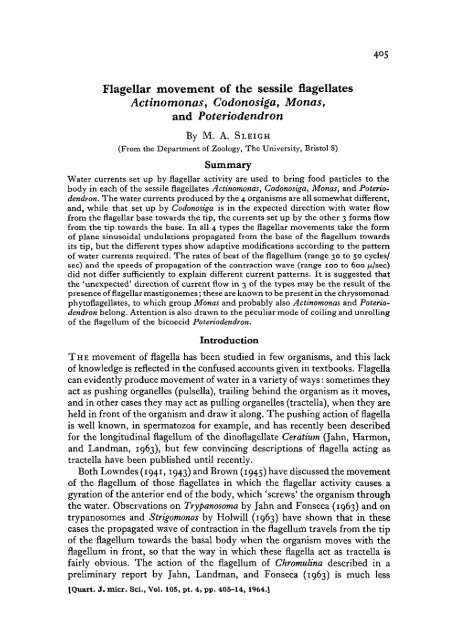
The form <strong>of</strong> <strong>the</strong> body and <strong>the</strong> distribution<br />
<strong>of</strong> <strong>the</strong> appendages are shown<br />
in fig. 1. The almost spherical body<br />
(diameter 8 to 10 p) carries a single flagellum<br />
(length 20 to 30 /JL) at one pole, and<br />
at <strong>the</strong> o<strong>the</strong>r pole <strong>the</strong>re is a very slender<br />
flagellum<br />
filopodia<br />
FIG. 1. <strong>Actinomonas</strong>, showing body form<br />
and <strong>the</strong> water currents caused by flagellar<br />
beating.<br />
stalk (up to at least 60 (j, long) which attaches <strong>the</strong> body to a piece <strong>of</strong> debris or<br />
plant material. This stalk is usually rigid, but is also contractile in <strong>the</strong> sense<br />
that it suddenly bends, usually near <strong>the</strong> end that is attached to <strong>the</strong> substrate,<br />
and carries <strong>the</strong> body to a new position. Contractions <strong>of</strong> <strong>the</strong> stalk were seen to<br />
be especially violent when <strong>the</strong> organism was subjected to <strong>the</strong> bright illumination<br />
required for dark-ground observations, and <strong>the</strong>y caused rapid jerky <strong>movement</strong>s<br />
<strong>of</strong> <strong>the</strong> whole body when <strong>the</strong> stalk was detached from <strong>the</strong> substrate. The<br />
very fine filopodia (length 10 to 15 fi) occur in two groups, a ring <strong>of</strong> 6 to 8<br />
around <strong>the</strong> flagellar base, and ano<strong>the</strong>r ring <strong>of</strong> about 4 above <strong>the</strong> stalk; <strong>the</strong>y<br />
are apparently straight and permanent even when food is being ingested.<br />
Both Kent and Greissmann observed that <strong>the</strong> flagellum was held out stiffly<br />
from <strong>the</strong> body and caused water <strong>movement</strong>s that can be seen in <strong>the</strong> <strong>movement</strong><br />
<strong>of</strong> particles. The water circulation caused is shown in fig. 1; currents travel
408 Sleigh—<strong>Flagellar</strong> <strong>movement</strong> <strong>of</strong> <strong>sessile</strong> <strong>flagellates</strong><br />
towards <strong>the</strong> body from <strong>the</strong> region <strong>of</strong> <strong>the</strong> flagellar tip, and food particles<br />
become entangled in <strong>the</strong> filopodia. Small amoebae were seen to be caught in<br />
this way and carried down <strong>the</strong> filopodia to be ingested in <strong>the</strong> body.<br />
The flagellar beat is rapid and regular, and under stroboscopic illumination<br />
<strong>the</strong> form <strong>of</strong> beat is seen to be a sine wave with almost constant wavelength and<br />
amplitude along its length (<strong>the</strong>re is a slight tendency to an increase in both<br />
towards <strong>the</strong> distal end). Sometimes <strong>the</strong> flagellum appears as shown in fig. i<br />
when <strong>the</strong> sine wave is seen from <strong>the</strong> side, and sometimes <strong>the</strong> flagellum appears<br />
as a straight line that is seen as a dotted line at a particular optical level, when<br />
<strong>the</strong> sine wave is seen from <strong>the</strong> edge; evidently <strong>the</strong> beat takes place in a single<br />
plane. In observations on a number <strong>of</strong> specimens <strong>the</strong> waves <strong>of</strong> bending were<br />
always seen to pass up <strong>the</strong> flagellum from base to tip; it is certain that this<br />
observation is not a stroboscopic artifact. The frequency <strong>of</strong> beat at i8° C was<br />
on average about 50 cycles/sec, and <strong>the</strong> length <strong>of</strong> <strong>the</strong> waves <strong>of</strong> contraction was<br />
about 7 to 10 JX, so that <strong>the</strong> contraction waves were propagated at about 350<br />
to 500 /x/sec.<br />
Codonosiga<br />
This organism is well known from <strong>the</strong> studies <strong>of</strong> Kent (1880) and Lapage<br />
(1925), and <strong>the</strong> species used here, C. botrytis, is a common fresh-water choan<strong>of</strong>lagellate.<br />
Lapage has thoroughly described <strong>the</strong> organism and <strong>the</strong> water<br />
currents that are produced by flagellar activity, but he did not venture to<br />
describe <strong>the</strong> <strong>movement</strong> <strong>of</strong> <strong>the</strong> flagellum in any detail because it was normally<br />
too rapid to be seen. Hollande (1952) has discussed evidence relating <strong>the</strong><br />
choan<strong>of</strong>lagellates to <strong>the</strong> chrysomonad phyt<strong>of</strong>lagellates, but found it inconclusive.<br />
The individuals observed were attached to debris or moss by slender rigid<br />
stalks up to 100 fj, or more long; frequently <strong>the</strong>re were several animals on a<br />
single stalk. Each body (about 10 /u, long) carried a single flagellum (length 25<br />
to 30 p), whose base was at <strong>the</strong> centre <strong>of</strong> a collar (about 8 to 10 JJL tall). It was<br />
long believed that <strong>the</strong> collar was a conical membrane, but electron microscope<br />
observations <strong>of</strong> Fjerdingstad (1961) have confirmed an alternative belief—that<br />
<strong>the</strong> collar is made up <strong>of</strong> a palisade <strong>of</strong> microvilli, a structure that is shared with<br />
<strong>the</strong> choanocyte cells <strong>of</strong> sponges.<br />
Particles borne by water currents can be seen to flow in a three-dimensional<br />
vortex in which <strong>the</strong> water moves in below <strong>the</strong> flagellar base and is swept away<br />
along <strong>the</strong> axis <strong>of</strong> <strong>the</strong> flagellum towards its distal end (fig. 2, A). It is believed<br />
that water passes through <strong>the</strong> meshwork <strong>of</strong> <strong>the</strong> collar, where particles are<br />
strained from <strong>the</strong> water by <strong>the</strong> microvilli and carried down to <strong>the</strong> body by<br />
protoplasmic flow, to be engulfed in food vacuoles near <strong>the</strong> base <strong>of</strong> <strong>the</strong><br />
collar.<br />
The flagellum beats about 30 times/sec at 18° C, with a wavelength <strong>of</strong> 15<br />
to 2011, giving a speed <strong>of</strong> 450 to 600 /it/sec for <strong>the</strong> propagation <strong>of</strong> <strong>the</strong> contraction<br />
waves. The waves take place in a single plane, as is evident when organisms<br />
turn so that <strong>the</strong> waves may be seen from <strong>the</strong> edge. The waves <strong>of</strong> contraction
Sleigh—<strong>Flagellar</strong> <strong>movement</strong> <strong>of</strong> <strong>sessile</strong> <strong>flagellates</strong> 409<br />
pass from <strong>the</strong> base <strong>of</strong> <strong>the</strong> flagellum to <strong>the</strong> tip, and are sinusoidal, though with<br />
perhaps some increase in amplitude <strong>of</strong> <strong>the</strong> broad waves as <strong>the</strong>y near <strong>the</strong> tip.<br />
Monas<br />
Many organisms <strong>of</strong> <strong>the</strong> ochromonad type were found in <strong>the</strong> stream water<br />
and among <strong>the</strong>se were <strong>sessile</strong> forms that lacked plastids and had <strong>the</strong> body-form<br />
shown in fig. 2, B ; <strong>the</strong>y are believed to belong to <strong>the</strong> genus Monas. The <strong>movement</strong><br />
<strong>of</strong> Monas has been discussed by Krijgsman (1925) and Lowndes (1944),<br />
FIG. 2. A, Codonosiga; B, Monas,<br />
and figures by both authors have been published in textbooks to illustrate<br />
flagellar <strong>movement</strong>. Krijgsman considered that <strong>the</strong> flagellum performed an<br />
oar-like rowing stroke, but Lowndes pointed out that this <strong>movement</strong> was<br />
unlikely to be <strong>the</strong> normal mode <strong>of</strong> progression. By <strong>the</strong> use <strong>of</strong> a stroboscope<br />
Lowndes found that <strong>the</strong> flagellar waves moved from base to tip with a frequency<br />
<strong>of</strong> about 19 cycles/sec, that <strong>the</strong> waves increased in amplitude towards<br />
<strong>the</strong> tip, and that <strong>the</strong> water current set up was in <strong>the</strong> direction from <strong>the</strong> tip <strong>of</strong><br />
<strong>the</strong> flagellum towards <strong>the</strong> base. He pointed out that <strong>the</strong> organisms on which<br />
<strong>the</strong>se observations were made were swimming at much less than maximum<br />
speed. The waves passing up <strong>the</strong> flagellum were believed to be spiral, and<br />
this helical component provided a basis for Lowndes's explanation <strong>of</strong> <strong>the</strong><br />
mechanism <strong>of</strong> <strong>movement</strong>. He believed that <strong>the</strong> organisms always swam in a
41 o Sleigh—<strong>Flagellar</strong> <strong>movement</strong> <strong>of</strong> <strong>sessile</strong> <strong>flagellates</strong><br />
spiral and that <strong>the</strong> rotation and gyration provided <strong>the</strong> forward component <strong>of</strong><br />
<strong>movement</strong>. The <strong>sessile</strong> monads studied here could not move, nor turn quickly,<br />
so that <strong>the</strong> motion <strong>of</strong> <strong>the</strong> flagellum is easily seen.<br />
The body <strong>of</strong> <strong>the</strong> organism (some 10 to 12 \x in diameter) is attached to debris<br />
or moss by a very fine strand (some 20 /J. long), which is flexible and noncontractile<br />
(fig. 2, B). The two flagella emerge close toge<strong>the</strong>r, <strong>the</strong> longer one<br />
(25 to 30 fx in length) being extended almost straight out from <strong>the</strong> body, and<br />
<strong>the</strong> shorter one (about 5 [x long) being held curved close to <strong>the</strong> body.<br />
Strong water currents reminiscent <strong>of</strong> those caused by <strong>Actinomonas</strong> flow<br />
around <strong>the</strong> active flagellum (fig. 2, B), differing slightly in that <strong>the</strong>y impinge<br />
on <strong>the</strong> surface <strong>of</strong> <strong>the</strong> body at <strong>the</strong> flagellar base. Food particles are seen to be<br />
engulfed by activity <strong>of</strong> <strong>the</strong> body surface near <strong>the</strong> flagellar base and carried<br />
down <strong>the</strong> side <strong>of</strong> <strong>the</strong> body and into vacuoles. Food objects as large as diatoms<br />
30 to 40 ix long were seen enclosed in <strong>the</strong> body; intense flagellar activity must<br />
be used to collect such large objects.<br />
When seen under <strong>the</strong> stroboscope, <strong>the</strong> longer flagellum shows a roughly<br />
sinusoidal beat, in which both <strong>the</strong> amplitude and wavelength increased<br />
markedly towards <strong>the</strong> tip. The waves travel from base to tip and are planar.<br />
The frequency <strong>of</strong> beat is about 50 beats/sec at 20 0 C, and <strong>the</strong> wavelength<br />
increases from about 6 fx at <strong>the</strong> base to 10 to 12 fx distally, so that <strong>the</strong> waves<br />
are propagated at 300 jtt/sec basally and 500 to 600 //,/sec distally.<br />
The small flagellum appeared to flicker, but its motion was not found to be<br />
regular enough for stroboscopic observation. It may be concerned with <strong>the</strong><br />
trapping and ingestion <strong>of</strong> food particles.<br />
Poteriodendron<br />
The <strong>movement</strong> <strong>of</strong> <strong>the</strong> flagellum <strong>of</strong> this colonial bicoecid has been briefly<br />
discussed by Geitler (1942), but without stroboscopic or cinematographic<br />
observations he was unable to see <strong>the</strong> true nature <strong>of</strong> <strong>the</strong> beating activity. Kent<br />
(1880) noted that free-swimming bicoecids swam with <strong>the</strong>ir flagella held in<br />
front, presumably acting as tractella. While Kent classed bicoecids with <strong>the</strong><br />
chrysomonad phyt<strong>of</strong>lagellates, Geitler believed <strong>the</strong>m to be choan<strong>of</strong>lagellates,<br />
and Grasse and Deflandre (1952) found that <strong>the</strong>ir systematic position was<br />
uncertain because <strong>of</strong> lack <strong>of</strong> knowledge <strong>of</strong> <strong>the</strong>ir structure.<br />
Individuals <strong>of</strong> Poteriodendron occupy a stalked campanulate lorica (fig. 3, A),<br />
which is attached to plant material or debris. The stalk is secreted first, and<br />
<strong>the</strong>n <strong>the</strong> lorica, which has a spiral structure (Robinow, 1956), is produced<br />
from a continuous thread <strong>of</strong> secreted material laid down by rotation <strong>of</strong> <strong>the</strong><br />
animal; in one case <strong>the</strong> animal was observed to rotate continuously clockwise<br />
during secretion <strong>of</strong> <strong>the</strong> lorica, performing one complete rotation every 7 or 8<br />
min. The fully-formed lorica is about 15 /x long at <strong>the</strong> end <strong>of</strong> a stalk 5 to 30 fx<br />
long.<br />
The animal is attached to <strong>the</strong> base <strong>of</strong> <strong>the</strong> inside <strong>of</strong> <strong>the</strong> lorica by a contractile<br />
'foot' which is said to be a modified flagellum. The body <strong>of</strong> <strong>the</strong> organism<br />
(about 8 to 12 ju, long) is held near <strong>the</strong> mouth <strong>of</strong> <strong>the</strong> lorica at <strong>the</strong> end <strong>of</strong> <strong>the</strong> foot,
Sleigh—<strong>Flagellar</strong> <strong>movement</strong> <strong>of</strong> <strong>sessile</strong> <strong>flagellates</strong> 411<br />
and from it projects <strong>the</strong> stiffly held, but slightly curved, flagellum, 25 to 40 /x<br />
long. The flagellum is attached near <strong>the</strong> end <strong>of</strong> <strong>the</strong> body, at <strong>the</strong> base <strong>of</strong> an<br />
apical bulge.<br />
<strong>Flagellar</strong> activity sets up water currents which impinge on <strong>the</strong> body surface<br />
near <strong>the</strong> base <strong>of</strong> <strong>the</strong> flagellum, in which region food particles are ingested. The<br />
currents are different from those in ei<strong>the</strong>r <strong>Actinomonas</strong> or Monas, since<br />
<strong>the</strong> water is funnelled in from a considerable distance, but only close to <strong>the</strong><br />
flagellum (fig. 3, B). Stroboscopic observations show <strong>the</strong> reason for this: <strong>the</strong><br />
amplitude <strong>of</strong> <strong>the</strong> bending waves <strong>of</strong> <strong>the</strong> flagellum is small, and <strong>the</strong> sinusoidal<br />
'/ water currents •<br />
FIG. 3. Poteriodendron. A, part <strong>of</strong> a colony; B, water currents around <strong>the</strong> flagellum; c, unrolling<br />
<strong>of</strong> <strong>the</strong> flagellum on relaxation after retraction <strong>of</strong> <strong>the</strong> animal into <strong>the</strong> lorica.<br />
waves have a small wavelength which tends to decrease distally. Examination<br />
<strong>of</strong> <strong>the</strong> animal while it is rotating during <strong>the</strong> secretion <strong>of</strong> <strong>the</strong> lorica shows<br />
alternately <strong>the</strong> edge and flat face <strong>of</strong> a plane sine wave envelope <strong>of</strong> <strong>movement</strong>,<br />
and this is especially obvious under <strong>the</strong> stroboscope. The frequency <strong>of</strong> beat<br />
is 40 to 50 cycles/sec, and <strong>the</strong> wavelength 3 to 5 fj,, so that <strong>the</strong> rate <strong>of</strong> propagation<br />
<strong>of</strong> <strong>the</strong> contraction waves from <strong>the</strong> base to <strong>the</strong> tip <strong>of</strong> <strong>the</strong> flagellum is 120<br />
to 250 /x/sec.<br />
On stimulation <strong>of</strong> <strong>the</strong> animal it was retracted to <strong>the</strong> base <strong>of</strong> <strong>the</strong> lorica by <strong>the</strong><br />
contraction <strong>of</strong> <strong>the</strong> foot, and at <strong>the</strong> same time <strong>the</strong> flagellum coiled up very<br />
rapidly to assume <strong>the</strong> shape <strong>of</strong> a watchspring. On relaxation, <strong>the</strong> animal<br />
emerged as <strong>the</strong> foot extended and <strong>the</strong> flagellum slowly uncoiled over a period<br />
<strong>of</strong> a second or two (fig. 3, c). The contraction <strong>of</strong> a flagellum into a plane spiral<br />
is most unusual, and <strong>the</strong> mechanism involved must be considerably different<br />
from that which produces normal contractile waves in <strong>the</strong> flagellum.
412 Sleigh—<strong>Flagellar</strong> <strong>movement</strong> <strong>of</strong> <strong>sessile</strong> <strong>flagellates</strong><br />
Discussion<br />
In all <strong>the</strong>se 4 <strong>flagellates</strong> <strong>the</strong> flagellum is <strong>of</strong> a similar size, and plane waves<br />
<strong>of</strong> approximately sinusoidal form are propagated from <strong>the</strong> base to <strong>the</strong> tip (fig.<br />
4), yet <strong>the</strong> results <strong>of</strong> flagellar activity are all somewhat different. From studies<br />
on <strong>the</strong> tails <strong>of</strong> spermatozoa one would expect that water currents would be<br />
set up which travel in <strong>the</strong> same direction as <strong>the</strong> propagated bending waves<br />
in <strong>the</strong> flagellum, and this is indeed what has been observed in Codonosiga. In<br />
<strong>the</strong> o<strong>the</strong>r 3 examples <strong>the</strong> water currents were observed to travel in <strong>the</strong> direction<br />
opposite to that in which <strong>the</strong> flagellar waves are propagated; this is not easily<br />
explained.<br />
<strong>Actinomonas</strong> Codonosiga. Monas Potehodendron<br />
FIG. 4. Comparison <strong>of</strong> <strong>the</strong> form <strong>of</strong> flagella during beating.<br />
At this point it is interesting to draw an analogy with <strong>the</strong> <strong>movement</strong> <strong>of</strong><br />
organisms <strong>of</strong> larger size. Comparison <strong>of</strong> <strong>the</strong> swimming <strong>of</strong> an eel with that<br />
<strong>of</strong> a polychaete worm like Nepktys is instructive. The eel propagates waves <strong>of</strong><br />
bending from <strong>the</strong> head to <strong>the</strong> tail as it moves through <strong>the</strong> water, while in<br />
Nephtys <strong>the</strong> waves <strong>of</strong> bending are propagated from <strong>the</strong> tail to <strong>the</strong> head as <strong>the</strong><br />
animal swims head-first. The difference here is <strong>the</strong> presence <strong>of</strong> <strong>the</strong> paddle-like<br />
parapodia situated in <strong>the</strong> plane <strong>of</strong> <strong>the</strong> oscillations in <strong>the</strong> polychaete, while <strong>the</strong><br />
eel is smooth-bodied. Taylor (1952) approached this problem from a ma<strong>the</strong>matical<br />
point <strong>of</strong> view, and found from <strong>the</strong>oretical calculations that, while <strong>the</strong><br />
propagation <strong>of</strong> sinusoidal waves along a smooth thin cylinder will cause<br />
<strong>movement</strong> <strong>of</strong> water in <strong>the</strong> same direction as <strong>the</strong> propagation <strong>of</strong> <strong>the</strong> waves, <strong>the</strong><br />
propagation <strong>of</strong> similar waves along a cylinder whose surface is sufficiently<br />
rough, and whose roughness has a directional character, may cause <strong>movement</strong><br />
in a direction opposite to <strong>the</strong> direction <strong>of</strong> propagation <strong>of</strong> <strong>the</strong> waves.<br />
It is possible that <strong>the</strong> flagellum <strong>of</strong> <strong>the</strong>se <strong>flagellates</strong> uses a similar mechanism.<br />
The presence <strong>of</strong> mastigonemes or flimmer filaments on many flagella is well<br />
established (see Pitelka, 1963), but <strong>the</strong>ir arrangement on <strong>the</strong> flagellar shaft is<br />
as yet unknown. Flimmer filaments appear to be <strong>of</strong> two distinct types: <strong>the</strong><br />
larger mastigoneme structures several micra long and 10 to 20 m/x in diameter,<br />
and finer filaments only 2 m/x or so in diameter. Light microscopists found that<br />
many flagella carry flimmer filaments, and Deflandre (1934) listed choan<strong>of</strong>lagellates<br />
and chrysomonad <strong>flagellates</strong> among those forms whose flagella<br />
carry lateral appendages; however, only by electron microscopy is it possible
Sleigh—<strong>Flagellar</strong> <strong>movement</strong> <strong>of</strong> <strong>sessile</strong> <strong>flagellates</strong> 413<br />
to determine <strong>the</strong> type and arrangement <strong>of</strong> <strong>the</strong> flimmer filaments adequately.<br />
The long flagellum <strong>of</strong> ochromonad <strong>flagellates</strong>, which no doubt has <strong>the</strong> same<br />
structure as that <strong>of</strong> Manas, has been found to carry both mastigonemes and<br />
fine filaments (Pitelka and Schooley, 1955), but no clear evidence is available<br />
about <strong>the</strong> o<strong>the</strong>r types studied here. Electron micrographs <strong>of</strong> Codonosiga by<br />
Petersen and Hansen (1954) show a fibrous zone around <strong>the</strong> flagellum which<br />
may represent poorly-preserved flimmer filaments, and Afzelius (1961) found<br />
that thin flimmer filaments were present in <strong>the</strong> plane <strong>of</strong> <strong>the</strong> central fibres <strong>of</strong><br />
<strong>the</strong> choanocyte flagellum <strong>of</strong> <strong>the</strong> sponge Microciona; <strong>the</strong> flimmer filaments <strong>of</strong><br />
choan<strong>of</strong>lagellates may well be <strong>of</strong> <strong>the</strong> same type. It is worth noticing that <strong>the</strong>se<br />
filaments are not situated in <strong>the</strong> presumed plane <strong>of</strong> oscillation <strong>of</strong> <strong>the</strong> sponge<br />
flagellum, but in <strong>the</strong> plane at right angles to this. It is possible that in <strong>Actinomonas</strong>,<br />
Monas, and Poteriodendron <strong>the</strong>re are mastigonemes situated in <strong>the</strong><br />
plane <strong>of</strong> <strong>the</strong> oscillations (ei<strong>the</strong>r in two rows, or all round like <strong>the</strong> hairs <strong>of</strong> a<br />
bottle brush), which may act to move water in <strong>the</strong> direction opposite to that<br />
expected, by providing <strong>the</strong> roughness mentioned in Taylor's <strong>the</strong>oretical consideration<br />
<strong>of</strong> <strong>the</strong> <strong>movement</strong> <strong>of</strong> undulating thin cylinders. These mastigonemes<br />
may perhaps be visualized as oars projecting from <strong>the</strong> flagellar shaft which row<br />
water towards <strong>the</strong> flagellar base as <strong>the</strong> crests <strong>of</strong> <strong>the</strong> waves <strong>of</strong> bending move up<br />
<strong>the</strong> flagellum.<br />
There is doubt as to <strong>the</strong> systematic position <strong>of</strong> both <strong>Actinomonas</strong> and <strong>the</strong><br />
bicoecids, but <strong>the</strong> similarity <strong>of</strong> <strong>the</strong>ir flagellar activity to that <strong>of</strong> Monas and<br />
o<strong>the</strong>r chrysomonads that I have studied suggest that <strong>the</strong>y may belong to this<br />
group; indeed Pascher has included <strong>Actinomonas</strong> with <strong>the</strong> chrysomonads for<br />
ano<strong>the</strong>r reason, and several probable bicoecids like Codonodendron are <strong>of</strong>ten<br />
placed in <strong>the</strong> same group <strong>of</strong> phyt<strong>of</strong>lagellates.<br />
It is interesting to note <strong>the</strong> adaptive modification <strong>of</strong> <strong>the</strong> form <strong>of</strong> flagellar<br />
beat in <strong>Actinomonas</strong>, Monas, and Poteriodendron. The food-catching pseudopodia<br />
<strong>of</strong> <strong>Actinomonas</strong> only require a broad stream <strong>of</strong> water-carrying particles,<br />
but in <strong>the</strong> o<strong>the</strong>r two forms <strong>the</strong> particles must hit <strong>the</strong> body near <strong>the</strong> flagellar<br />
base. In Monas this is achieved by funnelling <strong>the</strong> water current towards <strong>the</strong><br />
body by increasing <strong>the</strong> amplitude <strong>of</strong> <strong>the</strong> bending waves <strong>of</strong> <strong>the</strong> flagellum distally,<br />
and in Poteriodendron <strong>the</strong> amplitude <strong>of</strong> <strong>the</strong> waves is kept small so that <strong>the</strong><br />
main current is close to <strong>the</strong> flagellar shaft and impinges on <strong>the</strong> body surface.<br />
Evidently <strong>the</strong>re is a considerable variety <strong>of</strong> patterns <strong>of</strong> flagellar <strong>movement</strong>,<br />
and a full range <strong>of</strong> <strong>the</strong>se patterns must be studied before <strong>the</strong> mode <strong>of</strong> functioning<br />
<strong>of</strong> <strong>the</strong> contractile mechanism <strong>of</strong> <strong>the</strong> flagellum can be properly understood.<br />
The first observations were made at <strong>the</strong> University <strong>of</strong> Exeter, in <strong>the</strong> Department<br />
<strong>of</strong> Pr<strong>of</strong>essor L. A. Harvey, whom I should like to thank for his interest.<br />
I am also grateful to Pr<strong>of</strong>essor J. E. Harris, F.R.S., for reading <strong>the</strong> manuscript.
414 Sleigh—<strong>Flagellar</strong> <strong>movement</strong> <strong>of</strong> <strong>sessile</strong> <strong>flagellates</strong><br />
References<br />
Afzelius, B. A., 1961. Nature, Lond., 191, 1318.<br />
Brown, H. P., 1945. Ohio J. Sci., 45, 247.<br />
Deflandre, G., 1934. Ann. Protistol., 4, 31.<br />
Fjerdingstad, E. J., 1961. Z. Zellforsch, 54, 499.<br />
Geitler, L., 1942. Arch. Protistenk., 96, 119.<br />
Grasse, P.-P., and Deflandre, G., 1952. In Traite de Zoologie, ed. by P.-P. Grasses Tome I,<br />
fasc. I, p. 599. Paris (Masson).<br />
Gray, J., 1955. J. exp. Biol., 32, 775.<br />
1958. Ibid., 35, 96.<br />
Greissmann, K., 1914. Arch. Protistenk., 32, 1.<br />
Hollande, A., 1952. In Traite de Zoologie, ed. by P.-P. Grass£, Tome I, fasc. I, p. 471.<br />
Paris (Masson).<br />
Hoi will, M. E. J., 1963. Personal communication.<br />
Jahn, T. L., and Fonseca, J. R., 1963. J. Protozool., 10, suppl., p. 11.<br />
Harmon, W. M., and Landman, M., 1963. Ibid., 10, 358.<br />
Landman, M., and Fonseca, J. R., 1963. Proc. XVI int. Congr. Zool., 2, 292.<br />
Kent, W. S., 1880. A manual <strong>of</strong> <strong>the</strong> infusioria. London (Bogue).<br />
Krijgsman, B. J., 1925. Arch. Protistenk., 52, 478.<br />
Lapage, G., 1925. Quart. J. micr. Sci., 69, 471.<br />
Lowndes, A. G., 1941. Proc. zool. Soc. Lond. Ser. A., m, m.<br />
1943- Ibid., 113, 99.<br />
1944- Ibid., 114, 325.<br />
Pascher, A., 1918. Arch. Protistenk., 38, 1.<br />
Petersen, J. B., and Hansen, J. B., 1954. Bot. Tidsskr., 51, 281.<br />
Pitelka, D. R., 1963. Electron-microscopic structure <strong>of</strong> protozoa. Oxford (Pergamon).<br />
and Schooley, C. N., 1955. Univ. Calif. Publ. Zool., 61, 79.<br />
Robinow, C. F., 1956. J. biophys. biochem. Cytol., 2, suppl., p. 233.<br />
Roskin, G., 1931. Arch. Protistenk, 73, 203.<br />
Stokes, A. C, 1888. J. Trenton Nat. Hist. Soc, 1, 71.<br />
Taylor, G. I., 1952. Proc. Roy. Soc. A, 214, 158.<br />
Tregoub<strong>of</strong>f, G., 1953. In Traite de Zoologie, ed. by P.-P. Grasse^ Tome I, fasc. II, p. 437.<br />
Paris (Masson).


