Session WedAT1 Pegaso A Wednesday, October 10, 2012 ... - Lirmm
Session WedAT1 Pegaso A Wednesday, October 10, 2012 ... - Lirmm
Session WedAT1 Pegaso A Wednesday, October 10, 2012 ... - Lirmm
- TAGS
- pegaso
- october
- lirmm
- www2.lirmm.fr

You also want an ePaper? Increase the reach of your titles
YUMPU automatically turns print PDFs into web optimized ePapers that Google loves.
<strong>Session</strong> WedCT9 Fenix 1 <strong>Wednesday</strong>, <strong>October</strong> <strong>10</strong>, <strong>2012</strong>, 11:00–12:30<br />
Sensing in Medical Robotics<br />
Chair M. Cenk Cavusoglu, Case Western Res. Univ.<br />
Co-Chair<br />
11:00–11:15 WedCT9.1<br />
Scanning the surface of soft tissues with a<br />
micrometer precision thanks to endomicroscopy<br />
based visual servoing<br />
Benoît Rosa, Mustapha Suphi Erden, Jérôme Szewczyk, and<br />
Guillaume Morel<br />
ISIR, Université Pierre et Marie Curie, Paris, France<br />
Tom Vercauteren<br />
Mauna Kea Technologies, Paris, France<br />
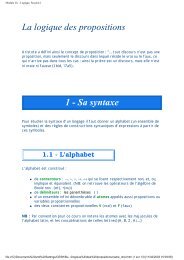
• Probe-based confocal endomicroscopy is a<br />
promising imaging modality for performing<br />
optical biopsies<br />
• Problem: tissue deformation while scanning for<br />
getting wide field of view mosaics<br />
• Solution proposed: visual servo control using<br />
the confocal images as a measurement of<br />
probe/tissue displacement<br />
• Ex vivo validation on different tissues and<br />
trajectories using a precision robot<br />
• Further work: in vivo trial with a dedicated<br />
laparoscopic instrument<br />
Mosaics from raster scans on<br />
liver tissue. Up: without visual<br />
servo control (light line is the<br />
robot trajectory). Down: using<br />
visual servo control.<br />
11:30–11:45 WedCT9.3<br />
Internal Bleeding Detection Algorithm Based on<br />
Determination of Organ Boundary by Low-<br />
Brightness Set Analysis<br />
Keiichiro Ito, Shigeki Sugano, Fellow IEEE<br />
Creative Science and Engineering, Waseda University, Japan<br />
Hiroyasu Iwata, Member IEEE<br />
Waseda Institute for Advanced Study, Waseda University, Japan<br />
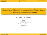
• This paper proposes an organ<br />
boundary determination method for<br />
detecting internal bleeding.<br />
• We developed method for extracting<br />
low-brightness areas and<br />
determining algorithms of organ<br />
boundaries by low-brightness set<br />
analysis, and we detect internal<br />
bleeding by combining these two<br />
methods.<br />
• Experimental results based on<br />
clinical US images of internal<br />
bleeding between Liver and Kidney<br />
showed that proposed algorithms<br />
had a sensitivity of 77.8% and<br />
specificity of 95.7%.<br />
Kidney<br />
Liver<br />
Gap between the organs<br />
(Internal bleeding)<br />
Internal Bleeding Detection Algorithm<br />
12:00–12:15 WedCT9.5<br />
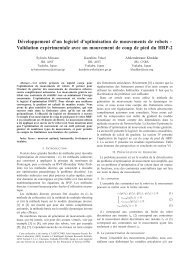
Heart motion measurement with three dimensional<br />
sonomicrometry and acceleration sensing<br />
Tetsuya Horiuchi and Ken Masamune<br />
Graduate School of Information Science and Technology, University of Tokyo,<br />
Japan<br />
Eser Erdem Tuna and Murat Cenk Çavuşoğlu<br />
Department of Electrical Engineering and Computer Science, Case Western<br />
Reserve University, USA<br />
• Point of Interest for Coronary<br />
artery bypass graft surgery.<br />
• Estimation by particle filter with<br />
position and acceleration sensor<br />
which has uncertain incline.<br />
• New estimation method,<br />
“Differential Probability Method”,<br />
which enhanced particle filter.<br />
• Reduce 27.2% RMS error from<br />
Conventional method.<br />
Overview of the system<br />
11:15–11:30 WedCT9.2<br />
Preliminary Evaluation of a Micro-Force Sensing<br />
Handheld Robot for Vitreoretinal Surgery<br />
Berk Gonenc, Marcin A. Balicki<br />
Russell H. Taylor and Iulian Iordachita<br />
ERC for Computer Integrated Surgery, Johns Hopkins University, USA<br />
James Handa and Peter Gehlbach<br />
Wilmer Eye Institute, The Johns Hopkins School of Medicine, USA<br />
Cameron N. Riviere<br />
Robotics Institute, Carnegie Mellon University, USA<br />
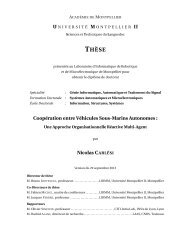
• A 2-DOF force sensing hook is<br />
integrated with a handheld robot,<br />
Micron, for superior performance<br />
in membrane peeling operations.<br />
• FBG based force sensing<br />
instrument could directly inform<br />
the surgeon of the extremely<br />
delicate peeling forces.<br />
• Preliminary tests were done on<br />
bandage phantom and inner shell<br />
membrane of raw chicken eggs.<br />
• The peeling forces were kept<br />
below 7 mN with a significant<br />
reduction in 2-20 Hz oscillations.<br />
11:45–12:00 WedCT9.4<br />
A Cyber-Physical System for Strain<br />
Measurements in the Cerebral Aneurysm Models<br />
Chaoyang Shi, Masahiro Kojima, Carlos Tercero, Seiichi Ikeda,<br />
Toshio Fukuda, Fumihito Arai<br />
Micro-Nano Systems Engineering, Nagoya University, Japan<br />
Makoto Negoro and Keiko Irie<br />
Department of Neurosurgery, Fujita Health University, Japan<br />
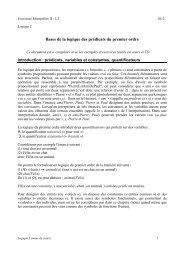
� Build a novel in-vitro<br />
experimental platform for the<br />
dynamic deformation measurements<br />
on the aneurysm<br />
� Justify a link between robotic<br />
technologies and this cyberphysical<br />
systems for the<br />
aneurysm diagnosis and<br />
prognosis<br />
� Realize the high resolution<br />
analysis by observing an<br />
enlarged silicone membrane<br />
aneurysm model under the<br />
microscope<br />
� Combine CFD (Computational<br />
Fluid Dynamics) simulation<br />
with experiments for validation<br />
<strong>2012</strong> IEEE/RSJ International Conference on Intelligent Robots and Systems<br />
–145–<br />
Experimental setup including the pump for<br />
blood flow simulation pump, the cerebral<br />
aneurysm model and vision system<br />
12:15–12:30 WedCT9.6<br />
Surface Texture and Pseudo Tactile Sensation<br />
Displayed by a MEMS-Based Tactile Display<br />
Junpei Watanabe, Hiroaki Ishikawa, and Arouette Xavier<br />
Department of Mechanical Engineering, Keio University, Japan<br />
Norihisa Miki<br />
Department of Mechanical Engineering, Keio University, Japan<br />
and<br />
JST PRESTO, Japan<br />
• We demonstrate display of artificial tactile<br />
feeling using large displacement MEMS<br />
actuator arrays.<br />
• We investigated the artificial tactile feeling<br />
projected onto the fingertip in contact with<br />
the display.<br />
• The actuator arrays could successfully<br />
display “rough” and “smooth” tactile feeling<br />
distinctly.<br />
• We experimentally deduced the conditions<br />
when the pseudo tactile sensation was<br />
generated.<br />
Schematic view of a tactile display