Olympus CK30/CK40 Culture Microscope Instructions
Olympus CK30/CK40 Culture Microscope Instructions
Olympus CK30/CK40 Culture Microscope Instructions

Create successful ePaper yourself
Turn your PDF publications into a flip-book with our unique Google optimized e-Paper software.
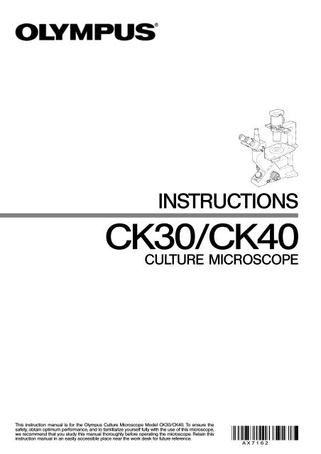
INSTRUCTIONS<br />
<strong>CK30</strong>/<strong>CK40</strong><br />
CULTURE MICROSCOPE<br />
This instruction manual is for the <strong>Olympus</strong> <strong>Culture</strong> <strong>Microscope</strong> Model <strong>CK30</strong>/<strong>CK40</strong>. To ensure the<br />
safety, obtain optimum performance, and to familiarize yourself fully with the use of this microscope,<br />
we recommend that you study this manual thoroughly before operating the microscope. Retain this<br />
instruction manual in an easily accessible place near the work desk for future reference.<br />
A X 7 1 6 2
The <strong>CK30</strong> and <strong>CK40</strong> <strong>Culture</strong> <strong>Microscope</strong>s have different system configurations. The differences are shown in the table below.<br />
@<br />
<strong>CK30</strong> <strong>CK40</strong><br />
Observation tube Built-in binocular tube Interchangeable*<br />
Stage plate – Interchangeable**<br />
<strong>CK40</strong>-RFL reflected light<br />
fluorescence attachment<br />
Not mountable Mountable<br />
* The CH3-BI45 binocular tube, CH3-TR45 trinocular tube, and <strong>CK40</strong>-TBI Tilting binocular tube are all<br />
mountable. The <strong>CK40</strong>-EPA eyepoint adjuster can also be used, but not in combination with the <strong>CK40</strong>-<br />
TBI. The only usable intermediate observation tube is the <strong>CK40</strong>-EPA. Relief phase contrast observation<br />
is not available when the <strong>CK40</strong>-EPA is used.<br />
** In addition to the standard stage plate, you can mount the <strong>CK40</strong>-CPG glass stage plate or IX-CP50<br />
stage plate ( 50). Only 20X-or-less objectives should be used with the <strong>CK40</strong>-CPG.<br />
SAFETY PRECAUTIONS<br />
Fig. 1<br />
Fig. 2<br />
³<br />
|<br />
²<br />
1. Install the microscope on a stable, horizontal table. Make sure the table is<br />
sturdy enough to support the microscope’s weight. (Weight: <strong>CK30</strong> – approx.<br />
8 kg (17.6 lb); <strong>CK40</strong> – approx. 8.6 kg (18.9 lb))<br />
2. When transporting the microscope, always hold the lower side @ of the<br />
observation tube and the illumination unit support ².<br />
3. If a culture solution or water is spilled on the stage, objective or observation<br />
tube, unplug the power cord and dry it off immediately. Failure to do<br />
so could cause equipment failure.<br />
4. The surface of the lamp socket ³ on the lamp housing support can get<br />
extremely hot. Make sure you leave sufficient space around the lamp<br />
socket, especially above it, to dissipate heat. (Fig. 1)<br />
5. To avoid potential shock hazards and burns when replacing the bulb,<br />
make sure the main switch is set to “\” (OFF), the power cord is unplugged<br />
from the outlet, and that the lamp and the area around the lamp<br />
socket have cooled sufficiently. (Fig. 2)<br />
Applicable bulb Halogen bulb, 6V 30WHAL (Philips 5761)<br />
6. Be sure to use an <strong>Olympus</strong>-specified power cord. Safety and performance<br />
cannot be guaranteed otherwise.<br />
7. Be sure to ground the unit. The designated electrical safety standard<br />
cannot be guaranteed otherwise.<br />
i
Safety Symbols<br />
The following symbols are found on the microscope. Study the meaning of the symbols, and always use the equipment in<br />
the safest possible manner.<br />
Warnings<br />
Symbol Explanation<br />
l<br />
\<br />
Indicates that the surface becomes hot, and should not be touched with bare hands.<br />
Before use, carefully read the instruction manual. Improper use could result in personal injury and/or<br />
damage to the equipment.<br />
Indicates that the main switch is ON.<br />
Indicates that the main switch is OFF.<br />
Warning indications are placed where special precautions are required when handling and using the unit.<br />
1 Getting Ready<br />
Warning indication position<br />
Lamp socket<br />
[Warning against high temperature]<br />
1. A microscope is a precision instrument. Handle it with care and avoid subjecting it to sudden or severe impacts.<br />
2. Do not expose the unit to direct sunlight, high temperature and humidity, dust or vibrations. (For operating conditions, refer<br />
to “8. Specifications” on page 26.)<br />
3. Use the tension adjustment ring to adjust the tension of the coarse adjustment knob.<br />
4. Use a flat-head screwdriver to set the voltage selector on the rear panel of the microscope body to the required voltage.<br />
}The selector is factory preset to the high-voltage side (110 – 120 V or 230 – 240 V).<br />
ii
2 Maintenance and Storage<br />
1. Clean all glass components by wiping gently with gauze. To remove fingerprints or oil smudges, wipe with gauze slightly<br />
moistened with a mixture of ether (70%) and alcohol (30%).<br />
# Do not use a mixture of ether (70%) and alcohol (30%) to clean the lower lens (made of optical plastic) of the<br />
eyepiece (NCWHK10X) because such solvents cloud the lens. If dust adheres to the lens, blow it off or wipe it away<br />
gently with a dry cloth.<br />
Since solvents such as ether and alcohol are highly flammable, they must be handled carefully. Be sure to keep<br />
these chemicals away from open flames or potential sources of electrical sparks —— for example, electrical<br />
equipment that is being switched on or off. Also remember to always use these chemicals only in a wellventilated<br />
room.<br />
2. Many parts of the exterior are made of plastic. Wipe the unit with a clean cloth only. Do not use organic solvents to clean<br />
non-optical components. If smudges are difficult to remove, wipe them with a soft cloth slightly moistened with a diluted<br />
neutral detergent.<br />
3. Be careful not to spill any liquid – such as a culture solution – on the unit. If you do spill anything, immediately set the<br />
main switch to “ ” (OFF) and unplug the power cord. Then wipe away any liquid on the objectives or under the objectives.<br />
4. If no objectives are mounted, be sure to cover the objective mounting threaded holes on the revolving nosepiece to<br />
prevent dust and spilled culture solution from getting on the lenses inside.<br />
5. Never disassemble any part of the unit. Doing so could cause malfunctions or reduced performance.<br />
6. When the unit is not in use, keep it covered with a dust cover. Make sure the lamp socket is cool before covering the unit.<br />
7. Using a device that radiates ultraviolet light such as a germicidal lamp near the unit may discolor (yellow) parts of the<br />
unit´s surface. The amount of discoloration depends on the radiation intensity of the ultraviolet light and the distance<br />
between the unit and radiation source. When not using the unit, cover it with the dust cover. We recommend that you also<br />
cover the unit with an impermeable sheet.<br />
3 Caution<br />
If the equipment is used in a manner not specified by this manual, the safety of the user may be imperiled. In addition, the<br />
equipment may also be damaged. Always use the equipment as outlined in this instruction manual.<br />
The following symbols are used to set off text in this instruction manual.<br />
: Indicates that failure to follow the instructions in the warning could result in bodily harm to the<br />
user and/or damage to equipment (including objects in the vicinity of the equipment).<br />
# : Indicates that failure to follow the instructions could result in damage to equipment.<br />
} : Indicates commentary (for ease of operation and maintenance).<br />
iii
1 NOMENCLATURE<br />
2 ASSEMBLY<br />
3 CONTROLS<br />
4 SUMMARY OF OBSERVATION PROCEDURES<br />
5 USING THE CONTROLS<br />
6 PHASE CONTRAST OBSERVATION<br />
7 PHOTOMICROGRAPHY<br />
8 SPECIFICATIONS<br />
9 TROUBLESHOOTING GUIDE<br />
PROPER SELECTION OF THE POWER SUPPLY CORD.............................................................................30<br />
<strong>CK30</strong>/<strong>CK40</strong><br />
2-1 Assembly Diagram ................................................................................................................................................................... 3<br />
2-2 Detailed Assembly Procedure ................................................................................................................................. 4<br />
5-1 <strong>Microscope</strong> Body ...................................................................................................................................................................... 12<br />
5-2 Stage ........................................................................................................................................................................................................... 13<br />
5-3 Observation Tube ..................................................................................................................................................................... 14<br />
5-4 Illumination Unit .......................................................................................................................................................................... 17<br />
5-5 Objectives ............................................................................................................................................................................................ 18<br />
1<br />
3<br />
9<br />
11<br />
12<br />
19<br />
23<br />
25<br />
27
1<br />
<strong>CK30</strong><br />
Eyepiece<br />
· NCWHK10X<br />
· WHK10X<br />
· WHK15X<br />
1 NOMENCLATURE<br />
Objective<br />
For observation:<br />
EDAch4X<br />
EDAch10X<br />
LWDCDAch20X<br />
# A dedicated objective is<br />
required for ordinary<br />
phase contrast observation<br />
and relief phase<br />
contrast observation.<br />
(See page 19.)<br />
Binocular tube<br />
(stationary)<br />
<strong>Microscope</strong> body<br />
<strong>CK30</strong>-F<br />
Stage (stationary)<br />
· Stage extension plate:<br />
CK2-SS<br />
· Mechanical stage:<br />
<strong>CK40</strong>-MVR<br />
Lamp socket<br />
U-LS30-3<br />
Revolving<br />
nosepiece<br />
(stationary)<br />
Quadruple<br />
revolving nosepiece<br />
Illumination unit<br />
support<br />
(stationary)<br />
Phase contrast<br />
slider<br />
· Pre-centered Ph slider:<br />
<strong>CK40</strong>-SLP<br />
· Centering Ph slider:<br />
<strong>CK40</strong>-SL<br />
· RP slider:<br />
<strong>CK40</strong>-RPSL<br />
Ultra-long working<br />
distance condenser
<strong>CK40</strong><br />
Eyepiece<br />
· NCWHK10X<br />
· WHK10X<br />
· WHK15X<br />
Objective<br />
For observation:<br />
EDAch4X<br />
EDAch10X<br />
LWDCDAch20X<br />
# A dedicated objective is<br />
required for ordinary<br />
phase contrast observation<br />
and relief phase<br />
contrast observation.<br />
Observation tube<br />
· Binocular tube: CH3-BI45<br />
· Trinocular tube: CH3-TR45<br />
· Tilting binocular tube:<br />
<strong>CK40</strong>-TBI<br />
Eyepoint adjuster<br />
· <strong>CK40</strong>-EPA<br />
# Cannot be combined with<br />
the <strong>CK40</strong>-TBI.<br />
<strong>Microscope</strong> body<br />
<strong>CK40</strong>-F Stage (stationary)<br />
· Stage extension plate:<br />
CK2-SS<br />
· Mechanical stage:<br />
<strong>CK40</strong>-MVR<br />
* Relief phase contrast observation is not possible with the <strong>CK40</strong>-EPA.<br />
Revolving<br />
nosepiece<br />
(stationary)<br />
Lamp socket<br />
U-LS30-3<br />
Quadruple<br />
revolving nosepiece<br />
<strong>CK30</strong>/<strong>CK40</strong><br />
Illumination unit<br />
support<br />
(stationary)<br />
Phase contrast<br />
slider<br />
· Ph precentering slider:<br />
<strong>CK40</strong>-SLP<br />
· Ph centering slider:<br />
<strong>CK40</strong>-SL<br />
· RP slider:<br />
<strong>CK40</strong>-RPSL *<br />
Ultra-long working<br />
distance condenser<br />
Stage plate<br />
· Standard stage plate<br />
· Glass stage plate:<br />
<strong>CK40</strong>-CPG<br />
· Stage plate ( 50):<br />
IX-CP50<br />
2
3<br />
ASSEMBLY<br />
2-1 Assembly Diagram<br />
The diagram below shows the assembly sequence for the various modules. The numbers indicate the order of assembly.<br />
# When assembling the equipment, make sure that all parts are free of dust and dirt. Avoid scratching any parts or glass<br />
surfaces.<br />
# Keep the provided Allen wrench on hand. You will need it when replacing the modules.<br />
Eyepiece<br />
· NCWHK10X<br />
· WHK10X<br />
· WHK15X<br />
Allen wrench<br />
Filter holder<br />
Required tool<br />
* Can also be mounted on the<br />
left side. However, the mechanical<br />
stage cannot be mounted<br />
in the same position as the<br />
stage extension plate.<br />
** The <strong>CK30</strong> is provided with stationary<br />
binocular tube.<br />
***The <strong>CK30</strong> is not provided with<br />
a stage plate.<br />
Filter<br />
Transmitted<br />
illumination<br />
unit<br />
Observation tube<br />
· CH3-BI45<br />
· CH3-TR45<br />
· <strong>CK40</strong>-TBI<br />
<strong>Microscope</strong> body<br />
· <strong>CK30</strong>-F<br />
· <strong>CK40</strong>-F<br />
Halogen bulb<br />
6V 30W HAL<br />
Stage extension plate<br />
CK2-SS<br />
Lamp socket<br />
U-LS30-3<br />
Stage plate<br />
· Standard stage plate<br />
· <strong>CK40</strong>-CPG<br />
· IX-CP50<br />
Power cord<br />
Objective<br />
Mechanical stage<br />
<strong>CK40</strong>-MVR
2-2 Detailed Assembly Procedure<br />
²<br />
ƒ<br />
³<br />
|<br />
Fig. 3<br />
Fig. 4<br />
³<br />
Fig. 5<br />
³<br />
@ ²<br />
@<br />
²<br />
|<br />
@<br />
<strong>CK30</strong>/<strong>CK40</strong><br />
1 Mounting and Replacing the Bulb (Fig. 3)<br />
}Use only the specified Philips 5761 halogen bulb, 6 V 30 W HAL.<br />
To prevent reduced bulb life or cracking, do not touch the bulb with<br />
bare hands. If fingerprints are accidentally left on the bulb, wipe the<br />
bulb with a soft cloth.<br />
1. Hold the bulb @ with gauze or other protective material and insert the<br />
bulb pins ² into the lamp socket’s pin holes ³ as far as they will go.<br />
# Insert the bulb gently. Squeezing too hard will damage the bulb.<br />
Bulb replacement during use or after use:<br />
The bulb and the lamp socket surfaces and vicinity will be extremely<br />
hot during use and right after use. Set the main switch to “ \ ” (OFF)<br />
and disconnect the power cord from the wall outlet. Then allow the<br />
old bulb, lamp housing and vicinity to cool before replacing the bulb.<br />
2 Installing the Transmitted Illumination Unit (Fig. 4)<br />
1. While aligning the indicator groove ² on the transmitted illumination unit<br />
@ with the notch ³ on the lamp housing support´s brace, insert the<br />
illumination unit @ gently into the support’s brace.<br />
2. Turn the illumination unit @ 90° clockwise so that “ AS ” on the filter<br />
holder faces directly to the front. Then tighten the clamping screw ƒ<br />
using the Allen wrench provided with the microscope body to fix it securely.<br />
3 Installing the Lamp Socket (Fig. 5)<br />
1. Connect the plug @ with the socket pin ². Then, while aligning the<br />
guide pins ³ with the condenser’s guide holes |, push the lamp socket<br />
gently onto the Illumination unit.<br />
4
5<br />
²<br />
Fig. 6<br />
Fig. 7<br />
Fig. 8<br />
@<br />
@<br />
4 Mounting the Objectives (Fig. 6 & 7)<br />
5<br />
#First raise the revolving nosepiece slightly to remove the transportation<br />
pad on the nosepiece’s base.<br />
}Keep the transportation pad in a safe place. You will need it when the<br />
equipment is sent for repair or transported to another location.<br />
1. Turn the coarse adjustment knob @ towards the back until the revolving<br />
nosepiece is set at its lower limit. (Fig. 6)<br />
2. Screw the objective with the lowest magnification into the revolving nosepiece<br />
from the left side. Then turn the nosepiece clockwise and mount<br />
the remaining objectives in order of magnification –– from low to high.<br />
}Mounting the objectives this way makes it easier to change magnification.<br />
}With the <strong>CK40</strong>, the objectives can be mounted through the opening on<br />
the stage.<br />
#Clean the objectives periodically. The objective tips on a culture<br />
microscope are susceptible to dust.<br />
#Be sure to cover any unused threaded holes with the objective cap<br />
² to prevent dirt and dust from getting inside. (Fig. 7)<br />
Mounting the Stage Extension<br />
Plate/Mechanical Stage<br />
(Fig. 8)<br />
}The stage extension plate can be mounted on the left or right side of the<br />
stage to expand the stage surface. However, the stage extension plate<br />
and mechanical plate cannot be used simultaneously on the same side.<br />
Mounting the CK2-SS Stage Extension Plate<br />
Screw the clamping screws @ into the stage extension plate and then<br />
into the plain stage from above on the right side or from below on the left<br />
side. Tighten them with a coin or similar tool until the plate is securely<br />
attached.<br />
Mounting the <strong>CK40</strong>-MVR Mechanical Stage<br />
}This can also be mounted on either the left or right side of the stage.<br />
Mount it in the same way as the stage extension plate.
²<br />
Fig. 9<br />
Fig. 10<br />
Fig. 11<br />
@<br />
@<br />
²<br />
³<br />
@<br />
<strong>CK30</strong>/<strong>CK40</strong><br />
6 Mounting the Stage Plate (<strong>CK40</strong> Only) (Fig. 9)<br />
Fit the standard stage plate @ into the opening on the stage.<br />
#Turn the standard stage plate so that the notch ² faces to the<br />
front for easy confirmation of an objective tip. When using the<br />
glass stage plate, set it in the direction so that its product code<br />
inscription “ <strong>CK40</strong>-CPG ” can be read from the front.<br />
7 Mounting the Observation Tube (<strong>CK40</strong> Only) (Fig. 10 & 11)<br />
Mounting the CH3-BI45/CH3-TR45<br />
1. Loosen the observation tube clamping knob @. (Fig. 10)<br />
2. Insert the circular dovetail at the bottom of the observation tube into the<br />
mount opening on the microscope body. Adjust the observation tube<br />
until the binocular eyepieces face directly to the front, and then tighten<br />
the clamping thumbscrew. (Fig. 10)<br />
Mounting the <strong>CK40</strong>-TBI<br />
1. Loosen the observation tube clamping knob @ as much as possible<br />
without it coming off. (Fig. 11)<br />
2. Carefully insert the relay lens tube ³ of the <strong>CK40</strong>-TBI ² into the observation<br />
tube mount opening. (Fig. 11)<br />
3. Insert the circular dovetail at the bottom of the <strong>CK40</strong>-TBI ² into the mount<br />
opening on the microscope body. Adjust the observation tube until the<br />
binocular eyepieces face directly to the front, and then tighten the clamping<br />
knob. (Fig. 11)<br />
# Do not mount the observation tube on the microscope body at an<br />
extremely oblique angle. If you do, the observation tube’s dovetail<br />
could hit the relay lens tube and damage it.<br />
# Use in combination the <strong>CK40</strong>-TBI ² and relay lens tube ³ from the<br />
same package.<br />
6
7<br />
²<br />
²<br />
@<br />
@<br />
Fig. 12<br />
Fig. 13<br />
Correct<br />
Incorrect<br />
Fig. 14<br />
8 Mounting the Eyepieces (Fig. 12)<br />
Insert the eyepiece @ into the eyepiece sleeve ² on the observation<br />
tube.<br />
9 Mounting the Filter (Fig. 13 & 14)<br />
Let the filter cool down sufficiently before replacing the filter.<br />
Remove the filter holder @ and mount the required filter ².<br />
#Push the filter down to the bottom as shown in Fig. 14 so that it does<br />
not tilt. If the filter is inclined or is not pushed down to the bottom, it<br />
may fall off the filter mount.<br />
}Filters can be stacked in the filter holder. You can mount as many as you<br />
like, as long as the total thickness does not exceed 11 mm.
³<br />
ƒ<br />
Fig. 15<br />
Fig. 16<br />
²<br />
†<br />
@<br />
|<br />
‡<br />
10<br />
Connecting the Lamp Cord Plug and<br />
Power Cord<br />
<strong>CK30</strong>/<strong>CK40</strong><br />
(Figs. 15 & 16)<br />
Do not subject the power cord to excessive force. Cables and cords<br />
are more susceptible to damage when bent or twisted.<br />
Make sure the main switch @ is set to “ \ ” (OFF) before connecting<br />
the power cord. (Fig. 15)<br />
1. Connect the lamp cord plug @ securely to the connector ² on the rear<br />
of the microscope body. (Fig. 15)<br />
Do not connect anything other than the lamp cord plug @ to the<br />
connector ². Doing so will cause equipment failure.<br />
Always use the power cord provided by <strong>Olympus</strong>. If no power cord<br />
is provided, please select the proper power cord by referring to the<br />
section “PROPER SELECTION OF THE POWER SUPPLY CORD” at<br />
the end of this instruction manual.<br />
2. Be sure to set the input voltage selector ³ to the required voltage. Input<br />
voltage is factory-set to 110 – 120 V or 230 – 240 V.<br />
3. Insert the power cord plug | securely into the connector ƒ. (Fig. 15)<br />
4. Insert the power cord plug † securely into the wall outlet ‡. (Fig. 16)<br />
Connect the power cord correctly and ensure that the ground terminal<br />
of the power supply and that of the wall outlet are properly connected.<br />
If the equipment is not grounded, <strong>Olympus</strong> can no longer<br />
warrant the electrical safety and performance of the equipment.<br />
8
9<br />
<strong>CK30</strong><br />
Diopter adjustment ring<br />
Interpupillary distance<br />
scale<br />
CONTROLS<br />
Brightness adjustment control<br />
Filter holder<br />
Aperture iris diaphragm lever<br />
Main switch<br />
Fine adjustment knob<br />
Coarse adjustment knob<br />
Coarse tension adjustment ring<br />
Phase contrast slider<br />
Slider centering lever<br />
storage holes
<strong>CK40</strong><br />
Diopter adjustment ring<br />
Interpupillary distance<br />
scale<br />
Knurled sliding grips<br />
Brightness adjustment control<br />
Filter holder<br />
Aperture iris diaphragm lever<br />
Main switch<br />
Fine adjustment knob<br />
Coarse adjustment knob<br />
Coarse tension adjustment ring<br />
<strong>CK30</strong>/<strong>CK40</strong><br />
Phase contrast slider<br />
Standard stage plate<br />
Slider centering lever<br />
storage holes<br />
10
11<br />
²<br />
|<br />
SUMMARY OF OBSERVATION PROCEDURES<br />
@<br />
³<br />
1. Set the main switch @ to “ I ” (ON) and turn the brightness adjustment<br />
control ² to obtain appropriate light intensity. (Page 12)<br />
2. When using the <strong>CK40</strong>, push in the light path selector ³ on the trinocular<br />
tube to set the light path at 100% for binocular observation. (Page 15)<br />
3. Place a specimen on the stage. (Page 13)<br />
4. Turn the revolving nosepiece to bring the 10X objective into the light path.<br />
Be sure to turn the revolving nosepiece until it clicks.<br />
5. Adjust the interpupillar distance of the eyepieces. (Page 14)<br />
6. Adjust the diopters of both eyepieces. (Page 15)<br />
7. Bring the required objective into the light path and focus on the specimen.<br />
8. When using the 40X objective provided with the correction collar, set the<br />
scale on the correction collar according to the thickness of the vessel<br />
bottom. (Page 18)<br />
}When performing phase contrast observation, refer to page 19 and subsequent<br />
pages for details.<br />
9. When observing an undyed specimen with brightfield, stop down the<br />
aperture iris diaphragm |. In phase contrast observation, set the aperture<br />
wide-open.<br />
10. Bring the required filter into the light path. (Page 17)<br />
In brightfield observation, use the LBD filter. In phase contrast observation,<br />
use the IF550 green filter as required.<br />
}When taking photographs, use of an infrared (IR) filter is recommended.
USING THE CONTROLS<br />
5-1 <strong>Microscope</strong> Body<br />
@<br />
Fig. 17<br />
Fig. 18<br />
Fig. 19<br />
@<br />
@<br />
<strong>CK30</strong>/<strong>CK40</strong><br />
1 Turning on the Light Source (Fig. 17)<br />
Set the main switch @ on the side panel of the microscope body to<br />
“ I ” (ON).<br />
2 Adjusting the Brightness Adjustment Control (Fig. 18)<br />
3<br />
Turn the brightness adjustment control @ clockwise to raise the voltage<br />
and increase the light intensity. Turn it counterclockwise to lower the voltage<br />
and decrease the light intensity.<br />
}The service life of the bulb can be extended by using the bulb at a lower<br />
voltage.<br />
Adjusting the Tension of the Coarse<br />
Adjustment Knob<br />
(Fig. 19)<br />
# Be sure to use the coarse tension adjustment ring @ to adjust the<br />
tension of the coarse adjustment knob.<br />
How to adjust the tension<br />
Turn the coarse tension adjustment ring @ with your fingers or using a<br />
flat-head screwdriver. When the ring is turned in the direction of the arrow,<br />
tension of the coarse adjustment knob increases. Turning the ring in the<br />
opposite direction decreases the tension.<br />
If the revolving nosepiece descends on its own or if the specimen gets<br />
out of focus quickly even when brought into focus using the fine adjustment<br />
knob, it means that the tension of the coarse adjustment knob is<br />
too low. Turn the ring in the direction of the arrow to increase the tension.<br />
12
13<br />
5-2 Stage<br />
@<br />
²<br />
³<br />
Fig. 20<br />
Fig. 21<br />
ƒ<br />
†<br />
…<br />
|<br />
1 Placing the Specimen (Fig. 20 & 21)<br />
Put the specimen in the center of the stage.<br />
#When the specimen is placed on slide glass, turn it over so that the<br />
cover glass faces the objective.<br />
When using a 35 mm petri dish<br />
}When the standard stage plate is mounted on the <strong>CK40</strong>, the 35 mm<br />
petri dish can be mounted directly on the stage.<br />
1. When using the <strong>CK30</strong>, put the provided 35 mm petri dish holder @ on the<br />
stage and mount the 35 mm petri dish on the opening in the center.<br />
2. To move the petri dish, slide the entire holder.<br />
Using the mechanical stage<br />
1. When using an 96-well or 24-well micro-titre plate, extend the specimen<br />
holder to directly hold the micro-titre plate. (Fig. 21)<br />
2. To hold any other type of plate, combine one of the following provided<br />
holders with the mechanical stage:<br />
· Terasaki holder ³ (AB4488) for Terasaki plate<br />
· Petri dish holder | (AD0675) for 35 petri dish<br />
· Slide glass holder ƒ (AB4489) for slide glass 54 petri dish<br />
3. Turn the transverse feeding ring … and longitudinal feeding ring † to<br />
move the specimen to the required position (stroke: 120 mm width, 78<br />
mm length).<br />
2 Moving the Specimen<br />
Turn the feeding rings on the mechanical stage or move the specimen<br />
directly by hand to the required position.<br />
# Be careful when changing objectives. When objectives are switched<br />
after observing the specimen with an objective with short working<br />
distance, the objective may interfere with the stage plate or petri<br />
dish holder.<br />
}When using the <strong>CK40</strong>, the IX-CP50 stage insert plate ( 50) has a wide<br />
range of use without interference.
5-3 Observation Tube<br />
Fig. 22<br />
Fig. 23<br />
²<br />
Fig. 24<br />
@<br />
<strong>CK30</strong>/<strong>CK40</strong><br />
1 Adjusting the Interpupillary Distance (Figs. 22 – 24)<br />
When using the <strong>CK30</strong><br />
While looking through the eyepieces, move both eyepieces until the left<br />
and right fields of view coincide fully. (Fig. 22)<br />
Make sure the two dots are horizontal. (Fig. 23)<br />
}Use the lines inscribed on the pivot to make the dots horizontal. The dots<br />
can be set anywhere between the inscribed interpupillary distances – 50,<br />
60, 70 and 75 – as long as they are horizontal. (Fig. 23)<br />
}Make a note of the interpupillary distance for easy readjustment in the<br />
next observation.<br />
When using the <strong>CK40</strong><br />
þ Binocular tube and tilting binocular tube ý<br />
Follow the procedure in “ When using the <strong>CK30</strong> ”.<br />
þ Trinocular tube ý<br />
1. While looking through the eyepieces, slide the knurled sliding grips @<br />
until the left and right fields of view fully coincide. (Fig. 24)<br />
}The knurled sliding grips @ are coupled with the interpupillary distance<br />
scale ². Make a note of the interpupillary distance for easy readjustment<br />
in the next observation. (Fig. 24)<br />
14
15<br />
Fig. 25<br />
²<br />
Fig. 26<br />
Fig. 27<br />
@<br />
@<br />
2 Adjusting the Diopter (Figs. 25 & 26)<br />
3<br />
When using the <strong>CK30</strong><br />
1. While looking through the left eyepiece with your left eye, turn the coarse<br />
and fine adjustment knobs to bring the specimen into focus.<br />
2. While looking through the right eyepiece with your right eye, turn only the<br />
diopter adjustment ring @ to focus on the specimen. (Fig. 25)<br />
When using the <strong>CK40</strong><br />
þ Binocular tube and tilting binocular tube ý<br />
1. While looking through the right eyepiece with your right eye, turn the<br />
coarse and fine adjustment knobs to bring the specimen into focus.<br />
2. While looking through the left eyepiece with your left eye, turn only the<br />
diopter adjustment ring ² to focus on the specimen. (Fig. 26)<br />
þ Trinocular tube ý<br />
Follow the procedure in “Binocular tube and tilting binocular tube”.<br />
Selecting the Light Path for the<br />
Observation Tube (CH3-TR45 Only)<br />
(Fig. 27)<br />
}Move the light path selector @ to select the required light path.<br />
}For normal observation, push in the light path selector. For video and<br />
photomicrography, pull out the selector.<br />
Light path selector Intensity ratio Application<br />
Pushed in 100% for binocular<br />
eyepieces<br />
Pulled out 20% for binocular<br />
eyepieces, 80% for<br />
Video/photomicro<br />
graphy<br />
Observation of dark<br />
specimens<br />
Observation of<br />
bright specimens,<br />
and Video/<br />
photomicrography<br />
#After extended exposure, ambient light in the room will enter through<br />
the observation tube and the eyepieces and may produce ghost<br />
images or flare. To block this extraneous light, dim the room or cap<br />
the eyepiece or the focusing telescope.
30°– 60°<br />
@<br />
Fig. 28<br />
Fig. 29<br />
<strong>CK30</strong>/<strong>CK40</strong><br />
4 Adjusting the Tilt (<strong>CK40</strong>-TBI Only) (Fig. 28)<br />
}The height and angle of the binocular tube can be adjusted to any<br />
position, giving you more freedom to find a comfortable observation<br />
posture.<br />
Hold the binocular assembly with both hands and move it up or<br />
down until the tube is tilted in a way that you find comfortable and<br />
facilitates easy observation.<br />
# Do not attempt to force the binocular assembly past the upper or<br />
lower stop positions. Applying excessive force could destroy the<br />
mechanism.<br />
# When using the <strong>CK40</strong>-TBI tilting binocular tube, the <strong>CK40</strong>-EPA eyepoint<br />
adjuster cannot be used.<br />
# The connectable eyepiece is the NCWHK10X only. Combination<br />
with any other eyepiece will result in insufficient illumination at the<br />
periphery of the viewing field.<br />
5 Using the Eyepoint Adjuster (<strong>CK40</strong> Only) (Fig. 29)<br />
#The <strong>CK40</strong>-TBI tilting binocular tube cannot be used with the<br />
<strong>CK40</strong>-EPA. Only the <strong>CK40</strong>-EPA eyepoint adjuster can be used as<br />
an intermediate observation tube.<br />
}Mount the <strong>CK40</strong>-EPA eyepoint adjuster @ between the observation tube<br />
and mount opening on the microscope body to raise the eyepoint by 32<br />
mm. The observation tube magnification is set at 1X by the built-in correction<br />
lens; therefore, no magnification correction is required.<br />
#The eyepoint adjuster cannot be used for relief phase contrast<br />
observation.<br />
16
17<br />
5-4 Illumination Unit<br />
Aperture iris<br />
diaphragm image<br />
Objective pupil<br />
@<br />
Fig. 30<br />
Fig. 31<br />
70 – 80%<br />
30 – 20%<br />
1 Using the Filter<br />
}Use filters as needed to increase the accuracy of observation and photomicrography.<br />
The LBD filter is especially recommended for observation<br />
and photomicrography since it achieves more neutral colors.<br />
}Filters can be stacked in the filter holder. (Maximum thickness: 11 mm)<br />
Filter Purpose<br />
IF550 Monochrome contrast filter (green)<br />
ND6, ND25 Light intensity adjustment filter<br />
(transmittance: 6%/25%)<br />
LBD Color temperature conversion filter<br />
(for observation/photomicrography)<br />
IR cut filter (for <strong>CK40</strong>) For exposure time compensation in photomicrography<br />
2 Aperture Iris Diaphragm (Fig. 30)<br />
} The aperture iris diaphragm determines the numerical aperture of the<br />
illumination system in brightfield observation. It enables you to adjust the<br />
depth of focus, contrast and resolution according to your requirements.<br />
· To confirm the aperture iris diaphragm, remove the eyepiece when necessary<br />
(and insert the CT-5 if you have one). Then look into the eyepiece<br />
sleeve; you will see the field of view as shown in Fig. 30. Now adjust the<br />
aperture iris diaphragm lever as required.<br />
· In general, when observing a dyed specimen, set the condenser aperture<br />
iris diaphragm to 70 – 80% of the N.A. of the objective in use. However,<br />
when observing a culture specimen – since it is not dyed – set the<br />
aperture iris diaphragm lever to “ ”.<br />
3 Removing the Condenser Lens (Fig. 31)<br />
}To provide more operation space, turn the condensor´s lower section @<br />
clockwise (when viewed from above) and remove it. When you do this,<br />
however, keep in mind that proper illumination cannot be achieved. Remove<br />
the condenser lens only when using a large culture vessel.
5-5 Objectives<br />
Fig. 32<br />
@<br />
<strong>CK30</strong>/<strong>CK40</strong><br />
1 Setting the Correction Collar (Fig. 32)<br />
A culture microscope is designed to observe specimens contained in<br />
vessels of various bottom thicknesses. In order to achieve optimum<br />
objective performance of the culture microscope, the high N . A . objectives<br />
are provided with a correction collar @. (Available on the<br />
LWDCDPlan40XPL-6 only.)<br />
The correction collar is effective with a vessel bottom from 0 to 2 mm<br />
thickness.<br />
1. If the thickness of the vessel bottom is known:<br />
Match the correction collar to the thickness of the vessel bottom using<br />
the collar scale provided.<br />
2. If the thickness of the vessel bottom is unknown:<br />
The optimum position for the correction collar can be obtained from the<br />
image resolution. If a satisfactorily sharp image is not obtained after focus<br />
adjustment, rotate the correction collar to the right and left so that<br />
you can compare the images at both sides. Reset the collar to the better<br />
image, then starting from this position, further rotate the collar to the right<br />
and left until both images can be obtained for comparison. By repeating<br />
this procedure several times, you will find best position for the correction<br />
collar. Refocus after rotating the correction collar.<br />
18
19<br />
PHASE CONTRAST OBSERVATION<br />
The following three units are available for phase contrast observation. Mount a slider onto the microscope and replace the<br />
objectives with phase-contrast-compatible objectives.<br />
Model name Description Compatible objectives<br />
Ph precentering slider<br />
<strong>CK40</strong>-SLP<br />
Ph centering slider<br />
<strong>CK40</strong>-SL<br />
RP slider<br />
<strong>CK40</strong>-RPSL<br />
· The light annuli are precentered, so no adjustment<br />
is required. (For 4X, for 10X/20X/40X &<br />
empty)<br />
· The empty opening can be used as a filter holder.<br />
· The light annulus has to be centered.<br />
(For 4X, for 10X/20X & empty)<br />
· The empty opening can accommodate the CK2-<br />
RS40 light annulus for the 40X objective or can<br />
be used as a filter holder.<br />
· The relief slit has to be centered.<br />
(For 4X, for 10X/20X & empty)<br />
· The empty opening can accommodate the<br />
<strong>CK40</strong>-RPS40 relief annuls for the 40X objective.<br />
SPlan4XPL-6<br />
DAch10XPL-6<br />
LWDCDAch20XPL-6<br />
LWDCDPlan40XFPL-6<br />
SPlan4XPL-6<br />
DAch10XPL-6<br />
LWDCDAch20XPL-6<br />
LWDCDPlan40XPL-6<br />
SPlan4XRP<br />
DAch10XRP<br />
LWDCDAch20XRP<br />
LWDCDPlan40XRP<br />
#During relief phase contrast observation using the RP slider, part of the field of view may get dark when the lamp<br />
voltage is lowered. This happens because the viewing field´s brightness is affected by fluorescent light from the<br />
ceiling. Should this happen, turn off the room light or raise the lamp voltage of the unit. When a 10X objective is used,<br />
the edges of the field of view may become dark.
1 Names of Parts<br />
Phase Slider<br />
Ph precentering slider <strong>CK40</strong>-SLP<br />
#Mountable 40X objective: LWDCDPlan 40XFPL-6 only.<br />
Relief PC Slider<br />
For 4X<br />
Ph centering slider <strong>CK40</strong>-SL<br />
#Mountable 40X objective: LWDCDPlan 40XPL-6 only.<br />
RP Slider RP slider <strong>CK40</strong>-RPSL<br />
For 4X<br />
For 4X<br />
For 10X & 20X<br />
For 10X & 20X<br />
For 10X, 20X & 40X<br />
Empty<br />
<strong>CK30</strong>/<strong>CK40</strong><br />
Light annulus for the<br />
40X objective<br />
CK2-RS40<br />
Centering<br />
levers<br />
Relief annuls for the<br />
40X objective<br />
<strong>CK40</strong>-RPS40<br />
Centering<br />
levers<br />
20
21<br />
²<br />
Fig. 33<br />
Fig. 34<br />
²<br />
@<br />
@<br />
2<br />
Mounting the Light Annulus for the<br />
40X Objective<br />
(Fig. 33)<br />
}When observing with the 40X objective, you must use a light annulus<br />
exclusively designed for the phase contrast slider you are using. The<br />
CK2-RS40 must be mounted on the <strong>CK40</strong>-SL and the <strong>CK40</strong>-RPS40 on<br />
the <strong>CK40</strong>-RPSL.<br />
· Hold the 40X light annulus @ face up (printed side up) and fit it into the<br />
empty opening ².<br />
#In relief phase contrast observation the direction of the specimen<br />
shadow varies depending on the opening direction of the relief slit.<br />
When mounting the <strong>CK40</strong>-RPS40, mount it in the direction shown<br />
in the figure on page 20 so that it is aligned with the 4X and 10X/<br />
20X relief slits.<br />
3 Mounting the Phase Slider (Fig. 34)<br />
1. Hold the phase slider @ face up (printed side up) with the finger hold on<br />
the right, and insert it into the illumination unit slot.<br />
2. When performing phase contrast observation, always set the aperture iris<br />
diaphragm lever ² to “ \ ” (wide-open).
²<br />
²<br />
|<br />
@<br />
³<br />
@<br />
<strong>CK40</strong>-SL<br />
<strong>CK40</strong>-RPSL<br />
Fig. 35<br />
Fig. 36<br />
4<br />
Centering the Light Annulus or<br />
Relief Slit<br />
<strong>CK30</strong>/<strong>CK40</strong><br />
(Figs. 35 & 36)<br />
# The <strong>CK40</strong>-SLP does not need to be centered.<br />
1. Place a specimen on the stage and bring it into focus.<br />
2. Replace the objective in the sleeve which does not have a diopter adjustment<br />
ring with the CT-5 centering telescope.<br />
3. Make sure the magnification of the objective in the light path matches<br />
that of the light annulus on the phase slider or that of the relief slit on the<br />
RP slider.<br />
4. While looking into the centering telescope, turn the knurled dial to focus<br />
on the phase annulus ² of the objective corresponding to the light annulus<br />
or relief slit @. (Fig. 35)<br />
5. Insert the centering levers | into the two centering tapped holes ³<br />
on the phase slider or RP slider. Tighten and loosen the centering<br />
levers until the light annulus or relief slit @ is superimposed on the<br />
phase annulus ². (Figs. 35 & 36)<br />
6. Repeat these steps to adjust the other objectives. Keep in mind, however,<br />
that 10X and 20X objectives share the same light annulus with the <strong>CK40</strong>-<br />
SL and <strong>CK40</strong>-RPSL. Put an uncentered objective into the light path to<br />
make absolutely sure the light annulus or relief slit @ is not deviating<br />
from the phase annulus ². If there is any deviation, perform the centering<br />
procedure again.<br />
# Optimum performance cannot be achieved if the light annulus or<br />
relief slit is not properly centered,<br />
# Ghost images of the light annulus or relief slit may sometimes emerge.<br />
If this happens, superimpose the brightest image with the phase<br />
annulus.<br />
# When a thick specimen is moved or replaced, the light annulus or<br />
relief slit and the phase annulus may deviate. This can reduce image<br />
contrast. If this happens, repeat Steps 1 – 5 for readjustment.<br />
# The centering procedure may have to be repeated in order to get<br />
the best possible contrast if a specimen slide or the bottom surface<br />
of a culture vessel is not flat. Center the light annulus or relief slit<br />
using objectives in the order of lower to higher magnifications.<br />
22
23<br />
PHOTOMICROGRAPHY<br />
7-1 Connecting to the Phtomicrographic Systems<br />
C-mount<br />
Video camera<br />
Video adapter<br />
MTV-3<br />
Photomicrographic<br />
system<br />
PM-10M<br />
OM camera back<br />
Photomicro adapter L<br />
MOM-L<br />
Photo eyepiece NFK<br />
NFK photo eyepiece adapter<br />
PM-ADF<br />
Trinocular tube<br />
CH3-TR45<br />
<strong>Microscope</strong> body<br />
<strong>CK40</strong><br />
}Use the CH-TR45 trinocular tube when performing photomicrography.<br />
# Use the 45HA heat absorbing filter when using the PM10, PM20, or PM30 photomicrogtraphic system.<br />
# Pay attention to the design and weight of a CCD camera when selecting one to use with this system. Stability and ease<br />
of observation can be interfered with by inappropriate cameras.<br />
1 Selecting the Light Path<br />
Automatic photomicrographic system<br />
PM-30<br />
Automatic photomicrographic system<br />
PM20<br />
Automatic exposure photomicrographic<br />
system PM-10AK<br />
Pull out he light path selector to choose the photomicrographic light<br />
path. For details, refer to “ 3 Selecting the Light Path for the Observation<br />
head ” in “ 5-3 Observation head ” on page 15.
Camera Unit<br />
Fig. 37<br />
|<br />
@<br />
²<br />
³<br />
<strong>CK30</strong>/<strong>CK40</strong><br />
2 Mounting the Camera Unit (Fig. 37)<br />
1. Loosen the clamping screw ² of the PM-ADF NFK photo eyepiece<br />
adapter @.<br />
2. Attach the adapter @ to the CH3-TR45 trinocular tube ³ and tighten the<br />
screw ².<br />
3. Insert 3.3X or 5X NFK photo eyepiece | into the adapter @.<br />
4. Mount the camera unit directly on the adapter @. Make sure the viewfinder<br />
on the camera unit faces sideways. If it faces to the front, the eyepieces<br />
will get in your way when you try to look into the camera unit’s viewfinder.<br />
If the viewfinder of the PM30 faces sideways, it will interfere with the<br />
connection cable. To avoid this, mount the PM30 on the adapter at about<br />
a 30° angle with respect to the eyepieces.<br />
When using the OM camera back<br />
Mount the MOM-L photomicro adapter onto the OM camera back in the<br />
same way as you would an interchangeable lens.<br />
· Use the 3.3X or 5X NFK photo eyepiece.<br />
· Look into the viewfinder on the camera back to focus on the specimen.<br />
· Photomicrographic magnification<br />
= Objective magnification X Photo eyepiece magnification.<br />
# When taking microscopic pictures with an SLR camera back, mirrorlockup<br />
shock occurs. To minimize the camera-shake associated with<br />
this shock, select a longer exposure time to obtain clearer pictures.<br />
If it is not possible to select a longer exposure time, use the ND filter<br />
to reduce the light intensity.<br />
3 Focusing<br />
Focusing is not coupled between the observation tube´s eyepiece and<br />
the film surface. Be sure to use the camera unit´s viewfinder to focus on<br />
the specimen when taking a photograph. For details, refer to the camera<br />
unit’s instruction manual.<br />
4 Color Temperature Adjustment<br />
}When taking color pictures using daylight type film, follow the procedures<br />
below.<br />
1. Mount the 45LBD-2N color temperature conversion filter in the filter holder.<br />
2. Turn the brightness adjustment control to set it at its maximum position.<br />
Illumination close to daylight will be obtained.<br />
24
25<br />
SPECIFICATIONS<br />
Item<br />
Specifications<br />
<strong>CK30</strong> <strong>CK40</strong><br />
1. Optical system LB optical system (finite-corrected system)<br />
2. Illumination 6 V 30 W HAL high-intensity halogen bulb<br />
Philips 5761<br />
(Average service life: Approx. 100 hrs. when used as directed)<br />
Output rating: 6 V 30 VA<br />
Input rating: 100V area –– 100/110-120 V 0.6 A, 50/60 Hz<br />
200V area –– 220/230-240 V 0.3 A, 50/60 Hz<br />
3. Focusing Vertical movement of revolving nosepiece (stage is fixed)<br />
Coarse/fine adjustment knobs (provided with tension adjustment mechanism)<br />
Stroke (from focal point on stage surface): 7 mm upward and 2 mm downward<br />
4. Revolving nosepiece Quadruple<br />
5. Observation tube Binocular tube<br />
CH3-BI45<br />
Trinocular tube<br />
CH3-TR45<br />
Tilting binocular tube<br />
<strong>CK40</strong>-TBI<br />
6. Eyepiece NCWHK10X Field of view no. 18<br />
WHK10X Field of view no. 20<br />
WHK15X Field of view no. 12<br />
7. Objectives For brightfield EDAch4X<br />
EDAch10X<br />
LWDCDAch20X<br />
Equivalent of CH3-BI45 incorporated in<br />
microscope body<br />
For phase contrast SPlan4XPL-6<br />
DAch10XPL-6<br />
LWDCDAch20XPL-6<br />
LWDCDPlan40XPL-6<br />
For relief phase<br />
contrast<br />
LWDCDPlan40XFPL-6<br />
SPlan4XRP<br />
DAch10XRP<br />
LWDCDAch20XRP<br />
LWDCDPlan40XRP<br />
Tube inclination: 45°<br />
Interpupillary distance adjustment:<br />
48 – 75 mm<br />
Tube inclination: 45°<br />
Interpupillary distance adjustment:<br />
53 – 72 mm<br />
Tube inclination: 30° – 60°<br />
Interpupillary distance adjustment:<br />
50 – 76 mm<br />
NCWHK10X objective can only be used<br />
N. A. 0.10 W.D. 29.0 mm<br />
N. A. 0.25 W.D. 6.3 mm<br />
N. A. 0.40 W.D. 5.4 mm<br />
N. A. 0.13 W.D. 15.5 mm<br />
N. A. 0.25 W.D. 7.18 mm<br />
N. A. 0.40 W.D. 5.4 mm<br />
N. A. 0.55 W.D. 2.04 mm with correction<br />
collar<br />
N. A. 0.55 W.D. 2.04 mm<br />
N. A. 0.13 W.D. 15.5 mm<br />
N. A. 0.25 W.D. 7.18 mm<br />
N. A. 0.40 W.D. 5.4 mm<br />
N. A. 0.55 W.D. 2.04 mm
Item<br />
Specifications<br />
<strong>CK30</strong> <strong>CK40</strong><br />
8. Stage Stage dimensions: 160 (W) X 250 (D) mm<br />
Stage extension plate dimensions: 70 (W) X 180 (D) mm<br />
Provided with ø35 mm petri dish holder (<strong>CK30</strong> only)<br />
<strong>CK30</strong>/<strong>CK40</strong><br />
9. Mechanical stage Traversing area: 120 (X) X 78 (Y) mm; coaxial low drive control knobs on right or<br />
left side of plain stage; provided with 3 culture vessel holder<br />
10. Condenser Ultra-long-working distance condenser N.A. 0.3, W.D. 72 mm; detachable<br />
11. Dimensions and weight 236 (W) X 469 (D) X 476 (H) mm<br />
8 kg (17.6 lb)<br />
236 (W) X 371 (D) X 476 (H) mm<br />
8.6 kg (18.9 lb)<br />
12. Operating environment · Indoor use<br />
· Altitude : Max. 2,000 m<br />
· Ambient temperature: 5°C to 40°C (41°F to 104°F)<br />
· Maximum relative humidity: 80% for temperatures up to 31°C (88°F) decreasing<br />
linearly through 70% at 34°C (93°F), 60% at 37°C (99°F), to 50% relative<br />
humidity at 40°C (104°F)<br />
· Main supply voltage fluctuations not to exceed ±10% of nominal voltage<br />
· Pollution Degree: 2 (in accordance with IEC664)<br />
· Installation/Overvoltage Category: II (in accordance with IEC664)<br />
26
27<br />
9 TROUBLESHOOTING GUIDE<br />
Under certain conditions, performance of this unit may be adversely affected by factors other than defects. If problems occur,<br />
please review the following list and take remedial action as needed. If you cannot solve the problem after checking the entire list,<br />
please contact your local <strong>Olympus</strong> representative for assistance.<br />
1. Optical System<br />
Trouble Cause Remedy Page<br />
a) Although the illumination is<br />
on, the field of view is dark.<br />
b) The edges of the field of view<br />
are shaded, or the field of view<br />
brightness is uneven.<br />
c) Dust and smudges are noticeable<br />
in the field of view.<br />
The socket pin is not connected to the<br />
illumination unit.<br />
Connect it securely.<br />
The bulb is burned out. Replace it with a new one.<br />
The brightness adjustment control is set<br />
too low.<br />
Set it to the appropriate position.<br />
Too many filters are stacked in the filter. Reduce them to the minimum required<br />
number.<br />
The mounted lamp is not the one specified.<br />
The input voltage selector is not set at<br />
the required position.<br />
The revolving nosepiece is not clicked into<br />
position.<br />
Use the specified 6 V, 30 W halogen lamp.<br />
Set it according to the line voltage.<br />
Turn the nosepiece slightly until it clicks<br />
into position.<br />
The filter is stopped halfway. Push it in all the way.<br />
The phase slider is not installed properly. Move the slider until it clicks into position.<br />
The specimen is dirty. Replace it with a clean specimen.<br />
The eyepiece is dirty. Clean the eyepiece.<br />
d) The image appears shiny. The aperture iris diaphragm is stopped<br />
down.<br />
e) Resolution problems<br />
· Images are not sharp.<br />
· Contrast is wrong.<br />
· Image details lack definition.<br />
The objective is not correctly positioned<br />
in the light path.<br />
The aperture iris diaphragm is opened<br />
or stopped down too much.<br />
You are using an objective with a correction<br />
collar that has not been adjusted.<br />
The condenser lens, objective, eyepiece,<br />
or specimen vessel is dirty.<br />
The thickness of the specimen slide or<br />
the bottom of the culture vessel exceeds<br />
2 mm.<br />
Open the aperture iris diaphragm.<br />
Turn the nosepiece slightly until it clicks<br />
into position.<br />
Adjust the aperture properly.<br />
While focusing on the specimen, turn the<br />
collar until the image looks best.<br />
Clean it.<br />
Use one with a bottom thickness of 2 mm<br />
or less.<br />
You are using a brightfield objective. Use a phase contrast objective.<br />
The light annulus of the condenser does<br />
not match the phase annulus of the objective.<br />
Use a light annulus that matches the<br />
phase annulus of objective.<br />
4<br />
4<br />
12<br />
17<br />
4<br />
8<br />
11<br />
7<br />
–<br />
–<br />
iii<br />
17<br />
11<br />
17<br />
18<br />
iii<br />
–<br />
19, 25<br />
19
<strong>CK30</strong>/<strong>CK40</strong><br />
Trouble Cause Remedy Page<br />
e) Resolution problems<br />
· Images are not sharp.<br />
· Contrast is wrong.<br />
· Image details lack definition.<br />
f ) The image is partially out of<br />
focus.<br />
2. Electric System<br />
a) The bulb flickers and the light<br />
intensity is unstable.<br />
3. Focusing<br />
a) The coarse adjustment knob<br />
is too diffcult to turn.<br />
b) The image goes out of focus<br />
during observation.<br />
4. Observation Tube<br />
a) The fields of view of the left<br />
and right eyepieces do not<br />
match.<br />
The light annulus and phase annulus are<br />
not centered.<br />
The objective is not compatible with<br />
phase contrast observation.<br />
When the edge of the culture vessel is<br />
viewed, the phase annulus and light annulus<br />
deviate from one another.<br />
The objective is not correctly positioned<br />
in the light path.<br />
The specimen is not correctly placed on<br />
the stage.<br />
The optical performance of the culture<br />
vessel is poor (e.g., surface regularity).<br />
When using the <strong>CK40</strong>-SL or <strong>CK40</strong>-RPSL<br />
center them correctly.<br />
Use one compatible with phase contrast<br />
observation.<br />
Move the vessel until phase contrast effect<br />
is achieved. Also remove the slider<br />
and set the aperture iris diaphragm lever<br />
to “ ”.<br />
Turn the nosepiece slightly until it clicks<br />
into position.<br />
Put it on the stage correctly.<br />
Use one with good surface regularity.<br />
The line voltage fluctuates. Use a voltage stabilizer.<br />
The bulb is almost burned out. Replace it with a new one.<br />
The power cord is not connected securely. Connect it securely.<br />
The coarse tension adjustment ring has<br />
been tightened too much.<br />
The coarse tension adjustment ring has<br />
been loosened too much.<br />
The interpupillary distance is not correctly<br />
adjusted.<br />
Loosen it appropriately.<br />
Tighten it appropriately.<br />
Adjust it correctly.<br />
The diopter is not corrected. Correct the diopter according to your eyesight.<br />
You are not accustomed to binocular vision. Do not try to gaze at the specimen right<br />
after looking into the eyepieces. Instead,<br />
look at the entire field of view. Or take your<br />
eyes away from the eyepieces briefly and<br />
look at something else, then look into the<br />
eyepieces again.<br />
22<br />
19<br />
17<br />
11<br />
13<br />
–<br />
–<br />
4<br />
8<br />
12<br />
12<br />
14<br />
15<br />
–<br />
28
29<br />
5. Photomicrography<br />
Trouble Cause Remedy Page<br />
a) The picture is out of focus. You are using the OM camera with the<br />
shutter speed set faster than 1/2 sec.<br />
b) The edges are blurred. You are using an achromatic objective.<br />
This type of objective cannot bring edges<br />
into sharp focus.<br />
c) The viewfinder image does<br />
not look sharp.<br />
d) A room window or fluorescent<br />
lamp was also shot.<br />
The correction collar of the LWDCDPlan<br />
40XPL-6 is not adjusted.<br />
Stray light entered through the eyepieces<br />
or the camera’s viewfinder.<br />
Use the ND filter and select a slower shutter<br />
speed.<br />
Use a Plan objective when necessary.<br />
Adjust the correction collar according to<br />
the thickness of the vessel bottom.<br />
Cap both eyepieces and the camera’s<br />
viewfinder.<br />
24<br />
–<br />
18<br />
–
PROPER SELECTION OF THE POWER SUPPLY CORD<br />
<strong>CK30</strong>/<strong>CK40</strong><br />
If no power supply cord is provided, please select the proper power supply cord for the equipment by referring to “ Specifications ” and<br />
“ Certified Cord ” below:<br />
CAUTION: In case you use a non-approved power supply cord for <strong>Olympus</strong> products, <strong>Olympus</strong> can no longer warrant the<br />
electrical safety of the equipment.<br />
Voltage Rating<br />
Current Rating<br />
Temperature Rating<br />
Length<br />
Fittings Configuration<br />
Specifications<br />
125V AC (for 100-120V AC area) or, 250V AC (for 220-240V AC area)<br />
6A minimum<br />
60°C minimum<br />
3.05 m maximum<br />
Grounding type attachment plug cap. Opposite terminates in molded-on IEC configuration<br />
appliance coupling.<br />
Table 1 Certified Cord<br />
A power supply cord should be certified by one of the agencies listed in Table 1 , or comprised of cordage marked with an<br />
agency marking per Table 1 or marked per Table 2. The fittings are to be marked with at least one of agencies listed in<br />
Table 1. In case you are unable to buy locally in your country the power supply cord which is approved by one of the<br />
agencies mentioned in Table 1, please use replacements approved by any other equivalent and authorized agencies in<br />
your country.<br />
Country Agency<br />
Argentina<br />
Australia<br />
Austria<br />
Belgium<br />
Canada<br />
Denmark<br />
Finland<br />
France<br />
Germany<br />
Ireland<br />
IRAM<br />
SAA<br />
ÖVE<br />
CEBEC<br />
CSA<br />
DEMKO<br />
FEI<br />
UTE<br />
VDE<br />
NSAI<br />
Certification<br />
Mark<br />
Italy<br />
Country Agency<br />
Japan<br />
Netherlands<br />
Norway<br />
Spain<br />
Sweden<br />
Switzerland<br />
United<br />
Kingdom<br />
U.S.A.<br />
IMQ<br />
MITI<br />
KEMA<br />
NEMKO<br />
AEE<br />
SEMKO<br />
SEV<br />
ASTA<br />
BSI<br />
UL<br />
Certification<br />
Mark<br />
30
31<br />
Table 2 HAR Flexible Cord<br />
APPROVAL ORGANIZATIONS AND CORDAGE HARMONIZATION MARKING METHODS<br />
Approval Organization<br />
Comite Electrotechnique Belge<br />
(CEBEC)<br />
Verband Deutscher Elektrotechniker<br />
(VDE) e.V. Prüfstelle<br />
Union Technique de l´Electricite´<br />
(UTE)<br />
Instituto Italiano del Marchio di<br />
Qualita´ (IMQ)<br />
British Approvals Service for Electric<br />
Cables (BASEC)<br />
N.V. KEMA<br />
SEMKO AB Svenska Elektriska<br />
Materielkontrollanstalter<br />
Österreichischer Verband für<br />
Elektrotechnik (ÖVE)<br />
Danmarks Elektriske Materialkontroll<br />
(DEMKO)<br />
National Standards Authority of Ireland<br />
(NSAI)<br />
Norges Elektriske Materiellkontroll<br />
(NEMKO)<br />
Asociacion Electrotecnica Y<br />
Electronica Espanola (AEE)<br />
Hellenic Organization for<br />
Standardization (ELOT)<br />
Instituto Portages da Qualidade<br />
(IPQ)<br />
Schweizerischer Elektro<br />
Technischer Verein (SEV)<br />
Elektriska Inspektoratet<br />
Printed or Embossed Harmonization<br />
Marking (May be located on<br />
jacket or insulation of internal wiring)<br />
CEBEC <br />
<br />
USE <br />
IEMMEQU <br />
BASEC <br />
KEMA-KEUR <br />
SEMKO <br />
<br />
<br />
<br />
NEMKO <br />
<br />
ELOT <br />
np <br />
SEV <br />
SETI <br />
Underwriters Laboratories Inc. (UL) SV, SVT, SJ or SJT, 3 X 18AWG<br />
Canadian Standards Association (CSA) SV, SVT, SJ or SJT, 3 X 18AWG<br />
Alternative Marking Utilizing<br />
Black-Red-Yellow Thread (Length<br />
of color section in mm)<br />
Black Red Yellow<br />
10 30 10<br />
30 10 10<br />
30 10 30<br />
10 30 50<br />
10 10 30<br />
10 30 30<br />
10 10 50<br />
30 10 50<br />
30 10 30<br />
30 30 50<br />
10 10 70<br />
30 10 70<br />
30 30 70<br />
10 10 90<br />
10 30 90<br />
10 30 90<br />
This device complies with the requirements of both directive 89/336/EEC concerning electromagnetic compatibility<br />
and directive 73/23/EEC concerning low voltage. The CE marking indicates compliance with the above directives.
MEMO
MEMO
2-43-2,Hatagaya, Shibuya-ku, Tokyo, Japan<br />
Postfach 10 49 08, 20034, Hamburg, Germany<br />
2 Corporate Center Drive, Melville, NY 11747-3157, U.S.A.<br />
491B River Valley Road, #12-01/04 Valley Point Office Tower, Singapore 248373<br />
2-8 Honduras Street, London EC1Y OTX, United Kingdom.<br />
104 Ferntree Gully Road, Oakleigh, Victoria, 3166, Australia<br />
This publication is printed on recycled paper.<br />
Printed in Japan 2001 11 M 030–@