

Primary Retinal Detachment
Primary Retinal Detachment
Primary Retinal Detachment

You also want an ePaper? Increase the reach of your titles
YUMPU automatically turns print PDFs into web optimized ePapers that Google loves.
84<br />
5 Vitrectomy for the <strong>Primary</strong> Management of <strong>Retinal</strong> <strong>Detachment</strong><br />
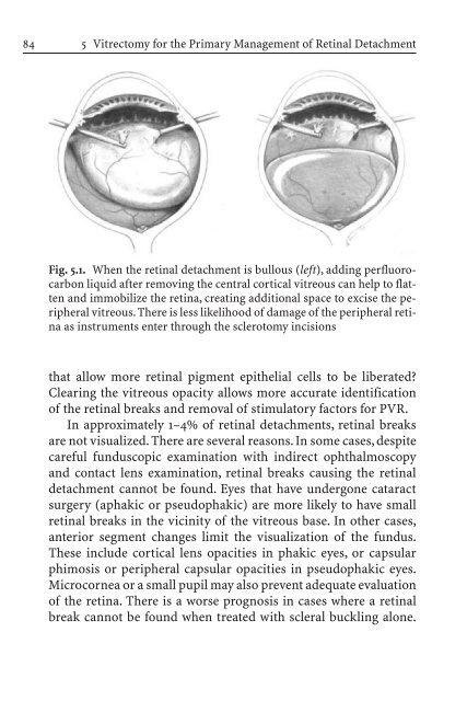
Fig. 5.1. When the retinal detachment is bullous (left), adding perfluorocarbon<br />
liquid after removing the central cortical vitreous can help to flatten<br />
and immobilize the retina, creating additional space to excise the peripheral<br />
vitreous. There is less likelihood of damage of the peripheral retina<br />
as instruments enter through the sclerotomy incisions<br />
that allow more retinal pigment epithelial cells to be liberated?<br />
Clearing the vitreous opacity allows more accurate identification<br />
of the retinal breaks and removal of stimulatory factors for PVR.<br />
In approximately 1–4% of retinal detachments, retinal breaks<br />
are not visualized. There are several reasons. In some cases, despite<br />
careful funduscopic examination with indirect ophthalmoscopy<br />
and contact lens examination, retinal breaks causing the retinal<br />
detachment cannot be found. Eyes that have undergone cataract<br />
surgery (aphakic or pseudophakic) are more likely to have small<br />
retinal breaks in the vicinity of the vitreous base. In other cases,<br />
anterior segment changes limit the visualization of the fundus.<br />
These include cortical lens opacities in phakic eyes, or capsular<br />
phimosis or peripheral capsular opacities in pseudophakic eyes.<br />
Microcornea or a small pupil may also prevent adequate evaluation<br />
of the retina. There is a worse prognosis in cases where a retinal<br />
break cannot be found when treated with scleral buckling alone.


