

Primary Retinal Detachment
Primary Retinal Detachment
Primary Retinal Detachment

You also want an ePaper? Increase the reach of your titles
YUMPU automatically turns print PDFs into web optimized ePapers that Google loves.
60<br />
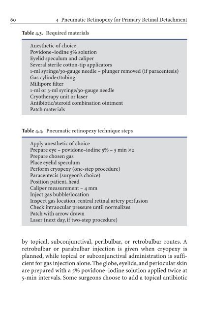
Table 4.3. Required materials<br />
4 Pneumatic Retinopexy for <strong>Primary</strong> <strong>Retinal</strong> <strong>Detachment</strong><br />
Anesthetic of choice<br />
Povidone–iodine 5% solution<br />
Eyelid speculum and caliper<br />
Several sterile cotton-tip applicators<br />
1-ml syringe/30-gauge needle – plunger removed (if paracentesis)<br />
Gas cylinder/tubing<br />
Millipore filter<br />
1-ml or 3-ml syringe/30-gauge needle<br />
Cryotherapy unit or laser<br />
Antibiotic/steroid combination ointment<br />
Patch materials<br />
Table 4.4. Pneumatic retinopexy technique steps<br />
Apply anesthetic of choice<br />
Prepare eye – povidone–iodine 5% – 5 min ¥2<br />
Prepare chosen gas<br />
Place eyelid speculum<br />
Perform cryopexy (one-step procedure)<br />
Paracentecis (surgeon’s choice)<br />
Position patient, head<br />
Caliper measurement – 4 mm<br />
Inject gas bubble/location<br />
Inspect gas location, central retinal artery perfusion<br />
Check intraocular pressure until normalizes<br />
Patch with arrow drawn<br />
Laser (next day, if two-step procedure)<br />
by topical, subconjunctival, peribulbar, or retrobulbar routes. A<br />
retrobulbar or parabulbar injection is given when cryopexy is<br />
planned, while topical or subconjunctival administration is sufficient<br />
for gas injection alone. The globe, eyelids, and periocular skin<br />
are prepared with a 5% povidone–iodine solution applied twice at<br />
5-min intervals. Some surgeons choose to add a topical antibiotic


