Journal <strong>of</strong> Agricultural and Food Chemistry ARTICLE Figure 4. HILIC separation pr<strong>of</strong>ile with UV detection at 316 nm. For peak identities refer to Table 4. The HILIC separation method is capable <strong>of</strong> detection <strong>of</strong> ferulated arab<strong>in</strong>o-oligosaccharides from DP 1 to at least DP 15 20. Analysis <strong>of</strong> LAOS did not show any molecules with<strong>in</strong> this size frame (data not shown), despite this fraction conta<strong>in</strong><strong>in</strong>g significant amounts <strong>of</strong> ferulic acid (Table 3). Accord<strong>in</strong>g to the HPAEC analysis, LAOS did conta<strong>in</strong> oligosaccharides with an approximate size <strong>of</strong> DP 5 10, but apparently the feruloyl substitution was present on oligosaccharides with higher DP than DP 20. (The term “oligosaccharides” is used for consistency.) The fact that only the relatively larger oligosaccharides were feruloyl substituted justifies the appearance <strong>of</strong> ferulated molecules <strong>in</strong> the water fraction, because the overall hydrophilicity <strong>of</strong>, for example, an arab<strong>in</strong>o-oligosaccharide with DP 20 and a s<strong>in</strong>gle feruloyl substitution would still fractionate <strong>in</strong>to the water fraction. In Vitro Fermentation. Quantitative real-time PCR from <strong>in</strong> vitro fermentations showed that fermentation on SAOS, LAOS, LFAOS, and the start<strong>in</strong>g material selectively <strong>in</strong>creased the density <strong>of</strong> Bifidobacterium spp. significantly (P < 0.05, P < 0.01 P < 0.001, and P < 0.05, respectively) when compared to the orig<strong>in</strong>al fecal sample (Figure 6). The densities <strong>of</strong> bifidobacteria after fermentation <strong>of</strong> the high molecular weight fractions, LAOS and LFAOS, were not significantly different from the densities obta<strong>in</strong>ed by fermentation <strong>of</strong> FOS, which is considered to be the “golden standard” with<strong>in</strong> the field <strong>of</strong> prebiotics. This result confirmed that the <strong>in</strong>duced growth was due to the arab<strong>in</strong>o-oligosaccharides and not a result <strong>of</strong> the presence <strong>of</strong> monomers. The f<strong>in</strong>d<strong>in</strong>g that arab<strong>in</strong>o-oligosaccharides are bifidogenic is <strong>in</strong> agreement with a recently patented discovery stat<strong>in</strong>g that branched arab<strong>in</strong>o-oligosaccharides with DP 2 15 are bifidogenic. 27 However, the patent 27 stated that sugar beet derived arab<strong>in</strong>ose-rich pect<strong>in</strong> oligosaccharides do not exert prebiotic effects <strong>in</strong> vitro. This statement may be due to the fact that ma<strong>in</strong>ly homogalacturonan sugar beet pect<strong>in</strong> oligosaccharides were evaluated <strong>in</strong> the patent as opposed to the arab<strong>in</strong>o-oligosaccharides described <strong>in</strong> the present work, which consisted <strong>of</strong> only 1 7% galacturonic acid and 87 96% arab<strong>in</strong>ose (Table 3). Fermentation <strong>of</strong> the LAOS and LFAOS fractions did not yield significantly different results, Table 4. Peak Identities from HILIC Separation (Fig 4) a peak aranFA aranFA2 I II DP2 III DP3 IV DP4 V DP5 DP7 VI DP6 DP8 VII DP6, DP7 DP9 VIII DP7, DP8 DP10 IX DP8, DP9 DP11, DP12 X DP9, DP10 DP13, DP14 a Underscore <strong>in</strong>dicates the mass with highest <strong>in</strong>tensity <strong>in</strong> the MALDI- TOF analysis (MALDI-TOF data not shown). <strong>in</strong>dicat<strong>in</strong>g that the size <strong>of</strong> the oligosaccharides was more important for selective bacterial stimulation than the amount <strong>of</strong> feruloyl substitutions. The f<strong>in</strong>d<strong>in</strong>gs that the high molecular weight fractions were more bifidogenic than the low molecular weight fractions further elaborates on the f<strong>in</strong>d<strong>in</strong>gs reported by Al-Tamimi et al. 8 which <strong>in</strong>dicated that the low molecular weight fractions were more selective for bifidobacteria than arab<strong>in</strong>an. In that study 8 only arab<strong>in</strong>o-oligosaccharides up to DP 8 were tested, and some fractions appeared to conta<strong>in</strong> ma<strong>in</strong>ly monosaccharides. The fact that feruloyl substitution was no h<strong>in</strong>drance to bifidogenic metabolism was <strong>in</strong> good correlation with the results by Funk et al. 13 They found that human <strong>in</strong>test<strong>in</strong>al microbial communities were able to degrade maize cell wall material regardless <strong>of</strong> ferulate levels. No significant changes were seen <strong>in</strong> the densities <strong>of</strong> Lactobacillus spp. and Firmicutes after fermentation. The relative amount <strong>of</strong> Bacteroidetes decreased significantly (P < 0.001) for all tested fractions, <strong>in</strong>clud<strong>in</strong>g FOS, as compared to the <strong>in</strong>oculum (Figure 6). This decrease was most likely due to an <strong>in</strong>crease <strong>in</strong> other types <strong>of</strong> bacteria. The relative balance between Firmicutes and Bacteroidetes is believed to play a role <strong>in</strong> obesity risk, both with respect to develop<strong>in</strong>g obesity as shown by <strong>in</strong>troduc<strong>in</strong>g “obese microbiota” <strong>in</strong> germ-free mice 28 and by the observation that the relative levels <strong>of</strong> Firmicutes and Bacteroidetes change, that is, the relative levels <strong>of</strong> Bacteroidetes <strong>in</strong>crease even though the levels <strong>of</strong> Firmicutes are still dom<strong>in</strong>ant, when the diet for obese humans is restricted. 29 In the study presented <strong>in</strong> this paper the relative level <strong>of</strong> Firmicutes rema<strong>in</strong>ed unchanged and the relative level <strong>of</strong> Bacteroidetes decreased significantly, thereby alter<strong>in</strong>g the relative balance toward a higher Firmicutes/Bacteroidetes ratio. These data suggest that all <strong>of</strong> the oligosaccharide fractions, <strong>in</strong>clud<strong>in</strong>g FOS, a commercial prebiotic, may <strong>in</strong>crease the risk <strong>of</strong> obesity and maybe, <strong>in</strong> turn, the risk <strong>of</strong> develop<strong>in</strong>g the metabolic syndrome associated with obesity. However, it is not clear if the extent <strong>of</strong> change observed after the <strong>in</strong> vitro fermentations has any significance, as the available <strong>in</strong> vivo evidence <strong>of</strong> an effect <strong>of</strong> change <strong>in</strong> the Firmicutes/Bacteroidetes ratio was recorded over a longer period, namely 1 year, and the change was accompanied by a change <strong>in</strong> body weight (<strong>in</strong> humans). 29 Clearly, the data therefore require further <strong>in</strong>vestigation. Besides the positive bifidogenic effects <strong>of</strong> the feruloylated arab<strong>in</strong>o-oligosaccharides, it was <strong>in</strong>vestigated whether these substrates could be fermented by an opportunistic <strong>in</strong>test<strong>in</strong>al pathogen, C. difficile. C. difficile <strong>in</strong>fection is l<strong>in</strong>ked to consumption <strong>of</strong> antibiotics, which disrupt the normal <strong>in</strong>test<strong>in</strong>al microbiota, allow<strong>in</strong>g C. difficile 6516 dx.doi.org/10.1021/jf200996h |J. Agric. Food Chem. 2011, 59, 6511–6519
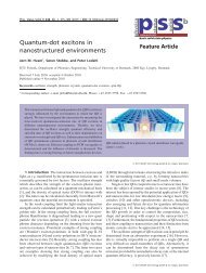
Journal <strong>of</strong> Agricultural and Food Chemistry ARTICLE Figure 5. MS/MS high-energy CID spectrum <strong>of</strong> a sodium adduct <strong>of</strong> ara5FA (fraction V, Figure 4), illustrat<strong>in</strong>g the fragmentation pattern and nomenclature, and two different proposed structures. Figure 6. Relative quantities <strong>of</strong> target genes <strong>in</strong> samples from orig<strong>in</strong>al fecal bacterial communities and after fermentation <strong>of</strong> oligosaccharides by these communities. Target genes encoded 16S rRNA from Bifidobacterium spp., Lactobacillus spp., Bacteroidetes, and Firmicutes. Fecal samples were obta<strong>in</strong>ed from the six healthy volunteers. The bars represent the average ( SEM <strong>of</strong> the response from six volunteers. Asterisks <strong>in</strong>dicate a significant difference between target density <strong>in</strong> the orig<strong>in</strong>al community and <strong>in</strong> the fermented samples: P < 0.05, /; P < 0.01, //; P < 0.001, ///. Pound signs <strong>in</strong>dicate a significant difference between target density after fermentation <strong>of</strong> FOS and after fermentation <strong>of</strong> the oligosaccharides: P < 0.05, #; P < 0.01, ##; P < 0.001, ###. 6517 dx.doi.org/10.1021/jf200996h |J. Agric. Food Chem. 2011, 59, 6511–6519
- Page 1:
Role of Intestinal Microbiota in Ul
- Page 4 and 5:
Role of Intestinal Microbiota in Ul
- Page 6 and 7:
Preface Preface This thesis present
- Page 8 and 9:
Summary Summary The microbiota of t
- Page 10 and 11:
Dansk sammendrag Dansk sammendrag M
- Page 12 and 13:
Introduction and objectives Introdu
- Page 14 and 15:
List of Manuscripts Not included in
- Page 16 and 17:
List of contents List of Centents P
- Page 18 and 19:
List of Centents Methodology append
- Page 21 and 22:
1. The intestinal environment Theor
- Page 23 and 24:
Theoretical part 5 1. The intestina
- Page 25 and 26:
2. The colonic environment Theoreti
- Page 27 and 28:
Theoretical part 9 2. The colonic e
- Page 29 and 30:
Table 1: The presence of glycoside
- Page 31 and 32:
Theoretical part Figure 3: The colo
- Page 33 and 34:
3. Inflammatory Bowel disease Theor
- Page 35 and 36:
Theoretical part 17 3. Inflammatory
- Page 37 and 38:
Theoretical part 19 4. Modulation o
- Page 39 and 40:
Theoretical part 21 4. Modulation o
- Page 41 and 42:
Theoretical part 23 4. Modulation o
- Page 43 and 44:
Table 4: Clinical trials on the pre
- Page 45 and 46:
Theoretical part 5. Production of p
- Page 47 and 48:
Theoretical part 5. Production of p
- Page 49 and 50:
Theoretical part 5. Production of p
- Page 51:
Methodology part
- Page 54 and 55:
Methodology part 6. Methodology, co
- Page 56 and 57:
Methodology part 6. Methodology, co
- Page 58 and 59:
Methodology part 6. Methodology, co
- Page 60 and 61:
Introduction Methodology part 42 Pa
- Page 62 and 63:
Abstract Background Detailed knowle
- Page 64 and 65:
depending the level of disease acti
- Page 66 and 67:
in 1 x TAE at 60 °C for 16 h at 36
- Page 68 and 69:
Statistics PCA were generated by SA
- Page 70 and 71:
The PCA of the Gram‐positive bact
- Page 72 and 73:
layer of UC patients and found that
- Page 74 and 75:
Acknowledgements The authors thank
- Page 76 and 77:
Table 2 ‐ 16S rRNA gene and 16S
- Page 78 and 79:
1. Firmicutes phylum 2. Bacteroidet
- Page 80 and 81:
Supplementary Figure S1. Dice clust
- Page 82 and 83:
Reference List 1. Ahmed S, Macfarla
- Page 84 and 85: 32. Matsuki T, Watanabe K, Fujimoto
- Page 87 and 88: Methodology part Paper 2 Fecal lact
- Page 89 and 90: Fecal lactobacilli and bifidobacter
- Page 91 and 92: Introduction The mucus layer lining
- Page 93 and 94: efore enrolment and there was no si
- Page 95 and 96: (Bio‐Rad Labs, Hercules, Californ
- Page 97 and 98: Microbial community analysis using
- Page 99 and 100: difference from the luminal microbi
- Page 101 and 102: that C. coccoides group and C. lept
- Page 103 and 104: Table 1 ‐ 16S rRNA gene of phylum
- Page 105 and 106: Table 2 ‐ Preference of bacterial
- Page 107 and 108: Figure 1. A) Schematic overview of
- Page 109 and 110: A. B. Figure 3. Principal component
- Page 111 and 112: 15. Fooks LJ, Gibson GR. (2002) In
- Page 113 and 114: 47. Ouwehand AC, Suomalainen T, Tol
- Page 115 and 116: Methodology part Paper 3 Paper 3 In
- Page 117 and 118: APPLIED AND ENVIRONMENTAL MICROBIOL
- Page 119 and 120: 8338 VIGSNÆS ET AL. APPL. ENVIRON.
- Page 121 and 122: 8340 VIGSNÆS ET AL. APPL. ENVIRON.
- Page 123 and 124: 8342 VIGSNÆS ET AL. APPL. ENVIRON.
- Page 125: 8344 VIGSNÆS ET AL. APPL. ENVIRON.
- Page 128 and 129: Methodology part Introduction The a
- Page 130 and 131: Journal of Agricultural and Food Ch
- Page 132 and 133: Journal of Agricultural and Food Ch
- Page 136 and 137: Journal of Agricultural and Food Ch
- Page 139 and 140: Methodology part Paper 5 Paper 5 Ma
- Page 141 and 142: Appl Microbiol Biotechnol (2011) 90
- Page 143 and 144: Appl Microbiol Biotechnol (2011) 90
- Page 145 and 146: Appl Microbiol Biotechnol (2011) 90
- Page 147 and 148: Appl Microbiol Biotechnol (2011) 90
- Page 149 and 150: Appl Microbiol Biotechnol (2011) 90
- Page 151 and 152: Appl Microbiol Biotechnol (2011) 90
- Page 153 and 154: Methodology part Paper 6 Tailored e
- Page 155 and 156: Process Biochemistry 46 (2011) 1039
- Page 157 and 158: Table 1 List of enzymes. Enzyme Sou
- Page 159 and 160: J. Holck et al. / Process Biochemis
- Page 161 and 162: Intensity 50 0 C1 175.05 J. Holck e
- Page 163 and 164: J. Holck et al. / Process Biochemis
- Page 165: [29] Bauer S, Vasu P, Persson S, Mo
- Page 168 and 169: Methodology part 150
- Page 170 and 171: Methodology part 152 Appendix 1 hor
- Page 172 and 173: Appendix 3 Methodology part Appendi
- Page 174 and 175: Methodology part 156 Appendix 3
- Page 177 and 178: 7. Discussion and perspectives Disc
- Page 179 and 180: Discussion and conclusion 161 7. Di
- Page 181 and 182: Discussion and conclusion 163 7. Di
- Page 183 and 184: Discussion and conclusion 165 7. Di
- Page 185 and 186:
References References Abreu,M.T., V
- Page 187 and 188:
References Derrien,M., Vaughan,E.E.
- Page 189 and 190:
References Haarman,M. and Knol,J. (
- Page 191 and 192:
References Lantz,P.G., Matsson,M.,
- Page 193 and 194:
References Matsuki,T., Watanabe,K.,
- Page 195 and 196:
References Pullan,R.D., Thomas,G.A.
- Page 197 and 198:
References Tannock,G.W. (2010). Ana
- Page 200:
National Food Institute Technical U