

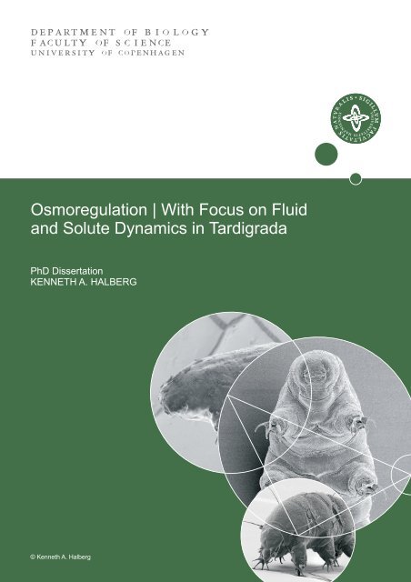
Osmoregulation | With Focus on Fluid and Solute Dynamics in ...
Osmoregulation | With Focus on Fluid and Solute Dynamics in ...
Osmoregulation | With Focus on Fluid and Solute Dynamics in ...

Create successful ePaper yourself
Turn your PDF publications into a flip-book with our unique Google optimized e-Paper software.
<str<strong>on</strong>g>Osmoregulati<strong>on</strong></str<strong>on</strong>g> | <str<strong>on</strong>g>With</str<strong>on</strong>g> <str<strong>on</strong>g>Focus</str<strong>on</strong>g> <strong>on</strong> <strong>Fluid</strong><br />
<strong>and</strong> <strong>Solute</strong> <strong>Dynamics</strong> <strong>in</strong> Tardigrada<br />
PhD Dissertati<strong>on</strong><br />
KENNETH A. HALBERG<br />
© Kenneth A. Halberg
UNIVERSITAS<br />
HAFNIENSIS<br />
2012<br />
FACULTY OF SCIENCE<br />
UNIVERSITY OF COPENHAGEN<br />
<str<strong>on</strong>g>Osmoregulati<strong>on</strong></str<strong>on</strong>g>│<str<strong>on</strong>g>With</str<strong>on</strong>g> <str<strong>on</strong>g>Focus</str<strong>on</strong>g> <strong>on</strong> <strong>Fluid</strong><br />
<strong>and</strong> <strong>Solute</strong> <strong>Dynamics</strong> <strong>in</strong> Tardigrada<br />
PhD Dissertati<strong>on</strong><br />
Kenneth A. Halberg<br />
Dissertati<strong>on</strong> submitted M<strong>on</strong>day the 14th of May 2012.<br />
Supervisor: Associate Professor Nadja Møbjerg, PhD.
Dissertati<strong>on</strong> presented at University of Copenhagen to be publicly exam<strong>in</strong>ed (provided acceptance <strong>in</strong> its<br />
current form) <strong>in</strong> Auditorium 1, August Krogh Build<strong>in</strong>g, Universitetsparken 13, Thursday, June 28, 2012<br />
at 14:00 for the degree of Doctor of Philosophy. The exam<strong>in</strong>ati<strong>on</strong> will be c<strong>on</strong>ducted <strong>in</strong> English.<br />
Abstract<br />
Halberg, K. A. 2012. <str<strong>on</strong>g>Osmoregulati<strong>on</strong></str<strong>on</strong>g> │<str<strong>on</strong>g>With</str<strong>on</strong>g> <str<strong>on</strong>g>Focus</str<strong>on</strong>g> <strong>on</strong> <strong>Fluid</strong> <strong>and</strong> <strong>Solute</strong> <strong>Dynamics</strong> <strong>in</strong><br />
Tardigrada.<br />
<str<strong>on</strong>g>Osmoregulati<strong>on</strong></str<strong>on</strong>g> is the regulated c<strong>on</strong>trol of water <strong>and</strong> solute compositi<strong>on</strong> <strong>in</strong> body fluid<br />
compartments. On <strong>on</strong>e h<strong>and</strong>, the <strong>in</strong>ternal compositi<strong>on</strong> must be kept with<strong>in</strong> optimal<br />
c<strong>on</strong>diti<strong>on</strong>s for metabolic processes <strong>in</strong> the face of external perturbati<strong>on</strong>. On the other<br />
h<strong>and</strong>, the nature of the liv<strong>in</strong>g state dem<strong>and</strong>s a c<strong>on</strong>t<strong>in</strong>uous traffic of compounds <strong>in</strong> <strong>and</strong><br />
out of the organism. These dem<strong>and</strong>s appear to be <strong>in</strong> fundamental c<strong>on</strong>tradicti<strong>on</strong><br />
however cells <strong>and</strong> animals achieve so-called “steady-state” by means of an array of<br />
transport prote<strong>in</strong>s, which provide a str<strong>in</strong>gent c<strong>on</strong>trol <strong>on</strong> the exchange of water <strong>and</strong><br />
solutes across body surfaces. The dist<strong>in</strong>ct mechanisms of solute transport have been<br />
studied <strong>in</strong> most animal groups, but there are still large gaps <strong>in</strong> our underst<strong>and</strong><strong>in</strong>g of<br />
how animals cope with osmotic stress. In the present thesis, osmoregulatory<br />
phenomena were studied <strong>in</strong> vertebrate <strong>and</strong> <strong>in</strong>vertebrate organism alike, with the ma<strong>in</strong><br />
focus be<strong>in</strong>g <strong>on</strong> fluid <strong>and</strong> solute dynamics <strong>in</strong> Tardigrada. For example, the <strong>in</strong>organic<br />
i<strong>on</strong> compositi<strong>on</strong> of several species was <strong>in</strong>vestigated, which revealed that tardigrades<br />
c<strong>on</strong>ta<strong>in</strong> roughly similar relative c<strong>on</strong>tributi<strong>on</strong>s of <strong>in</strong>organic i<strong>on</strong>s to total osmotic<br />
c<strong>on</strong>centrati<strong>on</strong>, when compared to closely related animal groups. Moreover, it was<br />
<strong>in</strong>ferred that cryptobiotic tardigrades (species able to enter a state of latent life)<br />
c<strong>on</strong>ta<strong>in</strong> a large fracti<strong>on</strong> of organic osmolytes. The mechanisms of organic ani<strong>on</strong><br />
transport <strong>in</strong> a mar<strong>in</strong>e species of tardigrade was <strong>in</strong>vestigated pharmacologically, <strong>and</strong><br />
compared to that of <strong>in</strong>sects. These data showed that organic ani<strong>on</strong> transport is<br />
localized to the midgut epithelium <strong>and</strong> that the transport is both active <strong>and</strong> transporter<br />
mediated with a pharmacological profile similar to that of <strong>in</strong>sects. Tardigrades survive<br />
<strong>in</strong> a variety of osmotic envir<strong>on</strong>ments (semi-terrestrial, limnic <strong>and</strong> mar<strong>in</strong>e habitats),<br />
why the ability to volume <strong>and</strong> osmoregulate was exam<strong>in</strong>ed. These studies dem<strong>on</strong>strated<br />
an ability to regulate total body volume dur<strong>in</strong>g both hypo- <strong>and</strong> hyperosmotic<br />
c<strong>on</strong>diti<strong>on</strong>s, <strong>and</strong> that the ability to hyper-regulate could be a general theme am<strong>on</strong>g<br />
members of eutardigrades. Thus, the work presented here<strong>in</strong>, have c<strong>on</strong>tributed to<br />
establish<strong>in</strong>g tardigrades as an important experimental group <strong>in</strong> which central<br />
physiological questi<strong>on</strong>s may be answered, <strong>in</strong>clud<strong>in</strong>g aspects of osmotic <strong>and</strong> i<strong>on</strong>ic<br />
regulati<strong>on</strong>.<br />
Keywords: osmoregulati<strong>on</strong>, volume regulati<strong>on</strong>, organic ani<strong>on</strong> transport, hyper-regulate,<br />
<strong>in</strong>organic i<strong>on</strong>s, organic osmolytes, tardigrade, <strong>in</strong>sect,<br />
Kenneth A. Halberg, The August Krogh Centre, Department of Biology,<br />
Universitetsparken 13, DK-2100 Copenhagen Ø, Denmark<br />
© Kenneth A. Halberg 2012
“Beautiful is what we see,<br />
More beautiful is what we perceive,<br />
Most beautiful is what we do not underst<strong>and</strong>”<br />
- Niels Stensen
List of Papers<br />
This thesis is based <strong>on</strong> the follow<strong>in</strong>g papers <strong>and</strong> manuscripts, which are referred to<br />
<strong>in</strong> the text by their Roman numerals.<br />
I. Halberg, K. A., Larsen, K. W., Jørgensen, A., Ramløv, H. & Møbjerg,<br />
N. Cryptobiotic tardigrades c<strong>on</strong>ta<strong>in</strong> large fracti<strong>on</strong> of unidentified<br />
organic solutes: A comparative study <strong>on</strong> <strong>in</strong>organic i<strong>on</strong> compositi<strong>on</strong> <strong>in</strong><br />
Tardigrada.<br />
II. Halberg, K. A., & Møbjerg, N. (2012). First evidence of epithelial<br />
transport <strong>in</strong> tardigrades: A comparative <strong>in</strong>vestigati<strong>on</strong> of organic ani<strong>on</strong><br />
transport. Journal of Experimental Biology, 215: 497-507.<br />
III. Møbjerg, N. M., Halberg, K. A., Jørgensen, A. Perss<strong>on</strong>., D., Bjørn M,<br />
Ramløv H & Kristensen R. M. (2011). Survival <strong>in</strong> extreme<br />
envir<strong>on</strong>ments – <strong>on</strong> current knowledge of adaptati<strong>on</strong>s <strong>in</strong> tardigrades.<br />
Acta Physiologica, 202: 409-420.<br />
IV. Haugen, B.M., Halberg, K.A., Jespersen, Å., Prehn, L.R. & Møbjerg,<br />
N. (2010). Functi<strong>on</strong>al characterizati<strong>on</strong> of the vertebrate primary ureter:<br />
Structure <strong>and</strong> i<strong>on</strong> transport mechanisms of the pr<strong>on</strong>ephric duct of<br />
axolotl larvae (Amphibia). BMC developmental Biology, 10: 56.<br />
V. Halberg, K. A., Perss<strong>on</strong>, D., Ramløv, H., Westh, P., Kristensen, R. M.<br />
& Møbjerg, N. (2009). Cyclomorphosis <strong>in</strong> Tardigrada: Adapti<strong>on</strong> to<br />
envir<strong>on</strong>mental c<strong>on</strong>stra<strong>in</strong>ts. Journal of Experimental Biology, 212:<br />
2803-2811.<br />
Additi<strong>on</strong>ally, the follow<strong>in</strong>g papers <strong>and</strong> manuscripts were prepared dur<strong>in</strong>g the<br />
course of my PhD studies, but are not <strong>in</strong>cluded <strong>in</strong> the thesis:<br />
VI. Halberg K. A., Jørgensen, A. <strong>and</strong> Møbjerg, N. (<strong>in</strong> prep.). Surviv<strong>in</strong>g<br />
without water: Tun formati<strong>on</strong> <strong>in</strong> tardigrades is an active process<br />
mediated by the musculature<br />
VII. Halberg K. A., Perss<strong>on</strong>, D., Jørgensen, A. Kristensen, R. M. <strong>and</strong><br />
Møbjerg, N. (submitted). Populati<strong>on</strong> dynamics of a mar<strong>in</strong>e tardigrade:<br />
Temperature limits geographic distributi<strong>on</strong> of Halobiotus crispae.<br />
Mar<strong>in</strong>e Biological Research
VIII. Perss<strong>on</strong>, D., Halberg K. A., Jørgensen A., Møbjerg N. & Kristensen<br />
R. M. (<strong>in</strong> review). Neuroanatomy of Halobiotus crispae (Eutardigrada:<br />
Hypsibiidae): Tardigrade bra<strong>in</strong> structure suggests <strong>in</strong>clusi<strong>on</strong> <strong>in</strong>to<br />
Panarthropoda. Journal of Morphology.<br />
IX. Perss<strong>on</strong>, D., Halberg K. A., Jørgensen A., Ricci C., Møbjerg N. &<br />
Kristensen R. M. (2010). Extreme stress tolerance <strong>in</strong> tardigrades:<br />
Surviv<strong>in</strong>g space c<strong>on</strong>diti<strong>on</strong>s <strong>in</strong> low earth orbit. Journal of Zoological<br />
Systematics <strong>and</strong> Evoluti<strong>on</strong>ary Research, 49: 90-97.<br />
X. Halberg, K. A., Perss<strong>on</strong> D., Møbjerg N., Wann<strong>in</strong>ger A. & Kristensen<br />
R. M. (2009). Myoanatomy of the Mar<strong>in</strong>e Tardigrade Halobiotus<br />
crispae (Eutardigrada: Hypsibiidae). Journal of Morphology, 270:<br />
996-1013.<br />
Lastly, follow<strong>in</strong>g paper provides important background knowledge for the work<br />
presented here<strong>in</strong>:<br />
XI. Møbjerg, N., A. Jørgensen, J. Eibye-Jacobsen, K. A. Halberg, D.<br />
Perss<strong>on</strong> & R. M. Kristensen (2007). New Records <strong>on</strong> cyclomorphosis<br />
<strong>in</strong> the mar<strong>in</strong>e eutardigrade Halobiotus crispae (Eutardigrada:<br />
Hypsibiidae). Journal of Limnology, 66 (suppl. 1): 132-140.
Repr<strong>in</strong>t <strong>and</strong> publicati<strong>on</strong> is made with permissi<strong>on</strong> from the respective copyright<br />
holders.<br />
Paper II, V © The Company of Biologists.<br />
Paper IV, is copyright of the authors.<br />
Paper III © Wiley-Blackwell<br />
Statement of authorship<br />
Paper I: KAH was deeply <strong>in</strong>volved <strong>in</strong> study design. KAH participated <strong>in</strong><br />
extract<strong>in</strong>g animals <strong>and</strong> i<strong>on</strong> chromatography. KAH performed nanoliter osmometry.<br />
KAH performed the data analysis, prepared the figures, participated <strong>in</strong> discussi<strong>on</strong>s<br />
<strong>and</strong> <strong>in</strong>terpretati<strong>on</strong> of the data, <strong>and</strong> drafted the manuscript.<br />
Paper II: KAH performed the major part of the experimental work <strong>and</strong> data<br />
analysis. He participated <strong>in</strong> plann<strong>in</strong>g of experiments, data <strong>in</strong>terpretati<strong>on</strong>, prepared<br />
the figures <strong>and</strong> drafted the manuscript.<br />
Paper III: KAH performed cell counts <strong>and</strong> provided images of tardigrades. He<br />
helped draft parts of the manuscript.<br />
Paper IV: KAH participated <strong>in</strong> immunosta<strong>in</strong><strong>in</strong>g experiments, performed CLSM<br />
<strong>and</strong> prepared the 3D images. KAH participated <strong>in</strong> discussi<strong>on</strong>s <strong>and</strong> <strong>in</strong>terpretati<strong>on</strong> of<br />
the data.<br />
Paper V: KAH participated <strong>in</strong> plann<strong>in</strong>g of experiments, sampl<strong>in</strong>g, stag<strong>in</strong>g,<br />
scann<strong>in</strong>g electr<strong>on</strong> microscopy, DSC experiments, experiments <strong>on</strong> cold hard<strong>in</strong>ess<br />
<strong>and</strong> osmotic stress tolerance, volume measurements, hemolymph sample<br />
collecti<strong>on</strong>s, <strong>and</strong> nanoliter osmometry. He furthermore participated <strong>in</strong> discussi<strong>on</strong>s<br />
<strong>and</strong> <strong>in</strong>terpretati<strong>on</strong> of data, prepared the figures <strong>and</strong> drafted the manuscript.<br />
Fr<strong>on</strong>t cover: Scann<strong>in</strong>g elecr<strong>on</strong> micrographs of the tardigrades Rictersius cor<strong>on</strong>ifer<br />
(top left), Halobiotus crispae (middle right), <strong>and</strong> Ech<strong>in</strong>iscus testudo (middle<br />
bottom).
C<strong>on</strong>tents<br />
Preface.......................................................................................................................... 9<br />
Introducti<strong>on</strong> ................................................................................................................ 11<br />
Ma<strong>in</strong>ta<strong>in</strong><strong>in</strong>g a stable <strong>in</strong>ternal envir<strong>on</strong>ment .......................................................... 11<br />
Osmoregulators <strong>and</strong> osmoc<strong>on</strong>formers ................................................................. 12<br />
Osmoregulatory organs........................................................................................ 12<br />
Filtrati<strong>on</strong>-Reabsorpti<strong>on</strong> systems ................................................................ 13<br />
Secreti<strong>on</strong>-Reabsorpti<strong>on</strong> systems ................................................................ 14<br />
Phylum Tardigrada..................................................................................................... 17<br />
General morphology ............................................................................................ 18<br />
Classificati<strong>on</strong> ....................................................................................................... 19<br />
Ecology ................................................................................................................ 21<br />
<strong>Fluid</strong> <strong>and</strong> solute dynamics – an overview .................................................................. 23<br />
Inorganic i<strong>on</strong> compositi<strong>on</strong> ................................................................................... 23<br />
Organic ani<strong>on</strong> transport ....................................................................................... 25<br />
Volume <strong>and</strong> osmoregulati<strong>on</strong>................................................................................ 26<br />
C<strong>on</strong>clusi<strong>on</strong>s <strong>and</strong> future perspectives .......................................................................... 27<br />
Dansk sammenfatn<strong>in</strong>g ................................................................................................ 29<br />
Acknowledgements .................................................................................................... 30<br />
References .................................................................................................................. 32
Preface<br />
The primary aim of this thesis was to address several aspects of the fluid <strong>and</strong><br />
solute dynamics <strong>in</strong> tardigrades, <strong>and</strong> hereby provide new <strong>in</strong>sight <strong>in</strong>to the general<br />
stress biology of these enigmatic creatures. This was d<strong>on</strong>e by adopt<strong>in</strong>g an<br />
<strong>in</strong>tegrative approach, i.e. apply<strong>in</strong>g advanced methods <strong>in</strong> biology, analytic<br />
biochemistry <strong>and</strong> physical chemistry, which offered functi<strong>on</strong>al data from different<br />
discipl<strong>in</strong>es to be obta<strong>in</strong>ed. The experimental work was carried out ma<strong>in</strong>ly at The<br />
August Krogh Centre, University of Copenhagen; however, additi<strong>on</strong>al<br />
experimental work was performed at the Danish Natural History Museum,<br />
University of Copenhagen <strong>and</strong> at the Department of Nature, Systems <strong>and</strong> Models,<br />
Roskilde University. Overall this thesis has c<strong>on</strong>tributed to c<strong>on</strong>vert<strong>in</strong>g tardigrades,<br />
from an almost exclusive tax<strong>on</strong>omic phenomen<strong>on</strong> <strong>in</strong>to an established <strong>and</strong><br />
important experimental group, <strong>in</strong> which central physiological questi<strong>on</strong>s can be<br />
<strong>in</strong>vestigated.<br />
This PhD-thesis comprises a short <strong>in</strong>troducti<strong>on</strong> to osmotic <strong>and</strong> i<strong>on</strong>ic<br />
regulati<strong>on</strong> <strong>in</strong> Metazoa, accompanied by a brief review <strong>on</strong> the general morphology,<br />
classificati<strong>on</strong> <strong>and</strong> ecology of tardigrades. Moreover, an overview of the results<br />
presented as well as c<strong>on</strong>clusi<strong>on</strong>s <strong>and</strong> future perspectives are presented. Five papers<br />
<strong>and</strong> manuscripts form the basis of this thesis, of which four are published <strong>in</strong> peer<br />
review journals (Papers II, III, IV <strong>and</strong> V), <strong>and</strong> <strong>on</strong>e is prepared for submissi<strong>on</strong><br />
(Paper I). I am the first author of three (Paper I, II <strong>and</strong> V) <strong>and</strong> sec<strong>on</strong>d author <strong>on</strong><br />
two (Papers III <strong>and</strong> IV) of these papers. Five additi<strong>on</strong>al papers <strong>and</strong> manuscripts<br />
were prepared dur<strong>in</strong>g the course of my PhD studies, but are not <strong>in</strong>cluded <strong>in</strong> the<br />
thesis. This work was funded by the 2008 Faculty of Science, University of<br />
Copenhagen Freja-Programme.<br />
Copenhagen, the 14 th of May 2012<br />
Kenneth A. Halberg<br />
9
Introducti<strong>on</strong><br />
Ma<strong>in</strong>ta<strong>in</strong><strong>in</strong>g a stable <strong>in</strong>ternal envir<strong>on</strong>ment<br />
“Life is as a th<strong>in</strong>g of macromolecular cohesi<strong>on</strong> <strong>in</strong> salty water” (Gilles & Delpire,<br />
1997). Albeit a crude statement, it frames the fact that the ability to c<strong>on</strong>trol salt<br />
<strong>and</strong> water balance is a fundamental prerequisite for both cellular <strong>and</strong> animal life.<br />
Indeed, the <strong>in</strong>ternal envir<strong>on</strong>ment must usually be kept with<strong>in</strong> relatively narrow<br />
limits, as substantial deviati<strong>on</strong>s <strong>in</strong> cell compositi<strong>on</strong> are <strong>in</strong>compatible with the<br />
optimal functi<strong>on</strong> of macromolecules (lipids, prote<strong>in</strong>s, RNA), <strong>and</strong> may ultimately<br />
modify the rate <strong>and</strong> extent of cellular reacti<strong>on</strong>s (Zhao, 2005). The overall<br />
mechanism by which animals c<strong>on</strong>serve a proper osmotic balance between cells,<br />
extracellular fluid <strong>and</strong> the envir<strong>on</strong>ment is termed osmoregulati<strong>on</strong>.<br />
The basis for osmoregulati<strong>on</strong> lies <strong>in</strong> the strict c<strong>on</strong>trol of the i<strong>on</strong>ic<br />
compositi<strong>on</strong> <strong>and</strong> the osmotic pressure of the <strong>in</strong>tra- <strong>and</strong> extracellular compartments<br />
through the regulated accumulati<strong>on</strong> <strong>and</strong> loss of <strong>in</strong>organic i<strong>on</strong>s <strong>and</strong> organic<br />
compounds (Daws<strong>on</strong> & Liu, 2009). This regulati<strong>on</strong> is achieved through the<br />
coord<strong>in</strong>ated activity of an array of transporter prote<strong>in</strong>s (both energy-c<strong>on</strong>sum<strong>in</strong>g<br />
<strong>and</strong> passive), which collectively ma<strong>in</strong>ta<strong>in</strong> the steady-state c<strong>on</strong>diti<strong>on</strong> of cells <strong>and</strong><br />
animals (Essig, 1968). Dur<strong>in</strong>g steady-state c<strong>on</strong>diti<strong>on</strong>s, compositi<strong>on</strong>s of the <strong>in</strong>tra-<br />
<strong>and</strong> extracellular compartments are ma<strong>in</strong>ta<strong>in</strong>ed <strong>in</strong> a n<strong>on</strong>-equilibrium state (Daws<strong>on</strong><br />
& Liu, 2009). This uneven distributi<strong>on</strong> of solutes is important for keep<strong>in</strong>g an<br />
optimal milieu for metabolic processes (Zhao, 2005). Accord<strong>in</strong>gly, the<br />
extracellular fracti<strong>on</strong> of the body fluids of animals are typically high <strong>in</strong> Na + <strong>and</strong> Cl -<br />
, <strong>and</strong> relatively low <strong>in</strong> the other major i<strong>on</strong>s (e.g. K + , Ca 2+ <strong>and</strong> Mg 2+ ), while the<br />
<strong>in</strong>tracellular envir<strong>on</strong>ment of most organisms is low <strong>in</strong> Na + but high <strong>in</strong> K + , PO4 3-<br />
<strong>and</strong> prote<strong>in</strong>s (e.g. Daws<strong>on</strong> & Lui, 2009; Paper I). As such, the plasma membrane<br />
of cells must ma<strong>in</strong>ta<strong>in</strong> i<strong>on</strong>ic, but not osmotic, differences, while specialized<br />
excretory organs – e.g. antennal gl<strong>and</strong>s of crustaceans, Malpighian tubules of<br />
<strong>in</strong>sects <strong>and</strong> tardigrades, rectal gl<strong>and</strong>s of sharks <strong>and</strong> rays, gills <strong>and</strong> <strong>in</strong>test<strong>in</strong>e of<br />
teleost fishes, salt gl<strong>and</strong>s of birds <strong>and</strong> reptiles, the kidneys of vertebrates etc. –<br />
often ma<strong>in</strong>ta<strong>in</strong> both i<strong>on</strong>ic <strong>and</strong> osmotic differences between animals <strong>and</strong> their<br />
envir<strong>on</strong>ments (Riegel, 1970; Peaker, 1971; Paper IV, Beyenbach & Piermar<strong>in</strong>i,<br />
2011; Reilly et al., 2011; Whittamore, 2012). In general, mechanisms that allowed<br />
organisms to resp<strong>on</strong>d <strong>and</strong> adapt to an osmotic challenge over the course of<br />
11
evoluti<strong>on</strong>, has been fundamental to the <strong>in</strong>vasi<strong>on</strong> of new osmotic hostile habitats,<br />
<strong>and</strong> such ecological divergence <strong>in</strong> turn is an important mechanism for the<br />
speciati<strong>on</strong> process (Schluter, 2009). Accord<strong>in</strong>gly, if the ability to osmoregulate<br />
had not evolved, life <strong>on</strong> the planet would look quiet different from how we know<br />
it.<br />
Osmoregulators <strong>and</strong> osmoc<strong>on</strong>formers<br />
Generally, animals are divided <strong>in</strong>to two broad categories <strong>in</strong> terms of their<br />
resp<strong>on</strong>ses to osmotic stress: osmoregulators, which ma<strong>in</strong>ta<strong>in</strong> an <strong>in</strong>ternal osmolarity<br />
different from that of the external envir<strong>on</strong>ment, <strong>and</strong> osmoc<strong>on</strong>formers, which<br />
c<strong>on</strong>form to the external medium <strong>in</strong> which they are immersed (Fig. 1). Most<br />
vertebrates are strict osmoregulators (e.g. Paper IV), as they ma<strong>in</strong>ta<strong>in</strong> i<strong>on</strong>ic <strong>and</strong><br />
osmotic balance with<strong>in</strong> narrow limits; although hagfish, a basal group of<br />
vertebrates, are a notable excepti<strong>on</strong> (Sardella et al., 2009). C<strong>on</strong>versely, mar<strong>in</strong>e<br />
<strong>in</strong>vertebrates are typically categorized as osmoc<strong>on</strong>formers, as many of them<br />
appear to be <strong>in</strong> osmotic balance with sea water over a range of sal<strong>in</strong>ities (Fig. 1).<br />
However, there are numerous obvious excepti<strong>on</strong>s <strong>in</strong>clud<strong>in</strong>g the mar<strong>in</strong>e tardigrade<br />
Halobiotus crispae as well as several members of Crustacea (e.g. Sarver et al.,<br />
1994; Normant et al., 2005; Paper V). In fact, the terms ‘strict osmoregulator’ <strong>and</strong><br />
‘strict osmoc<strong>on</strong>former’ must be used with cauti<strong>on</strong>, as typical osmoregulators are<br />
forced to c<strong>on</strong>form dur<strong>in</strong>g the most extreme c<strong>on</strong>diti<strong>on</strong>s (e.g. Dowd et al., 2010),<br />
whereas animals otherwise described as osmoc<strong>on</strong>formers actually ma<strong>in</strong>ta<strong>in</strong> slight<br />
differences between the <strong>in</strong>ternal <strong>and</strong> external envir<strong>on</strong>ment (e.g. van Weel, 1957).<br />
Although achieved through different mechanisms, both osmoregulat<strong>in</strong>g <strong>and</strong><br />
osmoc<strong>on</strong>form<strong>in</strong>g animals may tolerate wide ranges of external sal<strong>in</strong>ities, thus<br />
termed euryhal<strong>in</strong>e species, while animals <strong>in</strong>tolerant of large changes are called<br />
stenohal<strong>in</strong>e species.<br />
Osmoregulatory organs<br />
Osmoregulatory organs are specialized organs <strong>in</strong>volved <strong>in</strong> ma<strong>in</strong>ta<strong>in</strong><strong>in</strong>g i<strong>on</strong>ic <strong>and</strong><br />
osmotic homeostasis <strong>in</strong> the face of osmotic perturbati<strong>on</strong>, as well as <strong>in</strong> excret<strong>in</strong>g<br />
endobiotic <strong>and</strong> exobiotic waste products (Daws<strong>on</strong> & Lui, 2009; Papers II, IV).<br />
The specific organs mediat<strong>in</strong>g these processes may vary between different groups<br />
of animals (see above); however, the molecular basis <strong>and</strong> specific mechanism of<br />
solute <strong>and</strong> water transport often show a highly c<strong>on</strong>vergent/homologous pattern<br />
across different types of epithelia (e.g. Paper II). In general, two types of systems<br />
12
Fig. 1 Examples of osmotic performance of representative species from various groups<br />
exposed to sea/brackish water, expressed as the relati<strong>on</strong> between <strong>in</strong>ternal (extracellular) <strong>and</strong><br />
external (habitat) osmolality. These data show that osmoc<strong>on</strong>formaty (∆osm=0) is present <strong>in</strong><br />
<strong>in</strong>vertebrate <strong>and</strong> vertebrate species alike, albeit str<strong>on</strong>g hypo-regulators <strong>and</strong> the ability to<br />
produce a hyperosmotic ur<strong>in</strong>e appears restricted to vertebrates. It should be emphasized that<br />
the selected species not necessarily represent the osmotic performance of the entire group, as<br />
large difference may exist between even closely related species. For orig<strong>in</strong>al data see:<br />
Roberts<strong>on</strong>, 1949 a ; van Weel, 1957 e ; Dice, 1968 d ; Ligg<strong>in</strong>s <strong>and</strong> Grigg, 1985 j ; Diehl, 1986 b ;<br />
Normant et al., 2005 f ; Sardella et al., 2009 g ; Paper V c ;Reilly et al., 2011 h ; Whittamore,<br />
2012 i ).<br />
have evolved by which the <strong>in</strong>itial process of ur<strong>in</strong>e formati<strong>on</strong> takes place i) the<br />
filtrati<strong>on</strong>-reabsorpti<strong>on</strong> type <strong>and</strong> ii) the secreti<strong>on</strong>-reabsorpti<strong>on</strong> type.<br />
Filtrati<strong>on</strong>-Reabsorpti<strong>on</strong> systems<br />
The kidneys of vertebrates (fish, amphibians, reptiles, birds <strong>and</strong> mammals), <strong>and</strong><br />
the functi<strong>on</strong>al analogs of crusteans <strong>and</strong> molluscs, ma<strong>in</strong>ta<strong>in</strong> extracellular fluid<br />
homeostasis by produc<strong>in</strong>g ur<strong>in</strong>e through the filtrati<strong>on</strong> of plasma (ultrafiltrati<strong>on</strong>),<br />
which is subsequently modified by selective reabsorpti<strong>on</strong> <strong>and</strong> secreti<strong>on</strong> of i<strong>on</strong>s,<br />
organic molecules <strong>and</strong> water (Anders<strong>on</strong>, 1960; Schmidt-Nielsen, 1963; Riegel,<br />
13
1970; Møbjerg et al., 2004; Paper IV; Whittamore et al., 2012). In vertebrates,<br />
three temporally <strong>and</strong> spatially different kidney generati<strong>on</strong>s, the pr<strong>on</strong>ephroi,<br />
mes<strong>on</strong>ephroi <strong>and</strong> metanephroi, successively ma<strong>in</strong>ta<strong>in</strong> fluid <strong>and</strong> electrolyte<br />
homeostasis dur<strong>in</strong>g morphogenesis, with the pr<strong>on</strong>ephroi c<strong>on</strong>stitut<strong>in</strong>g the functi<strong>on</strong>al<br />
kidneys of fish <strong>and</strong> amphibian larvae (Paper IV). The functi<strong>on</strong>al unit of the<br />
vertebrate kidney is the nephr<strong>on</strong>, which is composed of a filtrati<strong>on</strong> unit <strong>and</strong> a renal<br />
tubule (Anders<strong>on</strong>, 1960; Møbjerg et al., 2004; Paper IV). The filtrati<strong>on</strong> process<br />
that takes place <strong>in</strong> the filtrati<strong>on</strong> unit (the glomerulus <strong>and</strong> Bowman’s capsule) is<br />
‘passive’ i.e. entirely driven by the hydrostatic pressure generated by the heart,<br />
whereas the reabsorpti<strong>on</strong> <strong>and</strong> secreti<strong>on</strong> processes take place over specialized<br />
epithelia of the renal tubule (Schmidt-Nielsen, 1963). The primary membrane<br />
transporter for energiz<strong>in</strong>g vertebrate tissue is the Na/K-ATPase (e.g. Paper IV).<br />
Accord<strong>in</strong>gly, vertebrate kidneys may produce both dilute, iso-osmotic <strong>and</strong><br />
c<strong>on</strong>centrated ur<strong>in</strong>e relative to the body fluids (Paper IV; Whittamore et al., 2012),<br />
which has been a dom<strong>in</strong>ant factor <strong>in</strong> allow<strong>in</strong>g vertebrates to penetrate <strong>in</strong>to all types<br />
of habitats <strong>on</strong> Earth. Filtrati<strong>on</strong>-reabsorpti<strong>on</strong> systems are capable of process<strong>in</strong>g<br />
large volumes of fluids, but are energetically costly, as any substance (e.g.<br />
glucose) that has been filtered rema<strong>in</strong>s <strong>in</strong> the ur<strong>in</strong>e unless subsequently reabsorbed<br />
(Schmidt-Nielsen, 1963). The advantage of such a system; however, is that new<br />
potentially toxic compounds are elim<strong>in</strong>ated without the need for develop<strong>in</strong>g<br />
dist<strong>in</strong>ct secretory pathways for each new compound, which may be necessary for<br />
the secreti<strong>on</strong>-reabsorpti<strong>on</strong> type system (Paper II).<br />
Secreti<strong>on</strong>-Reabsorpti<strong>on</strong> systems<br />
The Malpighian tubules of <strong>in</strong>sects, <strong>and</strong> possibly tardigrades (Møbjerg & Dahl,<br />
1996; Papers II, III, V), are the functi<strong>on</strong>al analogs of the vertebrate kidney, but<br />
c<strong>on</strong>stitute a secreti<strong>on</strong>-reabsorpti<strong>on</strong> system that produces ur<strong>in</strong>e <strong>in</strong> a fundamentally<br />
different way than the filtrati<strong>on</strong>-reabsorpti<strong>on</strong>-systems (Beyenbach & Piermar<strong>in</strong>i,<br />
2011). In the absence of blood vessels (i.e. a closed circulatory system), the<br />
hemolymph of <strong>in</strong>sects is circulated at pressures <strong>in</strong>sufficient for filtrati<strong>on</strong>, <strong>and</strong> the<br />
Malpighian tubules thus form the (primary) ur<strong>in</strong>e entirely by secreti<strong>on</strong> (Beyenbach<br />
& Piermar<strong>in</strong>i, 2011). The formati<strong>on</strong> of the primary ur<strong>in</strong>e is generally <strong>in</strong>itiated <strong>in</strong><br />
the distal segments (bl<strong>in</strong>d-ended tip) of the Malpighian tubule, <strong>and</strong> is essentially<br />
iso-osmotic (c<strong>on</strong>sist<strong>in</strong>g ma<strong>in</strong>ly of KCl <strong>and</strong> NaCl) to the hemolymph (Williams &<br />
Beyenbach, 1983). The subsequent reabsorpti<strong>on</strong> of water, i<strong>on</strong>s <strong>and</strong> metabolites <strong>in</strong><br />
proporti<strong>on</strong>s that ma<strong>in</strong>ta<strong>in</strong> extracellular homeostasis (a process analogous to that of<br />
vertebrates) takes place <strong>in</strong> downstream structures i.e. proximal tubule, h<strong>in</strong>dgut <strong>and</strong><br />
rectum (O’D<strong>on</strong>nell & Maddrell, 1995; Coast, 2007). The f<strong>in</strong>al ur<strong>in</strong>e compositi<strong>on</strong> is<br />
14
adjusted <strong>in</strong> the rectum (Coast, 2007) <strong>and</strong> may be either str<strong>on</strong>gly hypo- or<br />
hyperosmotic depend<strong>in</strong>g <strong>on</strong> the species <strong>and</strong> its physiological status (Maddrell &<br />
Phillips, 1975; Reynolds & Bellward, 1989). In c<strong>on</strong>trast to vertebrate epithelia, the<br />
V-type H + -ATPase is c<strong>on</strong>sidered ubiquitous <strong>in</strong> energiz<strong>in</strong>g <strong>in</strong>sect epithelia<br />
(Beyenbach & Piermar<strong>in</strong>i, 2011); although the Na/K-ATPase is still expressed <strong>and</strong><br />
functi<strong>on</strong>ally relevant for tubular functi<strong>on</strong> (Torrie et al., 2004; Paper II). In fact,<br />
energized by the H + -ATPase, some of the highest fluid secreti<strong>on</strong> rates per unit area<br />
membrane from any tissue have been reported from hematophagous <strong>in</strong>sects (e.g.<br />
Aedes aegypti, Rhodnius prolixus) after a blood meal (Williams & Beyenbach,<br />
1983; Maddrell & Phillips, 1975). In additi<strong>on</strong> to play<strong>in</strong>g a key role <strong>in</strong><br />
osmoregulati<strong>on</strong>, new properties of the Malpighian tubules of <strong>in</strong>sects have emerged<br />
<strong>in</strong> recent years, which suggest that Malpighian tubules are <strong>in</strong>volved <strong>in</strong> such diverse<br />
functi<strong>on</strong>s as renal detoxificati<strong>on</strong>, metabolism of tox<strong>in</strong>s <strong>and</strong> immune system<br />
resp<strong>on</strong>ses (Dow & Davies, 2006; Paper II).<br />
As holds for <strong>in</strong>sects, ultrastructural studies <strong>on</strong> the Malpighian tubules of<br />
tardigrades <strong>in</strong>dicate that they functi<strong>on</strong> as secreti<strong>on</strong>-reabsorpti<strong>on</strong> systems <strong>in</strong>volved<br />
<strong>in</strong> fluid <strong>and</strong> solute transport (Weglarska, 1987; Møbjerg <strong>and</strong> Dahl, 1996; Peltzer et<br />
al., 2007). They are positi<strong>on</strong>ed at the transiti<strong>on</strong> z<strong>on</strong>e between the midgut <strong>and</strong><br />
rectum of eutardigrades (Fig. 2), <strong>and</strong> the positi<strong>on</strong>al c<strong>on</strong>formity between <strong>in</strong>sects<br />
<strong>and</strong> eutardigrades has been used as a str<strong>on</strong>g argument for their homology (Greven,<br />
1982; Møbjerg <strong>and</strong> Dahl, 1996). However, at present no functi<strong>on</strong>al data exist<br />
relat<strong>in</strong>g the Malpighian tubules of tardigrades to an osmoregulatory role.<br />
Accord<strong>in</strong>gly, functi<strong>on</strong>al studies <strong>on</strong> the fluid <strong>and</strong> solute dynamics of tardigrades are<br />
greatly needed (Papers I, II, III, V), <strong>and</strong> due to the close aff<strong>in</strong>ity to the<br />
euarthropod complex (Agu<strong>in</strong>aldo et al., 1997), would be highly useful <strong>in</strong><br />
underst<strong>and</strong><strong>in</strong>g <strong>and</strong> rec<strong>on</strong>struct<strong>in</strong>g the evoluti<strong>on</strong> of osmoregulati<strong>on</strong> <strong>in</strong> Insects <strong>and</strong><br />
other arthropods.<br />
15
Fig. 2 Structure <strong>and</strong> organizati<strong>on</strong> of the Malpighian tubules of the mar<strong>in</strong>e eutardigrade<br />
Halobiotus crispae. dm, dorsal Malpighian tubule; dp, distal part; is, <strong>in</strong>itial segment; mg,<br />
midgut; mv, microvilli,; nu, nucleus; pp, proximal part; re, rectum; From: Møbjerg & Dahl,<br />
1996.<br />
16
Phylum Tardigrada<br />
Tardigrades, also known as water bears, are am<strong>on</strong>g the smallest multi-cellular<br />
animals <strong>on</strong> the planet (0.1-1.2 mm). They were discovered <strong>in</strong> 1773 by the German<br />
pastor J. A. E. Goeze, who described them as “kle<strong>in</strong>er Wasserbär”, or little water<br />
bear, due to their str<strong>on</strong>g resemblance to a t<strong>in</strong>y bear (Ramazzotti & Maucci 1982).<br />
Not l<strong>on</strong>g after the current name “Tardigrada” was given by the Italian naturalist<br />
Spallanzani <strong>in</strong> 1776 (Lat. tardus – slow, grado – walker). In resp<strong>on</strong>se to<br />
unfavorable envir<strong>on</strong>mental c<strong>on</strong>diti<strong>on</strong>s, many species of tardigrades have the ability<br />
to enter the ametabolic state of suspended animati<strong>on</strong>, also known as cryptobiosis,<br />
<strong>in</strong> which the organism is neither dead nor alive (Møbjerg et al., 2011; Fig. 3). The<br />
animal can rema<strong>in</strong> <strong>in</strong> this state for as much as 20 years (Jørgensen et al., 2007), yet<br />
<strong>on</strong>ce external c<strong>on</strong>diti<strong>on</strong>s aga<strong>in</strong> become favorable, the tardigrade resumes activity<br />
unaffected. This <strong>in</strong>credible ability is shared with selected species of nematodes,<br />
rotifers <strong>and</strong> arthropods (Glasheen & H<strong>and</strong>, 1988; Crowe & Madd<strong>in</strong>, 1974; Ricci et<br />
al., 2003). In 1962, Tardigrada was recognized as a phylum by Ramazzotti <strong>in</strong> Il<br />
Phylum Tardigrada. Presently, there are more than 1000 described species<br />
(Guidetti & Bertolani 2005; Degma & Guidetti, 2007; Degma et al., 2012);<br />
however, it has been estimated that several thous<strong>and</strong> species rema<strong>in</strong> undescribed<br />
(Paper III).<br />
Tardigrada is <strong>in</strong>cluded <strong>in</strong> the <strong>in</strong>vertebrate superclade Ecdysozoa<br />
(Agu<strong>in</strong>aldo et al. 1997); however, their precise phylogenetic positi<strong>on</strong> is still be<strong>in</strong>g<br />
debated. Both molecular <strong>and</strong> morphological <strong>in</strong>vestigati<strong>on</strong>s produce c<strong>on</strong>flict<strong>in</strong>g<br />
c<strong>on</strong>clusi<strong>on</strong>s, <strong>and</strong> it is currently unclear whether the group is more closely related to<br />
the nematodes <strong>and</strong> nematomorphs or to arthropods <strong>and</strong> <strong>on</strong>ychophorans (Agu<strong>in</strong>aldo<br />
et al. 1997; Dunn et al. 2008; Zantke et al., 2008; Edgecombe 2010; Rota-Stabelli<br />
et al., 2010; Cambell et al., 2011). Regardless, this group is closely related to <strong>on</strong>e<br />
of the two most species-rich <strong>and</strong> ec<strong>on</strong>omically important groups Nematoda or<br />
Arthropoda, <strong>and</strong> thus ma<strong>in</strong>ta<strong>in</strong>s a central positi<strong>on</strong> <strong>in</strong> relati<strong>on</strong> to the two major<br />
<strong>in</strong>vertebrate model organisms, i.e. Caenorhabditis elegans Maupas, 1900 <strong>and</strong><br />
Drosophila (Sophophora) melanogaster Meigen, 1830 (Gabriel et al. 2007;<br />
Goldste<strong>in</strong> <strong>and</strong> Blaxter 2002).<br />
17
Fig. 3 Scann<strong>in</strong>g electr<strong>on</strong> micrographs show<strong>in</strong>g the external morphology of Richtersius<br />
cor<strong>on</strong>ifer (Eutardigrada) A. lateral view of the active, hydrated state B. Dorsal view of the<br />
dehydrated, cryptobiotic state C. Ventral view of the dehydrated, cryptobiotic state. The<br />
pictures illustrate the morphological changes associated with entry <strong>in</strong>to an ametabolic state<br />
(i.e. cryptobiosis), which <strong>in</strong>clude the retracti<strong>on</strong> of head <strong>and</strong> limbs <strong>in</strong>to the body cavity, <strong>and</strong> the<br />
formati<strong>on</strong> of a compact shape – the tun. From: Paper VI.<br />
General morphology<br />
Tardigrades are bilaterally symmetric micrometazoans with a body divided <strong>in</strong>to<br />
five separate body segments, i.e. a cephalic segment, c<strong>on</strong>ta<strong>in</strong><strong>in</strong>g a mouth, eyespots<br />
18
<strong>and</strong> sensory organs (papillae cephalica or cirri <strong>and</strong> clavae), <strong>and</strong> four trunk<br />
segments (Nels<strong>on</strong>, 2002; Fig. 3A). The first three trunk segments each bear a pair<br />
of lateroventrally directed legs, while the term<strong>in</strong>al trunk segment bears a pair of<br />
posterioventrally directed legs (Figs. 3, 4). The legs typically term<strong>in</strong>ate <strong>in</strong> 4 to 13<br />
claws or sucti<strong>on</strong> discs (Nels<strong>on</strong>, 2002). Tardigrades are ventrally flattened with a<br />
c<strong>on</strong>vex dorsal side, <strong>and</strong> are covered by a segmented cut<strong>in</strong>ous cuticle, which is<br />
periodically shed dur<strong>in</strong>g molt<strong>in</strong>g – formati<strong>on</strong> of the new cuticle is ma<strong>in</strong>ta<strong>in</strong>ed by a<br />
s<strong>in</strong>gle layer of epidermal cells (Nels<strong>on</strong>, 2002). The digestive system c<strong>on</strong>sists of a<br />
foregut, midgut <strong>and</strong> a h<strong>in</strong>dgut with a pair of stylets <strong>and</strong> stylet gl<strong>and</strong>s flank<strong>in</strong>g the<br />
buccal tube. Three gl<strong>and</strong>s (the Malpighian tubules) are positi<strong>on</strong>ed at the transiti<strong>on</strong><br />
z<strong>on</strong>e between the midgut <strong>and</strong> h<strong>in</strong>dgut <strong>in</strong> eutardigrades (Weglarska, 1987; Møbjerg<br />
<strong>and</strong> Dahl, 1996; Peltzer et al., 2007). Tardigrades posses a hemocoel-type of fluidfilled<br />
body cavity, i.e. an open circulatory system as seen <strong>in</strong> arthropods <strong>and</strong><br />
nematodes, which likely functi<strong>on</strong>s <strong>in</strong> circulati<strong>on</strong> <strong>and</strong> respirati<strong>on</strong>. The somatic<br />
musculature is composed of structurally <strong>in</strong>dependent muscle fibers, which can be<br />
divided <strong>in</strong>to a dorsal, ventral, dorsoventral, <strong>and</strong> a lateral musculature <strong>in</strong> additi<strong>on</strong> to<br />
a dist<strong>in</strong>ct leg musculature (Schmidt-Rhaesa & Kulessa, 2007; Fig. 4). Moreover,<br />
the buccopharyngeal muscles, <strong>in</strong>test<strong>in</strong>al muscles <strong>and</strong> cloacal muscles comprise the<br />
animal’s visceral musculature. Whereas cross striati<strong>on</strong> of the somatic musculature<br />
is especially pr<strong>on</strong>ounced <strong>in</strong> Arthrotardigrada, the somatic muscles of Eutardigrada<br />
are described as an <strong>in</strong>termediate between smooth <strong>and</strong> obliquely striated (Walz,<br />
1974). The nervous system of tardigrades c<strong>on</strong>sists of an (at least) three lobed bra<strong>in</strong><br />
(Fig. 5) <strong>and</strong> a ventral nerve cord with four fused paired ganglia that shows a clear<br />
segmental organizati<strong>on</strong>.<br />
Classificati<strong>on</strong><br />
Orig<strong>in</strong>ally based <strong>on</strong> morphological characters, tardigrades are divided <strong>in</strong>to two<br />
ma<strong>in</strong> evoluti<strong>on</strong>ary l<strong>in</strong>es, represented by the extant l<strong>in</strong>eages Eutardigrada <strong>and</strong><br />
Heterotardigrada. The validity of a third class, Mesotardigrada, is currently<br />
c<strong>on</strong>sidered dubious (Ramazzotti <strong>and</strong> Maucci, 1983).<br />
Heterotardigrada c<strong>on</strong>sists of the orders Arthrotardigrada <strong>and</strong> Ech<strong>in</strong>iscoidea<br />
with arthrotardigrades possess<strong>in</strong>g the most plesiomorphic characters.<br />
Arthrotardigrada c<strong>on</strong>sists exclusively of mar<strong>in</strong>e species (Renaud-Mornant 1982;<br />
Jørgensen et al. 2010), <strong>and</strong> are morphologically the most diverse group. They are<br />
present <strong>in</strong> all oceans from <strong>in</strong>tertidal z<strong>on</strong>es to abyssal depths, <strong>and</strong> <strong>in</strong>habit various<br />
types of sediment. In c<strong>on</strong>trast, the Ech<strong>in</strong>iscoidea comprises both limno-terrestrial,<br />
limnic as well as mar<strong>in</strong>e species with the majority of the described species<br />
bel<strong>on</strong>g<strong>in</strong>g to the family Ech<strong>in</strong>iscidae. Tax<strong>on</strong>omically, the ma<strong>in</strong> characters<br />
19
Fig. 4 Tardigrade musculature as revealed by fluorescently coupled phalloid<strong>in</strong> <strong>in</strong> comb<strong>in</strong>ati<strong>on</strong><br />
with c<strong>on</strong>focal laser scann<strong>in</strong>g microscopy three-dimensi<strong>on</strong>al rec<strong>on</strong>structi<strong>on</strong> A. Lateral view of<br />
Halobiotus crispae (Eutardigrada), Paper VIII B. Ventral view of Ech<strong>in</strong>iscoides sigismundi<br />
(Heterotardigrada), unpublished data.<br />
separat<strong>in</strong>g the two groups from Eutardigrada <strong>in</strong>clude a separate g<strong>on</strong>opore, a closed<br />
three-lobed anus as well as well-developed cephalic-, trunk-, <strong>and</strong> leg appendages<br />
(Guidetti <strong>and</strong> Bertolani, 2005).<br />
The eutardigrades are divided <strong>in</strong>to the two orders; Apochela <strong>and</strong> Parachela.<br />
Both orders c<strong>on</strong>ta<strong>in</strong> ma<strong>in</strong>ly limno-terrestrial species, albeit with a few excepti<strong>on</strong>s –<br />
20
Fig. 5 C<strong>on</strong>ceptual draw<strong>in</strong>g based <strong>on</strong> immunofluorescent <strong>and</strong> ultrastructural data, show<strong>in</strong>g an<br />
<strong>in</strong>terpretati<strong>on</strong> of the bra<strong>in</strong> structure of Halobiotus crispae. A. Lateral view B. Fr<strong>on</strong>tal view.<br />
clg, claw gl<strong>and</strong>; co, c<strong>on</strong>nective; dc, dorsal commissure; ey, eye; g0, sub-pharyngeal gangli<strong>on</strong>;<br />
gI, first ventral trunk gangli<strong>on</strong>; ic, <strong>in</strong>ner c<strong>on</strong>nective; il, <strong>in</strong>ner lobe; lgg, leg gangli<strong>on</strong>; mg,<br />
median gangli<strong>on</strong>; mo, mouth open<strong>in</strong>g; oc, outer c<strong>on</strong>nective; ol, outer lobe; pc, papilla<br />
cephalica; pb, pharyngeal bulb; st, stylet; t, temporalia; vll, ventrolateral lobe. From: Paper<br />
VIII.<br />
most notably the mar<strong>in</strong>e genus Halobiotus (see Paper III). In general,<br />
eutardigrades are cyl<strong>in</strong>drically shaped with a more or less dist<strong>in</strong>ct segmentati<strong>on</strong>,<br />
<strong>and</strong> exhibit a relatively uniform morphology (Fig. 3A). The key characters of<br />
eutardigrades <strong>in</strong>clude a cloaca (comb<strong>in</strong>ed g<strong>on</strong>opore <strong>and</strong> anus), the presence of<br />
Malpighian tubules <strong>and</strong> a str<strong>on</strong>g reducti<strong>on</strong> of cephalic sensory structures (Guidetti<br />
<strong>and</strong> Bertolani, 2005). Structures such as the bucco-pharyngeal apparatus <strong>and</strong> claw<br />
shape are important tax<strong>on</strong>omic characters with<strong>in</strong> Eutardigrada.<br />
Ecology<br />
Tardigrades occupy a range of moisture regimes <strong>and</strong> often c<strong>on</strong>stitute a major<br />
comp<strong>on</strong>ent of meiofaunal communities <strong>in</strong> terrestrial, limnic <strong>and</strong> mar<strong>in</strong>e<br />
ecosystems throughout the globe. However, they are dist<strong>in</strong>ctly aquatic organisms,<br />
requir<strong>in</strong>g a film of water to be active. Tardigrades are predom<strong>in</strong>antly egg-lay<strong>in</strong>g,<br />
with both sexual <strong>and</strong> parthenogenetic modes of reproducti<strong>on</strong> described (Bertolani,<br />
2001). Molt<strong>in</strong>g occurs c<strong>on</strong>t<strong>in</strong>uously throughout their lifecycle, which may be<br />
between 3-30 m<strong>on</strong>ths (Nels<strong>on</strong>, 2002). Populati<strong>on</strong>s of tardigrades have been<br />
studied <strong>in</strong> a variety of habitats; <strong>in</strong>clud<strong>in</strong>g mosses, lichens, leaf litter <strong>and</strong> soil, <strong>and</strong><br />
21
Fig. 6 Populati<strong>on</strong> dynamics of the mar<strong>in</strong>e tardigrade Halobiotus crispae show<strong>in</strong>g a unimodal<br />
pattern of maximal frequency. Graphic representati<strong>on</strong> of sampl<strong>in</strong>g data (2006-2012)<br />
compar<strong>in</strong>g the temporal pattern <strong>in</strong> abundance of H. crispae to abiotic parameters<br />
(temperature, (─ ─); sal<strong>in</strong>ity, (- - -); pH, (- ─ -), <strong>and</strong> the seas<strong>on</strong>al appearance of the different<br />
cyclomorphic stages (shown <strong>on</strong> top), from the locality of Vellerup Vig, Isefjord, Denmark.<br />
Light grey area is the period <strong>in</strong> which exuvia c<strong>on</strong>ta<strong>in</strong><strong>in</strong>g eggs were found, <strong>and</strong> thus <strong>in</strong>dicates<br />
the period of sexual reproducti<strong>on</strong>. From: Paper VII.<br />
the life history <strong>and</strong> populati<strong>on</strong> dynamics have received some attenti<strong>on</strong> (Mart<strong>in</strong>ez,<br />
1975; Morgan 1977; Guidetti et al. 1999; Uhía <strong>and</strong> Bri<strong>on</strong>es 2002; Jöns<strong>on</strong> 2003;<br />
Suzuki 2003). Tardigrade populati<strong>on</strong> dynamics may show both unimodal <strong>and</strong><br />
bimodal patterns of annual variati<strong>on</strong> (Mart<strong>in</strong>ez, 1975; Morgan, 1977; Fig. 6),<br />
albeit the specific pattern appears to be both species <strong>and</strong> habitat specific. Factors<br />
such as temperature, moisture <strong>and</strong> food availability have been suggested to be<br />
correlated with populati<strong>on</strong> density (Hallas & Yeates, 1972; Morgan, 1977).<br />
However, other factors <strong>in</strong>clud<strong>in</strong>g competiti<strong>on</strong>, predati<strong>on</strong> <strong>and</strong> parasitism may play a<br />
role <strong>in</strong> c<strong>on</strong>troll<strong>in</strong>g populati<strong>on</strong> density <strong>and</strong> diversity (Nels<strong>on</strong>, 2002). Tardigrades<br />
possess an amaz<strong>in</strong>g reproductive capacity, as <strong>in</strong>dicated by the large changes <strong>in</strong><br />
animal density <strong>on</strong> short temporal scales (Morgan, 1977).<br />
22
<strong>Fluid</strong> <strong>and</strong> solute dynamics – an overview<br />
Inorganic i<strong>on</strong> compositi<strong>on</strong><br />
Knowledge of the compositi<strong>on</strong> as well as c<strong>on</strong>centrati<strong>on</strong>s of dissolved particles <strong>in</strong><br />
<strong>in</strong>ternal fluids of an organism, <strong>and</strong> how these change dur<strong>in</strong>g various exposures, is<br />
fundamental to the underst<strong>and</strong><strong>in</strong>g of its basic physiology. However, practically<br />
noth<strong>in</strong>g is known about these aspects <strong>in</strong> tardigrades, which has been a major<br />
obstacle to the study of the fluid <strong>and</strong> solute dynamics <strong>in</strong> these animals. In Paper I,<br />
the <strong>in</strong>organic i<strong>on</strong> c<strong>on</strong>tent of five different species (Ech<strong>in</strong>iscus testudo, Milnesium<br />
tardigradum, Richtersius cor<strong>on</strong>ifer, Macrobiotus cf. hufel<strong>and</strong>i <strong>and</strong> Halobiotus<br />
crispae) cover<strong>in</strong>g both a large phylogenetic <strong>and</strong> habitat spectrum was analyzed<br />
(Fig. 7A). These data dem<strong>on</strong>strated that Na + <strong>and</strong> Cl - are the pr<strong>in</strong>ciple <strong>in</strong>organic<br />
i<strong>on</strong>s <strong>in</strong> tardigrade fluids, albeit substantial c<strong>on</strong>centrati<strong>on</strong>s of K + , NH4 + , Ca 2+ ,<br />
Mg 2+ , F - , SO4 2- <strong>and</strong> PO4 3- were also detected. Moreover, tardigrades appear to<br />
c<strong>on</strong>ta<strong>in</strong> roughly similar relative c<strong>on</strong>tributi<strong>on</strong>s of the respective <strong>in</strong>organic i<strong>on</strong>s to<br />
total osmotic c<strong>on</strong>centrati<strong>on</strong>, when compared to selected species of arthropods,<br />
nematodes <strong>and</strong> <strong>on</strong>ychophorans (Hobs<strong>on</strong> et al., 1952; Sutcliffe, 1962; Campiglia,<br />
1975; Wilder et al., 1998; Normant et al., 2005), albeit a large relative c<strong>on</strong>tributi<strong>on</strong><br />
of calcium appears characteristic of tardigrade fluids. Inorganic i<strong>on</strong>s destabilize<br />
macromolecules <strong>and</strong> affect the rate <strong>and</strong> extent of metabolic reacti<strong>on</strong>s at high<br />
c<strong>on</strong>centrati<strong>on</strong>s, which <strong>in</strong>variably leads to impairment of cellular functi<strong>on</strong> (Zhao,<br />
2005; Yancey, 2005). However, apparently R. cor<strong>on</strong>ifer does not exclude<br />
<strong>in</strong>organic i<strong>on</strong>s dur<strong>in</strong>g dehydrati<strong>on</strong> (Fig. 7B), which suggests a c<strong>on</strong>comitant<br />
accumulati<strong>on</strong> of organic solutes. Moreover, as evidenced by the differences<br />
between the calculated osmotic c<strong>on</strong>centrati<strong>on</strong>s of the known i<strong>on</strong>s <strong>and</strong> the<br />
measured total osmolarity <strong>in</strong> the different species (Fig. 7A), it was <strong>in</strong>ferred that<br />
cryptobiotic tardigrades (<strong>in</strong> steady-state) c<strong>on</strong>ta<strong>in</strong> a large fracti<strong>on</strong> of unidentified<br />
organic osmolytes. Organic osmolytes can be divided <strong>in</strong>to a few major categories<br />
(sugars, polyols, am<strong>in</strong>o acids <strong>and</strong> various derivatives), <strong>and</strong> several of these groups<br />
possess known protective functi<strong>on</strong>s <strong>in</strong> relati<strong>on</strong> to osmotic stress (H<strong>in</strong>cha <strong>and</strong><br />
Hagemann, 2004; Yancey, 2005). Accord<strong>in</strong>gly, the future detecti<strong>on</strong> <strong>and</strong> analysis of<br />
such compounds are likely to provide new <strong>in</strong>sight <strong>in</strong>to the biochemistry <strong>and</strong><br />
physiology of superior tardigrade adaptati<strong>on</strong>s.<br />
23
Fig. 7 I<strong>on</strong>ic c<strong>on</strong>tributi<strong>on</strong>s to total osmotic c<strong>on</strong>centrati<strong>on</strong> A. C<strong>on</strong>centrati<strong>on</strong>s (mM) of the<br />
respective cati<strong>on</strong>s <strong>and</strong> ani<strong>on</strong>s measured <strong>in</strong> each <strong>in</strong>vestigated species, as well as the<br />
corresp<strong>on</strong>d<strong>in</strong>g total osmotic c<strong>on</strong>centrati<strong>on</strong> (mOsm/kg). The blank area represents the osmotic<br />
deficit (OD). B. C<strong>on</strong>centrati<strong>on</strong>s (mg/l) of the respective cati<strong>on</strong>s <strong>and</strong> ani<strong>on</strong>s measured <strong>in</strong><br />
hydrated, active specimens compared to dehydrated cryptobiotic animals of Richtersius<br />
cor<strong>on</strong>ifer. Data are expressed as mean ± s.d. From: Paper I.<br />
24
Organic ani<strong>on</strong> transport<br />
The ability to excrete endogenous<br />
waste products as well as envir<strong>on</strong>mental<br />
tox<strong>in</strong>s is an essential step to<br />
avoid these compounds reach toxic<br />
levels <strong>and</strong> to keep metabolic reacti<strong>on</strong>s<br />
with<strong>in</strong> optimal c<strong>on</strong>diti<strong>on</strong>s. One of the<br />
better known systems <strong>in</strong>volved <strong>in</strong> such<br />
an excreti<strong>on</strong> is the organic ani<strong>on</strong><br />
transport system, the functi<strong>on</strong> of<br />
which has been studied <strong>in</strong> several<br />
vertebrate (fish, amphibians, reptiles,<br />
birds <strong>and</strong> mammals) <strong>and</strong> <strong>in</strong>vertebrate<br />
(nematodes <strong>and</strong> <strong>in</strong>sects) model<br />
organisms (George et al., 1999;<br />
Dantzler, 2002; Dow <strong>and</strong> Davies,<br />
2006). In Paper II, these data were<br />
exp<strong>and</strong>ed as the sites, characteristics<br />
<strong>and</strong> pharmacological profile of the<br />
transepithelial transport of chlorophenol<br />
red (CPR), a prototypical<br />
Fig. 8 Tentative cellular model for the<br />
transepithelial transport of organic ani<strong>on</strong>s <strong>in</strong><br />
tardigrades. From: Paper II.<br />
substrate of the classic organic ani<strong>on</strong> secreti<strong>on</strong> pathway, was <strong>in</strong>vestigated <strong>in</strong> the<br />
tardigrade Halobiotus crispae Kristensen, 1982 <strong>and</strong> compared to corresp<strong>on</strong>d<strong>in</strong>g<br />
data from the desert locust Schistocerca gregaria Forskål, 1775. This was d<strong>on</strong>e by<br />
<strong>in</strong>troduc<strong>in</strong>g a new method for quantify<strong>in</strong>g n<strong>on</strong>-fluorescent dyes. Our study<br />
revealed i) that tardigrades posses an organic ani<strong>on</strong> transport system, ii) that it was<br />
localized to the midgut epithelium, <strong>and</strong> iii) that organic ani<strong>on</strong> secreti<strong>on</strong> was both<br />
active <strong>and</strong> transporter mediated, with possible members of the SLC21/SLCO<br />
transport families mediat<strong>in</strong>g the basolateral entry step <strong>in</strong> tardigrade midgut cells.<br />
Transport by <strong>in</strong>sect Malpighian tubules showed a similar pharmacological profile,<br />
but higher c<strong>on</strong>centrati<strong>on</strong>s of CPR were achieved. Based <strong>on</strong> the observed transport<br />
characteristics <strong>in</strong> the presence <strong>and</strong> absence of transport <strong>in</strong>hibitors, a tentative<br />
cellular model for the transepithelial transport of CPR <strong>in</strong> tardigrades was<br />
suggested (Fig. 8). Specifically, a large lumen positive potential generated by the<br />
H + -ATPase could provide the driv<strong>in</strong>g force for accumulati<strong>on</strong> of ani<strong>on</strong>s <strong>in</strong> the<br />
lumen, although the exact coupl<strong>in</strong>g between electrochemical gradients generated<br />
by the pumps <strong>and</strong> transport of i<strong>on</strong>s is unknown. This study was the first to provide<br />
evidence for epithelial transport <strong>in</strong> tardigrades.<br />
25
Volume <strong>and</strong> osmoregulati<strong>on</strong><br />
Tardigrades survive <strong>in</strong> a variety of osmotic envir<strong>on</strong>ments <strong>and</strong> must protect the<br />
<strong>in</strong>ternal tissues from the vagaries <strong>and</strong> extremes of the external envir<strong>on</strong>ment. The<br />
sec<strong>on</strong>dary mar<strong>in</strong>e species Halobiotus crispae col<strong>on</strong>izes habitats characterized by<br />
especially large fluctuati<strong>on</strong>s <strong>in</strong> external sal<strong>in</strong>ity, why the ability to resp<strong>on</strong>d to an<br />
osmotic challenge was <strong>in</strong>vestigated <strong>in</strong> this species (Paper V). Animals were<br />
exposed to both hypo- <strong>and</strong> hyperosmotic media <strong>and</strong> the subsequent changes <strong>in</strong><br />
total body volume <strong>and</strong> <strong>in</strong>ternal osmotic pressure were recorded. These data<br />
revealed that H. crispae is able to regulate total body volume back to c<strong>on</strong>trol<br />
values when immersed <strong>in</strong> hypot<strong>on</strong>ic soluti<strong>on</strong>s, yet was unable to do so <strong>in</strong><br />
c<strong>on</strong>centrated media. Instead a new steady-state was achieved significantly below<br />
c<strong>on</strong>trol c<strong>on</strong>diti<strong>on</strong>s. Animal activity was <strong>on</strong>ly markedly affected <strong>in</strong> very dilute<br />
media, suggest<strong>in</strong>g a possible effect <strong>on</strong> neuro-muscular functi<strong>on</strong> at low salt<br />
c<strong>on</strong>centrati<strong>on</strong>s. C<strong>on</strong>versely, when analyz<strong>in</strong>g the c<strong>on</strong>comitant changes <strong>in</strong><br />
hemolymph c<strong>on</strong>centrati<strong>on</strong>s, it appeared that H. crispae is a euryhal<strong>in</strong>e<br />
osmoc<strong>on</strong>former, <strong>in</strong> which the hemolymph osmotic pressure is largely governed by<br />
the external envir<strong>on</strong>ment. However, express<strong>in</strong>g the hemolymph osmolality<br />
measured dur<strong>in</strong>g exposure to dilute as well as c<strong>on</strong>centrated media as a functi<strong>on</strong> of<br />
external sal<strong>in</strong>ity revealed that H. crispae is <strong>in</strong> fact a str<strong>on</strong>g hyper-regulator (Fig.<br />
9A). Exp<strong>and</strong><strong>in</strong>g these studies to <strong>in</strong>clude the limno-terrestrial species Richtersius<br />
cor<strong>on</strong>ifer Richters, 1903 (Paper III) showed that this is could be a general feature<br />
of all eutardigrades (Fig. 9B), which was further supported by data presented <strong>in</strong><br />
Paper I. The ability to hyper-regulate <strong>in</strong>dicates the excreti<strong>on</strong> of dilute ur<strong>in</strong>e. The<br />
three gl<strong>and</strong>s positi<strong>on</strong>ed at the transiti<strong>on</strong> z<strong>on</strong>e between the midgut <strong>and</strong> rectum of<br />
eutardigrades, are generally c<strong>on</strong>sidered to have an excretory functi<strong>on</strong>. This<br />
assumpti<strong>on</strong> is based <strong>on</strong> the probable homology of the Malpighian tubules of<br />
eutardigrades <strong>and</strong> <strong>in</strong>sects (Greven, 1982; Møbjerg <strong>and</strong> Dahl, 1996), <strong>and</strong> <strong>on</strong> several<br />
ultrastructural studies that shows that the epithelium is likely <strong>in</strong>volved <strong>in</strong> fluid <strong>and</strong><br />
solute transport (Weglarska, 1987; Møbjerg <strong>and</strong> Dahl, 1996; Peltzer et al., 2007).<br />
However, data <strong>in</strong> support of an osmoregulatory functi<strong>on</strong> of both the rectum<br />
(Dewel <strong>and</strong> Dewel, 1979) <strong>and</strong> midgut (Paper II) also exists, which emphasizes the<br />
need for functi<strong>on</strong>al studies <strong>on</strong> these organs at both the molecular <strong>and</strong> cellular level.<br />
26
Fig. 9 Osmotic performance of A. Halobiotus crispae <strong>and</strong> B. Richtersius cor<strong>on</strong>ifer dur<strong>in</strong>g<br />
exposure to media of vary<strong>in</strong>g osmotic strength. From: Paper III.<br />
27
C<strong>on</strong>clusi<strong>on</strong>s <strong>and</strong> future perspectives<br />
This dissertati<strong>on</strong> has provided new <strong>in</strong>sight <strong>in</strong>to the fluid <strong>and</strong> solute dynamics of<br />
metazoans, particularly relat<strong>in</strong>g to <strong>on</strong>e of the most enigmatic groups <strong>on</strong> the planet<br />
– the tardigrades. Us<strong>in</strong>g a multi-discipl<strong>in</strong>ary approach, crucial <strong>in</strong>formati<strong>on</strong> was<br />
provided <strong>on</strong> both organs <strong>and</strong> systems of several species, represent<strong>in</strong>g vertebrates,<br />
arthropods <strong>and</strong> tardigrades, <strong>and</strong> general patterns <strong>in</strong> especially tardigrade<br />
physiology have emerged (e.g. Paper III). By compar<strong>in</strong>g our data <strong>on</strong> tardigrades<br />
to several evoluti<strong>on</strong>ary related groups, <strong>in</strong>clud<strong>in</strong>g nematodes, <strong>on</strong>ychophorans <strong>and</strong><br />
arthropods, basic physiological pr<strong>in</strong>ciples have been discovered (e.g. Paper II),<br />
which emphasizes the importance of comparative physiology.<br />
In this respect, future work <strong>on</strong> tardigrade physiology could encompass an<br />
extensi<strong>on</strong> of the work presented here<strong>in</strong>. Additi<strong>on</strong>al species from different habitats<br />
<strong>and</strong> evoluti<strong>on</strong>ary l<strong>in</strong>eages should be <strong>in</strong>vestigated, <strong>in</strong> order to further explore the<br />
diversity as well as comm<strong>on</strong> trends <strong>in</strong> tardigrade biology. In particular, studies <strong>on</strong><br />
mar<strong>in</strong>e cryptobi<strong>on</strong>ts (e.g. Ech<strong>in</strong>iscoides sigismundi) <strong>and</strong> additi<strong>on</strong>al n<strong>on</strong>cryptobiotic<br />
species would help clarify whether the osmotic deficits observed <strong>in</strong><br />
cryptobiotic animals (Fig. 5; Paper I) <strong>in</strong> fact are related to cryptobiotic ability or<br />
alternatively to habitat preference. Regardless, large scale analyses <strong>and</strong><br />
characterizati<strong>on</strong> of the organic solutes of cryptobiotic tardigrades should be<br />
performed, which surely would provide an enhanced resoluti<strong>on</strong> of several aspects<br />
relat<strong>in</strong>g to tardigrade stress resp<strong>on</strong>ses. Other studies could <strong>in</strong>clude a<br />
characterizati<strong>on</strong> of the volume <strong>and</strong> osmoregulatory capacity of heterotardigrade<br />
species. Our results <strong>on</strong> eutardigrades show a capacity to hyper-regulate over a<br />
broad range of external sal<strong>in</strong>ities (Fig. 7); however, do heterotardigrades without<br />
Malpighian tubules posses the same ability? True limnic species (e.g. Bertolanius<br />
nebulosus) should also be <strong>in</strong>vestigated. In additi<strong>on</strong> to the suggested whole animal<br />
experiments, studies <strong>on</strong> the molecular <strong>and</strong> cellular level are needed to fully<br />
underst<strong>and</strong> how fluid <strong>and</strong> electrolyte homeostasis is achieved <strong>in</strong> these animals. For<br />
example, electrophysiological <strong>in</strong>vestigati<strong>on</strong>s us<strong>in</strong>g s<strong>in</strong>gle cell glass microelectrode<br />
impalements <strong>on</strong> dissected native tissue would help characterize the cellular<br />
transporters <strong>in</strong>volved <strong>in</strong> e.g. ur<strong>in</strong>e formati<strong>on</strong>. Collectively, these studies would be<br />
important to elucidate how tardigrades functi<strong>on</strong>ally have solved col<strong>on</strong>iz<strong>in</strong>g every<br />
major type of habitat <strong>on</strong> the planet, <strong>and</strong> perhaps be useful <strong>in</strong> rec<strong>on</strong>struct<strong>in</strong>g how<br />
osmoregulati<strong>on</strong> has evolved <strong>in</strong> metazoans.<br />
28
Dansk sammenfatn<strong>in</strong>g<br />
Osmoreguler<strong>in</strong>g er k<strong>on</strong>trollen af kropsvæskernes sammensætn<strong>in</strong>g af v<strong>and</strong> og<br />
opløste stoffer. Foruden at opfylde de optimale bet<strong>in</strong>gelser for metaboliske<br />
processer under <strong>in</strong>dflydelse af eksterne påvirkn<strong>in</strong>ger, skal denne<br />
sammensætn<strong>in</strong>g samtidig imødekomme den k<strong>on</strong>t<strong>in</strong>uerlige transport af stoffer<br />
<strong>in</strong>d og ud af den levende organisme. Disse krav forekommer umiddelbart<br />
fundamentalt uforenelige, men celler og dyr opnår et såkaldt ”steady-state” ved<br />
hjælp af et spektrum af transportprote<strong>in</strong>er, der udøver streng k<strong>on</strong>trol over<br />
udveksl<strong>in</strong>gen af v<strong>and</strong> og opløste stoffer over forskellige kropsoverflader. De<br />
forskellige transportmekanismer ansvarlige for denne k<strong>on</strong>trol er blevet<br />
undersøgt i de fleste dyregrupper, men vores viden om hvorledes dyr håndterer<br />
osmotisk stress, er stadig ufuldstændig. I dette arbejde er osmoregulatoriske<br />
fænomener blevet undersøgt i såvel vertebrater som <strong>in</strong>vertebrater, omend der<br />
hovedsageligt er blevet fokuseret på dynamikkerne af v<strong>and</strong> og opløste stoffer i<br />
Tardigrada. Eksempelvis blev sammensætn<strong>in</strong>gen af uorganiske i<strong>on</strong>er undersøgt<br />
i flere forskellige arter af bjørnedyr, hvilket afslørede, at de relative bidrag af<br />
uorganiske i<strong>on</strong>er til den totale osmotiske k<strong>on</strong>centrati<strong>on</strong> overordnet set er ens i<br />
bjørnedyr og nært beslægtede dyregrupper. Desuden blev det udledt, at<br />
kryptobiotiske bjørnedyr (arter, der er i st<strong>and</strong> til <strong>in</strong>dtræde i et stadie af latent<br />
liv) <strong>in</strong>deholder en stor <strong>and</strong>el af organiske osmolytter. Mekanismerne for<br />
ani<strong>on</strong>transport blev undersøgt farmakologisk i en mar<strong>in</strong> art af bjørnedyr, og<br />
sammenlignet med de tilsvarende mekanismer i <strong>in</strong>sekter. I bjørnedyret blev den<br />
organiske ani<strong>on</strong>transport lokaliseret til epitelet i midttarmen, og viste sig at<br />
være en aktiv transport, med en farmakologisk profil svarende til den i <strong>in</strong>sekter.<br />
Bjørnedyr kan overleve et bredt spektrum af osmotiske miljøer (semiterrestrisk,<br />
limnisk og mar<strong>in</strong>e habitater), hvorfor evnen til at volumen- og osmoregulere<br />
blev undersøgt. Disse studier dem<strong>on</strong>strerede at bjørnedyr kan regulere den<br />
samlede kropsvolumen under såvel hypo- som hyperosmotiske forhold, samt<br />
<strong>in</strong>dikerede at hyperreguler<strong>in</strong>g kan være en gennemgående tendens hos<br />
eutardigrader. Indeværende arbejder har altså bidraget til at fastslå bjørnedyr<br />
som en vigtig eksperimentel gruppe, hvori centrale fysiologiske spørgsmål kan<br />
besvares, heribl<strong>and</strong>t aspekter af i<strong>on</strong>reguler<strong>in</strong>g og osmoreguler<strong>in</strong>g.<br />
29
Acknowledgements<br />
Dur<strong>in</strong>g the course of my PhD studies I was fortunate to have the generous help<br />
of many great people, which has made this work possible.<br />
First <strong>and</strong> foremost, I thank my supervisor Nadja Møbjerg for be<strong>in</strong>g an<br />
unwaver<strong>in</strong>g source of support, encouragement <strong>and</strong> guidance dur<strong>in</strong>g past years,<br />
but also for c<strong>on</strong>stantly challeng<strong>in</strong>g me to develop <strong>in</strong>tellectually as well as a<br />
researcher. I truly appreciate all you have d<strong>on</strong>e for me, <strong>and</strong> hope to be able to<br />
c<strong>on</strong>t<strong>in</strong>ue our collaborati<strong>on</strong> <strong>in</strong> the future.<br />
I also greatly appreciate the support of Re<strong>in</strong>hardt Møbjerg Kristensen<br />
(close to be<strong>in</strong>g my sec<strong>on</strong>d supervisor) who <strong>in</strong>itially <strong>in</strong>troduced me to strange<br />
world of tardigrades, <strong>and</strong> who took me under his w<strong>in</strong>g, offer<strong>in</strong>g me guidance<br />
<strong>and</strong> support (<strong>and</strong> equally important coffee). You have had enormous impact <strong>on</strong><br />
my educati<strong>on</strong> as a young researcher, <strong>and</strong> I have s<strong>in</strong>cerely enjoyed work<strong>in</strong>g with<br />
you.<br />
I have had the great fortune of work<strong>in</strong>g with many w<strong>on</strong>derful<br />
colleagues at University of Copenhagen, both at The August Krogh Center <strong>and</strong><br />
the Natural History Museum who all are warmly thanked. Thanks to you it has<br />
been a pleasure com<strong>in</strong>g to work every day. I am especially grateful to Erik<br />
Hviid Larsen with whom I shared many <strong>in</strong>terest<strong>in</strong>g discussi<strong>on</strong>s, <strong>and</strong> who was<br />
an endless source of <strong>in</strong>spirati<strong>on</strong>. Aslak Jørgensen is thanked for great<br />
collaborati<strong>on</strong>s <strong>and</strong> discussi<strong>on</strong>s over the years as well as for critical review of<br />
my thesis. Dennis Krogh Perss<strong>on</strong> is thanked for be<strong>in</strong>g a good colleague, but<br />
more importantly, a good friend. S<strong>in</strong>ce we started this journey <strong>in</strong> research<br />
together, I have come to depend <strong>on</strong> our collaborati<strong>on</strong>s <strong>and</strong> camaraderie, which<br />
has been an <strong>in</strong>valuable help al<strong>on</strong>g the way (by the way, it is your turn to buy<br />
cake). I thank Jette Lyby Michelsen for technical assistance over the years.<br />
Dur<strong>in</strong>g my visits to Roskilde University I was fortunate to collaborate<br />
with several great people. I especially thank Hans Ramløv for allow<strong>in</strong>g me to<br />
come work <strong>in</strong> his laboratory (<strong>on</strong> several occasi<strong>on</strong>s), which scientifically turned<br />
out to be quite fruitful. I have particularly enjoyed our c<strong>on</strong>versati<strong>on</strong>s <strong>and</strong><br />
discussi<strong>on</strong>s, <strong>and</strong> I thank you for both your encouragement <strong>and</strong> support. I also<br />
thank Krist<strong>in</strong>e Wulff Larsen for unparalleled project teamwork as well as our<br />
many c<strong>on</strong>versati<strong>on</strong>s <strong>on</strong> everyth<strong>in</strong>g <strong>and</strong> anyth<strong>in</strong>g. It was an absolute pleasure,<br />
30
which I hope to repeat some time <strong>in</strong> the future. Peter Westh is thanked for<br />
allow<strong>in</strong>g me to work <strong>in</strong> his laboratory <strong>and</strong> for his enormous expertise <strong>and</strong> help.<br />
Perhaps most importantly I thank the help <strong>and</strong> support of family <strong>and</strong><br />
friends. I especially thank my parents <strong>and</strong> parent <strong>in</strong>-laws for their <strong>in</strong>valuable<br />
support dur<strong>in</strong>g difficult times, <strong>and</strong> for help<strong>in</strong>g me <strong>and</strong> my family <strong>in</strong> more ways<br />
than I care to menti<strong>on</strong>. You’re the best! Lastly I thank my girlfriend Iben Rønn<br />
Vel<strong>and</strong> for help<strong>in</strong>g me, support<strong>in</strong>g me, tolerat<strong>in</strong>g me <strong>and</strong> lov<strong>in</strong>g me, but mostly<br />
for tak<strong>in</strong>g care of our s<strong>on</strong> when I was work<strong>in</strong>g – n<strong>on</strong>e of this was possible<br />
without your help! I also thank my little s<strong>on</strong> Hannibal for tolerat<strong>in</strong>g that I had<br />
to work sometimes, <strong>and</strong> couldn’t be home when you wanted me to. I am<br />
look<strong>in</strong>g forward to mak<strong>in</strong>g up for lost time. I love you both more than you<br />
know. Thank you.<br />
This work was funded by the 2008 Faculty of Science, University of<br />
Copenhagen Freja-Programme.<br />
31
References<br />
Anders<strong>on</strong>, E. (1960). The ultramicroscopic structure of the reptilian kidney. J Morph 106:205-<br />
240.<br />
Agu<strong>in</strong>aldo, A.M.A., Turbeville, J.M., L<strong>in</strong>ford, L.S., Rivera, M.C., Garey, J.R., Raff, R.A.<br />
& Lake, J.A. (1997). Evidence for a clade of nematodes, arthropods <strong>and</strong> other moult<strong>in</strong>g<br />
animals. Nature 387 (6632):489-493.<br />
Beyenbach, K. W. & Piermar<strong>in</strong>i P. M. (2011). Transcellular <strong>and</strong> paracellular pathways of<br />
transepithelial fluid secreti<strong>on</strong> <strong>in</strong> Malpighian (renal) tubules of the yellow fever mosquito<br />
Aedes aegypti. Acta Physiol 202:387-407.<br />
Bertolani, R (2001). Evoluti<strong>on</strong> of the reproductive mechanisms <strong>in</strong> tardigrades – a review. Zool<br />
Anz 240:247-252.<br />
Campbell, L.I., Rota-stabelli, O., Edgecombe, G.D., Marchioro, T., L<strong>on</strong>ghorn, S.J.,<br />
Telford, M.J., Philippe, H., Rebecchi, L., Peters<strong>on</strong>, K.J. & Pisani, D. (2011).<br />
MicroRNAs <strong>and</strong> phylogenomics resolve the relati<strong>on</strong>ships of Tardigrada <strong>and</strong> suggest that<br />
velvet worms are the sister group of Arthropoda. PNAS 108:15920-15924.<br />
Campiglia, S. (1976). The blood of Peripatus acacioi Marcus & Marcus (Onychophora) – III.<br />
The i<strong>on</strong>ic compositi<strong>on</strong> of the hemolymph. Comp Biochem Physiol 54(A):129-133.<br />
Coast, G. (2007). The endocr<strong>in</strong>e c<strong>on</strong>trol of salt balance <strong>in</strong> <strong>in</strong>sects. Gen Comp Endocr<strong>in</strong>ol<br />
152:332-338.<br />
Crowe, J. H. & Mad<strong>in</strong>, K. A. (1974). Anhydrobiosis <strong>in</strong> tardigrades <strong>and</strong> nematodes. Trans Am<br />
Microsc Soc 93:513-524.<br />
Dantzler, W. H. (2002). Renal organic ani<strong>on</strong> transport: a comparative <strong>and</strong> cellular perspective.<br />
Biochim Biophys Acta 1566:169-181.<br />
Daws<strong>on</strong>, D. C. & Liu, Xueh<strong>on</strong>g (2009). <str<strong>on</strong>g>Osmoregulati<strong>on</strong></str<strong>on</strong>g>: some pr<strong>in</strong>ciples of water <strong>and</strong> solute<br />
transport. In: David H. Evans (ed) Osmotic <strong>and</strong> i<strong>on</strong>ic regulati<strong>on</strong>: cells <strong>and</strong> animals. CRS<br />
Press, Florida, USA.<br />
Degma, P., Bertolani, R. & Guidetti, R. 2009-2012. Actual checklist of Tardigrada species.<br />
Ver. 20: 17-01-2012. http://www.tardigrada.modena.unimo.it/miscellanea/Actual checklist<br />
of Tardigrada.pdf<br />
Degma, P. & Guidetti, R. (2007). Notes to the current checklist of Tardigrada. Zootaxa,<br />
1579:41-53.<br />
Dewel, R. A. & Dewel, W. C. (1979). Studies <strong>on</strong> the tardigrades. J Morphol 161:79-110.<br />
Diehl, W. J. (1986). <str<strong>on</strong>g>Osmoregulati<strong>on</strong></str<strong>on</strong>g> <strong>in</strong> ech<strong>in</strong>oderms. Comp Biochem Physiol A 84:199-205.<br />
Dow, J. A. T. & Davies, S. A. (2006). The Malpighian tubule: Rapid <strong>in</strong>sights from postgenomic<br />
biology. J Insect Physiol 52:365-378.<br />
Dowd, W. W., Harris, B. N. Chech Jr, J. J. & Kültz, D (2010). Proteomic <strong>and</strong> physiological<br />
resp<strong>on</strong>ses of leopard sharks (Triakis semifasciata) to sal<strong>in</strong>ity change. J Exp Biol 213:210-<br />
224.<br />
32
Dunn, C.W., Hejnol, A., Matus, D.Q., Pang, K., Browne, W.E., Smith, S.A., Seaver, E.,<br />
Rouse, G.W. Obst, M., Edgecombe, G.D., Sørensen, M.V., Haddock, S.H.D.,<br />
Schmidt-Rhaesa, A., Okusu, A., Kristensen, R.M. & Wheeler, W.C. (2008). Broad<br />
phylogenomic sampl<strong>in</strong>g improves resoluti<strong>on</strong> of the animal tree of life. Nature 452:745-<br />
750.<br />
Edgecombe, G. D. (2010). Arthropod phylogeny: An overview from the perspective of<br />
morphology, molecular data <strong>and</strong> the fossil record. Arthropod Struct Dev 39:74-87.<br />
Essig, A. (1968). The “pump-leak“ model <strong>and</strong> exchange diffusi<strong>on</strong>. Biophys J 8(1):53-63.<br />
Gabriel, W. N. & Goldste<strong>in</strong>, B. (2007). Segmental expressi<strong>on</strong> of Pax3/7 <strong>and</strong> Engrailed<br />
homologs <strong>in</strong> tardigrade development. Dev Genes Evol 217:421-433.<br />
George, R. L., Wu, X., Huang, W., Fei, Y. J., Leibach, F. H. & Ganapathy, V. (1999).<br />
Molecular cl<strong>on</strong><strong>in</strong>g <strong>and</strong> functi<strong>on</strong>al characterizati<strong>on</strong> of a polyspecific organic ani<strong>on</strong><br />
transporter from Caenorhabditis elegans. J Pharmacol Exp Ther 291:596-603.<br />
Gilles, R. & Delpire, E. (1997). Variati<strong>on</strong>s <strong>in</strong> sal<strong>in</strong>ity, osmolarity <strong>and</strong> water availability. In:<br />
Dantzler, W. H. (ed.). H<strong>and</strong>book of comparative physiology. New York: Oxford<br />
University Press, USA.<br />
Glasheen, J. S. & H<strong>and</strong>, S. C. (1988). Anhydrobiosis <strong>in</strong> embryos of the br<strong>in</strong>e shrimp Artemia:<br />
characterizati<strong>on</strong> of metabolic arrest dur<strong>in</strong>g reducti<strong>on</strong>s <strong>in</strong> cell associated water. J Exp Biol<br />
135:363-380.<br />
Goldste<strong>in</strong>, B. & Blaxter, M. (2002). Tardigrades. Curr Biol 12:R475.<br />
Guidetti, R. & Bertolani, R. (2005). Tardigrade tax<strong>on</strong>omy: an updated check list of the taxa<br />
<strong>and</strong> a list of characters for their identificati<strong>on</strong>. Zootaxa 845:1-46.<br />
Guidetti, R., Bertolani, R. & Nels<strong>on</strong>, D. R. (1999). Ecological <strong>and</strong> faunistic studies <strong>on</strong><br />
tardigrades <strong>in</strong> leaf litter of beech forest. Zool Anz 238:215-223.<br />
Hallas T, Yeates G (1972) Tardigrades of the soil <strong>and</strong> litter of a Danish beech forest.<br />
Pedobiologia 12:287-304.<br />
H<strong>in</strong>cha, D. K. & Hagemann, M. (2004). Stabilizati<strong>on</strong> of model membranes dur<strong>in</strong>g dry<strong>in</strong>g by<br />
compatible solutes <strong>in</strong>volved <strong>in</strong> the stress tolerance of plants <strong>and</strong> microorganisms. Biochem<br />
J 383:277-283.<br />
Hobsen, A. D., Stephens<strong>on</strong>, W. & Eden A. (1952). Studies <strong>on</strong> the physiology of Ascaris<br />
lumbricoides: II. The <strong>in</strong>organic compositi<strong>on</strong> of the body fluid <strong>in</strong> relati<strong>on</strong> to that of the<br />
envir<strong>on</strong>ment. J Exp Biol 29:22-29.<br />
Jöns<strong>on</strong>, K. I. (2003). Populati<strong>on</strong> density <strong>and</strong> species compositi<strong>on</strong> of moss-liv<strong>in</strong>g tardigrades <strong>in</strong><br />
a boreo-nemoral forest. Ecography 26:356-364.<br />
Jørgensen, A., Faurby, S., Hansen, J. G., Møbjerg, N. & Kristensen, R. M. (2010).<br />
Molecular phylogeny of Arthrotardigrada (Tardigrada). Mol Phylogenet Evol 54:1006-<br />
1015.<br />
Jørgensen, A., Møbjerg, N. & Kristensen, R. M. (2007). A molecular study of the tardigrade<br />
Ech<strong>in</strong>iscus testudo (Ech<strong>in</strong>iscidae) reveals low DNA sequence diversity over a large<br />
geographical area. Proceed<strong>in</strong>gs of the Tenth Internati<strong>on</strong>l Symposium <strong>on</strong> Tardigrada. J<br />
limnol 66:77-83.<br />
Ligg<strong>in</strong>s, G. W. & Grigg, G. C. (1985). <str<strong>on</strong>g>Osmoregulati<strong>on</strong></str<strong>on</strong>g> of the cane toad Bufo mar<strong>in</strong>us, <strong>in</strong> salt<br />
water. Comp Biochem Physiol A 82:613-619.<br />
Macallum, A. B. (1910). The <strong>in</strong>organic compositi<strong>on</strong> of the blood <strong>in</strong> vertebrates <strong>and</strong><br />
<strong>in</strong>vertebrates, <strong>and</strong> its orig<strong>in</strong>. Proc R. Soc L<strong>on</strong>d B 82(559):602-624.<br />
33
Maddrell, S. H. P. & Phillips, J. E. (1975). Secreti<strong>on</strong> of hypo-osmotic fluid by the lower<br />
Malpighian tubules of Rhodnius prolixus. J Exp Biol 62:671-683.<br />
Mart<strong>in</strong>ez, E. A. (1975). Mar<strong>in</strong>e meiofauna of a New York City beach, with particular<br />
reference to Tardigrada. Est Coast Mar Sci 3:337-348.<br />
Morgan, C. I. (1977). Populati<strong>on</strong> dynamics of two species of Tardigrada, Macrobiotus<br />
hufel<strong>and</strong>ii (Schultze) <strong>and</strong> Ech<strong>in</strong>iscus (Ech<strong>in</strong>iscus) testudo (Doyère), <strong>in</strong> roof moss from<br />
Swansea. J Anim Ecol 46:263-279.<br />
Møbjerg, N & Dahl, C. (1996). Studies <strong>on</strong> the morphology <strong>and</strong> ultrastructure of the<br />
Malpighian tubules of Halobiotus crispae Kristensen, 1982 (Eutardigrada). Zool J L<strong>in</strong>n<br />
Soc 116:85-99.<br />
Møbjerg, N., Jespersen, Å & Wilk<strong>in</strong>s<strong>on</strong>, M. (2004). Morphology of the kidney <strong>in</strong> the West<br />
African caecilian, Geotrypetes seraph<strong>in</strong>i (Amphibia, Gymnophi<strong>on</strong>a, Caeciliidae). J Morph<br />
262(2):583-607.<br />
Nels<strong>on</strong>, D. R. (2002). Current status of Tardigrada: evoluti<strong>on</strong> <strong>and</strong> ecology. Integr Comp Biol<br />
42(3):652-659.<br />
Normant, M., Kubicka, M., Lapucki, T., Czarnowski, W. & Michalowska M. (2005).<br />
Osmotic <strong>and</strong> i<strong>on</strong>ic haemolymph c<strong>on</strong>centrati<strong>on</strong> <strong>in</strong> the Baltic Sea amphipod Gammarus<br />
oceanicus <strong>in</strong> relati<strong>on</strong> to water sal<strong>in</strong>ity. Comp Biochem Physiol A 141:94-99.<br />
O’D<strong>on</strong>nell, M. J. & Maddrell, S. H. P. (1995). <strong>Fluid</strong> reabsorpti<strong>on</strong> <strong>and</strong> i<strong>on</strong> transport by the<br />
lower Malpighian tubules of adult female Drosophila. J Exp Biol 198:1647-1653.<br />
Peaker, M (1971). Avian salt gl<strong>and</strong>s. Philos Trans R Soc L<strong>on</strong>d B 262:289-300.<br />
Pelzer, B., Dastych, H. & Greven, H. (2007). The osmoregulatory/excretory organs of the<br />
glacier-dwell<strong>in</strong>g eutardigrade Hypsibius klebelsbergi Mihelcic, 1959 (Tardigrada). Mitt.<br />
hamb. Zool Mus Inst 104:61-72.<br />
Ramazzotti, G. & Maucci W. (1983). Il Phylum Tardigrada. Terza edizi<strong>on</strong>e riveduta e<br />
corretta. Mem Insti Ital Idro Dott Marco De Marchi 41:1-1012.<br />
Reilly, D., Cramp, R. L., Wils<strong>on</strong>, J. M., Campbell, H. A. & Frankl<strong>in</strong>, C. E. (2011).<br />
Branchial osmoregulati<strong>on</strong> <strong>in</strong> the euryhal<strong>in</strong>e bull shark, Charcharh<strong>in</strong>us leucas: a molecular<br />
analysis of i<strong>on</strong> transporters. J Exp Biol 214:2883-2895.<br />
Renaud-Mornant, J. (1982). Species diversity <strong>in</strong> mar<strong>in</strong>e Tardigrada. In: Nels<strong>on</strong> (Ed.)<br />
Proceed<strong>in</strong>gs of the 3rd <strong>in</strong>ternati<strong>on</strong>al symposium <strong>on</strong> Tardigrada. East Tennessee State<br />
University Press, pp. 149-178.<br />
Reynolds, S. E. & Bellward, K. (1989). Water balance <strong>in</strong> M<strong>and</strong>uca sexta caterpillars: water<br />
recycl<strong>in</strong>g from the rectum. J Exp Bio 141:33-45.<br />
Ricci, C., Mel<strong>on</strong>e, G. Santo, N. & Caprioli, M. (2003). Morphological resp<strong>on</strong>se of a Bdelloid<br />
Rotifer to dessicati<strong>on</strong>. J Morphol 257:246-253.<br />
Riegel, J. A. (1970). A new model of transepithelial fluid movement with detailed applicati<strong>on</strong><br />
to fluid movement <strong>in</strong> the crayfish antennal gl<strong>and</strong>. Comp Biochem Physiol 36:403-410.<br />
Roberts<strong>on</strong>, J. D. (1949). I<strong>on</strong>ic regulati<strong>on</strong> <strong>in</strong> some mar<strong>in</strong>e <strong>in</strong>vertebrates. J Exp Biol 26:182-<br />
200.<br />
Rota-Stabelli O, Kayal E, Glees<strong>on</strong> D, Daub J, Boore J, Telford M, Pisani D, Blaxter M,<br />
Lavrov D. (2010). Ecdysozoan mitogenomics: evidence for a comm<strong>on</strong> orig<strong>in</strong> of the<br />
legged <strong>in</strong>vertebrates, the Panarthropoda. Genome Biol Evol 2:425-440.<br />
Sardella, B. A., Baker, D. W. & Brauner, C. J. (2009). The effects of variable water sal<strong>in</strong>ity<br />
<strong>and</strong> i<strong>on</strong>ic compositi<strong>on</strong> <strong>on</strong> the plasma status of the Pacific Hagfish (Eptatretus stoutii). J<br />
Comp Physiol B 179:721-728.<br />
34
Sarver, R. G., Flynn, M. A. & Holliday, C. W. (1994). Renal Na, K-ATPase <strong>and</strong><br />
osmoregulati<strong>on</strong> <strong>in</strong> the crayfish Procambarus clarkii. Comp Biochem Physiol (A) 107:349-<br />
356.<br />
Schluter, D. (2009). Evidence for ecological speciati<strong>on</strong> <strong>and</strong> its alternative. Science 323:737-<br />
741.<br />
Schmidt-Nielsen, K. (1963). Osmotic regulati<strong>on</strong> <strong>in</strong> higher vertebrates. Harvey lect 58:53-93.<br />
Schmidt-Rhaesa A, Kulessa J. (2007). Muscular architecture of Milnesium tardigradum <strong>and</strong><br />
Hypsibius sp. (Eutardigrada, Tardigrada) with some data <strong>on</strong> Ramazzottius oberhaeuseri.<br />
Zoomorphol 126:265-281.<br />
Sutcliffe, D. W. (1962). The compositi<strong>on</strong> of haemolymph <strong>in</strong> aquatic <strong>in</strong>sects. J Exp Biol<br />
39:325-343.<br />
Suzuki, A. C. (2003). Life history of Milnesium tardigradum Doyère (Tardigrada) under a<br />
rear<strong>in</strong>g envir<strong>on</strong>ment. Zool Sci 20:49-57.<br />
Torrie, L. S., Radford, J. C., Southall, T. D., Kean, L., D<strong>in</strong>smore, A. J., Davies, S. A. &<br />
Dow, J. A. T. (2004). Resoluti<strong>on</strong> of the <strong>in</strong>sect ouaba<strong>in</strong> paradox. PNAS 101: 13689-13693.<br />
Uhía, E. & Bri<strong>on</strong>es, M. J. I. (2002) Populati<strong>on</strong> dynamics <strong>and</strong> vertical distributi<strong>on</strong> of<br />
enchytraeids <strong>and</strong> tardigrades <strong>in</strong> resp<strong>on</strong>se to deforestati<strong>on</strong>. Acta Oecolog 23:349-359.<br />
Van Weel, P. B. (1957). Observati<strong>on</strong>s <strong>on</strong> the osmoregulati<strong>on</strong> <strong>in</strong> Aplysia juliana Pease<br />
(Aplysiidae, Mollusca). Z vergl Physiol 39:492-506.<br />
Walz, B. (1974). The F<strong>in</strong>e Structure of Somatic Muscles of Tardigrada. Cell Tiss Res 149:81-<br />
89.<br />
Weglarska, B. (1987). Studies <strong>on</strong> the excretory system of Isohypsibius granulifer Thul<strong>in</strong><br />
(Eutardigrada). In: Biology of tardigrades. Selected symposia <strong>and</strong> m<strong>on</strong>ographs (1) (ed.<br />
Bertolani, R.), pp. 15-24. Modena: U.Z.I. Mucchi.<br />
Whittamore, J. M. (2012). <str<strong>on</strong>g>Osmoregulati<strong>on</strong></str<strong>on</strong>g> <strong>and</strong> epithelial water transport: less<strong>on</strong>s from the<br />
<strong>in</strong>test<strong>in</strong>e of mar<strong>in</strong>e teleost fish. J Comp Physiol B 182:1-39.<br />
Wilder, M. N., Ikuta, K., Atmomars<strong>on</strong>o M., Hatta, T. & Komuro, K. (1998). Changes <strong>in</strong><br />
osmotic <strong>and</strong> i<strong>on</strong>ic c<strong>on</strong>centrati<strong>on</strong>s <strong>in</strong> the hemolymph of Macrobrachium rosenbergii<br />
exposed to vary<strong>in</strong>g sal<strong>in</strong>ities <strong>and</strong> correlati<strong>on</strong> to i<strong>on</strong>ic <strong>and</strong> crystall<strong>in</strong>e compositi<strong>on</strong> of the<br />
cuticle. Comp Biochem Physiol A 119:941-950.<br />
Williams, J. C. & Beyenbach, K. W. (1983). Differential effects of secretagogues <strong>on</strong> Na <strong>and</strong><br />
K secreti<strong>on</strong> <strong>in</strong> the Malpighian tubules of Aedes aegypti. J Comp Physiol B 149:511-517.<br />
Yancey, P. H. (2005). Organic osmolytes as compatible, metabolic <strong>and</strong> counteract<strong>in</strong>g<br />
cytoprotectants <strong>in</strong> high osmolarity <strong>and</strong> other stresses. J Exp Biol 208:2819-2830.<br />
Zantke, J., Wolff, C. & Scholtz, G. (2008). Three-dimensi<strong>on</strong>al rec<strong>on</strong>structi<strong>on</strong> of the central<br />
nervous system of Macrobiotus hufel<strong>and</strong>i (Eutardigrada, Parachela): implicati<strong>on</strong>s for the<br />
phylogenetic positi<strong>on</strong> of Tardigrada. Zoomorphol 127:21–36.<br />
Zhao, H. (2005). Effect of i<strong>on</strong>s <strong>and</strong> other compatible solutes <strong>on</strong> enzyme activity, <strong>and</strong> its<br />
implicati<strong>on</strong> for biocatalysis us<strong>in</strong>g i<strong>on</strong>ic liquids. J Mol Catal B: Enzymat 37:16-25.<br />
35
36<br />
UNIVERSITAS<br />
HAFNIENSIS<br />
2012
Paper I
Inorganic i<strong>on</strong> compositi<strong>on</strong> <strong>in</strong> Tardigrada: cryptobi<strong>on</strong>ts c<strong>on</strong>ta<strong>in</strong> large<br />
fracti<strong>on</strong> of unidentified organic solutes<br />
Kenneth Agerl<strong>in</strong> Halberg 1,* , Krist<strong>in</strong>e Wulff Larsen 2 , Aslak Jørgensen 3 , Hans Ramløv 2 <strong>and</strong><br />
Nadja Møbjerg 1<br />
1 Department of Biology, the August Krogh Centre, University of Copenhagen, Universitetsparken 13, DK-<br />
2100 Copenhagen Ø, Denmark, 2 Department of Nature, Systems <strong>and</strong> Models, University of Roskilde,<br />
Universitetsvej 1, DK-4000 Roskilde, Denmark <strong>and</strong> 3 Laboratory of Molecular Systematics, Natural History<br />
Museum of Denmark, University of Copenhagen, Sølvgade 83, DK-1307 Copenhagen K, Denmark<br />
*Author for corresp<strong>on</strong>dence (kahalberg@bio.ku.dk)<br />
Submitted June 2012<br />
SUMMARY<br />
Tardigrades are a group of micrometazoans known to tolerate extreme envir<strong>on</strong>mental stress. Significant<br />
efforts have been devoted to the field, however; mechanisms expla<strong>in</strong><strong>in</strong>g the extreme adaptati<strong>on</strong>s found<br />
am<strong>on</strong>g tardigrades is still lack<strong>in</strong>g. Here we present data <strong>on</strong> the <strong>in</strong>organic i<strong>on</strong> compositi<strong>on</strong> <strong>and</strong> total<br />
osmotic c<strong>on</strong>centrati<strong>on</strong> of five different species of tardigrades (E. testudo, M. tardigradum, R. cor<strong>on</strong>ifer, M.<br />
cf. hufel<strong>and</strong>i <strong>and</strong> H. crispae) us<strong>in</strong>g high-performance ani<strong>on</strong>-exchange chromatography <strong>and</strong> nanoliter<br />
osmometry. Quantificati<strong>on</strong> of the i<strong>on</strong>ic c<strong>on</strong>tent <strong>in</strong>dicates that Na + <strong>and</strong> Cl - are the pr<strong>in</strong>ciple <strong>in</strong>organic i<strong>on</strong>s<br />
<strong>in</strong> tardigrade fluids, albeit substantial c<strong>on</strong>centrati<strong>on</strong>s of K + , NH4 + , Ca 2+ , Mg 2+ , F - , SO4 2- <strong>and</strong> PO4 3- also<br />
were detected. In limno-terrestrial tardigrades, the respective i<strong>on</strong>s are c<strong>on</strong>centrated by a large factor<br />
compared to that of the external medium (Na + , ×70-800; K + , ×20-90; Ca 2+ <strong>and</strong> Mg 2+ , ×30-200; Cl - , ×20-50;<br />
SO4 2- , ×30-150), whereas <strong>in</strong> the mar<strong>in</strong>e species H. crispae Na + , Cl - <strong>and</strong> SO4 2- are almost <strong>in</strong> i<strong>on</strong>ic<br />
equilibrium with (brackish) salt water, while K + , Ca 2+ <strong>and</strong> Mg 2+ are slightly c<strong>on</strong>centrated (×2-10).<br />
However, there is an ani<strong>on</strong> deficit of ~120 mEq/l <strong>in</strong> M. tardigradum <strong>and</strong> H. crispae, <strong>in</strong>dicat<strong>in</strong>g that there<br />
are i<strong>on</strong>ic comp<strong>on</strong>ents that rema<strong>in</strong> unidentified <strong>in</strong> these species. Body fluid osmolality ranged from 361±49<br />
<strong>in</strong> R. cor<strong>on</strong>ifer to 961±43 mOsm/kg <strong>in</strong> H. crispae. C<strong>on</strong>centrati<strong>on</strong>s of most <strong>in</strong>organic i<strong>on</strong>s are largely<br />
identical between active <strong>and</strong> dehydrated groups of R. cor<strong>on</strong>ifer, suggest<strong>in</strong>g that this tardigrade does not<br />
exclude large amounts of i<strong>on</strong>s dur<strong>in</strong>g dehydrati<strong>on</strong>. The large osmotic <strong>and</strong> i<strong>on</strong>ic gradients ma<strong>in</strong>ta<strong>in</strong>ed by<br />
both limno-terrestrial <strong>and</strong> mar<strong>in</strong>e species are <strong>in</strong>dicative of a powerful i<strong>on</strong>-retentive mechanism <strong>in</strong><br />
Tardigrada. Moreover, our data <strong>in</strong>dicates that cryptobiotic tardigrades c<strong>on</strong>ta<strong>in</strong> a large fracti<strong>on</strong> of<br />
unidentified organic osmolytes, the identificati<strong>on</strong> of which is expected to provide <strong>in</strong>creased <strong>in</strong>sight <strong>in</strong>to<br />
the phenomen<strong>on</strong> of cryptobiosis.<br />
Key words: tardigrades, <strong>in</strong>organic i<strong>on</strong>s, i<strong>on</strong> chromatography, nanoliter osmometry, organic osmolytes,<br />
cryptobiosis<br />
INTRODUCTION<br />
Tardigrades are a group of m<strong>in</strong>ute multi-cellular<br />
animals that are known to tolerate extreme<br />
envir<strong>on</strong>mental stress (Guidetti et al., 2010;<br />
Møbjerg et al., 2011). This capacity derives<br />
ma<strong>in</strong>ly from their ability to enter a state latent of<br />
life, i.e. cryptobiosis, <strong>in</strong> which their resistances to<br />
adverse envir<strong>on</strong>mental c<strong>on</strong>diti<strong>on</strong>s are greatly<br />
<strong>in</strong>creased (Møbjerg et al., 2011). In recent years,<br />
<strong>in</strong>tensive research efforts have been devoted to the<br />
field, as the translati<strong>on</strong>al output associated with a<br />
detailed underst<strong>and</strong><strong>in</strong>g of their complex stress<br />
biology is expected to <strong>in</strong>clude new methods for<br />
preserv<strong>in</strong>g <strong>and</strong> stabiliz<strong>in</strong>g biological materials<br />
(Wełnicz et al., 2010). Several advances have been<br />
made <strong>in</strong> our underst<strong>and</strong><strong>in</strong>g of tardigrade stress<br />
resp<strong>on</strong>ses, especially regard<strong>in</strong>g i) the role of<br />
selective carbohydrates (trehalose), ii) the
K. A. Halberg <strong>and</strong> others<br />
differential expressi<strong>on</strong> of stress prote<strong>in</strong>s (heat<br />
shock prote<strong>in</strong>s <strong>and</strong> late embryogenesis abundant<br />
prote<strong>in</strong>s), <strong>and</strong> iii) the identificati<strong>on</strong> of antioxidant<br />
defenses <strong>and</strong> DNA repair mechanisms<br />
(see reviews by Guidetti et al., 2010; Wełnicz et<br />
al., 2010; Møbjerg et al., 2011). However, a<br />
general mechanism expla<strong>in</strong><strong>in</strong>g the extreme<br />
adaptati<strong>on</strong>s found am<strong>on</strong>g tardigrades is still<br />
lack<strong>in</strong>g. C<strong>on</strong>sequently, new approaches that may<br />
provide <strong>in</strong>creased <strong>in</strong>sight <strong>in</strong>to the superior stress<br />
adaptati<strong>on</strong>s of tardigrades are greatly needed.<br />
Knowledge of the compositi<strong>on</strong> as well as<br />
c<strong>on</strong>centrati<strong>on</strong>s of dissolved particles <strong>in</strong> <strong>in</strong>ternal<br />
fluids is fundamental to the underst<strong>and</strong><strong>in</strong>g of<br />
basic physiological processes, such as fluid <strong>and</strong><br />
electrolyte homeostasis, signal transducti<strong>on</strong> <strong>and</strong><br />
solute transport. Accord<strong>in</strong>gly, such data have<br />
been provided for most major groups of animals<br />
(cnidarians, ech<strong>in</strong>oderms, annelids, molluscs,<br />
crustaceans, <strong>in</strong>sects, chelicerates, tunicates, fish,<br />
amphibians <strong>and</strong> mammals) more than half a<br />
century ago (Macallum, 1910; Roberts<strong>on</strong>, 1949,<br />
1954; Sutcliffe, 1962; Hr<strong>on</strong>oski & Armstr<strong>on</strong>g,<br />
1977). However, practically noth<strong>in</strong>g is known<br />
about the chemical compositi<strong>on</strong> of tardigrades,<br />
which has been a major obstacle to the<br />
underst<strong>and</strong><strong>in</strong>g of the fluid <strong>and</strong> solute dynamics <strong>in</strong><br />
these animals (Halberg et al., 2009b; Møbjerg et<br />
al., 2011; Halberg & Møbjerg, 2012). Questi<strong>on</strong>s<br />
relat<strong>in</strong>g to this area of research are especially<br />
important to address, if we wish to unravel the<br />
biological mechanisms mediat<strong>in</strong>g the unique<br />
tolerance to extreme desiccati<strong>on</strong> (anhydrobiosis)<br />
– the most widespread form of cryptobiosis <strong>in</strong><br />
Tardigrada.<br />
In the present study, we use a comb<strong>in</strong>ati<strong>on</strong> of<br />
high-performance ani<strong>on</strong>-exchange chromatography<br />
(HPAEC) <strong>and</strong> nanoliter osmometry, to<br />
identify <strong>and</strong> quantify <strong>in</strong>organic cati<strong>on</strong>s <strong>and</strong> ani<strong>on</strong>s<br />
present <strong>in</strong> tardigrade homogenates, <strong>and</strong> to<br />
measure the total osmotic c<strong>on</strong>centrati<strong>on</strong>s of five<br />
different species of tardigrades, cover<strong>in</strong>g both a<br />
broad phylogenetic <strong>and</strong> habitat spectrum. Our<br />
study <strong>in</strong>dicates that tardigrades possess powerful<br />
i<strong>on</strong>-retentive <strong>and</strong> osmoregulatory capacities, <strong>and</strong><br />
that (<strong>on</strong>ly) cryptobiotic species c<strong>on</strong>ta<strong>in</strong> a large<br />
fracti<strong>on</strong> of organic solutes.<br />
MATERIALS AND METHODS<br />
Tardigrade sampl<strong>in</strong>g<br />
Specimens of Richtersius cor<strong>on</strong>ifer Richters,<br />
1908, Macrobiotus cf. hufel<strong>and</strong>i C.A.S. Schultze,<br />
1834 <strong>and</strong> Milnesium tardigradum Doyère, 1840<br />
were extracted from moss collected at Öl<strong>and</strong>,<br />
Sweden, while Ech<strong>in</strong>iscus testudo Doyère, 1840<br />
was found <strong>in</strong> moss collected at Nivå, Denmark.<br />
These species were extracted by wash<strong>in</strong>g the<br />
respective moss samples with tap water through<br />
six different sieves of progressively smaller mesh<br />
size, so as to c<strong>on</strong>centrate the tardigrades <strong>and</strong> to<br />
remove large debris. Specimens of Halobiotus<br />
crispae Kristensen, 1982 were isolated from<br />
mar<strong>in</strong>e algae <strong>and</strong> sediment collected from<br />
Vellerup Vig, Denmark accord<strong>in</strong>g to the method<br />
of Halberg <strong>and</strong> Møbjerg (2012). The total number<br />
of animals used <strong>in</strong> all experiments was 2220 R.<br />
cor<strong>on</strong>ifer, 326 M. tardigradum, 630 M. cf.<br />
hufel<strong>and</strong>i, 426 E. testudo <strong>and</strong> 268 H. crispae.<br />
Inorganic cati<strong>on</strong> <strong>and</strong> ani<strong>on</strong> analysis<br />
The dom<strong>in</strong>ant cati<strong>on</strong>s <strong>and</strong> ani<strong>on</strong>s present <strong>in</strong> the<br />
different tardigrade species were determ<strong>in</strong>ed by<br />
HPAEC us<strong>in</strong>g a Metrohm chromatography system<br />
(830 IC <strong>in</strong>terface, 818 IC pump, 819 IC<br />
c<strong>on</strong>ductivity detector, columns C4 - 150/4.0<br />
(cati<strong>on</strong>s) <strong>and</strong> A supp 5 150/4.0 (ani<strong>on</strong>s); Metrohm,<br />
Herisau, Switzerl<strong>and</strong>). The eluents (mobile<br />
phases) were made accord<strong>in</strong>g to manufacturer’<br />
<strong>in</strong>structi<strong>on</strong>s: For cati<strong>on</strong>s, the eluent c<strong>on</strong>sisted of<br />
0.7 mM C7H5NO4 + 1.7 mM (65%) HNO3. For<br />
ani<strong>on</strong>s, it c<strong>on</strong>sisted of 3.2 mM Na 2CO 3 + 1.0 mM<br />
NaHCO3. The eluents were filtered (mesh size: 45<br />
µm) prior to use. The analysis sett<strong>in</strong>gs employed<br />
were a flow-rate of 0.9 ml/m<strong>in</strong> (cati<strong>on</strong>s) <strong>and</strong> 0.7<br />
ml/m<strong>in</strong> (ani<strong>on</strong>s) with a pressure of ~6.4 MPa.<br />
Cati<strong>on</strong> analyses were performed n<strong>on</strong>-suppressed,<br />
whereas ani<strong>on</strong> detecti<strong>on</strong> was c<strong>on</strong>ducted us<strong>in</strong>g<br />
chemical suppressi<strong>on</strong>. Fluka multi-element cati<strong>on</strong><br />
<strong>and</strong> ani<strong>on</strong> st<strong>and</strong>ards (Sigma-Aldrich, St. Louis,<br />
MO, USA) were used to c<strong>on</strong>struct calibrati<strong>on</strong><br />
curves for the respective i<strong>on</strong>s bracket<strong>in</strong>g the<br />
c<strong>on</strong>centrati<strong>on</strong> range of <strong>in</strong>terest. Based <strong>on</strong> these<br />
calibrati<strong>on</strong> curves, the i<strong>on</strong> chromatography (IC)<br />
software (IC Net 2.3, Metrohm, Herisau,<br />
Switzerl<strong>and</strong>) calculated the i<strong>on</strong> c<strong>on</strong>centrati<strong>on</strong>s of<br />
all subsequent samples (mg/l), which were<br />
recalculated to a different unit of c<strong>on</strong>centrati<strong>on</strong><br />
(mM) <strong>and</strong> adjusted accord<strong>in</strong>g to the appropriate<br />
diluti<strong>on</strong> factor (see below). Representative<br />
chromatograms of both the cati<strong>on</strong>ic <strong>and</strong> ani<strong>on</strong>ic<br />
fracti<strong>on</strong>s are shown for all <strong>in</strong>vestigated species<br />
(Figs. 1, 2). The empirically determ<strong>in</strong>ed eluti<strong>on</strong><br />
order <strong>and</strong> retenti<strong>on</strong> times of the <strong>in</strong>vestigated i<strong>on</strong>s<br />
were Na + (tR = 5.37 m<strong>in</strong>), NH4 + (tR = 6.03 m<strong>in</strong>),
K + (tR = 7.73 m<strong>in</strong>), Ca 2+ (tR = 18.12 m<strong>in</strong>), Mg 2+<br />
(tR = 23.17 m<strong>in</strong>), F - (tR = 4.05 m<strong>in</strong>), Cl - (tR = 6.01<br />
3- 2-<br />
m<strong>in</strong>), PO4 (tR = 14.74 m<strong>in</strong>) <strong>and</strong> SO4 (tR = 16.58<br />
m<strong>in</strong>).<br />
Sample preparati<strong>on</strong><br />
Follow<strong>in</strong>g extracti<strong>on</strong>, specimens were washed<br />
repeatedly with ddH2O (Halobiotus crispae was<br />
washed <strong>in</strong> filtered salt water; SW, 20 ‰), <strong>and</strong><br />
subsequently transferred, us<strong>in</strong>g an Irw<strong>in</strong> loop, to<br />
sample tubes c<strong>on</strong>ta<strong>in</strong><strong>in</strong>g cati<strong>on</strong> eluent (75-100<br />
µl); samples dissolved <strong>in</strong> cati<strong>on</strong> eluent allows for<br />
a more precise quantificati<strong>on</strong> of cati<strong>on</strong>s due to<br />
<strong>in</strong>creased signal to noise ratio (pers. comm.;<br />
Metrohm Nordic, Denmark). Prior to transfer,<br />
surface water was removed by blott<strong>in</strong>g the<br />
animals with tissue paper <strong>in</strong> an attempt to avoid<br />
unwanted diluti<strong>on</strong> of the samples. Pilot<br />
experiments revealed that this process was critical<br />
to acquire reproducible data, <strong>and</strong> was accord<strong>in</strong>gly<br />
Inorganic i<strong>on</strong> compositi<strong>on</strong> <strong>in</strong> Tardigrada<br />
Fig. 1. Representati<strong>on</strong>al chromatograms reveal<strong>in</strong>g the pr<strong>in</strong>cipal <strong>in</strong>organic cati<strong>on</strong>s present <strong>in</strong> each <strong>in</strong>vestigated species:<br />
1, sodium; 2, amm<strong>on</strong>ium; 3, potassium; 4, calcium; 5, magnesium; nOAp, negative organic acid peak. Stitched square<br />
<strong>in</strong>dicates an unidentified compound (tR = 10.36 m<strong>in</strong>) that <strong>in</strong>creases app. two-fold <strong>in</strong> absolute c<strong>on</strong>centrati<strong>on</strong> <strong>in</strong><br />
dehydrated animals of Richtersius cor<strong>on</strong>ifer (data not shown). Column, Metrohm C4-150/4.0; mobile phase, 0.7 mM<br />
dipicol<strong>in</strong>ic acid + 1.7 mM (65%) nitric acid; flow-rate, 0.9 ml/m<strong>in</strong>; C<strong>on</strong>ductivity detector without suppressi<strong>on</strong>.<br />
Injecti<strong>on</strong> volume, 60 µl.<br />
performed as fast <strong>and</strong> uniform as possible. A total<br />
of 40-225 animals were transferred to each test<br />
tube <strong>and</strong> the sample was subsequently<br />
homogenized us<strong>in</strong>g a sterile plastic pestle; great<br />
care was taken to ensure complete homogenizati<strong>on</strong><br />
(visually c<strong>on</strong>firmed at 50× magnificati<strong>on</strong>), <strong>and</strong> the<br />
pestle was subsequently r<strong>in</strong>sed with a small<br />
volume of cati<strong>on</strong> eluent to ensure total transfer of<br />
i<strong>on</strong>s to the test tube. The number of animals per<br />
sample (N) varied accord<strong>in</strong>g to species size <strong>and</strong><br />
availability (Tables 1, 2). The entire sample was<br />
then centrifuged (10 m<strong>in</strong> at 5600 rpm) to remove<br />
solid particles (e.g. cuticle fragments), <strong>and</strong> the<br />
supernatant was filtered (mesh size: 0.20 µm)<br />
us<strong>in</strong>g a s<strong>in</strong>gle use syr<strong>in</strong>ge filter (Sartorius AG,<br />
Gött<strong>in</strong>gen, Germany). The samples were<br />
subsequently frozen at -20 ºC, if not quantified<br />
immediately. A total of five to seven samples were<br />
prepared for each species (Table 2).
K. A. Halberg <strong>and</strong> others<br />
Fig. 2. Representati<strong>on</strong>al chromatograms reveal<strong>in</strong>g the pr<strong>in</strong>cipal <strong>in</strong>organic ani<strong>on</strong>s present <strong>in</strong> each <strong>in</strong>vestigated species:<br />
6, fluoride; 7, chloride; 8, phosphate; 9, sulfate; W, negative water peak. Stitched square <strong>in</strong>dicates an unidentified<br />
compound (tR = 7.24 m<strong>in</strong>) that <strong>in</strong>creases app. two-fold <strong>in</strong> absolute c<strong>on</strong>centrati<strong>on</strong> <strong>in</strong> dehydrated animals of Richtersius<br />
cor<strong>on</strong>ifer (data not shown). Column, Metrohm A supp 5 150/4.0; mobile phase, 3.2 mM sodium carb<strong>on</strong>ate + 1.0 mM<br />
sodium hydrogen carb<strong>on</strong>ate; flow-rate, 0.7 ml/m<strong>in</strong>; C<strong>on</strong>ductivity detector with chemical suppressi<strong>on</strong>. Injecti<strong>on</strong><br />
volume, 60 µl.<br />
The i<strong>on</strong>ic c<strong>on</strong>centrati<strong>on</strong> <strong>and</strong> compositi<strong>on</strong> of<br />
the external media from the different habitats i.e.<br />
moss water (MW) <strong>and</strong> SW, were additi<strong>on</strong>ally<br />
determ<strong>in</strong>ed. Moss samples were rehydrated <strong>in</strong><br />
ddH 2O for several hours, <strong>and</strong> MW samples were<br />
subsequently collected from between the leafcovered<br />
stems. SW samples were prepared by<br />
dilut<strong>in</strong>g SW (1:200) collected at the locality.<br />
Samples were quantified <strong>in</strong> triplets us<strong>in</strong>g both<br />
vapor pressure osmometry (Vapro 5520, Wescor<br />
<strong>in</strong>c., UT, USA) <strong>and</strong> HPAEC (Table 3).<br />
In order to document whether changes <strong>in</strong><br />
<strong>in</strong>organic i<strong>on</strong> c<strong>on</strong>tent occur dur<strong>in</strong>g dehydrati<strong>on</strong><br />
from an active to a cryptobiotic state, samples of<br />
dehydrated Richtersius cor<strong>on</strong>ifer were<br />
additi<strong>on</strong>ally prepared. Groups of 75 animals were<br />
transferred to each sample tube, <strong>and</strong> excess water<br />
was removed by blott<strong>in</strong>g the animals with tissue<br />
paper. The animals were subsequently allowed to<br />
dehydrate over the ensu<strong>in</strong>g 24 h at ambient<br />
temperature <strong>and</strong> humidity. Follow<strong>in</strong>g complete<br />
dehydrati<strong>on</strong>, tissue paper saturated with ddH 2O<br />
was used to r<strong>in</strong>se the surface of the animals, <strong>and</strong> a<br />
dry tissue paper was used to remove excess<br />
moisture. This was d<strong>on</strong>e <strong>in</strong> order to remove<br />
potential solutes extruded <strong>on</strong> the surface of the<br />
animals – the surface of some animals was<br />
<strong>in</strong>accessible due to animal clump<strong>in</strong>g, <strong>and</strong> therefore<br />
could not be r<strong>in</strong>sed. A volume of 90 µl of cati<strong>on</strong><br />
eluent was added, <strong>and</strong> the animals were<br />
immediately homogenized. The samples were then<br />
prepared as described above with six samples<br />
prepared <strong>in</strong> total (Table 2). Data (mg/l) from this<br />
experiment was directly compared to that of<br />
hydrated animals (Table 4; Fig. 3B), as both sets<br />
of samples c<strong>on</strong>ta<strong>in</strong>ed near identical number of<br />
animals per unit volume (i.e. 0.81 <strong>and</strong> 0.83<br />
animals/µl eluent respectively), which<br />
circumvented the need for recalculati<strong>on</strong>s (see<br />
below). In order to test whether R. cor<strong>on</strong>ifer<br />
actually produced viable tuns dur<strong>in</strong>g the<br />
abovementi<strong>on</strong>ed c<strong>on</strong>diti<strong>on</strong>s, post-cryptobiotic
survival was assessed. Us<strong>in</strong>g four groups of 50<br />
specimens, a survival rate of 94 ± 4% (mean ±<br />
s.d.) was observed, which is comparable to the<br />
maximally reported survival rate of R. cor<strong>on</strong>ifer<br />
dehydrated <strong>on</strong> Whatman filters (Perss<strong>on</strong> et al.,<br />
2010).<br />
Calculati<strong>on</strong> of i<strong>on</strong> c<strong>on</strong>centrati<strong>on</strong>s<br />
The IC software expressed the <strong>in</strong>tegrati<strong>on</strong> of<br />
peaks as a c<strong>on</strong>centrati<strong>on</strong> (mg/l), which was<br />
recalculated us<strong>in</strong>g the respective molecular<br />
weights of each compound to a different<br />
c<strong>on</strong>centrati<strong>on</strong> (mM) prior to adjust<strong>in</strong>g for the<br />
diluti<strong>on</strong> factors.<br />
The volume of each <strong>in</strong>vestigated species was<br />
calculated accord<strong>in</strong>g to an adjusted method of<br />
Halberg et al. (2009b). In brief, micrographs were<br />
taken of N = 20 animals of each species, us<strong>in</strong>g a<br />
digital camera (C-5050, Olympus, Japan)<br />
mounted <strong>on</strong> an Olympus BX 51microscope<br />
(Olympus, Japan), <strong>and</strong> median length (h) <strong>and</strong><br />
width (2r) of the trunk <strong>and</strong> legs were measured.<br />
Approximat<strong>in</strong>g the geometric shape of the trunk<br />
<strong>and</strong> legs as a cyl<strong>in</strong>der, <strong>and</strong> adjust<strong>in</strong>g the volume<br />
of liquid accord<strong>in</strong>g to the gravimetrically<br />
measured water c<strong>on</strong>tent (based <strong>on</strong> Westh <strong>and</strong><br />
Kristensen, 1992; Halberg et al., 2009b), the fluid<br />
volume of an <strong>in</strong>dividual tardigrade of each<br />
species was calculated us<strong>in</strong>g equati<strong>on</strong> (1).<br />
(Eq 1) V <strong>in</strong>dividual = π(r 2 trunkh trunk + 8r 2 legh leg) × W<br />
Where V <strong>in</strong>dividual is the volume of an <strong>in</strong>dividual<br />
Inorganic i<strong>on</strong> compositi<strong>on</strong> <strong>in</strong> Tardigrada<br />
tardigrade, r is the radius <strong>and</strong> h the length of the<br />
trunk <strong>and</strong> h<strong>in</strong>d legs respectively, while W (0.72 i.e.<br />
mean fracti<strong>on</strong>al water c<strong>on</strong>tent of R. cor<strong>on</strong>ifer <strong>and</strong><br />
H. crispae) is the gravimetrically measured<br />
fracti<strong>on</strong>al water c<strong>on</strong>tent. Us<strong>in</strong>g these data (Table<br />
1), the total tardigrade test volume was calculated<br />
by multiply<strong>in</strong>g the volume of an <strong>in</strong>dividual with<br />
the number of animals <strong>in</strong>cluded <strong>in</strong> the sample<br />
accord<strong>in</strong>g to equati<strong>on</strong> (2).<br />
(Eq 2) V total = V <strong>in</strong>dividual × N<br />
V total is the total tardigrade test volume, <strong>and</strong> N the<br />
number of animals <strong>in</strong>cluded <strong>in</strong> the sample. Lastly,<br />
the c<strong>on</strong>centrati<strong>on</strong>s of the dom<strong>in</strong>ant cati<strong>on</strong>s <strong>and</strong><br />
ani<strong>on</strong>s <strong>in</strong> the <strong>in</strong>vestigated species of tardigrades<br />
were calculated by multiply<strong>in</strong>g the measured i<strong>on</strong><br />
c<strong>on</strong>centrati<strong>on</strong>s with the diluti<strong>on</strong> factor, which was<br />
calculated accord<strong>in</strong>g to equati<strong>on</strong> (3).<br />
(Eq 3) D = F / V total<br />
Where D is the diluti<strong>on</strong> factor <strong>and</strong> F is the f<strong>in</strong>al<br />
volume (i.e. volume of cati<strong>on</strong> eluent the<br />
tardigrades were transferred to + V total). Sample<br />
<strong>in</strong>formati<strong>on</strong> for the respective species is listed <strong>in</strong><br />
Table 2.<br />
Nanoliter osmometry<br />
The total osmotic c<strong>on</strong>centrati<strong>on</strong> of tardigrades<br />
from each <strong>in</strong>vestigated species was estimated<br />
us<strong>in</strong>g nanoliter osmometry. This was d<strong>on</strong>e <strong>in</strong> order<br />
to determ<strong>in</strong>e the fracti<strong>on</strong> that the identified<br />
Table 1. Volume estimati<strong>on</strong>s. Mean values of length (h) <strong>and</strong> width (2r) of the trunk <strong>and</strong> legs (N = 20 animals), as well<br />
as the calculated volume (Eq 1), of each <strong>in</strong>vestigated species. W is the average of the gravimetrically determ<strong>in</strong>ed<br />
water c<strong>on</strong>tent (72%) of Richtersius cor<strong>on</strong>ifer (Westh <strong>and</strong> Kristensen, 1992) <strong>and</strong> Halobiotus crispae (Halberg et al.,<br />
2009). Data are expressed as mean ± s.d.
K. A. Halberg <strong>and</strong> others<br />
<strong>in</strong>organic i<strong>on</strong>s c<strong>on</strong>stitute of the total osmotic<br />
c<strong>on</strong>centrati<strong>on</strong> <strong>in</strong> each species, <strong>and</strong> to provide an<br />
<strong>in</strong>dependent verificati<strong>on</strong> of our HPAEC data, i.e.<br />
total osmotic c<strong>on</strong>centrati<strong>on</strong> should be higher than<br />
the accumulated c<strong>on</strong>centrati<strong>on</strong> of the respective<br />
<strong>in</strong>organic i<strong>on</strong>s (Table 3). Us<strong>in</strong>g the same<br />
procedure for remov<strong>in</strong>g excess water as described<br />
above, <strong>in</strong>dividual specimens were transferred <strong>in</strong>to<br />
sample oil wells (load<strong>in</strong>g oil type B; cST=1250 ±<br />
10%; Cargille laboratories, Cedar grove, NJ<br />
07009, USA) of a calibrated nanoliter osmometer<br />
(Clift<strong>on</strong> Technical Physics, Hartford, NY, USA),<br />
<strong>and</strong> the osmolality (mOsm/kg) was determ<strong>in</strong>ed by<br />
freez<strong>in</strong>g po<strong>in</strong>t depressi<strong>on</strong> (FPD = 1.858<br />
°C/Osmol). Six to ten animals of each species<br />
were used <strong>in</strong> this experiment (Table 3).<br />
Statistics<br />
Significant changes <strong>in</strong> the <strong>in</strong>dividual <strong>in</strong>organic<br />
i<strong>on</strong> c<strong>on</strong>centrati<strong>on</strong>s between active <strong>and</strong><br />
cryptobiotic animals of Richtersius cor<strong>on</strong>ifer<br />
were tested us<strong>in</strong>g an unpaired, two-sample t-test<br />
with significance levels of P>0.05 (not<br />
significant, NS), P
Inorganic i<strong>on</strong> compositi<strong>on</strong> <strong>in</strong> Tardigrada<br />
Figure 3. Graphical representati<strong>on</strong> of the respective i<strong>on</strong>ic c<strong>on</strong>tributi<strong>on</strong>s to total osmotic c<strong>on</strong>centrati<strong>on</strong>. A.<br />
C<strong>on</strong>centrati<strong>on</strong>s (mM) of the respective cati<strong>on</strong>s <strong>and</strong> ani<strong>on</strong>s measured <strong>in</strong> each <strong>in</strong>vestigated species, as well as the<br />
corresp<strong>on</strong>d<strong>in</strong>g total osmotic c<strong>on</strong>centrati<strong>on</strong> (mOsm/kg), as measured by nanoliter osmometry (see also Table 3). The<br />
blank area represents the osmotic deficit (OD), i.e. other solutes. The phylogenetic positi<strong>on</strong> <strong>and</strong> habitat preference of<br />
each species is listed. The light micrographs of the animals are shown to scale (see Table 1 for average species size).<br />
B. C<strong>on</strong>centrati<strong>on</strong>s (mg/l) of the respective cati<strong>on</strong>s <strong>and</strong> ani<strong>on</strong>s measured <strong>in</strong> hydrated, active specimens compared to<br />
dehydrated cryptobiotic animals of Richtersius cor<strong>on</strong>ifer (see also Table 4). Data are expressed as mean ± s.d.
K. A. Halberg <strong>and</strong> others<br />
Halobiotus crispae c<strong>on</strong>ta<strong>in</strong><strong>in</strong>g the lowest <strong>and</strong><br />
highest c<strong>on</strong>centrati<strong>on</strong>s respectively (Fig. 3A;<br />
Table 3). There are notable differences <strong>in</strong> the Na + /<br />
Cl - ratio between the animals, i.e., the ratio is less<br />
than unity <strong>in</strong> the limno-terrestrial herbivores E.<br />
testudo (0.61), R. cor<strong>on</strong>ifer (0.60) <strong>and</strong> M. cf.<br />
hufel<strong>and</strong>i (0.33), higher than unity <strong>in</strong> the limnoterrestrial<br />
predator M. tardigradum (1.19) <strong>and</strong><br />
close to unity <strong>in</strong> the mar<strong>in</strong>e herbivore H. crispae<br />
(0.92).<br />
Compared with Na + <strong>and</strong> Cl - , generally, the<br />
[K + ] is relatively low <strong>in</strong> all species, rang<strong>in</strong>g from<br />
19-73 mM (Tab. 3). Thus, the Na + / K + ratio is<br />
higher than unity <strong>in</strong> E. testudo (2.39), M.<br />
tardigradum (2.05), R. cor<strong>on</strong>ifer (1.32) <strong>and</strong> H.<br />
crispae (6.12), however; lower than unity <strong>in</strong> M.<br />
cf. hufel<strong>and</strong>i (0.77). Interest<strong>in</strong>gly, the relative<br />
c<strong>on</strong>tributi<strong>on</strong> of K + to total osmotic c<strong>on</strong>centrati<strong>on</strong><br />
is low c<strong>on</strong>stitut<strong>in</strong>g
Inorganic i<strong>on</strong> compositi<strong>on</strong> <strong>in</strong> Tardigrada<br />
Table 3. I<strong>on</strong>ic compositi<strong>on</strong> <strong>and</strong> total osmotic c<strong>on</strong>centrati<strong>on</strong> of the <strong>in</strong>vestigated species of tardigrades <strong>and</strong> the corresp<strong>on</strong>d<strong>in</strong>g external media. C<strong>on</strong>centrati<strong>on</strong>s (mM) of<br />
cati<strong>on</strong>s <strong>and</strong> ani<strong>on</strong>s detected <strong>in</strong> each <strong>in</strong>vestigated species, as well as moss water (MS) <strong>and</strong> 20 ‰ salt water (SW) samples, <strong>in</strong> additi<strong>on</strong> to the corresp<strong>on</strong>d<strong>in</strong>g total osmotic<br />
c<strong>on</strong>centrati<strong>on</strong> (mOsm/kg), as measured by nanoliter osmometry or vapor pressure osmometry, respectively. In additi<strong>on</strong>, the osmotic deficits (calculated as the<br />
difference <strong>in</strong> i<strong>on</strong>ic c<strong>on</strong>centrati<strong>on</strong>, <strong>and</strong> total osmotic c<strong>on</strong>centrati<strong>on</strong>), as well as the observed charge deficits (calculated as the difference between positive <strong>and</strong> negative<br />
charges) are listed; the polarity of the charge deficits is <strong>in</strong>dicated <strong>in</strong> parenthesis. Numbers noted <strong>in</strong> brackets <strong>in</strong>dicates the number of samples tested. Data are expressed<br />
as mean ± s.d.
K. A. Halberg <strong>and</strong> others<br />
In the present study we provide data <strong>on</strong> the i<strong>on</strong>ic<br />
compositi<strong>on</strong> of five different species of<br />
tardigrades cover<strong>in</strong>g a large phylogenetic<br />
spectrum. Our study is represented by members<br />
of Heterotardigrada (Ech<strong>in</strong>iscoidea) <strong>and</strong><br />
Eutardigrada (Apochela <strong>and</strong> Parachela), four<br />
evoluti<strong>on</strong>ary distant families (Ech<strong>in</strong>iscidae,<br />
Milnesiidae, Macrobiotidae <strong>and</strong> Hypsibiidae), as<br />
well as both limno-terrestrial <strong>and</strong> mar<strong>in</strong>e habitats.<br />
Accord<strong>in</strong>gly, we will discuss the i<strong>on</strong>ic<br />
compositi<strong>on</strong>s of the respective species <strong>in</strong> relati<strong>on</strong><br />
to systematic positi<strong>on</strong> <strong>and</strong> habitat preference, as<br />
well as make comments <strong>on</strong> our data <strong>in</strong> relati<strong>on</strong> to<br />
hemolymph compositi<strong>on</strong> <strong>in</strong> representatives of<br />
phylogenetically related groups (i.e. Arthropoda<br />
<strong>and</strong> Onychophora).<br />
Ech<strong>in</strong>iscus testudo (Heterotardigrada:<br />
Ech<strong>in</strong>iscidae) bel<strong>on</strong>gs to another evoluti<strong>on</strong>ary<br />
l<strong>in</strong>eage than the other tardigrades <strong>in</strong> the present<br />
study. Compared to limno-terrestrial members of<br />
Eutardigrada, the i<strong>on</strong>ic compositi<strong>on</strong> of this<br />
heterotardigrade is characterized by a large<br />
c<strong>on</strong>tributi<strong>on</strong> of Na + <strong>and</strong> Cl - (~45%), <strong>and</strong> a very<br />
low c<strong>on</strong>tributi<strong>on</strong> of Mg 2+ (0.4%), SO4 2- (0.6%)<br />
<strong>and</strong> PO 4 3- (1.4%), respectively (Tab. 5). The large<br />
c<strong>on</strong>tributi<strong>on</strong> of Na + <strong>and</strong> Cl - to total osmotic<br />
c<strong>on</strong>centrati<strong>on</strong>, which is comparable to that seen <strong>in</strong><br />
the mar<strong>in</strong>e species H. crispae (Table 5), could<br />
reflect the supposed mar<strong>in</strong>e orig<strong>in</strong> of tardigrades<br />
(Jørgensen et al., 2010). This hypothesis can be<br />
tested by data <strong>on</strong> members of the ‘ancient’ <strong>and</strong><br />
exclusively mar<strong>in</strong>e Arthrotardigrada.<br />
The family Milnesiidae, represented by the<br />
predator Milnesium tardigradum, is currently<br />
c<strong>on</strong>sidered the sister-group of all other<br />
eutardigrades (Guidetti et al., 2009). M.<br />
tardigradum c<strong>on</strong>ta<strong>in</strong>s the highest total osmotic- as<br />
well as i<strong>on</strong>ic c<strong>on</strong>centrati<strong>on</strong> am<strong>on</strong>g the limnoterrestrial<br />
species, with c<strong>on</strong>spicuously high levels<br />
of both K + <strong>and</strong> Ca 2+ (Table 3). The high [K + ] <strong>in</strong><br />
M. tardigradum compared to the phytophagous<br />
species is somewhat surpris<strong>in</strong>g, as e.g.<br />
carnivorous <strong>in</strong>sects typically c<strong>on</strong>ta<strong>in</strong> low levels of<br />
K + (Sutcliffe, 1962). C<strong>on</strong>versely, phytophagous<br />
tardigrades are known to feed <strong>on</strong> bryophytes high<br />
<strong>in</strong> K + <strong>and</strong> low <strong>in</strong> Na + (Smith, 1978), <strong>and</strong> were,<br />
analogous to phytophagous <strong>in</strong>sects (Sutcliffe,<br />
1962), expected to reflect this relative i<strong>on</strong><br />
compositi<strong>on</strong> <strong>in</strong> their extracellular body fluids.<br />
Interest<strong>in</strong>gly, the relative i<strong>on</strong> c<strong>on</strong>tributi<strong>on</strong>s to<br />
total osmotic c<strong>on</strong>centrati<strong>on</strong> suggests that M.<br />
tardigradum resembles the heterotardigrades more<br />
than the other eutardigrades (Table 5)<br />
Sutcliffe (1962, 1963) argued that def<strong>in</strong>itive<br />
types of hemolymph are related to phylogenetic<br />
positi<strong>on</strong> with<strong>in</strong> Insecta. Apart from small<br />
differences <strong>in</strong> absolute c<strong>on</strong>centrati<strong>on</strong>s of Na + <strong>and</strong><br />
PO 4 3- , the i<strong>on</strong>ic compositi<strong>on</strong>s <strong>and</strong> relative<br />
c<strong>on</strong>tributi<strong>on</strong>s of the different comp<strong>on</strong>ents <strong>in</strong><br />
Richtersius cor<strong>on</strong>ifer <strong>and</strong> Macrobiotus cf.<br />
hufel<strong>and</strong>i (Eutardigrada: Macrobiotidae) are<br />
similar (Tables. 3 <strong>and</strong> 5). As a testable hypothesis,<br />
these similarities would suggest that the relative<br />
i<strong>on</strong> compositi<strong>on</strong> am<strong>on</strong>g the species relates to<br />
phylogeny <strong>and</strong> systematic positi<strong>on</strong> <strong>in</strong> Tardigrada.<br />
In c<strong>on</strong>trast to the other groups of limno-terrestrial<br />
tardigrades, the <strong>in</strong>organic c<strong>on</strong>tent of R. cor<strong>on</strong>ifer<br />
<strong>and</strong> M. cf. hufel<strong>and</strong>i is characterized by a<br />
relatively small c<strong>on</strong>tributi<strong>on</strong> of Na + (~2-7 times<br />
lower) <strong>and</strong> Cl - (~2-3 times lower), <strong>and</strong> c<strong>on</strong>versely,<br />
a relatively large c<strong>on</strong>tributi<strong>on</strong> of SO4 2- (~4-10<br />
times higher). The physiological significance of<br />
these variati<strong>on</strong>s is unknown.<br />
Halobiotus crispae (Eutardigrada:<br />
Hypsibiidae) is a truly mar<strong>in</strong>e species, <strong>and</strong> is the<br />
species with the highest total c<strong>on</strong>centrati<strong>on</strong> of both<br />
i<strong>on</strong>s <strong>and</strong> total solutes measured. Na + <strong>and</strong> Cl -<br />
account for more than 50% of its total osmotic<br />
c<strong>on</strong>centrati<strong>on</strong>. The divalent cati<strong>on</strong>s, Ca 2+ <strong>and</strong><br />
Mg 2+ , are also detected <strong>in</strong> high c<strong>on</strong>centrati<strong>on</strong>s,<br />
both absolute <strong>and</strong> relative. In c<strong>on</strong>trast to the<br />
limno-terrestrial species, the total osmotic<br />
c<strong>on</strong>centrati<strong>on</strong> of H. crispae is almost exclusively<br />
accounted for by the measured i<strong>on</strong>ic<br />
c<strong>on</strong>centrati<strong>on</strong>s, which becomes evident when<br />
c<strong>on</strong>sider<strong>in</strong>g the charge deficit <strong>in</strong>dicated <strong>in</strong> Table 3.<br />
The c<strong>on</strong>tributi<strong>on</strong> of the total diffusible i<strong>on</strong>s to<br />
the total osmotic c<strong>on</strong>centrati<strong>on</strong> of tardigrades is<br />
roughly similar to that of the hemolymph of<br />
arthropods, nematodes <strong>and</strong> <strong>on</strong>ychophorans (Table<br />
5). In fact, as Na + predom<strong>in</strong>antly is an<br />
extracellular i<strong>on</strong>, whereas K + <strong>and</strong> Ca 2+ ma<strong>in</strong>ly are<br />
<strong>in</strong>tracellular i<strong>on</strong>s, the i<strong>on</strong>ic compositi<strong>on</strong> of<br />
tardigrade hemolymph is expected to resemble<br />
that of closely related animal groups, e.g. other<br />
members of Panarthropoda (see Table 5).<br />
It is relevant to compare c<strong>on</strong>centrati<strong>on</strong>s of i<strong>on</strong>s<br />
<strong>in</strong> tardigrade body fluids with those of the<br />
respective external media (Table 3). The str<strong>on</strong>gest<br />
ability to c<strong>on</strong>centrate i<strong>on</strong>s is seen <strong>in</strong> limnoterrestrial<br />
species, which hyper-regulate by as<br />
much as ~350-750 mOsm/kg (Fig. 3A). The<br />
mar<strong>in</strong>e H. crispae ma<strong>in</strong>ta<strong>in</strong>s hemolymph osmotic
pressure ~300 mOsm/kg above that of the<br />
envir<strong>on</strong>ment, <strong>and</strong> does so over much larger range<br />
of external sal<strong>in</strong>ities compared to the limnoterrestrial<br />
species R. cor<strong>on</strong>ifer (Halberg et al.,<br />
2009b; Møbjerg et al., 2011). In this c<strong>on</strong>text it<br />
should be emphasized that the MW samples were<br />
acquired by rehydrat<strong>in</strong>g moss samples with<br />
ddH2O, <strong>and</strong> not precipitati<strong>on</strong> from the locality,<br />
however; as atmospheric precipitati<strong>on</strong> generally<br />
c<strong>on</strong>ta<strong>in</strong>s few dissolved particles, with a<br />
c<strong>on</strong>centrati<strong>on</strong> of less than 400 µM (Granat, 1972),<br />
this is likely to be of little c<strong>on</strong>sequence.<br />
Accord<strong>in</strong>gly, Na + is c<strong>on</strong>centrated by a factor of<br />
×70-800, K + by ×20-90, Ca 2+ <strong>and</strong> Mg 2+ by ×30-<br />
200, whereas Cl - is c<strong>on</strong>centrated by ×20-50, <strong>and</strong><br />
SO4 2- by ×30-150 <strong>in</strong> limno-terrestrial tardigrades.<br />
In c<strong>on</strong>trast, Na + , Cl - <strong>and</strong> SO 4 2- are basically <strong>in</strong><br />
i<strong>on</strong>ic equilibrium with respect to the external SW<br />
<strong>in</strong> H. crispae, while K + , Ca 2+ <strong>and</strong> Mg 2+ are<br />
c<strong>on</strong>centrated by a factor of ×2-10. The large<br />
osmotic <strong>and</strong> i<strong>on</strong>ic gradients ma<strong>in</strong>ta<strong>in</strong>ed by both<br />
limno-terrestrial <strong>and</strong> mar<strong>in</strong>e species are <strong>in</strong>dicative<br />
of powerful i<strong>on</strong> retentive mechanisms <strong>in</strong><br />
Tardigrada – functi<strong>on</strong>s that presumably are<br />
ma<strong>in</strong>ta<strong>in</strong>ed by such organ systems as the<br />
Malpighian tubules (<strong>on</strong>ly found <strong>in</strong> eutardigrades,<br />
see. e.g. Møbjerg <strong>and</strong> Dahl, 1996) <strong>and</strong> gut system<br />
Inorganic i<strong>on</strong> compositi<strong>on</strong> <strong>in</strong> Tardigrada<br />
Table 4. Changes <strong>in</strong> i<strong>on</strong>ic<br />
compositi<strong>on</strong> dur<strong>in</strong>g dehydrati<strong>on</strong>.<br />
C<strong>on</strong>centrati<strong>on</strong>s (mg/l) of<br />
the respective cati<strong>on</strong>s <strong>and</strong><br />
ani<strong>on</strong>s measured <strong>in</strong> hydrated,<br />
active specimens compared to<br />
dehydrated cryptobiotic animals<br />
of Richtersius cor<strong>on</strong>ifer. Both<br />
sets of samples c<strong>on</strong>ta<strong>in</strong>ed near<br />
identical number of animals per<br />
unit volume (i.e. 0.8 animals/µl<br />
eluent), <strong>and</strong> were accord<strong>in</strong>gly<br />
directly comparable. Numbers<br />
noted <strong>in</strong> brackets <strong>in</strong>dicates the<br />
number of samples tested. Data<br />
are expressed as mean ± s.d.<br />
Significant difference <strong>in</strong> the<br />
c<strong>on</strong>centrati<strong>on</strong> of the respective<br />
<strong>in</strong>organic i<strong>on</strong> c<strong>on</strong>centrati<strong>on</strong>s<br />
were tested us<strong>in</strong>g an unpaired,<br />
two-sample t-test with<br />
significance levels of P>0.05<br />
(not significant, NS), P
K. A. Halberg <strong>and</strong> others<br />
<strong>in</strong>tracellular osmotic potential so that osmotic<br />
equilibrium between <strong>in</strong>tra- <strong>and</strong> extracellular<br />
fluids is ma<strong>in</strong>ta<strong>in</strong>ed. Moreover, organic<br />
osmolytes are known to stabilize macromolecular<br />
structures by direct <strong>in</strong>teracti<strong>on</strong> with prote<strong>in</strong>s <strong>and</strong><br />
membrane lipids (Crowe et al., 1987; H<strong>in</strong>cha <strong>and</strong><br />
Hagemann, 2004; Yancey, 2005). As evidenced<br />
by the large osmotic deficits, which appears<br />
restricted to cryptobiotic tardigrades (Fig. 3A;<br />
Table 3), it is tempt<strong>in</strong>g to suggest that a large<br />
quantity of organic osmolytes are synthesized <strong>in</strong><br />
these species, thus enabl<strong>in</strong>g the animals to<br />
resp<strong>on</strong>d quickly to decreases <strong>in</strong> external water<br />
potential. Such a strategy seems favorable <strong>in</strong> light<br />
of the c<strong>on</strong>t<strong>in</strong>uous dehydrati<strong>on</strong>-rehydrati<strong>on</strong> cycles<br />
that may occur <strong>in</strong> limno-terrestrial habitats,<br />
additi<strong>on</strong>ally supported by the short time span<br />
(
Table 5. Total osmotic c<strong>on</strong>centrati<strong>on</strong> (mOsm/kg) of the external medium <strong>and</strong> <strong>in</strong>ternal body fluids, as well as the osmotic c<strong>on</strong>tributi<strong>on</strong> (%) of the respective i<strong>on</strong>s to the<br />
<strong>in</strong>ternal c<strong>on</strong>centrati<strong>on</strong> <strong>in</strong> each of the <strong>in</strong>vestigated species of tardigrades. Corresp<strong>on</strong>d<strong>in</strong>g data <strong>on</strong> hemolymph c<strong>on</strong>centrati<strong>on</strong> <strong>and</strong> compositi<strong>on</strong> of selected species of<br />
nematodes, crustaceans, <strong>in</strong>sects <strong>and</strong> <strong>on</strong>ychophorans is <strong>in</strong>cluded for comparative purposes. TR, terrestrial; ─, not measured.<br />
Inorganic i<strong>on</strong> compositi<strong>on</strong> <strong>in</strong> Tardigrada
K. A. Halberg <strong>and</strong> others<br />
We would like to thank Anne Lise Maarup for technical<br />
assistance <strong>and</strong> Re<strong>in</strong>hardt Møbjerg Kristensen (Natural History<br />
Museum of Denmark) for loan of the Olympus BX 51<br />
microscope. Stati<strong>on</strong> L<strong>in</strong>né (Porten til Alvaret), Öl<strong>and</strong>s Skogsby,<br />
Sweden is warmly thanked for accommodati<strong>on</strong> dur<strong>in</strong>g sampl<strong>in</strong>g<br />
of tardigrades. Fund<strong>in</strong>g came from the Carlsberg Foundati<strong>on</strong><br />
<strong>and</strong> the Freja-Programme (Faculty of Science, University of<br />
Copenhagen).<br />
LIST OF ABBREVIATIONS<br />
FPD freez<strong>in</strong>g po<strong>in</strong>t depressi<strong>on</strong><br />
HPAEC high-performance ani<strong>on</strong>-exchange chromatography<br />
IC i<strong>on</strong> chromatography<br />
MW moss water<br />
SW salt water<br />
REFERENCES<br />
Campiglia, S. (1976). The blood of Peripatus acacioi<br />
Marcus & Marcus (Onychophora) – III. The i<strong>on</strong>ic<br />
compositi<strong>on</strong> of the hemolymph. Comp. Biochem.<br />
Physiol., 54(A):129-133.<br />
Chen, T., Acker, J. P., Eroglu, A. flere authors<br />
(2001). Beneficial effect of <strong>in</strong>tracellular trehalose <strong>on</strong><br />
the membrane <strong>in</strong>tegrity of dried mammalian cells.<br />
Cryobiol. 43:168-181.<br />
Crowe, J. H., Crowe, L. M., Carpenter, J. F. <strong>and</strong><br />
Wistrom, C. A. (1987). Stabilizati<strong>on</strong> of dry<br />
phospholipid bilayers <strong>and</strong> prote<strong>in</strong>s by sugars.<br />
Biochem. J., 242:1-10.<br />
Bird, A. F., McClure, S. G. (1997). Compositi<strong>on</strong> of the<br />
stylets of the tardigrade, Macrobiotus cf.<br />
pseudohufel<strong>and</strong>i. Trans R Soc. S Aust., 121:43-50.<br />
Eagers, R. Y. (1969). Toxic properties of <strong>in</strong>organic<br />
fluoride compounds. Elsevier, Amsterdam L<strong>on</strong>d<strong>on</strong><br />
New York, 382 pp.<br />
Granat, L. (1972). On the relati<strong>on</strong> between pH <strong>and</strong> the<br />
chemical compositi<strong>on</strong> <strong>in</strong> atmospheric precipitati<strong>on</strong>.<br />
Tellus XXIV, 6.<br />
Guidetti, R., Altiero, T. & Rebbecchi, L. (2010). On<br />
dormancy strategies <strong>in</strong> tardigrades. J. Insec.<br />
Physiol., 57(5):567-576.<br />
Guidette, R., Schill, R. O., Bertolani, R., D<strong>and</strong>ekar<br />
T. & Wolf, M. (2009). New molecular data for<br />
tardigrade phylogeny, with the erecti<strong>on</strong> of<br />
Paramacrobiotus gen. nov. J. Zool. Syst. Ecol. Res.,<br />
47(4):315-321.<br />
Halberg, K. A., Perss<strong>on</strong> D., Møbjerg N., Wann<strong>in</strong>ger<br />
A. & Kristensen R. M. (2009a). Myoanatomy of<br />
the Mar<strong>in</strong>e Tardigrade Halobiotus crispae<br />
(Eutardigrada: Hypsibiidae). J. Morphol., 270:996-<br />
1013.<br />
Halberg, K. A., Perss<strong>on</strong>, D., Ramløv, H., Westh, P.,<br />
Kristensen, R. M. & Møbjerg, N. (2009b).<br />
Cyclomorphosis <strong>in</strong> Tardigrada: Adapti<strong>on</strong> to<br />
envir<strong>on</strong>mental c<strong>on</strong>stra<strong>in</strong>ts. Journal of Experimental<br />
Biology, 212:2803-2811.<br />
Halberg, K. A. & Møbjerg, N. (2012). First evidence of<br />
epithelial transport <strong>in</strong> tardigrades: Comparative<br />
<strong>in</strong>vestigati<strong>on</strong> of organic ani<strong>on</strong> transport. J. Exp. Biol.,<br />
215:497-507.<br />
Halberg K. A., Perss<strong>on</strong>, D., Jørgensen, A. Kristensen,<br />
R. M. <strong>and</strong> Møbjerg, N. submitted. Populati<strong>on</strong><br />
dynamics of a mar<strong>in</strong>e tardigrade: Temperature limits<br />
geographical distributi<strong>on</strong> of Halobiotus crispae.<br />
Mar. Biol. Res.<br />
Hengherr, S., Heyer, A. G., Köhler, H. R. & Schill, R.<br />
(2009). Trehalose <strong>and</strong> anhydrobiosis <strong>in</strong> tardigrades –<br />
evidence for divergence <strong>in</strong> resp<strong>on</strong>se to dehydrati<strong>on</strong>.<br />
FEBS. J., 275:281-288.<br />
H<strong>in</strong>cha, D. K. & Hagemann, M. (2004). Stabilizati<strong>on</strong><br />
of model membranes dur<strong>in</strong>g dry<strong>in</strong>g by compatible<br />
solutes <strong>in</strong>volved <strong>in</strong> the stress tolerance of plants <strong>and</strong><br />
microorganisms. Biochem. J. 383:277-283.<br />
Hobsen, A. D., Stephens<strong>on</strong>, W. <strong>and</strong> Eden A. (1952).<br />
Studies <strong>on</strong> the physiology of Ascaris lumbricoides:<br />
II. The <strong>in</strong>organic compositi<strong>on</strong> of the body fluid <strong>in</strong><br />
relati<strong>on</strong> to that of the envir<strong>on</strong>ment. J. Exp. Biol.,<br />
29:22-29.<br />
Hr<strong>on</strong>owski, L. & Armstr<strong>on</strong>g, J. B. (1977). I<strong>on</strong><br />
compositi<strong>on</strong> of the plasma of Ambystoma<br />
mexicanum. Comp. Biochem. Physiol. A, 58:181-183.<br />
Jönss<strong>on</strong>, K. I. & Perss<strong>on</strong>, O. (2010). Trehalose <strong>in</strong> three<br />
species of desiccati<strong>on</strong> tolerant tardigrades. Open<br />
Zool. J., 3:1-5.<br />
Jørgensen, A., Faurby, S., Hansen, J. G., Møbjerg, N.<br />
<strong>and</strong> Kristensen, R. M. (2010). Molecular phylogeny<br />
of Arthrotardigrada (Tardigrada). Mol. Phylogen.<br />
Evol., 54:1006-1015.<br />
Normant, M., Kubicka, M., Lapucki, T., Czarnowski,<br />
W. <strong>and</strong> Michalowska M. (2005). Osmotic <strong>and</strong> i<strong>on</strong>ic<br />
haemolymph c<strong>on</strong>centrati<strong>on</strong> <strong>in</strong> the Baltic Sea<br />
amphipod Gammarus oceanicus <strong>in</strong> relati<strong>on</strong> to water<br />
sal<strong>in</strong>ity. Comp. Biochem. Physiol. A, 141:94-99.<br />
Macallum, A. B. (1910). The <strong>in</strong>organic compositi<strong>on</strong> of<br />
the blood <strong>in</strong> vertebrates <strong>and</strong> <strong>in</strong>vertebrates, <strong>and</strong> its<br />
orig<strong>in</strong>. Proc. R. Soc. L<strong>on</strong>d. B, 82(559):602-624.<br />
Møbjerg N, Dahl C. (1996). Studies <strong>on</strong> the morphology<br />
<strong>and</strong> ultrastructure of the Malpighian tubules of<br />
Halobiotus crispae Kristensen, 1982 (Eutardigrada).<br />
Zool. J. L<strong>in</strong>n. Soc., 116:85-99<br />
Møbjerg, N., A. Jørgensen, J. Eibye-Jacobsen, K. A.<br />
Halberg, D. Perss<strong>on</strong> & R. M. Kristensen (2007).<br />
New Records <strong>on</strong> cyclomorphosis <strong>in</strong> the mar<strong>in</strong>e<br />
eutardigrade Halobiotus crispae (Eutardigrada:<br />
Hypsibiidae). J. Limnol., 66 (suppl. 1): 132-140.<br />
Møbjerg, N. M., Halberg, K. A., Perss<strong>on</strong>., D.,<br />
Jørgensen, A. & Kristensen R. M. (2011). Survival<br />
<strong>in</strong> extreme envir<strong>on</strong>ments – <strong>on</strong> current knowledge of<br />
adaptati<strong>on</strong>s <strong>in</strong> tardigrades. Acta Physiol., 202: 409-<br />
420.<br />
Perss<strong>on</strong>, D., Halberg K. A., Jørgensen A., Ricci C.,<br />
Møbjerg N. & Kristensen R. M. (2010). Extreme<br />
stress tolerance <strong>in</strong> tardigrades: Surviv<strong>in</strong>g space
c<strong>on</strong>diti<strong>on</strong>s <strong>in</strong> low earth orbit. J. Zool. Syst. Evol.<br />
Res., 49: 90-97.<br />
Perss<strong>on</strong>, D., Halberg K. A., Jørgensen A., Møbjerg<br />
N. & Kristensen R. M. (2012). Neuroanatomy of<br />
Halobiotus crispae: Tardigrade bra<strong>in</strong> structure<br />
suggests <strong>in</strong>clusi<strong>on</strong> <strong>in</strong>to Panarthropoda. J. Morphol.<br />
(<strong>in</strong> review).<br />
Roberts<strong>on</strong>, J. D. (1949). I<strong>on</strong>ic regulati<strong>on</strong> <strong>in</strong> some<br />
mar<strong>in</strong>e <strong>in</strong>vertebrates. J. Exp. Biol., 26:182-200.<br />
Roberts<strong>on</strong>, J. D. (1954). The chemical compositi<strong>on</strong> of<br />
the blood of some aquatic chordates, <strong>in</strong>clud<strong>in</strong>g<br />
members of the Tunicata, Cyclostomata <strong>and</strong><br />
Osteichthyes. J. Exp. Biol., 31:424-442.<br />
S<strong>and</strong>s, M, Nicol, S. & McM<strong>in</strong>n, A. (1998). Fluoride <strong>in</strong><br />
Antarctic mar<strong>in</strong>e crustaceans. Mar. Biol., 132:591-<br />
598.<br />
Smith R. I. L. (1978). Summer <strong>and</strong> w<strong>in</strong>ter<br />
c<strong>on</strong>centrati<strong>on</strong>s of sodium, potassium <strong>and</strong> calcium <strong>in</strong><br />
some maritime Antarctic cryptogams. J. Ecol., 66<br />
(3):891-909.<br />
Sutcliffe, D. W. (1962). The compositi<strong>on</strong> of<br />
haemolymph <strong>in</strong> aquatic <strong>in</strong>sects. J. Exp. Biol.,<br />
39:325-343.<br />
Sutcliffe, D. W. (1963). The chemical compositi<strong>on</strong> of<br />
haemolymph <strong>in</strong> <strong>in</strong>sects <strong>and</strong> some other arthropods,<br />
<strong>in</strong> relati<strong>on</strong> to their phylogeny. Comp. Biochem.<br />
Physiol., 9(2):121-135.<br />
Tentori E. <strong>and</strong> Lockwood, A. P. M. (1990).<br />
Haemolymph magnesium levels <strong>in</strong> some oceanic<br />
crustaceans. Comp. Biochem. Physiol. A, 4:545-548.<br />
Wilder, M. N., Ikuta, K., Atmomars<strong>on</strong>o M., Hatta,<br />
T. <strong>and</strong> Komuro, K. (1998). Changes <strong>in</strong> osmotic<br />
<strong>and</strong> i<strong>on</strong>ic c<strong>on</strong>centrati<strong>on</strong>s <strong>in</strong> the hemolymph of<br />
Macrobrachium rosenbergii exposed to vary<strong>in</strong>g<br />
sal<strong>in</strong>ities <strong>and</strong> correlati<strong>on</strong> to i<strong>on</strong>ic <strong>and</strong> crystall<strong>in</strong>e<br />
compositi<strong>on</strong> of the cuticle. Comp. Biochem. Physiol.<br />
A, 119:941-950.<br />
Wyatt, G. R. (1961). The biochemistry of <strong>in</strong>sect<br />
hemolymph. Ann. Rev. Entomol., 6:75-102.<br />
Westh, P. <strong>and</strong> Kristensen, R. M. (1992). Ice formati<strong>on</strong><br />
<strong>in</strong> the freeze-tolerant eutardigrades Adorybiotus<br />
cor<strong>on</strong>ifer <strong>and</strong> Amphibolus nebulosus studied by<br />
differential scann<strong>in</strong>g calorimetry. Polar Biol.<br />
12:693-699.<br />
Westh, P. <strong>and</strong> Ramløv, H. (1991). Trehalose<br />
accumulati<strong>on</strong> <strong>in</strong> the tardigrade Adorybiotus<br />
cor<strong>on</strong>ifer dur<strong>in</strong>g anhydrobiosis. J. Exp. Zool.<br />
258:303-311.<br />
Yancey, P. H. (2005). Organic osmolytes as<br />
compatible, metabolic <strong>and</strong> counteract<strong>in</strong>g<br />
cytoprotectants <strong>in</strong> high osmolarity <strong>and</strong> other<br />
stresses. J. Exp. Biol., 208:2819-2830.<br />
Inorganic i<strong>on</strong> compositi<strong>on</strong> <strong>in</strong> Tardigrada
Paper II
The Journal of Experimental Biology 215, 497-507<br />
© 2012. Published by The Company of Biologists Ltd<br />
doi:10.1242/jeb.065987<br />
INTRODUCTION<br />
The ability to excrete metabolic waste products as well as<br />
envir<strong>on</strong>mental tox<strong>in</strong>s (xenobiotics) is a fundamental prerequisite<br />
for animal life. One of the earliest identified systems <strong>in</strong>volved <strong>in</strong><br />
such an excreti<strong>on</strong> is the classic organic ani<strong>on</strong> transport system<br />
(Marshall <strong>and</strong> Vickers, 1923). In vertebrates, this system is<br />
(especially) well known <strong>in</strong> the proximal tubule where it rids the<br />
organism of various xenobiotic <strong>and</strong> endobiotic compounds,<br />
through an ATP-dependent, net transepithelial secretory pathway<br />
(reviewed by Dantzler, 2002; Burckardt <strong>and</strong> Burckardt, 2003;<br />
Russel, 2010). The importance <strong>and</strong> underst<strong>and</strong><strong>in</strong>g of similar<br />
transport activities <strong>in</strong> various <strong>in</strong>vertebrate phyla, however, is<br />
extremely limited. In this study, we exp<strong>and</strong> <strong>on</strong> the current<br />
knowledge by <strong>in</strong>vestigat<strong>in</strong>g organic ani<strong>on</strong> transport <strong>in</strong> the<br />
tardigrade Halobiotus crispae (Tardigrada), as compared with the<br />
desert locust Schistocerca gregaria (Arthropoda).<br />
The phylum Tardigrada c<strong>on</strong>sists of a group of m<strong>in</strong>ute, eight-legged,<br />
multicellular animals that, like arthropods <strong>and</strong> nematodes, bel<strong>on</strong>g to<br />
the <strong>in</strong>vertebrate superclade Ecdyzosoa (Agu<strong>in</strong>aldo et al., 1997). They<br />
are c<strong>on</strong>sidered essential to our underst<strong>and</strong><strong>in</strong>g of early metazoan<br />
evoluti<strong>on</strong>, yet fundamental questi<strong>on</strong>s c<strong>on</strong>cern<strong>in</strong>g their basic biology<br />
rema<strong>in</strong> unanswered (Møbjerg et al., 2011). In spite of their small size<br />
(~50–1200�mm), tardigrades are relatively complex animals; they are<br />
composed of >1000 cells, <strong>and</strong> possess a well-developed musculature<br />
<strong>and</strong> nervous system, as well as a complex alimentary canal <strong>and</strong><br />
RESEARCH ARTICLE<br />
First evidence of epithelial transport <strong>in</strong> tardigrades: a comparative <strong>in</strong>vestigati<strong>on</strong> of<br />
organic ani<strong>on</strong> transport<br />
Kenneth Agerl<strong>in</strong> Halberg* <strong>and</strong> Nadja Møbjerg<br />
Department of Biology, The August Krogh Build<strong>in</strong>g, University of Copenhagen, Universitetsparken 13, DK-2100 Copenhagen Ø,<br />
Denmark<br />
*Author for corresp<strong>on</strong>dence (kahalberg@bio.ku.dk)<br />
Accepted 31 October 2011<br />
SUMMARY<br />
We <strong>in</strong>vestigated transport of the organic ani<strong>on</strong> Chlorophenol Red (CPR) <strong>in</strong> the tardigrade Halobiotus crispae us<strong>in</strong>g a new method for<br />
quantify<strong>in</strong>g n<strong>on</strong>-fluorescent dyes. We compared the results acquired from the tardigrade with CPR transport data obta<strong>in</strong>ed from<br />
Malpighian tubules of the desert locust Schistocerca gregaria. CPR accumulated <strong>in</strong> the midgut lumen of H. crispae, <strong>in</strong>dicat<strong>in</strong>g that<br />
organic ani<strong>on</strong> transport takes place here. Our results show that CPR transport is <strong>in</strong>hibited by the mitoch<strong>on</strong>drial un-coupler DNP<br />
(1�mmol�l –1 ; 81% reducti<strong>on</strong>), the Na + /K + -ATPase <strong>in</strong>hibitor ouaba<strong>in</strong> (10�mmol�l –1 ; 21% reducti<strong>on</strong>) <strong>and</strong> the vacuolar H + -ATPase <strong>in</strong>hibitor<br />
bafilomyc<strong>in</strong> (5�mmol�l –1 ; 21% reducti<strong>on</strong>), <strong>and</strong> by the organic ani<strong>on</strong>s PAH (10�mmol�l –1 ; 44% reducti<strong>on</strong>) <strong>and</strong> probenecid (10�mmol�l –1 ; 61%<br />
reducti<strong>on</strong>, c<strong>on</strong>centrati<strong>on</strong>-dependent <strong>in</strong>hibiti<strong>on</strong>). Transport by locust Malpighian tubules exhibits a similar pharmacological profile,<br />
albeit with markedly higher c<strong>on</strong>centrati<strong>on</strong>s of CPR be<strong>in</strong>g reached <strong>in</strong> S. gregaria. Immunolocalizati<strong>on</strong> of the Na + /K + -ATPase �-subunit<br />
<strong>in</strong> S. gregaria revealed that this transporter is abundantly expressed <strong>and</strong> localized to the basal cell membranes. Immunolocalizati<strong>on</strong><br />
data could not be obta<strong>in</strong>ed from H. crispae. Our results <strong>in</strong>dicate that organic ani<strong>on</strong> secreti<strong>on</strong> by the tardigrade midgut is transporter<br />
mediated with likely c<strong>and</strong>idates for the basolateral entry step be<strong>in</strong>g members of the Oat <strong>and</strong>/or Oatp transporter families. From our<br />
results, we cautiously suggest that apical H + <strong>and</strong> possibly basal Na + /K + pumps provide the driv<strong>in</strong>g force for the transport; the exact<br />
coupl<strong>in</strong>g between electrochemical gradients generated by the pumps <strong>and</strong> transport of i<strong>on</strong>s, as well as the nature of the apical exit<br />
step, are unknown. This study is, to our knowledge, the first to show active epithelial transport <strong>in</strong> tardigrades.<br />
Key words: organic ani<strong>on</strong> transport, Chlorophenol Red, 2,4-d<strong>in</strong>itrophenol, ouaba<strong>in</strong>, bafilomyc<strong>in</strong>, probenecid, para-am<strong>in</strong>ohippuric acid, tardigrade,<br />
<strong>in</strong>sect, V-type H + -ATPase, Na + /K + -ATPase, Malpighian tubule.<br />
THE JOURNAL OF EXPERIMENTAL BIOLOGY<br />
497<br />
specialized reproductive <strong>and</strong> excretory organs (Dewel <strong>and</strong> Dewel,<br />
1979; Rebecchi <strong>and</strong> Bertolani, 1994; Møbjerg <strong>and</strong> Dahl, 1996;<br />
Schmidt-Raesa <strong>and</strong> Kulessa, 2007; Pelzer et al., 2007; Zantke et al.,<br />
2008; Halberg et al., 2009a; Møbjerg et al., 2011; Rost-Roszkowska<br />
et al., 2011). The alimentary canal can be divided <strong>in</strong>to several<br />
morphologically dist<strong>in</strong>ct regi<strong>on</strong>s i.e. bucco-pharyngeal apparatus,<br />
oesophagus, midgut <strong>and</strong> h<strong>in</strong>dgut, with the Malpighian tubules (MTs)<br />
of eutardigrades positi<strong>on</strong>ed at the juncti<strong>on</strong> between these last two<br />
secti<strong>on</strong>s. Interest<strong>in</strong>gly, the same basic organizati<strong>on</strong>al pattern is found<br />
<strong>in</strong> <strong>in</strong>sects, <strong>and</strong> has been used as a str<strong>on</strong>g argument for the homology<br />
of these two organ systems (Greven, 1982; Møbjerg <strong>and</strong> Dahl, 1996).<br />
Am<strong>on</strong>g multi-cellular animals, tardigrades exhibit an extraord<strong>in</strong>ary<br />
ability to resist envir<strong>on</strong>mental extremes, <strong>and</strong> are known to survive<br />
c<strong>on</strong>diti<strong>on</strong>s greatly exceed<strong>in</strong>g those encountered <strong>in</strong> their natural habitat<br />
– even <strong>in</strong> space (Jönss<strong>on</strong> et al., 2008; Rebecchi et al., 2008; Perss<strong>on</strong><br />
et al., 2011). The biochemical <strong>and</strong> physiological mechanisms<br />
mediat<strong>in</strong>g this unique tolerance, however, rema<strong>in</strong> largely unidentified.<br />
Previously, we have shown that the mar<strong>in</strong>e eutardigrade H. crispae<br />
tolerates large changes <strong>in</strong> external sal<strong>in</strong>ity surviv<strong>in</strong>g periods of osmotic<br />
stress by ma<strong>in</strong>ta<strong>in</strong><strong>in</strong>g haemolymph osmotic pressure above that of<br />
the external medium (Halberg et al., 2009b); an adaptive mechanism<br />
likely present <strong>in</strong> all eutardigrades (Møbjerg et al., 2011). Here, we<br />
identify organs <strong>in</strong>volved <strong>in</strong> transepithelial transport of organic ani<strong>on</strong>s<br />
<strong>and</strong> <strong>in</strong>vestigate the transport characteristics with the aim of provid<strong>in</strong>g<br />
a better underst<strong>and</strong><strong>in</strong>g of the unique biology <strong>in</strong> these animals.
498<br />
K. A. Halberg <strong>and</strong> N. Møbjerg<br />
Organic ani<strong>on</strong> transport has previously been described <strong>in</strong> other<br />
groups of <strong>in</strong>vertebrates (George et al., 1999; Torrie et al., 2004;<br />
Faria et al., 2010). Notably, the alimentary canal <strong>and</strong> MTs of <strong>in</strong>sects,<br />
which collectively form the functi<strong>on</strong>al analogue of the vertebrate<br />
kidney, have been studied (reviewed by Phillips, 1981; O’D<strong>on</strong>nell<br />
et al., 2003; Dow <strong>and</strong> Davies, 2006). As <strong>in</strong> vertebrates, the excretory<br />
organs of <strong>in</strong>sects transport a wide range of organic solutes <strong>and</strong><br />
exogenous tox<strong>in</strong>s through organic ani<strong>on</strong> transporters (Oats), organic<br />
ani<strong>on</strong>-transport<strong>in</strong>g peptides (Oatps), P-glycoprote<strong>in</strong>s (Mdr/P-gp) as<br />
well as multidrug resistance-associated prote<strong>in</strong>s (Mrps) (Maddrell<br />
et al., 1974; Bresler et al., 1990; Lann<strong>in</strong>g et al., 1996; L<strong>in</strong>t<strong>on</strong> <strong>and</strong><br />
O’D<strong>on</strong>nell, 2000; Torrie et al., 2004; Neufeld et al., 2005; Leader<br />
<strong>and</strong> O’D<strong>on</strong>nell, 2005; O’D<strong>on</strong>nell <strong>and</strong> Leader, 2006; Chah<strong>in</strong>e <strong>and</strong><br />
O’D<strong>on</strong>nell, 2009; Chah<strong>in</strong>e <strong>and</strong> O’D<strong>on</strong>nell, 2010). Organic ani<strong>on</strong>s<br />
are divided <strong>in</strong>to type I <strong>and</strong> type II organic ani<strong>on</strong>s, <strong>on</strong> the basis of<br />
structural <strong>and</strong> chemical properties (e.g. Wright <strong>and</strong> Dantzler, 2004).<br />
The different groups of transporters vary <strong>in</strong> their transport<br />
mechanisms, but overlap <strong>in</strong> their substrate specificity, as they<br />
transport carboxylates <strong>and</strong> sulph<strong>on</strong>ates <strong>in</strong>terchangeably (Neufeld et<br />
al., 2005; Chah<strong>in</strong>e <strong>and</strong> O’D<strong>on</strong>nell, 2009). Oats <strong>and</strong> Oatps (solute<br />
carriers bel<strong>on</strong>g<strong>in</strong>g to the SLC22 <strong>and</strong> SLC21/SLCO family) transport<br />
both small (450�Da) hydrophobic type II organic ani<strong>on</strong>s (Oatps),<br />
whereas Mdr/P-gp <strong>and</strong> Mrps (ABC transporters, ABCB <strong>and</strong> ABCC<br />
subfamilies) generally transport large (>500�Da) polyvalent type II<br />
organic ani<strong>on</strong>s (Wright <strong>and</strong> Dantzler, 2004; Russel, 2010). From<br />
the <strong>in</strong>sect’s perspective, the clearance of exogenous tox<strong>in</strong>s is of<br />
particular <strong>in</strong>terest, as <strong>in</strong>sects often live <strong>in</strong> envir<strong>on</strong>ments with high<br />
xenobiotic exposure, potentially at harmful or lethal c<strong>on</strong>centrati<strong>on</strong>s.<br />
Accord<strong>in</strong>gly, they are forced to process naturally occurr<strong>in</strong>g plant<br />
tox<strong>in</strong>s (Torrie et al., 2004; Neufeld et al., 2005), as well as<br />
anthropogenic c<strong>on</strong>tam<strong>in</strong>ants, such as <strong>in</strong>secticides (Lann<strong>in</strong>g et al.,<br />
1996; Neufeld et al., 2005; Buss <strong>and</strong> Callaghan, 2008). The<br />
physiological importance of this detoxificati<strong>on</strong> system – <strong>and</strong><br />
implicitly the MTs – is emphasized by the fact that transcripts for<br />
these transporters are enriched <strong>in</strong> the transcriptome of the MTs<br />
(Wang et al., 2004), <strong>and</strong> because the expressi<strong>on</strong> of several Oatp <strong>and</strong><br />
Mdr transporters is significantly upregulated up<strong>on</strong> dietary exposure<br />
to Oatp <strong>and</strong> Mdr substrates (Mulenga et al., 2008; Chah<strong>in</strong>e <strong>and</strong><br />
O’D<strong>on</strong>nell, 2009; Chah<strong>in</strong>e <strong>and</strong> O’D<strong>on</strong>nell, 2010). Indeed, it has been<br />
suggested that organic solute excreti<strong>on</strong> is the most significant<br />
functi<strong>on</strong> of the <strong>in</strong>sect MT (Dow <strong>and</strong> Davies, 2006). Nevertheless,<br />
<strong>in</strong> spite of significant efforts <strong>in</strong> the past decades, the mechanisms<br />
underly<strong>in</strong>g transepithelial transport of organic ani<strong>on</strong>s is far from<br />
understood, <strong>and</strong> our knowledge is limited to relatively few taxa.<br />
In this study we exam<strong>in</strong>ed epithelial transport <strong>in</strong> tardigrades.<br />
Us<strong>in</strong>g a comparative approach, we <strong>in</strong>vestigated the sites,<br />
characteristics <strong>and</strong> pharmacological profile of the net transepithelial<br />
transport of Chlorophenol Red (CPR; 3�,3�-dichlorophenolsulph<strong>on</strong>e-phthale<strong>in</strong>),<br />
a pH <strong>in</strong>dicator <strong>and</strong> a prototypic substrate of<br />
the classic organic ani<strong>on</strong> secretory pathway, <strong>in</strong> the tardigrade H.<br />
crispae <strong>and</strong> the desert locust S. gregaria. Our results show that the<br />
tardigrade midgut is the pr<strong>in</strong>cipal site of CPR transport, <strong>and</strong> that<br />
this transport is active <strong>and</strong> transporter mediated. Additi<strong>on</strong>ally, our<br />
data show that the pharmacological profiles of CPR transport <strong>in</strong> the<br />
tardigrade midgut <strong>and</strong> locust MT are surpris<strong>in</strong>gly similar.<br />
MATERIALS AND METHODS<br />
Quantificati<strong>on</strong> of CPR accumulati<strong>on</strong><br />
The quantificati<strong>on</strong> of CPR accumulati<strong>on</strong> was performed by<br />
<strong>in</strong>troduc<strong>in</strong>g a new method for quantify<strong>in</strong>g n<strong>on</strong>-fluorescent dyes. The<br />
method exploits the optical properties of CPR by relat<strong>in</strong>g relative<br />
Difference <strong>in</strong> red spectrum/green spectrum<br />
relati<strong>on</strong>ship (%)<br />
6000<br />
5000<br />
4000<br />
3000<br />
2000<br />
1000<br />
THE JOURNAL OF EXPERIMENTAL BIOLOGY<br />
0<br />
pH 6<br />
r 2 =0.994<br />
pH 7–10<br />
r 2 =0.987<br />
0 1 2 3 4 5<br />
[CPR] (mmol l –1 )<br />
pH 11<br />
r 2 =0.996<br />
Fig.�1. St<strong>and</strong>ard curves show<strong>in</strong>g the percentage difference <strong>in</strong> the red<br />
spectrum/green spectrum relati<strong>on</strong>ship as a functi<strong>on</strong> of Chlorophenol Red<br />
(CPR) c<strong>on</strong>centrati<strong>on</strong> at different pH values (pH�6, 7–10 <strong>and</strong> 11). The curves<br />
were fitted by regressi<strong>on</strong> analysis us<strong>in</strong>g Orig<strong>in</strong>Pro 7.5 with a third order<br />
polynomial provid<strong>in</strong>g the best fit with r 2 values close to unity.<br />
changes <strong>in</strong> spectral light to that of dye c<strong>on</strong>centrati<strong>on</strong> (Fig.�1). As<br />
CPR exhibits a pH-dependent shift <strong>in</strong> colour (from yellow to violet)<br />
between pH�4.6 <strong>and</strong> 7.0, <strong>and</strong> a sec<strong>on</strong>dary shift (to purple) at pH>10,<br />
a st<strong>and</strong>ard curve was c<strong>on</strong>structed at three discrete colours relevant<br />
to our <strong>in</strong>vestigati<strong>on</strong> (red, violet <strong>and</strong> purple corresp<strong>on</strong>d<strong>in</strong>g to<br />
pH�6–11). This was d<strong>on</strong>e <strong>in</strong> order to correct for potential pH<br />
dependent effects <strong>on</strong> CPR quantificati<strong>on</strong> caused by the tissue. The<br />
st<strong>and</strong>ard curves were c<strong>on</strong>structed from optical analysis of<br />
micrographs taken of samples (40�ml) with known dye c<strong>on</strong>centrati<strong>on</strong>s<br />
(rang<strong>in</strong>g from 0.1 to 5�mmol�l –1 ) at each respective pH value. The<br />
optical measurements provided relative, arbitrary values for the light<br />
<strong>in</strong>tensities of the red, green <strong>and</strong> blue spectrum of spectral light from<br />
each sample, <strong>and</strong> were acquired us<strong>in</strong>g the image analysis <strong>and</strong><br />
visualizati<strong>on</strong> software Imaris 6.4 (Bitplane, Zurich, Switzerl<strong>and</strong>).<br />
The st<strong>and</strong>ard curves are expressed as percentage difference <strong>in</strong> the<br />
red spectrum/green spectrum relati<strong>on</strong>ship as a functi<strong>on</strong> of CPR<br />
c<strong>on</strong>centrati<strong>on</strong> (Fig.�1). The curves were fitted by regressi<strong>on</strong> analysis<br />
us<strong>in</strong>g Orig<strong>in</strong>Pro 7.5 (Orig<strong>in</strong>Lab, Northampt<strong>on</strong>, MA, USA) with a<br />
third order polynomial provid<strong>in</strong>g the best fit with r 2 values close to<br />
unity. The CPR c<strong>on</strong>centrati<strong>on</strong> <strong>in</strong> a given tissue was subsequently<br />
calculated by employ<strong>in</strong>g the appropriate st<strong>and</strong>ard curve. The<br />
st<strong>and</strong>ard curve was chosen by visually compar<strong>in</strong>g the colour of the<br />
accumulated CPR with that of the respective st<strong>and</strong>ard curves.<br />
C<strong>on</strong>trary to our expectati<strong>on</strong>s, the midgut of H. crispae was the<br />
<strong>on</strong>ly organ <strong>in</strong> which CPR accumulati<strong>on</strong> was clearly visualized.<br />
No accumulati<strong>on</strong> was observed <strong>in</strong> the tardigrade MTs. As such,<br />
quantificati<strong>on</strong> of dye accumulati<strong>on</strong> was <strong>on</strong>ly performed from the<br />
tardigrade midgut, <strong>and</strong> compared with the accumulati<strong>on</strong> <strong>in</strong> the<br />
MTs of S. gregaria. Specifically, 6–12 regi<strong>on</strong>s from areas of the<br />
tardigrade midgut c<strong>on</strong>ta<strong>in</strong><strong>in</strong>g the highest dye <strong>in</strong>tensity were<br />
selected arbitrarily, <strong>in</strong> additi<strong>on</strong> to a similar number from the<br />
adjacent haemolymph, while four different regi<strong>on</strong>s were selected<br />
from each <strong>in</strong>sect MT. Each regi<strong>on</strong> selected represented a circle<br />
with a diameter of 5�mm for H. crispae <strong>and</strong> 25�mm for S. gregaria.<br />
The average light <strong>in</strong>tensity with<strong>in</strong> these circles was measured by<br />
the Imaris program. Averag<strong>in</strong>g the measured <strong>in</strong>tensity of all<br />
selected regi<strong>on</strong>s provided an overall average <strong>in</strong>tensity for the red,<br />
green <strong>and</strong> blue colour spectrum with<strong>in</strong> each <strong>in</strong>vestigated organ<br />
follow<strong>in</strong>g each exposure. As CPR predom<strong>in</strong>antly appears red (to<br />
violet) <strong>in</strong> the midgut of H. crispae <strong>and</strong> <strong>in</strong> the MTs of S. gregaria,
the red spectrum/green spectrum relati<strong>on</strong>ship offered an estimate<br />
of dye accumulati<strong>on</strong>, as calculated from the appropriate st<strong>and</strong>ard<br />
curve. The f<strong>in</strong>al CPR c<strong>on</strong>centrati<strong>on</strong> was normalized accord<strong>in</strong>g to<br />
the background light <strong>in</strong>tensity, i.e. the tardigrade haemolymph or<br />
untreated (c<strong>on</strong>trol) <strong>in</strong>sect MTs. For tardigrades, <strong>on</strong>ly regi<strong>on</strong>s<br />
without gut c<strong>on</strong>tent were selected for quantificati<strong>on</strong>, <strong>in</strong> order to<br />
avoid the <strong>in</strong>fluence of gut c<strong>on</strong>tent <strong>on</strong> the wavelength of captured<br />
light. Deviati<strong>on</strong>s <strong>in</strong> animal depth did not <strong>in</strong>fluence CPR<br />
quantificati<strong>on</strong> with<strong>in</strong> the variati<strong>on</strong> encountered <strong>in</strong> this study. The<br />
MTs of S. gregaria c<strong>on</strong>sist of three structurally dist<strong>in</strong>ct regi<strong>on</strong>s,<br />
i.e. proximal, middle <strong>and</strong> distal relative to the gut (Garret et al.,<br />
1988). Dye accumulati<strong>on</strong> was estimated from the proximal regi<strong>on</strong><br />
<strong>and</strong> parts of the middle regi<strong>on</strong> with<strong>in</strong> 5�mm of the juncti<strong>on</strong> with<br />
the gut, as dye accumulati<strong>on</strong> was highest here.<br />
Test soluti<strong>on</strong>s<br />
The haemolymph osmolality of H. crispae, kept at a sal<strong>in</strong>ity of<br />
20�p.p.t., was previously measured by nanolitre osmometry to<br />
~950�mOsm�kg –1 (Halberg et al., 2009b). At present we do not know<br />
the compositi<strong>on</strong> of tardigrade extracellular fluids. As such, the<br />
experimental soluti<strong>on</strong> (c<strong>on</strong>trol soluti<strong>on</strong>) was prepared from<br />
evaporative reducti<strong>on</strong> of seawater (SW; sal<strong>in</strong>ity 20�p.p.t., pH�8)<br />
collected at the locality, <strong>and</strong> 35�mmol�l –1 glucose was added to<br />
alleviate potential variati<strong>on</strong> <strong>in</strong> experiments caused by differences<br />
<strong>in</strong> nutrient availability. This yielded a f<strong>in</strong>al measured osmolality of<br />
950±3�mOsm�kg –1 (N�3). For S. gregaria an <strong>in</strong>sect sal<strong>in</strong>e (c<strong>on</strong>trol<br />
soluti<strong>on</strong>) was prepared c<strong>on</strong>ta<strong>in</strong><strong>in</strong>g (<strong>in</strong> mmol�l –1 ): 130 NaCl, 10 KCl,<br />
4 NaHCO3, 2 MgSO4, 2 CaCl2, 6 NaH2PO4, 35 glucose <strong>and</strong> 5 Hepes,<br />
titrated to pH�7.2, with a measured osmolality of 336±2�mOsm�kg –1<br />
(N�3).<br />
In order to explore the k<strong>in</strong>etics of CPR transport by MTs of S.<br />
gregaria, a c<strong>on</strong>centrati<strong>on</strong>–resp<strong>on</strong>se curve was c<strong>on</strong>structed over a<br />
5000-fold range of CPR c<strong>on</strong>centrati<strong>on</strong>s (1�mmol�l –1 to 5�mmol�l –1 )<br />
(Fig.�2). These data revealed that CPR transport is saturated at an<br />
external CPR c<strong>on</strong>centrati<strong>on</strong> of ~1.6�mmol�l –1 (Vmax�1.58�mmol�l –1 ,<br />
Km�81.8�mmol�l –1 ; Fig.�2). However, the MTs are unable to<br />
c<strong>on</strong>centrate the dye at high c<strong>on</strong>centrati<strong>on</strong>s of CPR (>1�mmol�l –1 ;<br />
Fig.�2, <strong>in</strong>set). Experiments, <strong>on</strong> both animals, were performed at a<br />
c<strong>on</strong>centrati<strong>on</strong> of 1�mmol�l –1 CPR.<br />
Test soluti<strong>on</strong>s were prepared from the two c<strong>on</strong>trol soluti<strong>on</strong>s<br />
c<strong>on</strong>ta<strong>in</strong><strong>in</strong>g 1�mmol�l –1 CPR, <strong>and</strong> <strong>on</strong>e of the follow<strong>in</strong>g <strong>in</strong>hibitors: 2,4d<strong>in</strong>itrophenol<br />
(DNP, 1�mmol�l –1 ), ouaba<strong>in</strong> (10�mmol�l –1 ), bafilomyc<strong>in</strong><br />
A1 (5�mmol�l –1 ), para-am<strong>in</strong>ohippuric acid (PAH; 10�mmol�l –1 ) or<br />
probenecid (0.1–10�mmol�l –1 ). The f<strong>in</strong>al osmolality of the soluti<strong>on</strong>s<br />
was measured <strong>on</strong> a Vapro 5520 vapour pressure osmometer (Wescor,<br />
Logan, UT, USA). All soluti<strong>on</strong>s were titrated with NaOH to pH�8<br />
for H. crispae <strong>and</strong> pH�7.2 for S. gregaria.<br />
Experimental animals<br />
Specimens of H. crispae Kristensen 1982 were collected <strong>on</strong> 11<br />
February 2008 <strong>and</strong> 17 January 2010 at Vellerup Vig, Isefjord,<br />
Denmark (55°44.206�N, 11°51.258�E) (see Fig.�3A). Bottom samples<br />
were collected with a m<strong>in</strong>i van Veen grab at a depth of 1–2�m (sal<strong>in</strong>ity<br />
~20�p.p.t., pH�8). Rocks, algae <strong>and</strong> sediment collected with the grab<br />
were freshwater shocked. The debris was decanted <strong>in</strong>to a c<strong>on</strong>ical net<br />
(mesh size 63�mm) <strong>and</strong> subsequently transferred to SW from the<br />
locality <strong>and</strong> kept at 4°C. Animals <strong>in</strong> the active stage (see Kristensen,<br />
1982; Møbjerg et al., 2007; Halberg et al., 2009b) were identified<br />
us<strong>in</strong>g a dissecti<strong>on</strong> microscope. The tardigrades were kept for a period<br />
of up to 6�m<strong>on</strong>ths at 4°C <strong>in</strong> SW (sal<strong>in</strong>ity 20�p.p.t., pH�8) <strong>and</strong> regularly<br />
supplied with fresh substrate, c<strong>on</strong>sist<strong>in</strong>g ma<strong>in</strong>ly of sediment, organic<br />
debris, filamentous algae <strong>and</strong> diatoms.<br />
A<br />
B<br />
[CPR]lumen (mmol l –1 )<br />
2.2<br />
2.0<br />
1.8<br />
1.6<br />
1.4<br />
1.2<br />
1.0<br />
0.8<br />
0.6<br />
0.4<br />
0.2<br />
THE JOURNAL OF EXPERIMENTAL BIOLOGY<br />
0<br />
tc<br />
lu<br />
Epithelial transport <strong>in</strong> tardigrades<br />
1 μmol l –1 10 μmol l –1 100 μmol l –1 1 mmol l –1 5 mmol l –1<br />
tc<br />
lu<br />
Km=81.8 μmol l –1<br />
Vmax=1.58 mmol l –1<br />
Lumen/bath ratio<br />
tc<br />
200<br />
150<br />
100<br />
499<br />
Specimens of S. gregaria Forskål 1775 were acquired from a<br />
specialized animal shop (www.exopark.dk), <strong>and</strong> kept at room<br />
temperature (RT) with light:dark periods of 16�h:8�h <strong>and</strong> fed annual<br />
meadow grass (Poa annua) ad libitum.<br />
Exposure to test soluti<strong>on</strong>s<br />
Halobiotus crispae<br />
Evaluati<strong>on</strong> of CPR transport by tardigrade epithelia was performed<br />
<strong>on</strong> whole animals immersed <strong>in</strong> the dye soluti<strong>on</strong>. Data obta<strong>in</strong>ed from<br />
82 animals were used for the study – no dist<strong>in</strong>cti<strong>on</strong> was made<br />
between male <strong>and</strong> female specimens. Initial observati<strong>on</strong>s revealed<br />
that the animals did not take up dye through the mouth or cuticle<br />
(CPR n<strong>on</strong>-punctured, Fig.�3) <strong>and</strong> test soluti<strong>on</strong>s were therefore<br />
<strong>in</strong>troduced to the haemolymph of s<strong>in</strong>gle specimens (length<br />
300–500�mm) through a small hole made <strong>in</strong> the cuticle <strong>in</strong> the anterior<br />
part of the animal. The animal was <strong>in</strong>cubated for a period of 60�m<strong>in</strong><br />
at RT <strong>in</strong> the respective test soluti<strong>on</strong>, <strong>and</strong> quickly washed <strong>in</strong> SW<br />
prior to photography. Light micrographs of the specimens were taken<br />
<strong>in</strong> bright-field at a �40 magnificati<strong>on</strong>, us<strong>in</strong>g an Olympus DP20<br />
camera mounted <strong>on</strong> an Olympus BX50 microscope (Olympus,<br />
Hamburg, Germany). Additi<strong>on</strong>ally, <strong>in</strong> order to <strong>in</strong>vestigate whether<br />
the test soluti<strong>on</strong>s were <strong>in</strong>gested dur<strong>in</strong>g the experimental period, <strong>in</strong>tact<br />
n<strong>on</strong>-punctured animals were <strong>in</strong>cubated <strong>in</strong> the CPR test soluti<strong>on</strong> for<br />
a corresp<strong>on</strong>d<strong>in</strong>g period. The animals were washed <strong>and</strong> photographed<br />
as described above.<br />
50<br />
lu<br />
tc<br />
[CPR]bath (mmol l –1 )<br />
lu<br />
0<br />
0 0.5 1<br />
0 1 2 3 4 5<br />
Fig.�2. Accumulati<strong>on</strong> of CPR as a functi<strong>on</strong> of external CPR c<strong>on</strong>centrati<strong>on</strong> <strong>in</strong><br />
Malpighian tubules (MTs) of Schistocerca gregaria. (A)�Representative light<br />
micrographs of the MTs follow<strong>in</strong>g exposure to different c<strong>on</strong>centrati<strong>on</strong>s<br />
(1�mmol�l –1 , 10�mmol�l –1 , 100�mmol�l –1 , 1�mmol�l –1 <strong>and</strong> 5�mmol�l –1 ) of CPR.<br />
Scale bars, 100�mm. lu, lumen; tc, trachea. (B)�Lum<strong>in</strong>al CPR c<strong>on</strong>centrati<strong>on</strong><br />
as a functi<strong>on</strong> of bath CPR c<strong>on</strong>centrati<strong>on</strong>, reveal<strong>in</strong>g the k<strong>in</strong>etic parameters<br />
Km <strong>and</strong> Vmax for CPR transport. Each po<strong>in</strong>t shows the mean ± s.d. for<br />
N�4–6 animals with 3–5 MTs provid<strong>in</strong>g the estimate for each animal. The<br />
solid l<strong>in</strong>e represents the fit to the Michaelis–Menten equati<strong>on</strong> by n<strong>on</strong>-l<strong>in</strong>ear<br />
regressi<strong>on</strong> analysis (us<strong>in</strong>g error as weight). Insert shows the lumen/bath<br />
ratio of CPR as a functi<strong>on</strong> of external CPR c<strong>on</strong>centrati<strong>on</strong>.<br />
lu<br />
5
500<br />
B<br />
[CPR]lum<strong>in</strong>al (mmol l –1 )<br />
1.6<br />
1.4<br />
1.2<br />
1.0<br />
0.8<br />
0.6<br />
0.4<br />
0.2<br />
0<br />
K. A. Halberg <strong>and</strong> N. Møbjerg<br />
SW<br />
c<strong>on</strong>trol<br />
mg<br />
C<br />
(12)<br />
SW c<strong>on</strong>trol<br />
CPR n<strong>on</strong>punctured<br />
mg<br />
(6)<br />
CPR n<strong>on</strong>-punctured<br />
mg<br />
CPR<br />
(13)<br />
1 mmol l –1 CPR<br />
+DNP<br />
–81%<br />
A separate experiment was performed to evaluate whole-organism<br />
activity of H. crispae follow<strong>in</strong>g puncture of the cuticle <strong>and</strong> exposure<br />
to a pure SW c<strong>on</strong>trol <strong>and</strong> the various test soluti<strong>on</strong>s, follow<strong>in</strong>g the<br />
procedure described above. The animals were scored with a<br />
numerical value accord<strong>in</strong>g to their activity, which was def<strong>in</strong>ed as:<br />
0, no movement discerned; +1, small movement of leg or claw; +2,<br />
clear movement of body <strong>and</strong> extremities. Thirty specimens were<br />
used for each test soluti<strong>on</strong>, <strong>and</strong> a cumulative score was given for<br />
each group (see Fig.�4).<br />
Schistocerca gregaria<br />
The entire alimentary canal <strong>in</strong>clud<strong>in</strong>g ~233 MTs (Garret el al., 1988)<br />
was dissected from adult specimens (see Fig. 5A) (imago females<br />
<strong>and</strong> males; 2–10 days post-fifth <strong>in</strong>star nymph) under <strong>in</strong>sect sal<strong>in</strong>e; 94<br />
animals were used <strong>in</strong> total. Prior to transfer <strong>in</strong>to the test soluti<strong>on</strong>s,<br />
several MTs were r<strong>and</strong>omly chosen <strong>and</strong> removed, <strong>and</strong> micrographs<br />
were taken <strong>in</strong> order to acquire an estimate of the background light<br />
<strong>in</strong>tensity for each animal. The background light <strong>in</strong>tensity was later<br />
used for normaliz<strong>in</strong>g the estimate of the CPR c<strong>on</strong>centrati<strong>on</strong> (see<br />
above). The gut systems were <strong>in</strong>cubated <strong>in</strong> the respective test<br />
soluti<strong>on</strong>s for 60�m<strong>in</strong> at RT. Follow<strong>in</strong>g a quick r<strong>in</strong>se <strong>in</strong> <strong>in</strong>sect sal<strong>in</strong>e,<br />
<strong>in</strong>dividual MTs representative of overall dye <strong>in</strong>tensity were<br />
subsequently isolated under microscope (Zeiss Stemi 200-CS, Carl<br />
Zeiss Internati<strong>on</strong>al, Oberkochen, Germany), <strong>and</strong> prepared for<br />
photography. MTs obviously damaged dur<strong>in</strong>g dissecti<strong>on</strong> (i.e. that did<br />
not accumulate CPR) were avoided. Details perta<strong>in</strong><strong>in</strong>g to the method<br />
of photography were as described above. Five to 11 locusts with 3–5<br />
MTs per specimen were used <strong>in</strong> estimati<strong>on</strong>s of the CPR c<strong>on</strong>centrati<strong>on</strong><br />
for each exposure; a m<strong>in</strong>imum of 20 <strong>and</strong> a maximum of 40 MTs<br />
were analysed <strong>in</strong> total for each test soluti<strong>on</strong>.<br />
Midgut <strong>and</strong> MT structure <strong>and</strong> immunosta<strong>in</strong><strong>in</strong>g<br />
Whole animals of H. crispae <strong>and</strong> MTs dissected from S. gregaria<br />
were prepared for structural <strong>in</strong>vestigati<strong>on</strong> <strong>and</strong> immunosta<strong>in</strong><strong>in</strong>g.<br />
mg<br />
(5) **<br />
+1 mmol l –1 DNP<br />
+Bafilomyc<strong>in</strong><br />
+Ouaba<strong>in</strong> +DMSO<br />
–21%<br />
n<br />
–21%<br />
n<br />
mg<br />
*<br />
(6)<br />
+10 mmol l –1 ouaba<strong>in</strong><br />
mg<br />
*<br />
(8)<br />
+5 μmol l –1 bafilomyc<strong>in</strong><br />
A<br />
+PAH<br />
–44%<br />
mg<br />
*<br />
*<br />
**<br />
(11)<br />
+10 mmol l –1 PAH<br />
mg<br />
*<br />
Leg 4<br />
+Probenecid<br />
–61%<br />
mg<br />
Leg 1<br />
Leg 2<br />
Leg 3<br />
**<br />
(10)<br />
+10 mmol l –1 probenecid<br />
Fig.�3. CPR accumulati<strong>on</strong> <strong>in</strong> the gut lumen of Halobiotus crispae,<br />
<strong>and</strong> the effect of <strong>in</strong>hibitors <strong>on</strong> dye accumulati<strong>on</strong>. (A)�Light<br />
micrograph of H. crispae from a ventral view show<strong>in</strong>g the animalʼs<br />
basic morphology. mg, midgut. Asterisks <strong>in</strong>dicate MTs. Scale bar,<br />
100�mm. (B)�Representative light micrographs of the midgut (mg)<br />
follow<strong>in</strong>g exposure to the various test soluti<strong>on</strong>s. Seawater c<strong>on</strong>trol<br />
shows the midgut of a punctured animal follow<strong>in</strong>g immersi<strong>on</strong> <strong>in</strong><br />
seawater, while CPR n<strong>on</strong>-punctured shows the midgut of an <strong>in</strong>tact<br />
animal after immersi<strong>on</strong> <strong>in</strong> CPR soluti<strong>on</strong>s. Estimati<strong>on</strong>s of lum<strong>in</strong>al<br />
CPR c<strong>on</strong>centrati<strong>on</strong> were performed solely from areas devoid of gut<br />
c<strong>on</strong>tent, i.e. brown material <strong>in</strong> the gut. The percentage change <strong>in</strong><br />
CPR c<strong>on</strong>centrati<strong>on</strong>, compared with experiments <strong>on</strong> CPR al<strong>on</strong>e, is<br />
noted. Scale bars, 50�mm. (C)�Corresp<strong>on</strong>d<strong>in</strong>g lum<strong>in</strong>al c<strong>on</strong>centrati<strong>on</strong>s<br />
of CPR. Data are depicted as means ± s.d. Asterisks refer to a<br />
significant difference from CPR al<strong>on</strong>e (*P
Microsystems, Wetzlar, Germany) with glass knives <strong>and</strong><br />
subsequently sta<strong>in</strong>ed with Toluid<strong>in</strong>e Blue.<br />
For immunocytochemistry, whole animals of H. crispae <strong>and</strong> MTs<br />
of S. gregaria were fixed <strong>in</strong> 3% paraformaldehyde <strong>in</strong> 0.1�mol�l –1<br />
sodium cacodylate buffer (pH�7.4) for 60�m<strong>in</strong> <strong>and</strong> subsequently<br />
transferred to 0.1�mol�l –1 sodium cacodylate buffer. The tissue was<br />
then dehydrated through a graded series of ethanol <strong>and</strong> xylene,<br />
embedded <strong>in</strong> paraff<strong>in</strong> <strong>and</strong> secti<strong>on</strong>ed <strong>in</strong>to ~10�mm secti<strong>on</strong>s, or<br />
transferred to PBS <strong>and</strong> used as whole mounts for immunosta<strong>in</strong><strong>in</strong>g.<br />
Paraff<strong>in</strong> secti<strong>on</strong>s were deparaff<strong>in</strong>ized through a graded series of xylene<br />
<strong>and</strong> alcohol, washed <strong>in</strong> sal<strong>in</strong>e (c<strong>on</strong>trol soluti<strong>on</strong>; see ‘Test soluti<strong>on</strong>s’<br />
above) <strong>and</strong> blocked with 10% normal goat serum (Invitrogen,<br />
Carlsbad, CA, USA) for 30�m<strong>in</strong>, prior to <strong>in</strong>cubati<strong>on</strong> with primary<br />
antibody. Paraff<strong>in</strong> secti<strong>on</strong>s, as well as whole mounts, were <strong>in</strong>cubated<br />
overnight at 4°C <strong>in</strong> <strong>in</strong>sect sal<strong>in</strong>e (MTs) or PBS (tardigrades) c<strong>on</strong>ta<strong>in</strong><strong>in</strong>g<br />
10% normal goat serum, 0.1% Trit<strong>on</strong>-X <strong>and</strong> primary antibody. The<br />
Na + /K + -ATPase �-subunit m<strong>on</strong>ocl<strong>on</strong>al mouse primary antibody �5-<br />
IgG (10�mg�ml –1 ) was developed by D. M. Famborough, <strong>and</strong> obta<strong>in</strong>ed<br />
from the Developmental Studies Hybridoma Bank (University of<br />
Iowa, Iowa City, IA, USA). This antibody has been used to identify<br />
the Na + /K + -ATPase <strong>in</strong> numerous excretory tissues, <strong>in</strong>clud<strong>in</strong>g MTs of<br />
Drosophila melanogaster (Lebovitz et al., 1989; Torrie et al., 2004),<br />
the gills of the blue crab Call<strong>in</strong>ectes sapidus (Towle et al., 2001) <strong>and</strong><br />
the pr<strong>on</strong>ephros of Ambystoma mexicanum (Haugen et al., 2010).<br />
Follow<strong>in</strong>g an extensive wash <strong>in</strong> sal<strong>in</strong>e, the tissue was <strong>in</strong>cubated with<br />
(anti-mouse) Alexa Fluor 594 (1:100) sec<strong>on</strong>dary antibody (Invitrogen)<br />
overnight at 4°C. The tissue was r<strong>in</strong>sed with sal<strong>in</strong>e then countersta<strong>in</strong>ed<br />
with Alexa Fluor 488-c<strong>on</strong>jugated phalloid<strong>in</strong> (lum<strong>in</strong>al marker; 1:40;<br />
Invitrogen) <strong>and</strong> DAPI (50�mg�ml –1 ; Invitrogen) for 2�h, washed <strong>and</strong><br />
mounted <strong>on</strong> glass coverslips <strong>in</strong> Vectashield (Vector Laboratories Inc.,<br />
Burl<strong>in</strong>game, CA, USA). Images were acquired us<strong>in</strong>g a Leica DM<br />
RXE 6 TL microscope equipped with a Leica TCS SP2 AOBS<br />
c<strong>on</strong>focal laser scann<strong>in</strong>g unit (Leica Microsystems, Wetzlar, Germany).<br />
The tissue was scanned employ<strong>in</strong>g sequential scann<strong>in</strong>g (sett<strong>in</strong>g:<br />
between frames) us<strong>in</strong>g the 488�nm l<strong>in</strong>e of an arg<strong>on</strong>/krypt<strong>on</strong> laser <strong>and</strong><br />
the 594�nm l<strong>in</strong>e of a helium laser, <strong>in</strong> additi<strong>on</strong> to the 405�nm UV laser<br />
l<strong>in</strong>e. The image series was processed <strong>and</strong> edited us<strong>in</strong>g Imaris software.<br />
C<strong>on</strong>focal images are based <strong>on</strong> 240 optical secti<strong>on</strong>s of a Z-series<br />
performed at <strong>in</strong>tervals of 0.7�mm. Experiments were c<strong>on</strong>ducted<br />
multiple times with corresp<strong>on</strong>d<strong>in</strong>g results. All c<strong>on</strong>trol preparati<strong>on</strong>s<br />
without primary antibody were negative for immunosta<strong>in</strong><strong>in</strong>g.<br />
Chemicals<br />
All chemicals were obta<strong>in</strong>ed from Sigma-Aldrich (St Louis, MO,<br />
USA). Bafilomyc<strong>in</strong> was dissolved <strong>and</strong> stored <strong>in</strong> dimethyl sulphoxide<br />
(DMSO). Inhibitors were allowed to dissolve <strong>in</strong> the CPR soluti<strong>on</strong><br />
overnight prior to use.<br />
Statistics<br />
Data are expressed as means ± s.d. unless otherwise stated. The<br />
statistical significance of differences between the various exposures<br />
was tested us<strong>in</strong>g <strong>on</strong>e-way ANOVA followed by a Tukey multiple<br />
comparis<strong>on</strong>s of means. The statistical tests were performed us<strong>in</strong>g<br />
the data analysis program Orig<strong>in</strong>Pro 7.5 (Orig<strong>in</strong>Lab). Significance<br />
levels were P>0.05 (not significant), P
502<br />
[CPR]lumen (mmol l –1 )<br />
A<br />
1.6<br />
1.4<br />
1.2<br />
1.0<br />
0.8<br />
0.6<br />
0.4<br />
0.2<br />
0<br />
K. A. Halberg <strong>and</strong> N. Møbjerg<br />
mg<br />
B<br />
CPR<br />
(13)<br />
1 mmol l –1 CPR<br />
*<br />
mg<br />
**<br />
(5)<br />
+DMSO (0.5%)<br />
+DMSO<br />
+Bafilomyc<strong>in</strong><br />
+DMSO<br />
–14% –21%<br />
CPR<br />
*<br />
gm<br />
(8)<br />
+5 μmol l –1 bafilomyc<strong>in</strong><br />
+DMSO (0.5%)<br />
dye accumulati<strong>on</strong> was readily visible <strong>in</strong> the midgut lumen,<br />
suggest<strong>in</strong>g that organic ani<strong>on</strong> transport is mediated by the midgut<br />
epithelium; CPR c<strong>on</strong>centrati<strong>on</strong> <strong>in</strong> the midgut lumen reached a mean<br />
of 0.68±0.1�mmol�l –1 (Fig.�3B,C). Similarly, dye accumulati<strong>on</strong> was<br />
evident <strong>in</strong> the lumen of the <strong>in</strong>sect MTs, reach<strong>in</strong>g a mean CPR<br />
c<strong>on</strong>centrati<strong>on</strong> of 1.3±0.13�mmol�l –1 (Fig.�5B,C).<br />
DNP is a mitoch<strong>on</strong>drial un-coupler that <strong>in</strong>hibits mitoch<strong>on</strong>drial<br />
ATP producti<strong>on</strong>. For H. crispae, ~81% reducti<strong>on</strong> <strong>in</strong> the mean CPR<br />
c<strong>on</strong>centrati<strong>on</strong> was detected <strong>in</strong> experiments us<strong>in</strong>g DNP, result<strong>in</strong>g <strong>in</strong><br />
a midgut CPR c<strong>on</strong>centrati<strong>on</strong> of <strong>on</strong>ly 0.1±0.07�mmol�l –1 (Fig.�3). The<br />
tardigrades almost all became passive (Fig.�4), <strong>in</strong>dicat<strong>in</strong>g that a range<br />
of tissues was affected by the DNP applicati<strong>on</strong>. In comparis<strong>on</strong>, a<br />
similar reducti<strong>on</strong> of ~80% was observed <strong>in</strong> the MTs of S. gregaria,<br />
with CPR c<strong>on</strong>centrati<strong>on</strong>s of 0.26±0.12�mmol�l –1 (Fig.�5). This f<strong>in</strong>d<strong>in</strong>g<br />
shows that CPR accumulati<strong>on</strong> is ATP dependent. The Na + /K + -<br />
ATPase <strong>and</strong> vacuolar H + -ATPase are likely ATP-c<strong>on</strong>sum<strong>in</strong>g<br />
c<strong>and</strong>idates, which could energize transepithelial dye transport. We<br />
therefore <strong>in</strong>vestigated CPR transport dur<strong>in</strong>g applicati<strong>on</strong>s of known<br />
pump <strong>in</strong>hibitors.<br />
Soluti<strong>on</strong>s c<strong>on</strong>ta<strong>in</strong><strong>in</strong>g ouaba<strong>in</strong>, a plant alkaloid that b<strong>in</strong>ds to <strong>and</strong><br />
<strong>in</strong>hibits the Na + /K + -ATPase, revealed a significant effect <strong>on</strong> CPR<br />
transport <strong>in</strong> both H. crispae <strong>and</strong> S. gregaria. The mean CPR<br />
c<strong>on</strong>centrati<strong>on</strong> was 0.44±0.11�mmol�l –1 (Fig.�3) <strong>in</strong> H. crispae, which<br />
is a 21% reducti<strong>on</strong> <strong>in</strong> lum<strong>in</strong>al CPR c<strong>on</strong>centrati<strong>on</strong>, <strong>and</strong><br />
1.04±0.19�mmol�l –1 (Fig.�5) <strong>in</strong> S. gregaria, corresp<strong>on</strong>d<strong>in</strong>g to ~20%<br />
reducti<strong>on</strong>. Activity of H. crispae was not affected by this test soluti<strong>on</strong><br />
(Fig.�4).<br />
The vacuolar H + -ATPase <strong>in</strong>hibitor bafilomyc<strong>in</strong> A1 similarly<br />
affected CPR transport significantly, show<strong>in</strong>g a mean CPR<br />
c<strong>on</strong>centrati<strong>on</strong> of 0.44±0.13�mmol�l –1 (Fig.�3) <strong>in</strong> H. crispae, which is<br />
a 21% reducti<strong>on</strong> compared with animals treated with CPR al<strong>on</strong>e<br />
(note, bafilomyc<strong>in</strong> was dissolved <strong>in</strong> DMSO, <strong>and</strong> a c<strong>on</strong>trol<br />
C<br />
1.6<br />
1.4<br />
1.2<br />
1.0<br />
0.8<br />
0.6<br />
0.4<br />
0.2<br />
tc lu<br />
0<br />
D<br />
1 mmol l –1 CPR<br />
+DMSO<br />
+5%<br />
lu tc<br />
**<br />
(11) (10)<br />
+DMSO (0.5%)<br />
+Bafilomyc<strong>in</strong><br />
+DMSO<br />
–26%<br />
**<br />
lu<br />
(8)<br />
+5 μmol l –1 bafilomyc<strong>in</strong><br />
+DMSO (0.5%)<br />
THE JOURNAL OF EXPERIMENTAL BIOLOGY<br />
tc<br />
Fig.�6. The effect of bafilomyc<strong>in</strong> <strong>and</strong> dimethylsulphoxide (DMSO) <strong>on</strong><br />
CPR accumulati<strong>on</strong>. (A)�Representative light micrographs of the<br />
tardigrade midgut (mg) follow<strong>in</strong>g exposure to the test soluti<strong>on</strong>s.<br />
The percentage change <strong>in</strong> CPR c<strong>on</strong>centrati<strong>on</strong>, compared with<br />
experiments with CPR al<strong>on</strong>e, is noted. Scale bars, 50�mm.<br />
(B)�Corresp<strong>on</strong>d<strong>in</strong>g lum<strong>in</strong>al c<strong>on</strong>centrati<strong>on</strong> of CPR <strong>in</strong> midgut lumen.<br />
(C)�Representative light micrographs of locust MTs follow<strong>in</strong>g<br />
exposure to the test soluti<strong>on</strong>s. The percentage change <strong>in</strong> CPR<br />
c<strong>on</strong>centrati<strong>on</strong>, compared with experiments <strong>on</strong> CPR al<strong>on</strong>e, is noted.<br />
Scale bars, 100�mm. (D)�Corresp<strong>on</strong>d<strong>in</strong>g lum<strong>in</strong>al c<strong>on</strong>centrati<strong>on</strong> of<br />
CPR <strong>in</strong> the MTs. Asterisks refer to significant difference from<br />
1�mmol�l –1 CPR al<strong>on</strong>e (*P
A<br />
[CPR]lumen (mmol l –1 )<br />
gregaria, with a mean c<strong>on</strong>centrati<strong>on</strong> of 0.31±0.07�mmol�l –1 (Fig.�3)<br />
<strong>and</strong> 0.78±0.19�mmol�l –1 (Fig.�5), respectively. In soluti<strong>on</strong>s c<strong>on</strong>ta<strong>in</strong><strong>in</strong>g<br />
10�mmol�l –1 probenecid, dye accumulati<strong>on</strong> was drastically reduced<br />
<strong>in</strong> H. crispae by ~61% compared with soluti<strong>on</strong>s c<strong>on</strong>ta<strong>in</strong><strong>in</strong>g CPR<br />
<strong>on</strong>ly, averag<strong>in</strong>g merely 0.21±0.05�mmol�l –1 <strong>in</strong> lum<strong>in</strong>al c<strong>on</strong>centrati<strong>on</strong><br />
(Fig.�3). Dye accumulati<strong>on</strong> was even more reduced <strong>in</strong> S. gregaria<br />
(by ~77%), averag<strong>in</strong>g 0.31±0.1�mmol�l –1 <strong>in</strong> lum<strong>in</strong>al c<strong>on</strong>centrati<strong>on</strong><br />
of probenecid-exposed tubules (Fig.�5). Halobiotus crispae became<br />
A<br />
B<br />
C<br />
1.6<br />
1.4<br />
1.2<br />
1.0<br />
0.8<br />
0.6<br />
0.4<br />
0.2<br />
0<br />
mg<br />
B<br />
(13)<br />
1 mmol l –1 CPR<br />
+0.1 mmol l<br />
CPR<br />
–1<br />
CPR –26%<br />
**<br />
mg<br />
** **<br />
(7)<br />
+0.1 mmol l –1 probenecid<br />
mu. f.<br />
lu mv<br />
lu<br />
mv<br />
+1 mmol l –1<br />
–56%<br />
*<br />
mg<br />
**<br />
+1 mmol l –1 probenecid<br />
mu. f.<br />
mu. f.<br />
D<br />
E<br />
F<br />
+10 mmol l –1<br />
–61%<br />
mg<br />
(4) (10)<br />
+10 mmol l –1 probenecid<br />
mv<br />
lu<br />
mv<br />
lu<br />
lu<br />
mu. f.<br />
C<br />
G<br />
tc<br />
tc<br />
lu lu<br />
1.6 D ** **<br />
(11) **<br />
1.4<br />
1.2<br />
1.0<br />
0.8<br />
0.6<br />
0.4<br />
0.2<br />
0<br />
CPR<br />
1 mmol l –1 CPR<br />
mu. f.<br />
+0.1 mmol l –1<br />
–9%<br />
(5)<br />
+0.1 mmol l –1 probenecid<br />
+1 mmol l –1<br />
–47%<br />
lu<br />
(5)<br />
+1 mmol l –1 probenecid<br />
+10 mmol l –1<br />
–77%<br />
tc<br />
**<br />
lu<br />
(9)<br />
+10 mmol l –1 probenecid<br />
Epithelial transport <strong>in</strong> tardigrades<br />
503<br />
Fig.�7. Titrati<strong>on</strong> of the <strong>in</strong>hibitory effect of<br />
probenecid. (A)�Representative light<br />
micrographs of the tardigrade midgut (mg)<br />
follow<strong>in</strong>g exposure to the different<br />
c<strong>on</strong>centrati<strong>on</strong>s (0.1, 1 <strong>and</strong> 10�mmol�l –1 ) of<br />
probenecid. The percentage change <strong>in</strong> CPR<br />
c<strong>on</strong>centrati<strong>on</strong>, compared with experiments<br />
with CPR al<strong>on</strong>e, is noted. Scale bars, 50�mm.<br />
(B)�Corresp<strong>on</strong>d<strong>in</strong>g lum<strong>in</strong>al c<strong>on</strong>centrati<strong>on</strong> of<br />
CPR <strong>in</strong> midgut lumen. (C)�Representative<br />
light micrographs of the <strong>in</strong>sect MTs follow<strong>in</strong>g<br />
exposure to the different c<strong>on</strong>centrati<strong>on</strong>s (0.1,<br />
1 <strong>and</strong> 10�mmol�l –1 ) of probenecid. The<br />
percentage change <strong>in</strong> CPR c<strong>on</strong>centrati<strong>on</strong>,<br />
compared with experiments <strong>on</strong> CPR al<strong>on</strong>e, is<br />
noted. Scale bars, 100�mm. (D)�Corresp<strong>on</strong>d<strong>in</strong>g<br />
lum<strong>in</strong>al c<strong>on</strong>centrati<strong>on</strong> of CPR <strong>in</strong> the MTs.<br />
Asterisks refer to significant difference from<br />
1�mmol�l –1 CPR al<strong>on</strong>e (*P
504<br />
K. A. Halberg <strong>and</strong> N. Møbjerg<br />
probenecid c<strong>on</strong>centrati<strong>on</strong> <strong>and</strong> CPR accumulati<strong>on</strong> (<strong>and</strong> activity; data<br />
not shown). At a c<strong>on</strong>centrati<strong>on</strong> of 0.1�mmol�l –1 probenecid, CPR<br />
c<strong>on</strong>centrati<strong>on</strong> was reduced by 26% with a lum<strong>in</strong>al c<strong>on</strong>centrati<strong>on</strong> of<br />
0.42±0.07�mmol�l –1 <strong>in</strong> H. crispae, <strong>and</strong> by 9% corresp<strong>on</strong>d<strong>in</strong>g to a<br />
c<strong>on</strong>centrati<strong>on</strong> of 1.2±0.14�mmol�l –1 <strong>in</strong> S. gregaria. When applied at<br />
a c<strong>on</strong>centrati<strong>on</strong> of 1�mmol�l –1 , probenecid reduced CPR c<strong>on</strong>centrati<strong>on</strong><br />
by 56% corresp<strong>on</strong>d<strong>in</strong>g to a lum<strong>in</strong>al c<strong>on</strong>centrati<strong>on</strong> of<br />
0.25±0.05�mmol�l –1 <strong>in</strong> H. crispae, <strong>and</strong> by 47% equivalent to<br />
0.7±0.16�mmol�l –1 <strong>in</strong> S. gregaria (Fig.�7).<br />
Midgut <strong>and</strong> MT structure <strong>and</strong> immunosta<strong>in</strong><strong>in</strong>g<br />
Our transport studies <strong>in</strong>dicate that CPR transport is active <strong>and</strong><br />
transporter mediated <strong>in</strong> the midgut of H. crispae as well as the MTs<br />
of S. gregaria. It is well established that the vacuolar H + -ATPase<br />
is important for energiz<strong>in</strong>g transepithelial transport <strong>in</strong> <strong>in</strong>sects,<br />
whereas (<strong>and</strong> <strong>in</strong> c<strong>on</strong>trast to vertebrate literature) the role of the<br />
Na + /K + -ATPase is c<strong>on</strong>troversial (e.g. Torrie et al., 2004; Beyenbach<br />
et al., 2010). The data obta<strong>in</strong>ed <strong>in</strong> the present study us<strong>in</strong>g ouaba<strong>in</strong><br />
would suggest that the Na + /K + -ATPase is important for CPR<br />
transport <strong>in</strong> both animals. C<strong>on</strong>versely, ouaba<strong>in</strong> could act as a<br />
competitive <strong>in</strong>hibitor, be<strong>in</strong>g transported by the same transporters as<br />
CPR. We therefore <strong>in</strong>vestigated whether the Na + /K + pump is<br />
expressed <strong>in</strong> the epithelia.<br />
Unfortunately, our attempt to localize Na + /K + -ATPase <strong>in</strong><br />
tardigrades was unsuccessful, both <strong>on</strong> whole mounts <strong>and</strong> <strong>on</strong> paraff<strong>in</strong><br />
secti<strong>on</strong>s, <strong>and</strong> also <strong>in</strong> attempts at antigen retrieval.<br />
Our observati<strong>on</strong>s <strong>on</strong> the MTs of S. gregaria generally corresp<strong>on</strong>d<br />
with those previously described (Garret et al., 1988), when focus<strong>in</strong>g<br />
<strong>on</strong> the proximal regi<strong>on</strong> <strong>and</strong> parts of the middle regi<strong>on</strong> used <strong>in</strong> the<br />
present study. In general, we observed a prom<strong>in</strong>ent brushborder<br />
(Fig.�8B,C) <strong>and</strong> at least two cell types. One type, a pr<strong>in</strong>cipal-like cell,<br />
has a s<strong>in</strong>gle nucleus, while the other is a very large double-nucleated<br />
cell (dashed circles <strong>in</strong> Fig.�8). Cross-striated muscle fibers are present<br />
<strong>in</strong> both the transverse (Fig.�8A,B) <strong>and</strong> l<strong>on</strong>gitud<strong>in</strong>al directi<strong>on</strong><br />
(Fig.�8E,G). The transverse muscle fibers were <strong>on</strong>ly observed <strong>in</strong> the<br />
proximal-most regi<strong>on</strong> of the MTs, whereas the l<strong>on</strong>gitud<strong>in</strong>al muscle<br />
extended the entire length of the tubule. Visual observati<strong>on</strong>s revealed<br />
that the transverse muscles aid the clearance of lum<strong>in</strong>al c<strong>on</strong>tent <strong>in</strong>to<br />
the midgut through peristaltic c<strong>on</strong>tracti<strong>on</strong>s. Our immunocytochemical<br />
analysis shows that the Na + /K + -ATPase is expressed <strong>in</strong> the MTs of<br />
S. gregaria, <strong>and</strong> is localized to the basal plasma membranes<br />
(Fig.�8D–G), although to a lesser extent <strong>in</strong> the double-nucleated cells<br />
(Fig.�8D,G). The localizati<strong>on</strong> was c<strong>on</strong>firmed by countersta<strong>in</strong><strong>in</strong>g of<br />
nuclei by DAPI <strong>and</strong> sta<strong>in</strong><strong>in</strong>g apical microvilli with phalloid<strong>in</strong>. This<br />
is, to our knowledge, the first study show<strong>in</strong>g immunolocalizati<strong>on</strong> of<br />
the Na + /K + -ATPase <strong>in</strong> S. gregaria MTs. Interest<strong>in</strong>gly, the localizati<strong>on</strong><br />
of the transporter substantiates previously published data <strong>on</strong> isolated<br />
basal membrane fracti<strong>on</strong>s from MTs <strong>and</strong> h<strong>in</strong>dgut from S. gregaria,<br />
which show a prom<strong>in</strong>ent Na + /K + -ATPase activity (Al-Fifi, 2007).<br />
DISCUSSION<br />
Our study is significant <strong>in</strong> two ma<strong>in</strong> aspects: (i) we are the first to<br />
provide evidence for active epithelial transport <strong>in</strong> tardigrades, <strong>and</strong><br />
(ii) our data show that tardigrades possess an organic ani<strong>on</strong> transport<br />
system. C<strong>on</strong>sequently, our study is important for underst<strong>and</strong><strong>in</strong>g the<br />
evoluti<strong>on</strong> of transport systems.<br />
CPR is categorized as a sulph<strong>on</strong>ate, but is known to compete with<br />
substrates for both solute carriers (SLC22 <strong>and</strong> SLC21/SLCO) <strong>and</strong><br />
ABC transporters <strong>in</strong> a number of organisms (Pritchard et al., 1999;<br />
L<strong>in</strong>t<strong>on</strong> <strong>and</strong> O’D<strong>on</strong>nell, 2000; Chah<strong>in</strong>e <strong>and</strong> O’D<strong>on</strong>nell, 2009). In H.<br />
crispae, CPR transport is ATP dependent (str<strong>on</strong>g <strong>in</strong>hibiti<strong>on</strong> by DNP),<br />
probably energized by both the Na + /K + -ATPase <strong>and</strong> the V-type H + -<br />
THE JOURNAL OF EXPERIMENTAL BIOLOGY<br />
ATPase (bafilomyc<strong>in</strong> <strong>and</strong> ouaba<strong>in</strong> reduce CPR accumulati<strong>on</strong>), <strong>and</strong><br />
possibly transporter mediated (<strong>in</strong>hibited by the prototypic organic<br />
ani<strong>on</strong>s PAH <strong>and</strong> probenecid). The latter suggests that CPR transport<br />
is (at least partly) transcellular. Compar<strong>in</strong>g the transport characteristics<br />
of CPR transport between the tardigrade midgut <strong>and</strong> the <strong>in</strong>sect MT<br />
reveals a surpris<strong>in</strong>gly similar overall pharmacological profile of the<br />
<strong>in</strong>vestigated tissues, albeit with markedly higher c<strong>on</strong>centrati<strong>on</strong>s of<br />
CPR observed <strong>in</strong> the <strong>in</strong>sect MT. In additi<strong>on</strong> to transcellular transport<br />
of the dye, fluid secreti<strong>on</strong> may augment transport of organic ani<strong>on</strong>s<br />
by c<strong>on</strong>vective secreti<strong>on</strong> through the paracellular pathway, <strong>and</strong>/or<br />
reduce diffusive back-flux of organic ani<strong>on</strong>s from the tubule lumen<br />
to the haemolymph (O’D<strong>on</strong>nell <strong>and</strong> Leader, 2006; Chah<strong>in</strong>e <strong>and</strong><br />
O’D<strong>on</strong>nell, 2010). C<strong>on</strong>sequently, without a measure of fluid secreti<strong>on</strong><br />
rates, <strong>and</strong> thereby a measure of net CPR secreted, we cannot c<strong>on</strong>clude<br />
whether the difference <strong>in</strong> relative c<strong>on</strong>centrati<strong>on</strong> of CPR <strong>in</strong> the gut<br />
lumen of H. crispae, compared with the MTs of S. gregaria, is a<br />
c<strong>on</strong>sequence of a lower dye transport capacity, or whether it reflects<br />
differences <strong>in</strong> fluid transport rates. Also, we cannot be certa<strong>in</strong> that<br />
the c<strong>on</strong>centrati<strong>on</strong> of CPR <strong>in</strong> the haemolymph of H. crispae is exactly<br />
the same as that of the surround<strong>in</strong>g bath – although, the relatively<br />
high dye c<strong>on</strong>centrati<strong>on</strong> used al<strong>on</strong>g with the small diffusi<strong>on</strong> distances<br />
<strong>in</strong>volved, the relatively l<strong>on</strong>g exposure time (60�m<strong>in</strong>) <strong>and</strong> the animal<br />
movements that facilitate fluid exchange, would make potential<br />
c<strong>on</strong>centrati<strong>on</strong> differences negligible.<br />
A vast number of papers have <strong>in</strong>vestigated various aspects of<br />
organic ani<strong>on</strong> transport <strong>in</strong> <strong>in</strong>sect MTs. In the present study we<br />
observed a mean CPR c<strong>on</strong>centrati<strong>on</strong> of 1.3±0.13�mmol�l –1 when no<br />
<strong>in</strong>hibitors were added. This c<strong>on</strong>centrati<strong>on</strong> is elevated above the bath<br />
c<strong>on</strong>centrati<strong>on</strong> (1�mmol�l –1 ), albeit by a much smaller factor than that<br />
reported from other <strong>in</strong>sects (Maddrell et al., 1974; Bresler et al.,<br />
1990; L<strong>in</strong>t<strong>on</strong> <strong>and</strong> O’D<strong>on</strong>nell, 2000; Leader <strong>and</strong> O’D<strong>on</strong>nell, 2005).<br />
However, when exposed to external CPR c<strong>on</strong>centrati<strong>on</strong>s of 1, 10<br />
<strong>and</strong> 100�mmol�l –1 , the mean lum<strong>in</strong>al CPR c<strong>on</strong>centrati<strong>on</strong>s were<br />
0.16±0.03, 0.73±0.09 <strong>and</strong> 0.89±0.07�mmol�l –1 , respectively, which<br />
is a factor of ~160, 70 <strong>and</strong> 9 above bath c<strong>on</strong>centrati<strong>on</strong>s (Fig.�2).<br />
When exposed to a bath c<strong>on</strong>centrati<strong>on</strong> of 5�mmol�l –1 CPR, the mean<br />
lum<strong>in</strong>al c<strong>on</strong>centrati<strong>on</strong> was 1.79±0.33�mmol�l –1 , a factor of 2.8 below<br />
external c<strong>on</strong>centrati<strong>on</strong>s. C<strong>on</strong>sequently, the lum<strong>in</strong>al CPR<br />
c<strong>on</strong>centrati<strong>on</strong> is maximally elevated (~160-fold) above that <strong>in</strong> the<br />
bath<strong>in</strong>g medium when the latter c<strong>on</strong>ta<strong>in</strong>s CPR at a c<strong>on</strong>centrati<strong>on</strong> of<br />
1�mmol�l –1 . This c<strong>on</strong>centrative ability is am<strong>on</strong>g the highest measured<br />
for <strong>in</strong>sects, <strong>and</strong> is an additi<strong>on</strong>al c<strong>on</strong>firmati<strong>on</strong> of active transport of<br />
CPR by the <strong>in</strong>sect MTs.<br />
Ouaba<strong>in</strong>, a well-characterized, potent <strong>in</strong>hibitor of the Na + /K + -<br />
ATPase, reduced the CPR c<strong>on</strong>centrati<strong>on</strong> <strong>in</strong> both the tardigrade<br />
midgut <strong>and</strong> the <strong>in</strong>sect MTs by ~20–21%. A similar 23% reducti<strong>on</strong><br />
<strong>in</strong> PAH secreti<strong>on</strong> <strong>in</strong> the presence of 1�mmol�l –1 ouaba<strong>in</strong> was reported<br />
from MTs of D. melanogaster (L<strong>in</strong>t<strong>on</strong> <strong>and</strong> O’D<strong>on</strong>nell, 2000), while<br />
fluoresce<strong>in</strong> uptake was reduced <strong>in</strong> MTs of Blaberus giganteus by<br />
30% <strong>and</strong> <strong>in</strong> MTs of Locusta migratoria by 20%, when ouaba<strong>in</strong> was<br />
used at 1 <strong>and</strong> 0.1�mmol�l –1 , respectively (Bresler et al., 1990). Our<br />
immunocytochemical <strong>in</strong>vestigati<strong>on</strong> <strong>on</strong> the MTs of S. gregaria<br />
revealed expressi<strong>on</strong> of the Na + /K + -ATPase <strong>in</strong> the basal cell<br />
membranes, although to a lesser extent <strong>in</strong> the double-nucleated cells.<br />
A basal localizati<strong>on</strong> of the Na + /K + -ATPase <strong>in</strong> pr<strong>in</strong>cipal cells of MTs<br />
of D. melanogaster has previously been reported (Torrie et al., 2004).<br />
The fact that there is expressi<strong>on</strong> of the Na + /K + -ATPase <strong>in</strong> the basal<br />
membranes, <strong>and</strong> that CPR transport is ouaba<strong>in</strong> sensitive, suggests<br />
that the pump is important for CPR transport. Alternatively, it could<br />
be argued that the <strong>in</strong>hibitory effects of ouaba<strong>in</strong> are due to competitive<br />
<strong>in</strong>hibiti<strong>on</strong>, rather than to n<strong>on</strong>-competitive <strong>in</strong>hibiti<strong>on</strong>, given the fact<br />
that ouaba<strong>in</strong> is actively transported by members of the SLC21/SLCO
subfamily (Oatps) <strong>in</strong> D. melanogaster (Torrie et al., 2004). Whether<br />
this is ubiquitous <strong>in</strong> <strong>in</strong>sects is at present not known; however, several<br />
members of the Oatp family are known to transport ouaba<strong>in</strong> <strong>in</strong><br />
human tissue (see Hagenbuch <strong>and</strong> Gui, 2008). Competiti<strong>on</strong> studies<br />
us<strong>in</strong>g fluorescently labeled ouaba<strong>in</strong> would help <strong>in</strong> clarify<strong>in</strong>g this<br />
matter. C<strong>on</strong>sider<strong>in</strong>g the strik<strong>in</strong>g similarities between the<br />
pharmacological profiles of the tardigrade midgut <strong>and</strong> <strong>in</strong>sect MT,<br />
it seems reas<strong>on</strong>able to assume that the Na + /K + -ATPase is similarly<br />
present <strong>in</strong> the basal cell membranes of the tardigrade midgut (<strong>in</strong><br />
spite of failed attempts to localize this transporter). In support of<br />
this <strong>in</strong>terpretati<strong>on</strong> is the fact that transcripts for the �-subunit of the<br />
Na + /K + -ATPase were found <strong>in</strong> the expressed sequence tags (EST)<br />
library from Hybsibius dujard<strong>in</strong>i Doyère, 1840 (TardiBASE cluster<br />
ID: HDC01733 TardiBASE; http://xyala.cap.ed.ac.uk/research/<br />
tardigrades/tardibase.shtml).<br />
Bafilomyc<strong>in</strong> is a specific <strong>in</strong>hibitor of the V-type H + -ATPase <strong>and</strong><br />
was found to reduce CPR accumulati<strong>on</strong> <strong>in</strong> both H. crispae <strong>and</strong> S.<br />
gregaria. This observati<strong>on</strong> is c<strong>on</strong>sistent with the fact that the V-type<br />
H + -ATPase is viewed as be<strong>in</strong>g central to the transport activities of<br />
the MT <strong>in</strong> <strong>in</strong>sects (Weng et al., 2003; Beyenbach et al., 2010) <strong>and</strong><br />
perhaps also for transport <strong>in</strong> tardigrades; the B-, C-, D-, E-, G- <strong>and</strong><br />
H-subunits of the V-type H + -ATPase were found <strong>in</strong> the EST library<br />
of H. dujard<strong>in</strong>i (TardiBASE). Indeed, visual changes to CPR colour<br />
were observed <strong>in</strong> the midgut of H. crispae (CPR became purple; data<br />
not shown) 10�m<strong>in</strong> post-<strong>in</strong>cubati<strong>on</strong>, suggest<strong>in</strong>g an alkalizati<strong>on</strong> of the<br />
midgut c<strong>on</strong>tent. This observati<strong>on</strong> is <strong>in</strong> accordance with specific<br />
<strong>in</strong>hibiti<strong>on</strong> of an apical V-type H + -ATPase <strong>in</strong> the tardigrade midgut.<br />
PAH is a prototypical substrate of the classic organic ani<strong>on</strong> transport<br />
system (i.e. the SLC22 subfamily) <strong>and</strong> was shown to be an effective<br />
<strong>in</strong>hibitor of CPR transport <strong>in</strong> both H. crispae <strong>and</strong> S. gregaria (~40%<br />
reducti<strong>on</strong> <strong>in</strong> both animals). This f<strong>in</strong>d<strong>in</strong>g is a str<strong>on</strong>g <strong>in</strong>dicati<strong>on</strong> that<br />
PAH (carboxylate) <strong>and</strong> CPR (sulph<strong>on</strong>ate) transport are mediated by<br />
a comm<strong>on</strong> transporter, or alternatively through two separate transport<br />
systems overlapp<strong>in</strong>g <strong>in</strong> aff<strong>in</strong>ity, <strong>in</strong> both <strong>in</strong>vestigated epithelia. Whether<br />
carboxylates (e.g. PAH <strong>and</strong> probenecid) <strong>and</strong> sulph<strong>on</strong>ates (e.g. CPR)<br />
are h<strong>and</strong>led by a comm<strong>on</strong> transporter (Bresler et al., 1990), or by two<br />
separate transport systems (Maddrell et al., 1974; L<strong>in</strong>t<strong>on</strong> <strong>and</strong><br />
O’D<strong>on</strong>nell, 2000; Chah<strong>in</strong>e <strong>and</strong> O’D<strong>on</strong>nell, 2009), appears to be highly<br />
variable <strong>and</strong>/or species specific. Indeed, transport of PAH <strong>and</strong><br />
probenecid (carboxylates) <strong>and</strong> CPR <strong>and</strong> methotrexate (MTX;<br />
sulph<strong>on</strong>ates) are all mediated by <strong>on</strong>e tranporter (OAT1) <strong>in</strong> humans,<br />
but not <strong>in</strong> rats (Burckardt <strong>and</strong> Burckardt, 2003), whereas MTX uptake<br />
is competitively <strong>in</strong>hibited by CPR <strong>and</strong> probenecid, but not PAH <strong>in</strong><br />
D. mel<strong>on</strong>gaster (Cah<strong>in</strong>e <strong>and</strong> O’D<strong>on</strong>nell, 2009).<br />
Probenecid is implicitly regarded as a competitive <strong>in</strong>hibitor of<br />
organic ani<strong>on</strong> transport, <strong>and</strong> has been shown to compete with both<br />
solute carrier <strong>and</strong> ABC transporter substrates (Bresler et al., 1990;<br />
L<strong>in</strong>t<strong>on</strong> <strong>and</strong> O’D<strong>on</strong>nell, 2000; Neufeld et al., 2005; Leader <strong>and</strong><br />
O’D<strong>on</strong>nell, 2005). It is typically applied <strong>in</strong> a c<strong>on</strong>centrati<strong>on</strong> of<br />
1�mmol�l –1 ; here, we exam<strong>in</strong>ed the effects of probenecid <strong>on</strong> CPR<br />
transport <strong>in</strong> the range 0.1–10�mmol�l –1 . In H. crispae, probenecid<br />
reduced CPR accumulati<strong>on</strong> by 26%, 56% <strong>and</strong> 61% at a c<strong>on</strong>centrati<strong>on</strong><br />
of 0.1, 1 <strong>and</strong> 10�mmol�l –1 , respectively, whereas the corresp<strong>on</strong>d<strong>in</strong>g<br />
reducti<strong>on</strong> <strong>in</strong> CPR c<strong>on</strong>centrati<strong>on</strong> was 9%, 47% <strong>and</strong> 77% <strong>in</strong> S.<br />
gregaria. In the presence of very high probenecid c<strong>on</strong>centrati<strong>on</strong>s<br />
(10�mmol�l –1 ), we observed an almost total loss of motility <strong>in</strong> H.<br />
crispae. Indeed, the level of animal movement was comparable to<br />
that <strong>in</strong> treatments c<strong>on</strong>ta<strong>in</strong><strong>in</strong>g DNP. This observati<strong>on</strong> suggests that<br />
the drug affected processes <strong>in</strong> additi<strong>on</strong> to CPR transport at the given<br />
c<strong>on</strong>centrati<strong>on</strong>. In fact, when applied at c<strong>on</strong>centrati<strong>on</strong>s of 1�mmol�l –1<br />
or above, probenecid is reported to <strong>in</strong>duce a range of n<strong>on</strong>-specific<br />
effects presumably <strong>in</strong>itiated by the uncoupl<strong>in</strong>g of mitoch<strong>on</strong>drial<br />
Haemolymph<br />
Epithelial transport <strong>in</strong> tardigrades<br />
Na + /K + -ATPase<br />
K +<br />
Pi+ADP<br />
Tardigrade<br />
midgut cell<br />
Na +<br />
H +<br />
Pi+ADP ATP<br />
V-type H + -ATPase<br />
Lumen<br />
THE JOURNAL OF EXPERIMENTAL BIOLOGY<br />
<strong>Solute</strong> carrier<br />
(SLC21/SLCO)<br />
ATP<br />
Nucleus<br />
OA –<br />
OA –<br />
Basal lam<strong>in</strong>a<br />
Apical juncti<strong>on</strong><br />
complex<br />
505<br />
Fig.�9. Tentative model of the tardigrade midgut cell derived from the<br />
current study <strong>on</strong> H. crispae. Based <strong>on</strong> the pharmacological profile of the<br />
tardigrade midgut epithelium, both the Na + /K + -ATPase <strong>and</strong> the V-type H + -<br />
ATPase are potential c<strong>and</strong>idates for provid<strong>in</strong>g an electrochemical driv<strong>in</strong>g<br />
force for the transepithelial movement of organic ani<strong>on</strong>s. Transport<br />
characteristics <strong>and</strong> the presence <strong>in</strong> tardigrade EST libraries suggest that a<br />
member of the SLC21/SLCO transporter family may mediate the<br />
basolateral entry of organic ani<strong>on</strong>s <strong>in</strong> tardigrades. The exact coupl<strong>in</strong>g<br />
between electrochemical gradients generated by the pumps <strong>and</strong> transport<br />
of i<strong>on</strong>s, as well as the nature of the apical exit step, are not known.<br />
oxidative phosphorylati<strong>on</strong> (Masereeuw et al., 2000). It is therefore<br />
possible that the reducti<strong>on</strong> <strong>in</strong> CPR transport is n<strong>on</strong>-specific when<br />
the drug is applied <strong>in</strong> c<strong>on</strong>centrati<strong>on</strong>s ≥1�mmol�l –1 . At lower<br />
c<strong>on</strong>centrati<strong>on</strong>s of probenecid (
506<br />
K. A. Halberg <strong>and</strong> N. Møbjerg<br />
ID: HDC00004, HDC02687 <strong>and</strong> HDC03352) (ABCB <strong>and</strong> ABCC<br />
prote<strong>in</strong> families) are present <strong>and</strong> expressed <strong>in</strong> tardigrades – just as<br />
they are <strong>in</strong> <strong>in</strong>sects (Maddrell et al., 1974; Lann<strong>in</strong>g et al., 1996;<br />
Bresler et al., 1990; L<strong>in</strong>t<strong>on</strong> <strong>and</strong> O’D<strong>on</strong>nell, 2000; Torrie et al., 2004;<br />
Neufeld et al., 2005; Leader <strong>and</strong> O’D<strong>on</strong>nell, 2005; O’D<strong>on</strong>nell <strong>and</strong><br />
Leader, 2006; Chah<strong>in</strong>e <strong>and</strong> O’D<strong>on</strong>nell, 2009). Compar<strong>in</strong>g the<br />
transport characteristics of the different groups of transporters reveals<br />
that it is unlikely that members of the ABC transporter family (i.e.<br />
P-gps <strong>and</strong> Mrps) mediate the basolateral entry of CPR <strong>in</strong> H. crispae<br />
<strong>and</strong> S. gregaria. This assumpti<strong>on</strong> is based <strong>on</strong> the fact that P-gp<br />
transporters predom<strong>in</strong>antly recognize large cati<strong>on</strong>ic species (Russel,<br />
2010), <strong>and</strong> because Mrp transporters ma<strong>in</strong>ly transport large<br />
(>500�Da) polyvalent type II organic ani<strong>on</strong>s (Wright <strong>and</strong> Dantzler,<br />
2004; Russel, 2010) – the apical transporter MRP2, however, was<br />
shown to transport PAH <strong>in</strong> humans (see below). In additi<strong>on</strong>, PAH<br />
does not compete with MTX (an Mrp substrate) <strong>in</strong> D. mel<strong>on</strong>gaster,<br />
suggest<strong>in</strong>g dist<strong>in</strong>ct transport systems for these compounds <strong>in</strong> <strong>in</strong>sects<br />
(Chah<strong>in</strong>e <strong>and</strong> O’D<strong>on</strong>nell, 2009). In c<strong>on</strong>trast, both members of the<br />
SLC22 <strong>and</strong> SLC21/SLCO prote<strong>in</strong> families (Oats <strong>and</strong> Oatps) have<br />
been reported to transport all the <strong>in</strong>vestigated organic ani<strong>on</strong>s,<br />
<strong>in</strong>clud<strong>in</strong>g probenecid, PAH <strong>and</strong> CPR (Pritchard et al., 1999; Lee<br />
<strong>and</strong> Kim, 2004; Torrie et al., 2004). Therefore, it seems probable<br />
that an Oat or Oatp homologue mediates the basolateral entry of<br />
CPR <strong>in</strong> the tardigrade midgut cell as well as the <strong>in</strong>sect MTs (Fig.�9).<br />
At present, our data do not allow us to make assumpti<strong>on</strong>s <strong>on</strong> the<br />
nature of the lum<strong>in</strong>al exit. Evidence suggests that MRP2 is <strong>in</strong>volved<br />
<strong>in</strong> the efflux of organic ani<strong>on</strong>s across the brush-border membrane<br />
<strong>in</strong> the human kidney proximal tubule (Leier et al., 2000), <strong>and</strong> a<br />
similar situati<strong>on</strong> could exist <strong>in</strong> tardigrades <strong>and</strong> <strong>in</strong>sects. Our data<br />
tentatively suggest that the V-type H + -ATPase, <strong>and</strong> perhaps also<br />
the Na + /K + -ATPase, provide the driv<strong>in</strong>g force for the transepithelial<br />
transport of organic ani<strong>on</strong>s <strong>in</strong> both H. crispae <strong>and</strong> S. gregaria<br />
(Fig.�9). It is likely that a large lumen-positive transepithelial<br />
potential, generated by an apical H + -ATPase provides a significant<br />
driv<strong>in</strong>g force for the accumulati<strong>on</strong> of ani<strong>on</strong>s <strong>in</strong> the lumen. However,<br />
the exact coupl<strong>in</strong>g between electrochemical gradients generated by<br />
the pumps <strong>and</strong> transport of the i<strong>on</strong>s is not known.<br />
In future studies, it would be of <strong>in</strong>terest to <strong>in</strong>vestigate whether<br />
substrates of Mrps (e.g. Texas Red <strong>and</strong> MTX) also accumulate <strong>in</strong><br />
the midgut of tardigrades, <strong>and</strong> whether transport of these ani<strong>on</strong>s<br />
occurs via a separate or a comm<strong>on</strong> transporter to CPR. Similarly,<br />
an underst<strong>and</strong><strong>in</strong>g of the electrophysiological properties of the<br />
midgut epithelium would be highly relevant <strong>in</strong> our <strong>on</strong>go<strong>in</strong>g struggle<br />
to underst<strong>and</strong> the complex biology of these amaz<strong>in</strong>g animals.<br />
ACKNOWLEDGEMENTS<br />
We would like to thank Re<strong>in</strong>hardt M. Kristensen for the use of the Olympus BX50<br />
stereomicroscope, Jette Lyby Michelsen for technical assistance <strong>and</strong> Dennis K.<br />
Perss<strong>on</strong> <strong>and</strong> Aslak Jørgensen for help dur<strong>in</strong>g sampl<strong>in</strong>g.<br />
FUNDING<br />
Fund<strong>in</strong>g came from the 2008 Faculty of Science, University of Copenhagen Freja-<br />
Programme <strong>and</strong> from the Carlsberg Foundati<strong>on</strong>.<br />
REFERENCES<br />
Agu<strong>in</strong>aldo, A. M. A., Turbeville, J. M., L<strong>in</strong>ford, L. S., Rivera, M. C., Garey, J. R., Raff,<br />
R. A. <strong>and</strong> Lake, J. A. (1997). Evidence for a clade of nematodes, arthropods <strong>and</strong> other<br />
moult<strong>in</strong>g animals. Nature 387, 489-493.<br />
Al-Fifi, Z. I. A. (2007). Comparative of effect of <strong>in</strong>hibitors <strong>on</strong> the ATPase from the<br />
excretory systems of the usherhopper, Poekilocerus buf<strong>on</strong>ius <strong>and</strong> desert locust,<br />
Schistocerca gregaria. Am. J. Cell Biol. 2, 11-22.<br />
Beyenbach, K. W., Skaer, H. <strong>and</strong> Dow, J. A. T. (2010). The developmental, molecular,<br />
<strong>and</strong> transport biology of Malpighian tubules. Annu. Rev. Entomol. 55, 351-374.<br />
Bresler, V. M., Belyaeva, E. A. <strong>and</strong> Mozhayeva, M. G. (1990). A comparative study <strong>on</strong><br />
the system of active transport of organic acids <strong>in</strong> Malpighian tubules of <strong>in</strong>sects. J. Insect<br />
Physiol. 36, 259-270.<br />
THE JOURNAL OF EXPERIMENTAL BIOLOGY<br />
Burckardt, B. C. <strong>and</strong> Burckardt, G. (2003). Transport of organic ani<strong>on</strong>s across the<br />
basolateral membrane of proximal tubule cells. Rev. Physiol. Biochem. Pharmacol. 146,<br />
95-158.<br />
Buss, D. S. <strong>and</strong> Callaghan, A. (2008). Interacti<strong>on</strong> of pesticides with p-glycoprote<strong>in</strong> <strong>and</strong><br />
other ABC prote<strong>in</strong>s: A survey of the possible importance to <strong>in</strong>secticides, herbicide <strong>and</strong><br />
fungicide resistance. Pestic. Biochem. Physiol. 90, 141-153.<br />
Chah<strong>in</strong>e, S. <strong>and</strong> OʼD<strong>on</strong>nell, M. J. (2009). Physiological <strong>and</strong> molecular characterizati<strong>on</strong> of<br />
methotrexate transport by Malpighian tubules of adult Drosophila melanogaster. J. Insect<br />
Physiol. 55, 927-935.<br />
Chah<strong>in</strong>e, S. <strong>and</strong> OʼD<strong>on</strong>nell, M. J. (2010). Effects of acute or chr<strong>on</strong>ic exposure to dietary<br />
organic ani<strong>on</strong>s <strong>on</strong> secreti<strong>on</strong> of methotrexate <strong>and</strong> salicylate by Malpighian tubules of<br />
Drosophila melanogaster larvae. Arch. Insect Biochem. Physiol. 73, 128-147.<br />
Dantzler, W. H. (2002). Renal organic ani<strong>on</strong> transport: a comparative <strong>and</strong> cellular<br />
perspective. Biochim. Biophys. Acta 1566, 169-181.<br />
Dewel, R. A. <strong>and</strong> Dewel, W. C. (1979). Studies <strong>on</strong> the tardigrades. J. Morphol. 161, 79-<br />
110.<br />
Dow, J. A. T. <strong>and</strong> Davies, S. A. (2006). The Malpighian tubule: rapid <strong>in</strong>sights from postgenomic<br />
biology. J. Insect. Physiol. 52, 365-378.<br />
Faria, M., Navarro, A., Luckenbach, T., Piña, B. <strong>and</strong> Barata, C. (2010). Characterizati<strong>on</strong><br />
of the multixenobiotic resistance (mxr) mechanism <strong>in</strong> embryos <strong>and</strong> larvae of the zebra<br />
mussel (Dreissena polymorpha) <strong>and</strong> studies <strong>on</strong> its role <strong>in</strong> tolerance to s<strong>in</strong>gle <strong>and</strong> mixture<br />
comb<strong>in</strong>ati<strong>on</strong>s of toxicants. Aqua. Toxicol. 101, 78-87.<br />
Garret, M. A., Bradley, T. J., Meredith, J. E. <strong>and</strong> Phillips, J. E. (1988). Ultrastructure of<br />
the Malpighian tubules of Schistocerca gregaria. J. Morphol. 195, 313-325.<br />
George, R. L., Wu, X., Huang, W., Fei, Y. J., Leibach, F. H. <strong>and</strong> Ganapathy, V. (1999).<br />
Molecular cl<strong>on</strong><strong>in</strong>g <strong>and</strong> functi<strong>on</strong>al characterizati<strong>on</strong> of a polyspecific organic ani<strong>on</strong><br />
transporter from Caenorhabditis elegans. J. Pharmacol. Exp. Ther. 291, 596-603.<br />
Greven, H. (1982). Homologues or analogues? A survey of some structural patterns <strong>in</strong><br />
Tardigrada. In Proceed<strong>in</strong>gs of the Third Internati<strong>on</strong>al Symposium <strong>on</strong> the Tardigrada (ed.<br />
D. R. Nels<strong>on</strong>), pp. 55-76. Johns<strong>on</strong> City, Tennessee: East Tennessee State University<br />
Press.<br />
Hagenbuch, B. <strong>and</strong> Gui, C. (2008). Xenobiotic transporters of human organic ani<strong>on</strong><br />
transport<strong>in</strong>g polypeptides (OATP) family. Xenobiotica 38, 778-801.<br />
Halberg, K. A., Perss<strong>on</strong>, D., Møbjerg, N., Wann<strong>in</strong>ger, A. <strong>and</strong> Kristensen, R. M.<br />
(2009a). Myoanatomy of the mar<strong>in</strong>e Tardigrade Halobiotus crispae (Eutardigrada:<br />
Hypsibiidae). J. Morphol. 270, 996-1013.<br />
Halberg, K. A., Perss<strong>on</strong>, D., Ramløv, H., Westh, P., Kristensen, R. M. <strong>and</strong> Møbjerg, N.<br />
(2009b). Cyclomorphosis <strong>in</strong> Tardigrada: adapti<strong>on</strong> to envir<strong>on</strong>mental c<strong>on</strong>stra<strong>in</strong>ts. J. Exp.<br />
Biol. 212, 2803-2811.<br />
Haugen, B. M., Halberg, K. A., Jespersen, Å., Prehn, L. R. <strong>and</strong> Møbjerg, N. (2010).<br />
Functi<strong>on</strong>al characterizati<strong>on</strong> of the vertebrate primary ureter: structure <strong>and</strong> i<strong>on</strong> transport<br />
mechanisms of the pr<strong>on</strong>ephric duct of axolotl larvae (Amphibia). BMC Develop. Biol. 10,<br />
56.<br />
Jönss<strong>on</strong>, K. I., Rabbow, E., Schill, R. O., R<strong>in</strong>gdahl, M. H. <strong>and</strong> Rettberg, P. (2008).<br />
Tardigrades survive exposure to space <strong>in</strong> low Earth orbit. Curr. Biol. 18, R729-R731.<br />
Kristensen, R. M. (1982). The first record of cyclomorphosis <strong>in</strong> Tardigrada based <strong>on</strong> a<br />
new genus <strong>and</strong> species from Arctic meiobenthos. Z. zool. Syst. Evolut.-forsch. 20, 249-<br />
270.<br />
Lann<strong>in</strong>g, C. L., F<strong>in</strong>e, R. L., Corcoran, J. J., Ayad, H. M., Rose, R. L. <strong>and</strong> Abou-D<strong>on</strong>ia,<br />
M. B. (1996). Tobacco budworm P-glycoprote<strong>in</strong>: biochemical characterizati<strong>on</strong> <strong>and</strong> its<br />
<strong>in</strong>volvement <strong>in</strong> pesticide resistance. Biochim. Biophys. Acta. 1291, 155-162.<br />
Leader, J. P. <strong>and</strong> OʼD<strong>on</strong>nell, M. J. (2005). Transepithelial transport of fluorescent pglycoprote<strong>in</strong><br />
<strong>and</strong> MRP2 substrates by <strong>in</strong>sect Malpighian tubules: c<strong>on</strong>focal microscopic<br />
analysis of secreted fluid droplets. J. Exp. Biol. 208, 4363-4376.<br />
Lebovitz, R. M., Takeyasu, K. <strong>and</strong> Fambrough, D. M. (1989). Molecular characterizati<strong>on</strong><br />
<strong>and</strong> expressi<strong>on</strong> of the (Na + + K + )-ATPase alpha-subunit <strong>in</strong> Drosophila melanogaster.<br />
EMBO J. 8, 193-202.<br />
Lee, W. <strong>and</strong> Kim, R. B. (2004). Transport <strong>and</strong> renal drug elim<strong>in</strong>ati<strong>on</strong>. Annu. Rev.<br />
Pharmacol. Toxicol. 44, 137-166.<br />
Leier, I., Hummel-Eisenbeiss, J., Cui, Y. <strong>and</strong> Keppler, D. (2000). ATP-dependent paraam<strong>in</strong>ohippurate<br />
transport by apical multidrug resistance prote<strong>in</strong> MRP2. Kidney Int. 57,<br />
1636-1642.<br />
L<strong>in</strong>t<strong>on</strong>, S. M. <strong>and</strong> OʼD<strong>on</strong>nell, M. J. (2000). Novel aspects of the transport of organic<br />
ani<strong>on</strong>s by Malpighian tubules of Drosophila melanogaster. J. Exp. Biol. 203, 3575-3584.<br />
Maddrell, S. H. P., Gard<strong>in</strong>er, B. O. C., Pilcher, D. E. M. <strong>and</strong> Reynolds, S. E. (1974).<br />
Active transport by <strong>in</strong>sect Malpighian tubules of acidic dyes <strong>and</strong> of acylamides. J. Exp.<br />
Biol. 61, 357-377.<br />
Marshall, E. K., Jr <strong>and</strong> Vickers, J. L. (1923). The mechanism of the elim<strong>in</strong>ati<strong>on</strong> of<br />
phenolsulph<strong>on</strong>ephthale<strong>in</strong> by the kidney - a proof of secreti<strong>on</strong> by the c<strong>on</strong>voluted tubules.<br />
Bull. Johns Hopk<strong>in</strong>s Hosp. 34, 1-6.<br />
Masereeuw, R., van-Pelt, A. P., van-Os, S. H. G., Willems, P. H. G. M., Smits, P. <strong>and</strong><br />
Russel, F. G. M. (2000). Probenecid <strong>in</strong>terferes with renal oxidative metabolism: a<br />
potential pitfall <strong>in</strong> its use as an <strong>in</strong>hibitor of drug transport. Br. J. Pharmacol. 131, 57-62.<br />
Møbjerg, N. <strong>and</strong> Dahl, C. (1996). Studies <strong>on</strong> the morphology <strong>and</strong> ultrastructure of the<br />
Malpighian tubules of Halobiotus crispae Kristensen 1982 (Eutardigrada). Zool. J. L<strong>in</strong>n.<br />
Soc. 116, 85-99.<br />
Møbjerg, N., Jørgensen, A., Eibye-Jacobsen, J., Halberg, K. A., Perss<strong>on</strong>, D. <strong>and</strong><br />
Kristensen, R. M. (2007): New records <strong>on</strong> cyclomorphosis <strong>in</strong> the mar<strong>in</strong>e eutardigrade<br />
Halobiotus crispae (Eutardigrada: Hypsibiidae). J. Limnol. 66 Suppl.1, 132-140.<br />
Møbjerg, N. M., Halberg, K. A., Jørgensen, A., Perss<strong>on</strong>. D., Bjørn, M., Ramløv, H. <strong>and</strong><br />
Kristensen, R. M. (2011). Survival <strong>in</strong> extreme envir<strong>on</strong>ments – <strong>on</strong> the current knowledge<br />
of adaptati<strong>on</strong>s <strong>in</strong> tardigrades. Acta Physiologica 202, 409-420.<br />
Mulenga, A., Khumth<strong>on</strong>g, R., Chalaire, K. C., Strey, O. <strong>and</strong> Teel, P. (2008). Molecular<br />
<strong>and</strong> biological characterizati<strong>on</strong> of the Amblyomma americanum organic ani<strong>on</strong> transporter<br />
polypeptide. J. Exp. Biol. 211, 3401-3408.<br />
Neufeld, D. S. G., Kaufmann, R. <strong>and</strong> Kurtz, Z. (2005). Specificity of the fluoresce<strong>in</strong><br />
transport process <strong>in</strong> Malpighian tubules of the cricket Acheta domesticus. J. Exp. Biol.<br />
208, 2227-2236.<br />
OʼD<strong>on</strong>nell, M. J. <strong>and</strong> Leader, J. P. (2006). Changes <strong>in</strong> fluid secreti<strong>on</strong> rate alter net<br />
transepithelial transport of MRP2 <strong>and</strong> p-glycoprote<strong>in</strong> substrates <strong>in</strong> Malpighian tubules of<br />
Drosophila melanogaster. Arch. Insect Biochem. Physiol. 63, 123-134.
OʼD<strong>on</strong>nell, M. J., Ianowski, J. P., L<strong>in</strong>t<strong>on</strong>, S. T. <strong>and</strong> Rheault, M. R. (2003). Inorganic <strong>and</strong><br />
organic ani<strong>on</strong> transport by <strong>in</strong>sect renal epithelia. Biochim. Biophys. Acta 1618, 194-206.<br />
Pelzer, B., Dastych, H. <strong>and</strong> Greven, H. (2007). The osmoregulatory/excretory organs of<br />
the glacier-dwell<strong>in</strong>g eutardigrade Hypsibius klebelsbergi Mihelcic, 1959 (Tardigrada).<br />
Mitt. Hamb. Zool. Mus. Inst. 104, 61-72.<br />
Perss<strong>on</strong>, D., Halberg, K. A., Jørgensen, A., Ricci, C., Møbjerg, N. <strong>and</strong> Kristensen, R.<br />
M. (2011). Extreme stress tolerance <strong>in</strong> tardigrades: surviv<strong>in</strong>g space c<strong>on</strong>diti<strong>on</strong>s <strong>in</strong> low<br />
earth orbit. J. Zool. Syst. Evol. Res. 49, 90-97.<br />
Phillips, J. (1981). Comparative physiology of <strong>in</strong>sect renal functi<strong>on</strong>. Am. J. Physiol. Regul.<br />
Integr. Comp. Physiol, 241, R241-R257.<br />
Pritchard, J. B., Sweet, D. H., Miller, D. S. <strong>and</strong> Walden, R. (1999). Mechanism of<br />
organic ani<strong>on</strong> transport across the apical membrane of choroid plexus. J. Biol. Chem.<br />
274, 33382-33387.<br />
Rebecchi, L. <strong>and</strong> Bertolani, R. (1994). Maturative pattern of ovary <strong>and</strong> testis <strong>in</strong><br />
eutardigrades of fresh-water <strong>and</strong> terrestrial habitats. Invert. Rep. Dev. 26, 107-117.<br />
Rebecchi, L., Altiero, T., Guidetti, R., Cesari, M., Bertolani, R., Negr<strong>on</strong>i, M. <strong>and</strong><br />
Rizzo, A. M. (2008). Tardigrade resistance to space effects: first results of<br />
experiments <strong>on</strong> the LIFE-TARSE missi<strong>on</strong> <strong>on</strong> FOTON-M3 (September 2007).<br />
Astrobiology 6, 581-591.<br />
Rost-Roszkowska, M. M., Poprawa, I., Wójtowicz, M. <strong>and</strong> Kaczmare, L. (2011).<br />
Ultrastructural changes of the midgut epithelium <strong>in</strong> Isohypsibius granulifer granulifer<br />
Thul<strong>in</strong>, 1928 (Tardigrada: Eutardigrada) dur<strong>in</strong>g oogenesis. Protoplasma 248, 405-<br />
414.<br />
THE JOURNAL OF EXPERIMENTAL BIOLOGY<br />
Epithelial transport <strong>in</strong> tardigrades<br />
507<br />
Russel, F. G. M. (2010). Transporters: importance <strong>in</strong> drug absorpti<strong>on</strong>, distributi<strong>on</strong>, <strong>and</strong><br />
removal. In Enzyme- <strong>and</strong> Transporter-Based Drug-Drug Interacti<strong>on</strong>s (ed. K. S. Pang, A.<br />
Rodrigues <strong>and</strong> R. M. Peter), pp. 27-49. Heidelberg: Spr<strong>in</strong>ger.<br />
Schmidt-Rhaesa, A. <strong>and</strong> Kulessa, J. (2007). Muscular architecture of Milnesium<br />
tardigradum <strong>and</strong> Hypsibius sp. (Eutardigrada, Tardigrada) with some data <strong>on</strong><br />
Ramazzottius oberhaeuseri. Zoomorphology 126, 265-281.<br />
Torrie, L. S., Radford, J. C., Southall, T. D., Kean, L., D<strong>in</strong>smore, A. J., Davies, S. A.<br />
<strong>and</strong> Dow, J. A. T. (2004). Resoluti<strong>on</strong> of the <strong>in</strong>sect ouaba<strong>in</strong> paradox. Proc. Natl. Acad.<br />
Sci. USA. 101, 13689-13693.<br />
Towle, D. W., Paulsen, R. S., Weihrauch, D., Kordylewski, M., Salvador, C., Lignot, J.<br />
H. <strong>and</strong> Spann<strong>in</strong>gs-Pierrrot, C. (2001). Na + +K + -ATPase <strong>in</strong> gills of the blue crab<br />
Call<strong>in</strong>ectes sapidus: cDNA sequenc<strong>in</strong>g <strong>and</strong> sal<strong>in</strong>ity-related expressi<strong>on</strong> of �-subunit<br />
mRNA <strong>and</strong> prote<strong>in</strong>. J. Exp. Biol. 204, 4005-4012.<br />
Wang, J., Kean, L., Yang, J., Allan, A. K., Davies, S. A., Herzyk, P. <strong>and</strong> Dow, J. A. T.<br />
(2004). Functi<strong>on</strong>-<strong>in</strong>formed transcriptome analysis of Drosophila renal tubule. Gen. Biol.<br />
5, R69.<br />
Weng, X.-H., Huss, M., Wieczorek, H. <strong>and</strong> Beyenbach, K. W. (2003). The V-type H + -<br />
ATPase <strong>in</strong> Malpighian tubules of Aedes aegypti: localizati<strong>on</strong> <strong>and</strong> activity. J. Exp. Biol.<br />
206, 2211-2219.<br />
Wright, S. W. <strong>and</strong> Dantzler, W. H. (2004). Molecular <strong>and</strong> cellular physiology of renal<br />
organic cati<strong>on</strong> <strong>and</strong> ani<strong>on</strong> transport. Physiol. Rev. 84, 987-1049.<br />
Zantke, J., Wolff, C. <strong>and</strong> Scholtz, G. (2008). Three-dimensi<strong>on</strong>al rec<strong>on</strong>structi<strong>on</strong> of the<br />
central nervous system of Macrobiotus hufel<strong>and</strong>i (Eutardigrada: Parachela): implicati<strong>on</strong>s<br />
for the phylogenetic positi<strong>on</strong> of Tardigrada. Z. Morphol. 127, 21-36.
Paper III
Acta Physiol 2011, 202, 409–420<br />
REVIEW<br />
Survival <strong>in</strong> extreme envir<strong>on</strong>ments – <strong>on</strong> the current<br />
knowledge of adaptati<strong>on</strong>s <strong>in</strong> tardigrades<br />
N. Møbjerg, 1 K. A. Halberg, 1 A. Jørgensen, 2 D. Perss<strong>on</strong>, 1,2 M. Bjørn, 3 H. Ramløv 3 <strong>and</strong><br />
R. M. Kristensen 2<br />
1 Department of Biology, University of Copenhagen, Copenhagen, Denmark<br />
2 Natural History Museum of Denmark, University of Copenhagen, Copenhagen, Denmark<br />
3 Department of Science, Systems <strong>and</strong> Models, University of Roskilde, Roskilde, Denmark<br />
Received 17 October 2010,<br />
revisi<strong>on</strong> requested 13 November<br />
2010,<br />
revisi<strong>on</strong> received 6 January 2011,<br />
accepted 10 January 2011<br />
Corresp<strong>on</strong>dence: N. Møbjerg,<br />
Department of Biology, August<br />
Krogh Build<strong>in</strong>g, Universitetsparken<br />
13, DK-2100 Copenhagen Ø,<br />
Denmark.<br />
E-mail: nmobjerg@bio.ku.dk<br />
Tardigrades, also known as water bears, are microscopic<br />
metazoans (approx. 0.1–1.2 mm). They were<br />
discovered <strong>in</strong> the 18th Century with the development of<br />
early microscopes <strong>and</strong> were first described by the<br />
German zoologist Goeze <strong>in</strong> 1773, who named them<br />
‘kle<strong>in</strong>er Wasserbär’ or little water bear because of their<br />
str<strong>on</strong>g resemblance to a little bear (see e.g. Ramazzotti<br />
& Maucci 1983, Nels<strong>on</strong> 2001, Schill 2010). Shortly<br />
after <strong>in</strong> 1776, the current name, Tardigrada (from Lat<strong>in</strong><br />
tardigradus, slow-mov<strong>in</strong>g), was given by the Italian<br />
natural scientist Spallanzani (see e.g. Rebecchi et al.<br />
Abstract<br />
Tardigrades are microscopic animals found worldwide <strong>in</strong> aquatic as well as<br />
terrestrial ecosystems. They bel<strong>on</strong>g to the <strong>in</strong>vertebrate superclade Ecdysozoa,<br />
as do the two major <strong>in</strong>vertebrate model organisms: Caenorhabditis elegans<br />
<strong>and</strong> Drosophila melanogaster. We present a brief descripti<strong>on</strong> of the tardigrades<br />
<strong>and</strong> highlight species that are currently used as models for physiological<br />
<strong>and</strong> molecular <strong>in</strong>vestigati<strong>on</strong>s. Tardigrades are uniquely adapted to a<br />
range of envir<strong>on</strong>mental extremes. Cryptobiosis, currently referred to as a<br />
reversible ametabolic state <strong>in</strong>duced by e.g. desiccati<strong>on</strong>, is comm<strong>on</strong> especially<br />
am<strong>on</strong>g limno-terrestrial species. It has been shown that the entry <strong>and</strong> exit of<br />
cryptobiosis may <strong>in</strong>volve synthesis of bioprotectants <strong>in</strong> the form of selective<br />
carbohydrates <strong>and</strong> prote<strong>in</strong>s as well as high levels of antioxidant enzymes <strong>and</strong><br />
other free radical scavengers. However, at present a general scheme of<br />
mechanisms expla<strong>in</strong><strong>in</strong>g this phenomen<strong>on</strong> is lack<strong>in</strong>g. Importantly, recent<br />
research has shown that tardigrades even <strong>in</strong> their active states may be extremely<br />
tolerant to envir<strong>on</strong>mental stress, h<strong>and</strong>l<strong>in</strong>g extreme levels of i<strong>on</strong>iz<strong>in</strong>g<br />
radiati<strong>on</strong>, large fluctuati<strong>on</strong> <strong>in</strong> external sal<strong>in</strong>ity <strong>and</strong> avoid<strong>in</strong>g freez<strong>in</strong>g by<br />
supercool<strong>in</strong>g to below )20 °C, presumably rely<strong>in</strong>g <strong>on</strong> efficient DNA repair<br />
mechanisms <strong>and</strong> osmoregulati<strong>on</strong>. This review summarizes the current<br />
knowledge <strong>on</strong> adaptati<strong>on</strong>s found am<strong>on</strong>g tardigrades, <strong>and</strong> presents new data<br />
<strong>on</strong> tardigrade cell numbers <strong>and</strong> osmoregulati<strong>on</strong>.<br />
Keywords cell numbers, cryptobiosis, evoluti<strong>on</strong>, osmoreglati<strong>on</strong>, supercool<strong>in</strong>g,<br />
tardigrade.<br />
2007). Tardigrades are excepti<strong>on</strong>al am<strong>on</strong>g metazoans <strong>in</strong><br />
their adaptati<strong>on</strong>s to the most extreme envir<strong>on</strong>ments. As<br />
is also known from selected species of arthropods,<br />
nematodes <strong>and</strong> rotifers, many species have the ability to<br />
enter cryptobiosis; a state of suspended animati<strong>on</strong><br />
believed to be ametabolic (Keil<strong>in</strong> 1959, Clegg 2001).<br />
Corti already noted these adaptati<strong>on</strong>s <strong>in</strong> tardigrades <strong>in</strong><br />
1774, when he observed that these animals could be<br />
revived after desiccati<strong>on</strong> (K<strong>in</strong>ch<strong>in</strong> 1994). In 1962,<br />
Tardigrada was recognized as a phylum by Ramazzotti<br />
<strong>in</strong> Il Phylum Tardigrada (Ramazzotti 1962). There are<br />
Ó 2011 The Authors<br />
Acta Physiologica Ó 2011 Sc<strong>and</strong><strong>in</strong>avian Physiological Society, doi: 10.1111/j.1748-1716.2011.02252.x 409
Adaptati<strong>on</strong> to extreme envir<strong>on</strong>ments <strong>in</strong> tardigrades Æ N. Møbjerg et al. Acta Physiol 2011, 202, 409–420<br />
currently approx. 1000 described species (Guidetti &<br />
Bertolani 2005, Degma et al. 2010), but this is likely far<br />
from the real number of tardigrade species, as especially<br />
the mar<strong>in</strong>e arthrotardigrades rema<strong>in</strong> relatively unexplored.<br />
Tardigrada bel<strong>on</strong>gs to the <strong>in</strong>vertebrate superclade<br />
Ecdysozoa, however; their precise phylogenetic positi<strong>on</strong><br />
is still debated <strong>and</strong> it is presently not clear whether the<br />
group is more closely related to arthropods <strong>and</strong><br />
<strong>on</strong>ychophorans or to the nematodes <strong>and</strong> nematomorphs<br />
(Fig. 1) (Agu<strong>in</strong>aldo et al. 1997, Dunn et al. 2008,<br />
Edgecombe 2010). In any case, as noticed <strong>in</strong> recent<br />
papers, this group has a central positi<strong>on</strong> placed <strong>in</strong><br />
PROTOSTOMIA<br />
DEUTEROSTOMIA<br />
ECDYSOZOA<br />
LOPHOTROCHOZOA<br />
between the two major <strong>in</strong>vertebrate model organisms –<br />
the nematode Caenorhabditis elegans Maupas, 1900<br />
<strong>and</strong> the arthropod Drosophila (Sophophora) melanogaster<br />
Meigen, 1830 (Goldste<strong>in</strong> & Blaxter 2002, Gabriel<br />
& Goldste<strong>in</strong> 2007).<br />
Little is known about the physiological mechanisms<br />
underly<strong>in</strong>g adaptati<strong>on</strong>s to extreme envir<strong>on</strong>mental c<strong>on</strong>diti<strong>on</strong>s<br />
<strong>in</strong> tardigrades. Past centuries of tardigrade<br />
research have ma<strong>in</strong>ly focused <strong>on</strong> species descripti<strong>on</strong>s<br />
<strong>and</strong> morphological <strong>in</strong>vestigati<strong>on</strong>s related to phylogenetic<br />
analysis. In recent years, however, research <strong>in</strong> the<br />
field has taken advantage of new molecular tools <strong>and</strong> an<br />
<strong>in</strong>creas<strong>in</strong>g number of scientists f<strong>in</strong>d the group fasci-<br />
?<br />
Loricifera<br />
K<strong>in</strong>orhyncha<br />
Priapulida<br />
Nematomorpha<br />
Nematoda*<br />
Tardigrada*<br />
Arthropoda*<br />
Onychophora<br />
Brachiopoda<br />
Annelida<br />
Mollusca<br />
Rotifera*<br />
Platyhelm<strong>in</strong>thes<br />
Chordata<br />
Ech<strong>in</strong>odermata<br />
Cnidaria<br />
Figure 1 Evoluti<strong>on</strong>ary positi<strong>on</strong> of tardigrades <strong>in</strong> the Animal K<strong>in</strong>gdom. Phylogeny of the Metazoa (animals) based <strong>on</strong><br />
Agu<strong>in</strong>aldo et al. (1997) <strong>and</strong> Dunn et al. (2008) show<strong>in</strong>g selected phyla with emphasis <strong>on</strong> the positi<strong>on</strong> of the Tardigrada. The<br />
<strong>in</strong>ferred positi<strong>on</strong> of the tardigrades is based <strong>on</strong> EST sequences from Richtersius cor<strong>on</strong>ifer <strong>and</strong> Hypsibius dujard<strong>in</strong>i. Ecdysozoa<br />
<strong>in</strong>cludes all molt<strong>in</strong>g animals <strong>and</strong> is <strong>on</strong>e of the two protostome superclades. The marked phyla have cryptobiotic species.<br />
410<br />
Porifera<br />
Ó 2011 The Authors<br />
Acta Physiologica Ó 2011 Sc<strong>and</strong><strong>in</strong>avian Physiological Society, doi: 10.1111/j.1748-1716.2011.02252.x
Acta Physiol 2011, 202, 409–420 N. Møbjerg et al. Æ Adaptati<strong>on</strong> to extreme envir<strong>on</strong>ments <strong>in</strong> tardigrades<br />
nat<strong>in</strong>g. In particular, the mechanisms underly<strong>in</strong>g the<br />
ability to enter cryptobiosis have attracted c<strong>on</strong>siderable<br />
scientific <strong>in</strong>terest. This <strong>in</strong>terest is no doubt associated<br />
with the suspected translati<strong>on</strong>al output related to a<br />
detailed underst<strong>and</strong><strong>in</strong>g of the complex stress physiology<br />
of tardigrades, i.e. <strong>in</strong> c<strong>on</strong>necti<strong>on</strong> with cryopreservati<strong>on</strong><br />
<strong>and</strong> dehydrati<strong>on</strong> of biological material. Noticeably, the<br />
phenomen<strong>on</strong> cryptobiosis touches up<strong>on</strong> our c<strong>on</strong>cepti<strong>on</strong><br />
of life <strong>and</strong> death; <strong>on</strong>e of the largest enigmas be<strong>in</strong>g how<br />
metabolism is restarted after years of suspensi<strong>on</strong>. This<br />
review puts focus <strong>on</strong> physiological <strong>and</strong> molecular<br />
adaptati<strong>on</strong>s to extreme c<strong>on</strong>diti<strong>on</strong>s found am<strong>on</strong>g tardigrades.<br />
We additi<strong>on</strong>ally present a brief descripti<strong>on</strong> of<br />
the phylum <strong>and</strong> highlight species that are currently used<br />
as models for these <strong>in</strong>vestigati<strong>on</strong>s.<br />
Phylum Tardigrada<br />
Tardigrade phylogeny <strong>and</strong> evoluti<strong>on</strong><br />
About 35 extant animal groups have body plans <strong>and</strong><br />
genes that are dist<strong>in</strong>ct enough to warrant elevati<strong>on</strong> to<br />
phylum status (Nielsen 2001). The tardigrades, compris<strong>in</strong>g<br />
the phylum Tardigrada, are <strong>on</strong>e of these groups<br />
(Fig. 1). Tardigrades are microscopic <strong>in</strong>vertebrates with<br />
a well developed organizati<strong>on</strong> <strong>in</strong>clud<strong>in</strong>g bra<strong>in</strong> <strong>and</strong><br />
sensory organs, muscles, a complex feed<strong>in</strong>g apparatus<br />
<strong>and</strong> alimentary tract, reproductive <strong>and</strong> osmoregulatory<br />
organs (see e.g. Rebecchi & Bertolani 1994, Dewel &<br />
br<br />
gI<br />
c.gl.<br />
Leg 1<br />
pb<br />
eo<br />
gII<br />
c.gl.<br />
Leg 2<br />
Dewel 1996, Møbjerg & Dahl 1996, Eibye-Jacobsen<br />
1997, Jørgensen et al. 1999, Greven 2007, Halberg<br />
et al. 2009a). They are found worldwide <strong>in</strong> aquatic as<br />
well as terrestrial envir<strong>on</strong>ments, but depend <strong>on</strong> free<br />
water to be <strong>in</strong> their active, reproduc<strong>in</strong>g state. It has been<br />
suggested that tardigrades, like e.g. nematodes, have<br />
eutely, but detailed studies <strong>on</strong> the subject are still<br />
lack<strong>in</strong>g. Cell counts based <strong>on</strong> nuclear sta<strong>in</strong><strong>in</strong>g with<br />
DAPI (4¢,6-diamid<strong>in</strong>o-2-phenyl<strong>in</strong>dole) <strong>in</strong> four active<br />
stage adults of the mar<strong>in</strong>e eutardigrade Halobiotus<br />
crispae Kristensen, 1982 revealed a total cell number of<br />
around 1060 cells, when exclud<strong>in</strong>g gametes (Fig. 2).<br />
This number could represent a slight underestimate of<br />
the total somatic cell number as body cavity cells (also<br />
known as storage cells) may have escaped two of the<br />
specimens, which were punctured prior to the sta<strong>in</strong><strong>in</strong>g<br />
(Fig. 2). We did not observe cell divisi<strong>on</strong>s (mitosis)<br />
dur<strong>in</strong>g the counts. Mitosis has, however, previously<br />
been reported <strong>in</strong> post-embry<strong>on</strong>ic eutardigrades (Bertolani<br />
1970a,b, 1982).<br />
There are two ma<strong>in</strong> evoluti<strong>on</strong>ary l<strong>in</strong>es with<strong>in</strong> the<br />
tardigrades, represented by the classes Eutardigrada <strong>and</strong><br />
Heterotardigrada (Fig. 3) (see e.g. Jørgensen & Kristensen<br />
2004). The validity of a third class, Mesotardigrada,<br />
is currently uncerta<strong>in</strong>. Mesotardigrada <strong>on</strong>ly<br />
c<strong>on</strong>ta<strong>in</strong>s a s<strong>in</strong>gle species, Thermozodium esakii Rahm,<br />
1937 orig<strong>in</strong>ally found <strong>in</strong> a hot spr<strong>in</strong>g <strong>in</strong> Japan. The type<br />
specimens of T. esakii no l<strong>on</strong>ger exist <strong>and</strong> the type<br />
locality was apparently destroyed <strong>in</strong> an earthquake<br />
gIII<br />
c.gl.<br />
Leg 3<br />
mg<br />
go<br />
gIV<br />
A P<br />
Figure 2 Cell numbers <strong>in</strong> Halobiotus crispae. 3-D rec<strong>on</strong>structi<strong>on</strong> of cell arrangement <strong>in</strong> the eutardigrade H. crispae based <strong>on</strong><br />
c<strong>on</strong>focal laser scann<strong>in</strong>g microscopy of a DAPI sta<strong>in</strong>ed specimen. In order to obta<strong>in</strong> an estimate of somatic cell numbers <strong>in</strong> this<br />
species, specimens were relaxed <strong>in</strong> CO 2-enriched water, fixed <strong>in</strong> 4% paraformaldehyde <strong>and</strong> ultras<strong>on</strong>icated. Two of the four<br />
specimens were additi<strong>on</strong>ally delicately punctured with a f<strong>in</strong>e needle. The specimens were subsequently <strong>in</strong>cubated with DAPI <strong>and</strong><br />
thoroughly r<strong>in</strong>sed before mount<strong>in</strong>g. Image acquisiti<strong>on</strong> was performed us<strong>in</strong>g a Leica DM RXE 6 TL microscope equipped with a<br />
Leica TCS SP2 AOBS c<strong>on</strong>focal laser scann<strong>in</strong>g unit. Cell counts <strong>and</strong> image process<strong>in</strong>g were performed us<strong>in</strong>g the software program<br />
Imaris (Bitplane, Zurich, Switzerl<strong>and</strong>). The total number of somatic cells <strong>in</strong> H. crispae was estimated at approx. 1060, based <strong>on</strong><br />
sta<strong>in</strong><strong>in</strong>gs of four male specimens (mean SD: 1058 53). This number could represent a slight underestimate of the total somatic<br />
cell number as so-called body cavity cells may have escaped the two specimens that were punctured prior to sta<strong>in</strong><strong>in</strong>g (cell counts for<br />
the punctured specimens: 998 <strong>and</strong> 1088; counts for the n<strong>on</strong>-punctured specimens: 1036 <strong>and</strong> 1112). The largest number of cells is<br />
clearly present <strong>in</strong> the anterior part of the animal c<strong>on</strong>ta<strong>in</strong><strong>in</strong>g the bra<strong>in</strong> <strong>and</strong> buccopharyngeal apparatus. A, anterior; P, posterior; br,<br />
bra<strong>in</strong>; c.gl., claw gl<strong>and</strong>; eo, esophagus; gI–IV, ventral ganglia I–IV; mg, midgut; go, g<strong>on</strong>ad; pb, pharyngeal bulb. Scale bar: 50 lm.<br />
Ó 2011 The Authors<br />
Acta Physiologica Ó 2011 Sc<strong>and</strong><strong>in</strong>avian Physiological Society, doi: 10.1111/j.1748-1716.2011.02252.x 411<br />
c.gl.<br />
Leg 4
Adaptati<strong>on</strong> to extreme envir<strong>on</strong>ments <strong>in</strong> tardigrades Æ N. Møbjerg et al. Acta Physiol 2011, 202, 409–420<br />
Ech<strong>in</strong>iscoides<br />
Ech<strong>in</strong>iscoidae<br />
Arthrotardigrada<br />
mar<strong>in</strong>e<br />
n<strong>on</strong>-cryptobi<strong>on</strong>ts<br />
Ech<strong>in</strong>iscus<br />
Ech<strong>in</strong>iscidae<br />
Ech<strong>in</strong>iscoidea<br />
limno-terrestrial<br />
Intertidal<br />
Cryptobi<strong>on</strong>ts<br />
Milnesium*<br />
Milnesiidae<br />
Apochela<br />
limno-terrestrial<br />
Cryptobi<strong>on</strong>ts<br />
Heterotardigrada Mesotardigrada (?) Eutardigrada<br />
(Nels<strong>on</strong> 2002). However, a thorough re-sampl<strong>in</strong>g for<br />
this species has to our knowledge not been performed.<br />
Tardigrades most likely evolved with<strong>in</strong> the mar<strong>in</strong>e<br />
envir<strong>on</strong>ment, <strong>and</strong> mar<strong>in</strong>e species are especially numerous<br />
with<strong>in</strong> the heterotardigrade order Arthrotardigrada<br />
(Renaud-Mornant 1982, Maas & Waloszek 2001,<br />
Jørgensen et al. 2010). Arthrotardigrades are present<br />
<strong>in</strong> all oceans from <strong>in</strong>tertidal z<strong>on</strong>es to abyssal depths,<br />
<strong>in</strong>habit<strong>in</strong>g different sediment types. In additi<strong>on</strong>, mar<strong>in</strong>e<br />
species are found with<strong>in</strong> the other ma<strong>in</strong> heterotardigrade<br />
order, Ech<strong>in</strong>iscoidea, represented by the <strong>in</strong>tertidal<br />
Ech<strong>in</strong>iscoides sigismundi (M. Schultze, 1865). This<br />
species may very well be the toughest creature <strong>on</strong><br />
Earth, hav<strong>in</strong>g to endure periods of desiccati<strong>on</strong> <strong>and</strong> low<br />
oxygen tensi<strong>on</strong> as well as large perturbati<strong>on</strong>s <strong>in</strong> sal<strong>in</strong>ity<br />
<strong>and</strong> freez<strong>in</strong>g (Kristensen & Hallas 1980). Nevertheless,<br />
the exact range of this tardigrade’s tolerances rema<strong>in</strong>s<br />
to be <strong>in</strong>vestigated. It may be hypothesized that an<br />
Ech<strong>in</strong>iscoides-like tardigrade <strong>in</strong>vaded the freshwater/<br />
terrestrial envir<strong>on</strong>ment <strong>and</strong> gave rise to the almost<br />
exclusively limno-terrestrial eutardigrades (K<strong>in</strong>ch<strong>in</strong><br />
1994). This is however currently not supported by<br />
molecular data (S<strong>and</strong>s et al. 2008, Jørgensen et al.<br />
2010; Jørgensen et al. 2011).<br />
The eutardigrades are divided <strong>in</strong>to two orders;<br />
Apochela <strong>and</strong> Parachela. Two genera with<strong>in</strong> the latter<br />
order, Ramajendas represented by Ramajendas renaudi<br />
(Ramazzotti 1972) <strong>in</strong> the Southern Hemisphere <strong>and</strong><br />
Halobiotus <strong>in</strong> the Northern Hemisphere, have sec<strong>on</strong>darily<br />
<strong>in</strong>vaded the mar<strong>in</strong>e envir<strong>on</strong>ment (Ramazzotti<br />
1972, Kristensen 1982, Møbjerg et al. 2007). Although<br />
cryptobiosis is comm<strong>on</strong> <strong>in</strong> most eutardigrades, our<br />
recent f<strong>in</strong>d<strong>in</strong>gs <strong>in</strong> H. crispae suggest that am<strong>on</strong>g these<br />
sec<strong>on</strong>dary mar<strong>in</strong>e species, adaptati<strong>on</strong>s are present that<br />
are quite extraord<strong>in</strong>ary (Halberg et al. 2009b). The<br />
tardigrades stay active while experienc<strong>in</strong>g large fluctuati<strong>on</strong>s<br />
<strong>in</strong> abiotic factors, fluctuati<strong>on</strong>s that <strong>in</strong> other<br />
tardigrades would <strong>in</strong>duce cryptobiosis.<br />
412<br />
Halobiotus<br />
Isohypsibioidea<br />
Richtersius<br />
Paramacrobiotus<br />
Macrobiotoidea<br />
Parachela<br />
limno-terrestrial<br />
cryptobi<strong>on</strong>ts<br />
n<strong>on</strong>-cryptobi<strong>on</strong>ts<br />
Hypsibius*<br />
Ramazzottius*<br />
Hypsibioidea<br />
Tardigrada *Genome projects<br />
Tardigrade genomes<br />
Figure 3 Tardigrade phylogeny. Phylogeny<br />
of tardigrades show<strong>in</strong>g major clades<br />
<strong>and</strong> positi<strong>on</strong> of model/discussed species.<br />
The phylogeny is based <strong>on</strong> S<strong>and</strong>s et al.<br />
(2008).<br />
There is a huge variati<strong>on</strong> <strong>in</strong> the genome size of<br />
tardigrades rang<strong>in</strong>g from about 75–100 Mb <strong>in</strong> Hypsibius<br />
<strong>and</strong> Ramazzottius to 800 Mb <strong>in</strong> Bertolanius<br />
(Gregory 2010, C-values c<strong>on</strong>verted from picograms to<br />
base pairs us<strong>in</strong>g the c<strong>on</strong>versi<strong>on</strong> 1 pg = 978 Mb accord<strong>in</strong>g<br />
to Dolezel et al. (2003), Bertolanius was previously<br />
named Amphibolus). For comparis<strong>on</strong>, the genome sizes<br />
of Caenorhabditis elegans <strong>and</strong> Drosophila melanogaster<br />
are about 100 Mb <strong>and</strong> 175 Mb respectively (Gregory<br />
2010). The general diploid chromosome numbers of<br />
the eutardigrades are 10–12 <strong>and</strong> 14 for the heterotardigrade<br />
Ech<strong>in</strong>iscus (Bertolani 1982). Polyploidy with<br />
up to 24 chromosomes is comm<strong>on</strong> <strong>in</strong> eutardigrades<br />
(Bertolani 1982). Three major sequenc<strong>in</strong>g projects are<br />
currently <strong>on</strong>go<strong>in</strong>g with<strong>in</strong> Tardigrada. However, all<br />
three projects are <strong>in</strong>vestigat<strong>in</strong>g eutardigrade species;<br />
no data are presently available for the other ma<strong>in</strong><br />
tardigrade group – the heterotardigrades. The <strong>in</strong>ternati<strong>on</strong>al<br />
collaborative Ecdysozoan Sequenc<strong>in</strong>g Project is<br />
assembl<strong>in</strong>g the genome of Hypsibius dujard<strong>in</strong>i (Doyère,<br />
1840) as part of an <strong>in</strong>vestigati<strong>on</strong> <strong>in</strong>to the ancestral<br />
genome of the Ecdysozoa l<strong>in</strong>eage. Prior to this project,<br />
the Ed<strong>in</strong>burgh based TardiBASE project, generated<br />
more than 5000 EST sequences for H. dujard<strong>in</strong>i<br />
(GenBank 2010). The German based FUNCRYPTA<br />
project was focused <strong>on</strong> <strong>in</strong>vestigat<strong>in</strong>g cryptobiosis <strong>in</strong><br />
Milnesium tardigradum Doyère, 1840 through studies<br />
of gene <strong>and</strong> prote<strong>in</strong> expressi<strong>on</strong> (Förster et al. 2009,<br />
Mali et al. 2010, Schokraie et al. 2010). The project<br />
had <strong>in</strong> 2010 generated approx. 7000 quality EST<br />
sequences <strong>and</strong> aimed at advanc<strong>in</strong>g the basic underst<strong>and</strong><strong>in</strong>g<br />
of prote<strong>in</strong> expressi<strong>on</strong> <strong>in</strong> tardigrades through transcriptomic<br />
<strong>and</strong> proteomic studies (Mali et al. 2010). The<br />
Japanese based Kumamushi Genome Project is assembl<strong>in</strong>g<br />
the genome of Ramazzottius varieornatus Bertolani<br />
<strong>and</strong> K<strong>in</strong>ch<strong>in</strong>, 1993 <strong>and</strong> has prelim<strong>in</strong>arily predicted<br />
Ó 2011 The Authors<br />
Acta Physiologica Ó 2011 Sc<strong>and</strong><strong>in</strong>avian Physiological Society, doi: 10.1111/j.1748-1716.2011.02252.x
Acta Physiol 2011, 202, 409–420 N. Møbjerg et al. Æ Adaptati<strong>on</strong> to extreme envir<strong>on</strong>ments <strong>in</strong> tardigrades<br />
about 20 000 c<strong>and</strong>idate genes <strong>in</strong> this species (Horikawa<br />
et al. 2008, Katayama et al. 2009). The great challenge<br />
<strong>in</strong> the years to come will be to correlate the descripti<strong>on</strong> of<br />
tardigrade genes with studies <strong>on</strong> the functi<strong>on</strong> of these<br />
genes as nicely illustrated <strong>in</strong> a recent study <strong>on</strong> a<br />
pur<strong>in</strong>ergic P2X receptor from H. dujard<strong>in</strong>i (Bavan et al.<br />
2009).<br />
Adaptati<strong>on</strong> to extreme envir<strong>on</strong>ments<br />
Tardigrades are extraord<strong>in</strong>ary <strong>in</strong> their tolerance to<br />
extremes, <strong>in</strong>clud<strong>in</strong>g limno-terrestrial habitats that frequently<br />
dry out, habitats that freeze <strong>and</strong> habitats that<br />
experience large fluctuati<strong>on</strong>s <strong>in</strong> e.g. osmotic pressure<br />
<strong>and</strong> oxygen tensi<strong>on</strong>. Cryptobiosis, referred to as a<br />
reversible ametabolic state <strong>in</strong>duced by unfavorable<br />
envir<strong>on</strong>mental c<strong>on</strong>diti<strong>on</strong>s, is a comm<strong>on</strong> adaptati<strong>on</strong><br />
especially am<strong>on</strong>g limno-terrestrial tardigrade species<br />
(see e.g. Wright 2001). Four cryptobiosis <strong>in</strong>duc<strong>in</strong>g<br />
physical extremes are traditi<strong>on</strong>ally recognized: dehydrati<strong>on</strong><br />
(anhydrobiosis), extremely low temperatures<br />
(cryobiosis), lack of oxygen (anoxybiosis) <strong>and</strong> high salt<br />
c<strong>on</strong>centrati<strong>on</strong> (osmobiosis) (Keil<strong>in</strong> 1959), with desiccati<strong>on</strong><br />
<strong>in</strong>duced anhydrobiosis <strong>and</strong> freez<strong>in</strong>g <strong>in</strong>duced cryobiosis<br />
be<strong>in</strong>g the most extensively studied states.<br />
Anhydrobiosis <strong>and</strong> cryobiosis are not equivalent phenomena<br />
<strong>and</strong> likely <strong>in</strong>volve different mechanisms for<br />
protecti<strong>on</strong> of cells <strong>and</strong> tissues (Crowe et al. 1990,<br />
1992). Little is known of cryobiosis <strong>in</strong> tardigrades<br />
(Westh et al. 1991, Ramløv & Westh 1992, Westh &<br />
Kristensen 1992, Halberg et al. 2009b, Hengherr et al.<br />
2009, 2010), whereas a great deal of attenti<strong>on</strong> has been<br />
paid to anhydrobiosis (Wright et al. 1992, Wright<br />
2001, Rebecchi et al. 2007, Schill 2010). The anhydrobiotic<br />
state is characterized by the formati<strong>on</strong> of a socalled<br />
tun with withdrawn legs <strong>and</strong> a l<strong>on</strong>gitud<strong>in</strong>ally<br />
c<strong>on</strong>tracted body (see e.g. Bertolani et al. 2004). Tun<br />
formati<strong>on</strong> is also seen <strong>in</strong> bdelloid rotifers, whereas<br />
desiccati<strong>on</strong> tolerant nematodes coil <strong>in</strong>to a tight spiral.<br />
Obviously, the ability to pack <strong>in</strong>ternal organs dur<strong>in</strong>g<br />
tun formati<strong>on</strong> is an important adaptati<strong>on</strong> to desiccati<strong>on</strong>.<br />
Importantly, tun formati<strong>on</strong> is an active, regulated<br />
event <strong>and</strong> not merely an effect of water removal (Crowe<br />
1972). Al<strong>on</strong>g this l<strong>in</strong>e, our unpublished data show that<br />
when we expose the active state of the cryptobiotic<br />
tardigrade Richtersius cor<strong>on</strong>ifer (Richters, 1903) to<br />
water c<strong>on</strong>ta<strong>in</strong><strong>in</strong>g high levels of chemical substances, the<br />
tardigrades will resp<strong>on</strong>d by c<strong>on</strong>tract<strong>in</strong>g their bodies<br />
<strong>in</strong>itiat<strong>in</strong>g tun formati<strong>on</strong> thus undergo<strong>in</strong>g chemobiosis–a<br />
cryptobiotic resp<strong>on</strong>se to envir<strong>on</strong>mental tox<strong>in</strong>s. The<br />
formati<strong>on</strong> of the tun is a critical <strong>and</strong> necessary event<br />
for tardigrades enter<strong>in</strong>g anhydrobiosis. Much more<br />
work needs to be d<strong>on</strong>e <strong>in</strong> order to underst<strong>and</strong> the<br />
processes undertaken dur<strong>in</strong>g transformati<strong>on</strong> to this<br />
ametabolic state, from the molecular to whole organism<br />
level, anatomically as well as physiologically. In the tun<br />
state tardigrades may not <strong>on</strong>ly st<strong>and</strong> l<strong>on</strong>g periods of<br />
desiccati<strong>on</strong> <strong>and</strong> exposure to toxic chemicals but also<br />
very low subzero temperatures, vacuum, high pressure,<br />
radiati<strong>on</strong>, extreme pH, anoxia <strong>and</strong> to some extent high<br />
temperature (see e.g. Wright 2001, Rebecchi et al.<br />
2007). It has been suggested <strong>and</strong> to some degree shown<br />
that adaptati<strong>on</strong>s to these extremes may <strong>in</strong>volve synthesis<br />
of bioprotectants <strong>in</strong> the form of selective carbohydrates<br />
<strong>and</strong> prote<strong>in</strong>s, high levels of antioxidant enzymes<br />
<strong>and</strong> other free radical scavengers, biological membranes<br />
c<strong>on</strong>ta<strong>in</strong><strong>in</strong>g specific phospholipids as well as powerful<br />
DNA repair mechanisms (Westh & Ramløv 1991, Schill<br />
et al. 2004, Jönss<strong>on</strong> et al. 2005, Rizzo et al. 2010).<br />
In additi<strong>on</strong> to cryptobiotic tardigrade species, we also<br />
f<strong>in</strong>d species that form cysts <strong>and</strong> enter diapause (see e.g.<br />
Møbjerg et al. 2007, Guidetti et al. 2008). Importantly,<br />
it has been shown that tardigrades even <strong>in</strong> their active<br />
states may be extremely tolerant to envir<strong>on</strong>mental stress<br />
(May et al. 1964, Jönss<strong>on</strong> et al. 2005, Horikawa et al.<br />
2006, Halberg et al. 2009b). Virtually noth<strong>in</strong>g is known<br />
about the normal physiology of tardigrades, <strong>and</strong><br />
answers to how they tolerate these extremes may<br />
actually be found here. We have recently shown that<br />
the littoral eutardigrade Halobiotus crispae h<strong>and</strong>les<br />
large fluctuati<strong>on</strong> <strong>in</strong> external sal<strong>in</strong>ity <strong>and</strong> avoids freez<strong>in</strong>g<br />
by supercool<strong>in</strong>g to around )20 °C <strong>in</strong> its active stage<br />
(Halberg et al. 2009b). This species is characterized by<br />
seas<strong>on</strong>al cyclic changes <strong>in</strong> morphology <strong>and</strong> physiology<br />
known as cyclomorphosis, <strong>on</strong>e of the cyclomorphic<br />
stages be<strong>in</strong>g freeze tolerant (Kristensen 1982, Møbjerg<br />
et al. 2007, Halberg et al. 2009b). The morphological<br />
changes occurr<strong>in</strong>g dur<strong>in</strong>g cyclomorphosis <strong>in</strong> H. crispae<br />
<strong>in</strong> some respects resemble the formati<strong>on</strong> of dauer larvae<br />
<strong>in</strong> C. elegans (see e.g. Cassada & Russell 1975, Burnell<br />
et al. 2005).<br />
Cryptobiosis <strong>in</strong> tardigrades<br />
Many experiments <strong>on</strong> tardigrade cryptobiosis have been<br />
performed <strong>on</strong> the eutardigrade Richtersius cor<strong>on</strong>ifer.<br />
R. cor<strong>on</strong>ifer, also known as the giant yellow water bear,<br />
has a body length of up to 1 mm (Fig. 4a–c). Both males<br />
<strong>and</strong> females are present <strong>in</strong> some populati<strong>on</strong>s, but, as<br />
comm<strong>on</strong>ly found am<strong>on</strong>g eutardigrades, several populati<strong>on</strong>s<br />
reproduce by parthenogenesis. R. cor<strong>on</strong>ifer lives <strong>in</strong><br />
moss <strong>in</strong> alp<strong>in</strong>e <strong>and</strong> arctic envir<strong>on</strong>ments <strong>and</strong> is furthermore<br />
numerous <strong>in</strong> moss <strong>on</strong> carb<strong>on</strong>ated bedrock <strong>in</strong> dry<br />
Swedish lowl<strong>and</strong> areas known as Alvar (see e.g. Westh<br />
& Kristensen 1992, Jönss<strong>on</strong> et al. 2001). R. cor<strong>on</strong>ifer is<br />
a true cryptobi<strong>on</strong>t, tolerat<strong>in</strong>g extreme desiccati<strong>on</strong> as<br />
imposed by e.g. space vacuum c<strong>on</strong>diti<strong>on</strong>s (Jönss<strong>on</strong> et al.<br />
2008, Perss<strong>on</strong> et al. 2011) <strong>and</strong> it additi<strong>on</strong>ally survives<br />
exposures to very low temperatures as encountered by<br />
transfers <strong>in</strong>to liquid nitrogen (approx. )196 °C) <strong>in</strong> the<br />
Ó 2011 The Authors<br />
Acta Physiologica Ó 2011 Sc<strong>and</strong><strong>in</strong>avian Physiological Society, doi: 10.1111/j.1748-1716.2011.02252.x 413
Adaptati<strong>on</strong> to extreme envir<strong>on</strong>ments <strong>in</strong> tardigrades Æ N. Møbjerg et al. Acta Physiol 2011, 202, 409–420<br />
(a) (b) (c)<br />
st<br />
pb<br />
eo<br />
tun as well as active hydrated state, however,with the<br />
tuns tolerat<strong>in</strong>g c<strong>on</strong>siderable l<strong>on</strong>ger time of exposure<br />
(Ramløv & Westh 1992, Perss<strong>on</strong> et al. 2011). The<br />
anhydrobiotic state survives temperatures of up to<br />
approx. 70 °C for 1 h, but survival rapidly decreases<br />
when the temperature exceeds 70 °C, <strong>and</strong> no specimens<br />
survives exposure to 100 °C (Ramløv & Westh 2001).<br />
The <strong>in</strong>crease <strong>in</strong> life span offered by anhydrobiosis <strong>in</strong> this<br />
species seems to be restricted to approx. 5 years (Westh<br />
& Kristensen 1992). This is less than the cryptobiotic<br />
life expansi<strong>on</strong> observed for the heterotardigrade Ech<strong>in</strong>iscus<br />
testudo (Doyère, 1840), for which we have<br />
revived specimens from moss cushi<strong>on</strong>s dried for approx.<br />
20 years (Jørgensen et al. 2007). What then sets the<br />
limits for cryptobiotic survival? Probably accumulated<br />
damage to DNA <strong>and</strong> other molecules as well as to<br />
tissues <strong>and</strong> organs obta<strong>in</strong>ed dur<strong>in</strong>g the ametabolic state.<br />
This <strong>in</strong>cludes damage obta<strong>in</strong>ed through oxidative processes<br />
as well as predati<strong>on</strong>, bacterial <strong>and</strong> fungal<br />
mg<br />
*<br />
*<br />
*<br />
A<br />
P<br />
st<br />
pb mg<br />
(d) (e) (f)<br />
st<br />
pb<br />
mg<br />
*<br />
*<br />
*<br />
st<br />
go<br />
mg<br />
pb<br />
* *<br />
*<br />
A<br />
P<br />
eo eo<br />
Figure 4 Cryptobiotic <strong>and</strong> n<strong>on</strong>-cryptobiotic survival <strong>in</strong> extreme envir<strong>on</strong>ments. (a–c): Microscopy of Richtersius cor<strong>on</strong>ifer from<br />
Stora Alvar <strong>on</strong> the Swedish isl<strong>and</strong> Öl<strong>and</strong> <strong>in</strong> the Baltic Sea. Richtersius cor<strong>on</strong>ifer has been a model tardigrade for <strong>in</strong>vestigati<strong>on</strong>s <strong>on</strong><br />
cryptobiotic survival. (a) Active state; (b) light microscopy <strong>and</strong> (c) scann<strong>in</strong>g electr<strong>on</strong> microscopy of the cryptobiotic tun state<br />
<strong>in</strong>duced by desiccati<strong>on</strong>. In this state R. cor<strong>on</strong>ifer e.g. tolerates complete desiccati<strong>on</strong> as experienced by vacuum c<strong>on</strong>diti<strong>on</strong> <strong>and</strong><br />
freez<strong>in</strong>g <strong>in</strong> liquid nitrogen. (d–f): Microscopy of Halobiotus crispae from the Danish populati<strong>on</strong> at Vellerup Vig, Denmark. This<br />
tardigrade is <strong>on</strong>e of a few species of eutardigrades that have sec<strong>on</strong>darily <strong>in</strong>vaded the mar<strong>in</strong>e envir<strong>on</strong>ment. Halobiotus crispae <strong>in</strong> the<br />
active stage (d) uniquely adapted to cope with profound changes <strong>in</strong> ambient sal<strong>in</strong>ity occurr<strong>in</strong>g <strong>in</strong> tidal <strong>and</strong> subtidal habitats. Up<strong>on</strong><br />
transfers to dilute salt water soluti<strong>on</strong>s, active stage H. crispae swell (e) <strong>and</strong> subsequently regulate their body volume to near c<strong>on</strong>trol<br />
c<strong>on</strong>diti<strong>on</strong>s. Halobiotus crispae is characterized by appear<strong>in</strong>g <strong>in</strong> different cyclomorphic stages. The pseudosimplex 1 stage (f) is freeze<br />
tolerant. eo, esophagus; mg, midgut; pb, pharyngeal bulb; st, stylet. Asterisks mark the positi<strong>on</strong> of three Malpighian tubules<br />
presumably <strong>in</strong>volved <strong>in</strong> osmoregulati<strong>on</strong>. Scale bars: 50 lm.<br />
414<br />
mg<br />
st<br />
pb<br />
* *<br />
*<br />
<strong>in</strong>fecti<strong>on</strong>s. A recent <strong>in</strong>vestigati<strong>on</strong> <strong>in</strong> Paramacrobiotus<br />
richtersi (Murray, 1911) has shown that regulati<strong>on</strong> of<br />
antioxidant metabolism likely plays an important role<br />
<strong>in</strong> defense aga<strong>in</strong>st the potential oxidative damage<br />
associated with dehydrati<strong>on</strong> (Rizzo et al. 2010). It has<br />
l<strong>on</strong>g been known that there is a positive correlati<strong>on</strong><br />
between the time spent <strong>in</strong> the anhydrobiotic state <strong>and</strong><br />
the time required for recover<strong>in</strong>g active life after rehydrati<strong>on</strong><br />
(Crowe & Higg<strong>in</strong>s 1967). Recent <strong>in</strong>vestigati<strong>on</strong>s<br />
<strong>in</strong>dicate that the l<strong>on</strong>ger the time spent <strong>in</strong> anhydrobiosis,<br />
the more damage is <strong>in</strong>flicted to DNA (Neumann et al.<br />
2009, Rebecchi et al. 2009b), which would expla<strong>in</strong> the<br />
prol<strong>on</strong>ged recovery time, <strong>in</strong>dicat<strong>in</strong>g that c<strong>on</strong>siderable<br />
repair of DNA (<strong>and</strong> other molecules) occurs <strong>in</strong> the postanhydrobiotic<br />
state.<br />
Trehalose accumulati<strong>on</strong>. Current knowledge suggests<br />
that specific carbohydrates <strong>and</strong> prote<strong>in</strong>s are important<br />
<strong>in</strong> protect<strong>in</strong>g the cells from damage encountered dur<strong>in</strong>g<br />
Ó 2011 The Authors<br />
Acta Physiologica Ó 2011 Sc<strong>and</strong><strong>in</strong>avian Physiological Society, doi: 10.1111/j.1748-1716.2011.02252.x<br />
A<br />
P
Acta Physiol 2011, 202, 409–420 N. Møbjerg et al. Æ Adaptati<strong>on</strong> to extreme envir<strong>on</strong>ments <strong>in</strong> tardigrades<br />
entry <strong>and</strong> exit of cryptobiosis. Bioprotectants may<br />
<strong>in</strong>teract directly with macromolecular structures such<br />
as membranes, DNA <strong>and</strong> prote<strong>in</strong>s, or they may act as<br />
osmolytes dur<strong>in</strong>g osmotic or dehydrati<strong>on</strong> stress. As has<br />
been shown for other cryptobiotic <strong>in</strong>vertebrates, noticeably<br />
<strong>in</strong> the cysts of the br<strong>in</strong>e shrimp Artemia franciscana<br />
Kellogg, 1906, some, but not all, cryptobiotic<br />
tardigrades accumulate the dissacharide trehalose dur<strong>in</strong>g<br />
desiccati<strong>on</strong> (Clegg 1965, Westh & Ramløv 1991,<br />
Hengherr et al. 2008, Jönss<strong>on</strong> & Perss<strong>on</strong> 2010). Trehalose<br />
has been proposed to act as a molecular stabilizer<br />
replac<strong>in</strong>g water <strong>and</strong> to further stabilize cellular structure<br />
through the formati<strong>on</strong> of amorphous glasses, a<br />
process known as vitrificati<strong>on</strong> (Clegg 2001). The highest<br />
trehalose c<strong>on</strong>centrati<strong>on</strong>s measured <strong>in</strong> anhydrobiotic<br />
tardigrades ranges from 2.3% d.w. <strong>in</strong> R. cor<strong>on</strong>ifer<br />
<strong>and</strong> Macrobiotus krynauwi Dastych <strong>and</strong> Harris, 1995<br />
to 2.9% d.w. <strong>in</strong> Macrobiotus isl<strong>and</strong>icus Richters, 1904<br />
(Westh & Ramløv 1991, Jönss<strong>on</strong> & Perss<strong>on</strong> 2010).<br />
These trehalose values are, however, relatively low<br />
when compared with the above menti<strong>on</strong>ed crustacean,<br />
which accumulates the disaccharide <strong>in</strong> c<strong>on</strong>centrati<strong>on</strong>s of<br />
around 15% d.w. Moreover, trehalose levels are barely<br />
measurable <strong>and</strong> do not <strong>in</strong>crease dur<strong>in</strong>g dehydrati<strong>on</strong> <strong>in</strong><br />
the cryptobiotic Milnesium tardigradum (see Hengherr<br />
et al. 2008, Jönss<strong>on</strong> & Perss<strong>on</strong> 2010). The latter<br />
<strong>in</strong>vestigati<strong>on</strong>s, analys<strong>in</strong>g trehalose c<strong>on</strong>tents <strong>in</strong> eutardigrades<br />
as well as heterotardigrades, show that<br />
trehalose accumulati<strong>on</strong> does not represent a universal<br />
protective mechanism enabl<strong>in</strong>g tardigrades to undergo<br />
cryptobiosis.<br />
Expressi<strong>on</strong> of heat-shock prote<strong>in</strong>s. Cryptobiosis has<br />
been suggested to rely <strong>on</strong> the synthesis of molecular<br />
chaper<strong>on</strong>es such as heat-shock-prote<strong>in</strong>s, which may<br />
assist fold<strong>in</strong>g of newly synthesized prote<strong>in</strong>s, c<strong>on</strong>trol<br />
their f<strong>in</strong>al <strong>in</strong>tracellular locati<strong>on</strong>, as well as protect them<br />
from stress-associated denaturati<strong>on</strong> <strong>and</strong> aid <strong>in</strong> renaturati<strong>on</strong><br />
(Clegg 2001). Dur<strong>in</strong>g entrance <strong>in</strong>to anhydrobiosis<br />
Ramløv & Westh (2001) described the upregulati<strong>on</strong> of<br />
a prote<strong>in</strong> with a molecular weight of approx. 71 kDa <strong>in</strong><br />
R. cor<strong>on</strong>ifer <strong>and</strong> proposed that this prote<strong>in</strong> might<br />
bel<strong>on</strong>g to the heat-shock-prote<strong>in</strong> (Hsp70) family. Schill<br />
et al. (2004) subsequently <strong>in</strong>vestigated RNA expressi<strong>on</strong><br />
patterns of three Hsp70 isoforms <strong>in</strong> active <strong>and</strong> anhydrobiotic<br />
states of M. tardigradum as well as dur<strong>in</strong>g<br />
entrance <strong>in</strong>to <strong>and</strong> exit out of anhydrobiosis. Only <strong>on</strong>e<br />
of these isoforms (isoform 2) was significantly <strong>in</strong>duced<br />
by the transiti<strong>on</strong> from the active to cryptobiotic state<br />
<strong>and</strong> <strong>in</strong>terest<strong>in</strong>gly, it showed a c<strong>on</strong>siderable <strong>in</strong>crease <strong>in</strong><br />
expressi<strong>on</strong> dur<strong>in</strong>g the post-cryptobiotic phase, while<br />
the other isoforms were down regulated dur<strong>in</strong>g cryptobiosis<br />
as well as <strong>in</strong> transiti<strong>on</strong>al states. Jönss<strong>on</strong> &<br />
Schill (2007) used an immuno-westernblot method to<br />
quantify the <strong>in</strong>ducti<strong>on</strong> of Hsp70 <strong>in</strong> R. cor<strong>on</strong>ifer <strong>in</strong><br />
resp<strong>on</strong>se to desiccati<strong>on</strong>, i<strong>on</strong>iz<strong>in</strong>g radiati<strong>on</strong> <strong>and</strong> heat<strong>in</strong>g.<br />
They reported elevated levels of Hsp70 follow<strong>in</strong>g both<br />
heat <strong>and</strong> radiati<strong>on</strong> treatment as well as <strong>in</strong> tardigrades<br />
rehydrated after a period of desiccati<strong>on</strong>. Noticeably,<br />
the authors found that tardigrades <strong>in</strong> the desiccated<br />
state had reduced Hsp70 levels as compared to the n<strong>on</strong>treated<br />
c<strong>on</strong>trol group <strong>and</strong> accord<strong>in</strong>gly suggested that<br />
Hsp70 may be <strong>in</strong>volved <strong>in</strong> the repair processes after<br />
desiccati<strong>on</strong> rather than act<strong>in</strong>g as a biochemical stabilizer<br />
<strong>in</strong> the dry state. Based <strong>on</strong> the M. tardigradum EST<br />
library, several additi<strong>on</strong>al heat-shock prote<strong>in</strong>s have<br />
been identified, <strong>in</strong>clud<strong>in</strong>g two a-crystall<strong>in</strong>e heat-shock<br />
prote<strong>in</strong>s, <strong>and</strong> the relative abundance of the transcripts<br />
cod<strong>in</strong>g for these stress prote<strong>in</strong>s have been <strong>in</strong>vestigated<br />
dur<strong>in</strong>g phases of dehydrati<strong>on</strong> <strong>and</strong> rehydrati<strong>on</strong> (Reuner<br />
et al. 2010). The results obta<strong>in</strong>ed suggested a limited<br />
role for heat-shock prote<strong>in</strong>s <strong>in</strong> the desiccati<strong>on</strong> tolerance<br />
of M. tardigradum. The authors found a variable<br />
pattern of expressi<strong>on</strong> with most of the c<strong>and</strong>idate genes<br />
be<strong>in</strong>g down regulated, <strong>and</strong> <strong>on</strong>ly <strong>on</strong>e of the genes (Mthsp90),<br />
be<strong>in</strong>g significantly upregulated <strong>in</strong> the dehydrated<br />
state (Reuner et al. 2010). Comparable studies<br />
<strong>in</strong> Paramacrobiotus richtersi did not show evidence for<br />
an <strong>in</strong>creased expressi<strong>on</strong> of either Hsp70 or Hsp90<br />
between hydrated <strong>and</strong> dehydrated animals (Rizzo et al.<br />
2010). Additi<strong>on</strong>ally, specimens of P. richtersi sent <strong>in</strong>to<br />
space (Fot<strong>on</strong>-M3 missi<strong>on</strong>) revealed no significant<br />
change as compared to ground c<strong>on</strong>trols <strong>in</strong> the expressi<strong>on</strong><br />
of these heat-shock prote<strong>in</strong>s (Rebecchi et al.<br />
2009a). These c<strong>on</strong>trast<strong>in</strong>g results <strong>in</strong>dicate that Hsp<br />
expressi<strong>on</strong> is species specific. A general role <strong>in</strong> the<br />
cryptobiotic survival of tardigrades can at present not<br />
be attributed to these stress prote<strong>in</strong>s. Nevertheless, a<br />
synergistic effect between HSPs <strong>and</strong> other bioprotectants<br />
such as trehalose or LEA (late-embryogenesis<br />
abundant) prote<strong>in</strong>s might exist (see e.g. Goyal et al.<br />
2005). Notably, LEA prote<strong>in</strong>s have been described <strong>in</strong><br />
many other desiccati<strong>on</strong> tolerant organisms from plants<br />
to arthropods <strong>and</strong> might analogously be functi<strong>on</strong>ally<br />
important <strong>in</strong> tardigrades (see e.g. Schill 2010, Warner<br />
et al. 2010). Indeed, a putative LEA prote<strong>in</strong> has<br />
recently been detected <strong>in</strong> M. tardigradum (Schokraie<br />
et al. 2010). C<strong>on</strong>sequently, future studies <strong>on</strong> these <strong>and</strong><br />
other bioprotectants might offer additi<strong>on</strong>al clues <strong>in</strong> our<br />
search to underst<strong>and</strong> the phenomen<strong>on</strong> cryptobiosis.<br />
Obviously, cryptobiosis is far from understood <strong>and</strong><br />
much more research is needed <strong>in</strong> order to underst<strong>and</strong><br />
the mechanisms underly<strong>in</strong>g this state. Interest<strong>in</strong>gly, a<br />
prelim<strong>in</strong>ary study from the Kumamushi Genome<br />
Project <strong>on</strong> R. varieornatus suggests that comparative<br />
metabolome profil<strong>in</strong>g of active <strong>and</strong> anhydrobiotic<br />
states may provide a novel <strong>in</strong>sight <strong>in</strong>to anhydrobiosis<br />
(Arakawa et al. 2009). Additi<strong>on</strong>ally, clues to cryptobiotic<br />
survival may be found <strong>in</strong> the extreme tolerance<br />
seen <strong>in</strong> active state tardigrades.<br />
Ó 2011 The Authors<br />
Acta Physiologica Ó 2011 Sc<strong>and</strong><strong>in</strong>avian Physiological Society, doi: 10.1111/j.1748-1716.2011.02252.x 415
Adaptati<strong>on</strong> to extreme envir<strong>on</strong>ments <strong>in</strong> tardigrades Æ N. Møbjerg et al. Acta Physiol 2011, 202, 409–420<br />
N<strong>on</strong>-cryptobiotic survival <strong>in</strong> tardigrades<br />
While tardigrades are well known for their abilities to<br />
cope with extreme envir<strong>on</strong>mental c<strong>on</strong>diti<strong>on</strong>s by enter<strong>in</strong>g<br />
cryptobiosis, little focus has been <strong>on</strong> their ability to<br />
susta<strong>in</strong> metabolism <strong>and</strong> rema<strong>in</strong> active dur<strong>in</strong>g fluctuat<strong>in</strong>g<br />
external circumstances. In this c<strong>on</strong>text it is <strong>in</strong>terest<strong>in</strong>g to<br />
note that tardigrades are genu<strong>in</strong>e aquatic animals – they<br />
are dependent <strong>on</strong> free water to be <strong>in</strong> their active feed<strong>in</strong>g<br />
<strong>and</strong> reproduc<strong>in</strong>g states. Truly terrestrial c<strong>on</strong>diti<strong>on</strong>s are<br />
<strong>on</strong>ly survived follow<strong>in</strong>g entry <strong>in</strong>to the cryptobiotic<br />
state. Furthermore, limno-terrestrial tardigrades that<br />
have the ability to enter the tun state may do so <strong>in</strong><br />
resp<strong>on</strong>se to envir<strong>on</strong>mental challenges (e.g. exposure to<br />
chemical substances) that <strong>in</strong> mar<strong>in</strong>e species would not<br />
force the animals <strong>in</strong>to cryptobiosis. In the follow<strong>in</strong>g we<br />
discuss what is currently known about the physiology<br />
<strong>and</strong> stress tolerance of tardigrades <strong>in</strong> their active states.<br />
Radiati<strong>on</strong> tolerance. Investigati<strong>on</strong>s <strong>on</strong> radiati<strong>on</strong> tolerance<br />
<strong>in</strong> Richtersius cor<strong>on</strong>ifer have revealed that exposure<br />
to c-radiati<strong>on</strong> at doses up to 1 kGy does not affect<br />
survival of desiccated nor hydrated animals, with<br />
hydrated animals tolerat<strong>in</strong>g doses of up to 5 kGy (Jönss<strong>on</strong><br />
et al. 2005). Horikawa <strong>and</strong> co-workers revealed that<br />
both hydrated <strong>and</strong> desiccated Milnesium tardigradum<br />
survive doses of c-radiati<strong>on</strong> of more than 5 kGy, <strong>and</strong> up to<br />
8 kGy of heavy i<strong>on</strong> radiati<strong>on</strong> (Horikawa et al. 2006).<br />
These <strong>and</strong> previous studies <strong>in</strong>dicate that hydrated animals<br />
are just as good or even better at tolerat<strong>in</strong>g radiati<strong>on</strong>. This<br />
<strong>in</strong>dicates that radiati<strong>on</strong> tolerance is not due to biochemical<br />
protectants associated with the cryptobiotic state, but<br />
suggests that tardigrades rely <strong>on</strong> efficient <strong>and</strong> yet unidentified<br />
mechanisms of DNA repair (Jönss<strong>on</strong> et al. 2005,<br />
Horikawa et al. 2006). Several recent studies <strong>in</strong>volved<br />
with the BIOPAN 6/Fot<strong>on</strong>-M3 missi<strong>on</strong> <strong>in</strong> 2007, funded<br />
by the European Space Agency, have <strong>in</strong>vestigated the<br />
impact <strong>on</strong> survival of tardigrades exposed to space<br />
c<strong>on</strong>diti<strong>on</strong>s (Jönss<strong>on</strong> et al. 2008, Rebecchi et al. 2009a,<br />
Perss<strong>on</strong> et al. 2011). Dur<strong>in</strong>g this missi<strong>on</strong>, cryptobiotic<br />
tardigrades (as well as nematodes <strong>and</strong> rotifers) were sent<br />
<strong>in</strong>to low earth orbit <strong>and</strong> exposed to space vacuum <strong>and</strong><br />
cosmic radiati<strong>on</strong>. The three studies revealed discrepancies<br />
<strong>in</strong> survival rates likely reflect<strong>in</strong>g differences between<br />
tardigrade species <strong>and</strong> experimental setups, however;<br />
they unanimously c<strong>on</strong>clude that tardigrades can survive<br />
the rigors of space, <strong>and</strong> that M. tardigradum is likely the<br />
most resistant species with embryos as well as adults<br />
tolerat<strong>in</strong>g space c<strong>on</strong>diti<strong>on</strong>s (see discussi<strong>on</strong> <strong>in</strong> Perss<strong>on</strong><br />
et al. 2011). Detailed knowledge of the life history of this<br />
predatory tardigrade is available as a result of a comprehensive<br />
study by Suzuki (2003).<br />
<str<strong>on</strong>g>Osmoregulati<strong>on</strong></str<strong>on</strong>g>. Experiments <strong>on</strong> the mar<strong>in</strong>e eutardigrade<br />
Halobiotus crispae have revealed an extraord<strong>in</strong>ary<br />
416<br />
tolerance to perturbati<strong>on</strong>s <strong>in</strong> body volume <strong>and</strong> osmotic<br />
pressure (Halberg et al. 2009b). The tidal habitat <strong>in</strong><br />
which this tardigrade lives is characterized by large<br />
fluctuati<strong>on</strong>s <strong>in</strong> abiotic factors; most noticeably, alterati<strong>on</strong>s<br />
are seen <strong>in</strong> sal<strong>in</strong>ity <strong>and</strong> temperature (Møbjerg et al.<br />
2007). One way of cop<strong>in</strong>g with these extremes would be<br />
to enter cryptobiosis. However, H. crispae is not a<br />
cryptobi<strong>on</strong>t. In the active stage the tardigrade h<strong>and</strong>les<br />
extremes by expend<strong>in</strong>g energy <strong>on</strong> active regulatory<br />
mechanisms. In additi<strong>on</strong>, the so-called pseudosimplex 1<br />
(P1) stage of this animal is freeze tolerant (Halberg et al.<br />
2009b); it is not, however, a cryobi<strong>on</strong>t as the P1 stage does<br />
not tolerate gradual freez<strong>in</strong>g to )80 °C. Cryobiosis is<br />
def<strong>in</strong>ed by the apparent absence of a lower lethal<br />
temperature (Wright 2001). The P1 stage is dist<strong>in</strong>ctly<br />
characterized by a double cuticle <strong>and</strong> closed mouth <strong>and</strong><br />
cloaca (Kristensen 1982, Møbjerg et al. 2007).<br />
Halobiotus crispae kept at a sal<strong>in</strong>ity of 20 ppt have an<br />
extensive ability to supercool (avoid freez<strong>in</strong>g) down to<br />
around )20 °C, enabl<strong>in</strong>g active stage animals to withst<strong>and</strong><br />
subzero temperatures without freez<strong>in</strong>g (Halberg<br />
et al. 2009b). Similar supercool<strong>in</strong>g po<strong>in</strong>ts have been<br />
reported from limno-terrestrial eutardigrades (Macrobiotus,<br />
Paramacrobiotus <strong>and</strong> Milnesium), whereas heterotardigades<br />
(Ech<strong>in</strong>iscus) had slightly higher po<strong>in</strong>ts<br />
(Hengherr et al. 2009). Much higher supercool<strong>in</strong>g<br />
po<strong>in</strong>ts of )6.7 °C <strong>and</strong> )7.4 °C were orig<strong>in</strong>ally observed<br />
<strong>in</strong> respectively Richtersius cor<strong>on</strong>ifer <strong>and</strong> Bertolanius<br />
nebulosus (Westh & Kristensen 1992). These high<br />
supercool<strong>in</strong>g po<strong>in</strong>ts are likely the result of the presence<br />
of ice-nucleat<strong>in</strong>g agents <strong>in</strong>itiat<strong>in</strong>g the freez<strong>in</strong>g process<br />
(Westh et al. 1991). Figure 5 shows thermograms of H.<br />
crispae kept <strong>in</strong> seawater with a sal<strong>in</strong>ity of 20 ppt<br />
(Fig. 5a) <strong>and</strong> distilled water (Fig. 5b) prior to differential<br />
scann<strong>in</strong>g calorimetry (DSC). Water c<strong>on</strong>tent <strong>in</strong> the<br />
animals kept at 20 ppt <strong>and</strong> <strong>in</strong> distilled water was<br />
respectively 73% <strong>and</strong> 81%. The latter group of tardigrades<br />
had a higher supercool<strong>in</strong>g po<strong>in</strong>t as compared to<br />
the group kept <strong>in</strong> 20 ppt saltwater, illustrat<strong>in</strong>g the<br />
expected correlati<strong>on</strong> between water c<strong>on</strong>tent <strong>and</strong> crystallizati<strong>on</strong><br />
temperature <strong>in</strong> species without ice-nucleat<strong>in</strong>g<br />
agents (see e.g. Hengherr et al. 2009). Thus, the extent<br />
of supercool<strong>in</strong>g <strong>and</strong> thereby the ability to avoid freez<strong>in</strong>g,<br />
is coupled to osmoregulati<strong>on</strong>.<br />
Halobiotus crispae has a large capacity to tolerate<br />
perturbati<strong>on</strong>s <strong>in</strong> ambient sal<strong>in</strong>ity mak<strong>in</strong>g it an ideal<br />
model for the study of osmoregulati<strong>on</strong> <strong>and</strong> volume<br />
regulati<strong>on</strong> <strong>in</strong> tardigrades. Experiments <strong>on</strong> this species<br />
revealed tolerance to a wide range of sal<strong>in</strong>ities, with<br />
specimens of the Greenl<strong>and</strong>ic populati<strong>on</strong> rema<strong>in</strong><strong>in</strong>g<br />
active <strong>in</strong> soluti<strong>on</strong>s rang<strong>in</strong>g from distilled water to<br />
saltwater with osmolalities up to 2000 mOsm kg )1<br />
(Halberg et al. 2009b). Dur<strong>in</strong>g experiments with transfers<br />
to str<strong>on</strong>g hypot<strong>on</strong>ic soluti<strong>on</strong>s, active stage H.<br />
crispae swell with up to 60% before regulat<strong>in</strong>g back to<br />
Ó 2011 The Authors<br />
Acta Physiologica Ó 2011 Sc<strong>and</strong><strong>in</strong>avian Physiological Society, doi: 10.1111/j.1748-1716.2011.02252.x
Acta Physiol 2011, 202, 409–420 N. Møbjerg et al. Æ Adaptati<strong>on</strong> to extreme envir<strong>on</strong>ments <strong>in</strong> tardigrades<br />
(a) 15<br />
Differential heat flow (mW)<br />
Differential heat flow (mW)<br />
7.5<br />
0<br />
(b) 15<br />
7.5<br />
0<br />
Exotherm Exotherm<br />
Cool<strong>in</strong>g<br />
Heat<strong>in</strong>g<br />
near c<strong>on</strong>trol values (Fig. 4e). Similarly, specimens<br />
transferred <strong>in</strong>to hypert<strong>on</strong>ic soluti<strong>on</strong>s shr<strong>in</strong>k <strong>and</strong> thereafter<br />
resp<strong>on</strong>d by regulat<strong>in</strong>g body volume. In a series of<br />
experiments with transfers <strong>in</strong>to saltwater soluti<strong>on</strong>s with<br />
a sal<strong>in</strong>ity between 2 ppt (62 mOsm kg )1 ) <strong>and</strong> 40 ppt<br />
(1245 mOsm kg )1 ) the active stage tardigrades hyperregulated<br />
at all times (Fig. 6a) (Halberg et al. 2009b).<br />
Hyperregulati<strong>on</strong> is likely a general feature of at least the<br />
eutardigrades, as our data <strong>on</strong> R. cor<strong>on</strong>ifer reveal that<br />
this species also keeps its body fluids hyperosmotic as<br />
compared to the surround<strong>in</strong>gs (Fig. 6b). Our data<br />
<strong>in</strong>dicate that this limno-terrestrial cryptobi<strong>on</strong>t is less<br />
tolerant of high sal<strong>in</strong>ity soluti<strong>on</strong>s than the littoral H.<br />
crispae. When exposed to water with <strong>in</strong>creas<strong>in</strong>g salt<br />
Endotherm<br />
20 ppt.<br />
–40 –35 –30 –25 –20 –15 –10 –5 0 5<br />
Cool<strong>in</strong>g<br />
Heat<strong>in</strong>g<br />
Endotherm<br />
0 ppt.<br />
–40 –35 –30 –25 –20 –15 –10 –5 0 5<br />
Temperature (°C)<br />
Figure 5 Supercool<strong>in</strong>g <strong>in</strong> Halobiotus crispae. (a) Supercool<strong>in</strong>g<br />
<strong>in</strong> H. crispae kept <strong>in</strong> seawater with a sal<strong>in</strong>ity of 20 ppt.<br />
Thermogram modified from Halberg et al. (2009b). (b) Halobiotus<br />
crispae kept <strong>in</strong> distilled water. Cool<strong>in</strong>g <strong>and</strong> subsequent<br />
reheat<strong>in</strong>g of a sample c<strong>on</strong>ta<strong>in</strong><strong>in</strong>g 46 specimens of H. crispae<br />
from Vellerup Vig, Denmark kept <strong>in</strong> distilled water for 45 m<strong>in</strong><br />
prior to the transfer <strong>in</strong>to pans for differential scann<strong>in</strong>g calorimetry<br />
(DSC). Freez<strong>in</strong>g ()12.2 °C) <strong>and</strong> melt<strong>in</strong>g ()1.2 °C)<br />
temperatures were estimated as <strong>on</strong>sets of peaks of respectively<br />
exothermic <strong>and</strong> endothermic events us<strong>in</strong>g DSC7 software<br />
(cool<strong>in</strong>g rate 2 °C m<strong>in</strong> )1 ) (DSC7; Perk<strong>in</strong> Elmer Inc., Wellesley,<br />
MA, USA).<br />
(a) 1400<br />
Hemolymph osmolality (mOsm kg –1 ) Hemolymph osmolality (mOsm kg –1 )<br />
1200<br />
1000<br />
800<br />
600<br />
400<br />
200<br />
(b) 1400<br />
1200<br />
1000<br />
H. crispae<br />
Osmotic performance<br />
Isoosmotic l<strong>in</strong>e<br />
0<br />
0 200 400 600 800 1000 1200 1400<br />
800<br />
600<br />
400<br />
200<br />
R. cor<strong>on</strong>ifer<br />
Osmotic performance<br />
Isoosmotic l<strong>in</strong>e<br />
0 200 400 600 800 1000 1200 1400<br />
External Osmolality (mOsm kg –1 0<br />
)<br />
Figure 6 <str<strong>on</strong>g>Osmoregulati<strong>on</strong></str<strong>on</strong>g> <strong>in</strong> eutardigrades. (a) Measured hemolymph<br />
osmolality of active stage Halobiotus crispae from<br />
Vellerup Vig, Denmark as a functi<strong>on</strong> of external osmolality.<br />
Figure modified from Halberg et al. (2009b). (b) Hemolymph<br />
osmolality of Richtersius cor<strong>on</strong>ifer from Öl<strong>and</strong>, Sweden as a<br />
functi<strong>on</strong> of external osmolality. External soluti<strong>on</strong>s with osmotic<br />
c<strong>on</strong>centrati<strong>on</strong>s of 100, 200, 300 <strong>and</strong> 500 mOsm kg )1 were<br />
prepared from artificial seawater salt (Tropic Mar<strong>in</strong>, Dr Biener<br />
GmbH, Germany). Each po<strong>in</strong>t <strong>on</strong> the graph represents<br />
mean SD of hemolymph osmolality measurements (nanolitre<br />
osmometry; Clift<strong>on</strong> Technical Physics, Hartford, NY, USA)<br />
made <strong>in</strong> tardigrades kept <strong>in</strong> dem<strong>in</strong>eralized water <strong>and</strong> the different<br />
salt water soluti<strong>on</strong> for 30 m<strong>in</strong>. Five animals were used for<br />
hemolymph determ<strong>in</strong>ati<strong>on</strong> at each of the experimental soluti<strong>on</strong>s.<br />
Arrow <strong>in</strong>dicates the upper lethal l<strong>in</strong>e for R. cor<strong>on</strong>ifer.<br />
c<strong>on</strong>centrati<strong>on</strong>s, R. cor<strong>on</strong>ifer will become <strong>in</strong>active <strong>and</strong><br />
eventually die, exhibit<strong>in</strong>g an upper lethal limit of<br />
around 500 mOsm kg )1 (Fig. 6b). In soluti<strong>on</strong>s rang<strong>in</strong>g<br />
from dem<strong>in</strong>eralized water to salt water with an osmolality<br />
of 500 mOsm kg )1 , the tardigrade, however,<br />
ma<strong>in</strong>ta<strong>in</strong>s a c<strong>on</strong>sistent osmotic gradient of around<br />
170 mOsm kg )1 above that of the external envir<strong>on</strong>ment<br />
(Fig. 6b). In comparis<strong>on</strong>, H. crispae exposed to the<br />
same sal<strong>in</strong>ity range ma<strong>in</strong>ta<strong>in</strong>s an osmotic gradient of<br />
around 300 mOsm kg )1 above that of the surround<strong>in</strong>gs<br />
(Fig. 6a). These data <strong>on</strong> two different species of<br />
tardigrades <strong>in</strong>dicate that eutardigrades have a relatively<br />
Ó 2011 The Authors<br />
Acta Physiologica Ó 2011 Sc<strong>and</strong><strong>in</strong>avian Physiological Society, doi: 10.1111/j.1748-1716.2011.02252.x 417
Adaptati<strong>on</strong> to extreme envir<strong>on</strong>ments <strong>in</strong> tardigrades Æ N. Møbjerg et al. Acta Physiol 2011, 202, 409–420<br />
high water turnover, <strong>and</strong> that they excrete a hypoosmotic<br />
fluid – the likely organs <strong>in</strong>volved <strong>in</strong> this<br />
excreti<strong>on</strong> be<strong>in</strong>g Malpighian tubules <strong>and</strong> the gut system<br />
(Fig. 4) (Møbjerg & Dahl 1996, Halberg et al. 2009b).<br />
At present the compositi<strong>on</strong> of tardigrade extracellular<br />
fluids (as well as <strong>in</strong>tracellular fluids) are unknown.<br />
Obviously, <strong>in</strong>formati<strong>on</strong> <strong>on</strong> the compositi<strong>on</strong> of the<br />
hemolymph is much needed for our underst<strong>and</strong><strong>in</strong>g of<br />
tardigrade physiology <strong>and</strong> future discussi<strong>on</strong>s <strong>on</strong> how<br />
tardigrades regulate their body fluids <strong>and</strong> especially<br />
which osmolytes they are regulat<strong>in</strong>g. It is likely that the<br />
osmotic pressure obta<strong>in</strong>ed through hyperregulati<strong>on</strong> is<br />
partly built by organic solutes, which <strong>in</strong> turn may act as<br />
bioprotectants or may be important for e.g. the above<br />
menti<strong>on</strong>ed ability to supercool.<br />
In summary, recent research has shown that tardigrades<br />
<strong>in</strong> their active state may tolerate large fluctuati<strong>on</strong>s<br />
<strong>in</strong> abiotic factors <strong>and</strong> cope with envir<strong>on</strong>mental<br />
alterati<strong>on</strong>s by stay<strong>in</strong>g active <strong>and</strong> ma<strong>in</strong>ta<strong>in</strong><strong>in</strong>g high<br />
metabolic rates. It would seem that these extraord<strong>in</strong>ary<br />
organisms have two ways of h<strong>and</strong>l<strong>in</strong>g envir<strong>on</strong>mental<br />
extremes; <strong>on</strong>e way is enter<strong>in</strong>g a dormant state, i.e.<br />
cryptobiosis or diapause/encystment <strong>and</strong> the other,<br />
likely requir<strong>in</strong>g a high metabolic rate, is relay<strong>in</strong>g <strong>on</strong><br />
e.g. osmoregulati<strong>on</strong> <strong>and</strong> DNA repair, while stay<strong>in</strong>g<br />
active. The mechanisms enabl<strong>in</strong>g tardigrades to withst<strong>and</strong><br />
envir<strong>on</strong>mental extremes <strong>in</strong> their active state may<br />
provide clues to the much debated mechanism underly<strong>in</strong>g<br />
the phenomen<strong>on</strong> of cryptobiosis. Intrigu<strong>in</strong>gly,<br />
adaptati<strong>on</strong>s to some of the harshest envir<strong>on</strong>ments <strong>on</strong><br />
Earth have given tardigrades the ability to survive<br />
c<strong>on</strong>diti<strong>on</strong>s that by far exceed the extremes presented by<br />
the envir<strong>on</strong>ment (e.g. extreme levels of i<strong>on</strong>iz<strong>in</strong>g radiati<strong>on</strong><br />
<strong>and</strong> extremely low temperatures), present<strong>in</strong>g an<br />
unresolved puzzle for c<strong>on</strong>temporary biology.<br />
C<strong>on</strong>flict of <strong>in</strong>terest<br />
There are no c<strong>on</strong>flicts of <strong>in</strong>terest.<br />
We would like to thank Peter Westh for help <strong>in</strong> generat<strong>in</strong>g the<br />
DSC data presented <strong>in</strong> Figure 5. Special thanks are due to the<br />
organizers of the August Krogh Symposium 2010: Erik Hviid<br />
Larsen, Ylva Hellsten <strong>and</strong> Jørgen Wojtaszewski. Fund<strong>in</strong>g came<br />
from the 2008 Faculty of Science, University of Copenhagen<br />
Freja-Programme <strong>and</strong> from the Carlsberg Foundati<strong>on</strong>.<br />
References<br />
Agu<strong>in</strong>aldo, A.M.A., Turbeville, J.M., L<strong>in</strong>ford, L.S., Rivera,<br />
M.C., Garey, J.R., Raff, R.A. & Lake, J.A. 1997. Evidence<br />
for a clade of nematodes, arthropods <strong>and</strong> other moult<strong>in</strong>g<br />
animals. Nature 387, 489–493.<br />
Arakawa, K., Ito, T., Kunieda, T., Horikawa, D., Soga, T. &<br />
Tomita, M. 2009. Comparative metabolome profil<strong>in</strong>g of<br />
active <strong>and</strong> anhydrobiotic states of tardigrade Ramazzottius<br />
418<br />
varieornatus. 11th Internati<strong>on</strong>al Symposium <strong>on</strong> Tardigrada.<br />
Tüb<strong>in</strong>gen, Germany. 3–6 August 2009, C<strong>on</strong>ference Guide, p.<br />
37 (Abstract).<br />
Bavan, S., Straub, V.A., Blaxter, M.L. & Enni<strong>on</strong>, S.J. 2009. A<br />
P2X receptor from the tardigrade species Hypsibius dujard<strong>in</strong>i<br />
with fast k<strong>in</strong>etics <strong>and</strong> sensitivity to z<strong>in</strong>c <strong>and</strong> copper.<br />
BMC Evol Biol 9, 17.<br />
Bertolani, R. 1970a. Mitosi somatiche e costanza cellulare<br />
numerica nei Tardigradi. Atti Accad Naz L<strong>in</strong>cei Rend Ser 8a,<br />
739–742.<br />
Bertolani, R. 1970b. Variabilità numerica cellulare <strong>in</strong> alcuni<br />
tessuti di Tardigradi. Atti Accad Naz L<strong>in</strong>cei Rend Ser 8a,<br />
442–445.<br />
Bertolani, R. 1982. Cytology <strong>and</strong> reproductive mechanisms <strong>in</strong><br />
tardigrades. In: D.R. Nels<strong>on</strong> (ed.) Proceed<strong>in</strong>gs of the Third<br />
Internati<strong>on</strong>al Symposium <strong>on</strong> the Tardigrada, pp. 93–114.<br />
East Tennessee State University Press, Johns<strong>on</strong> City, TN.<br />
Bertolani, R., Guidetti, R., Jönss<strong>on</strong>, K.I., Altiero, T., Bosch<strong>in</strong>i,<br />
D. & Rebecchi, L. 2004. Experiences with dormancy <strong>in</strong><br />
tardigrades. J Limnol 63, 16–25.<br />
Burnell, A.M., Houthoofd, K., O’Hanl<strong>on</strong>, K. & Vanfleteren, J.R.<br />
2005. Alternate metabolism dur<strong>in</strong>g the dauer stage of the<br />
nematodeCaenorhabditiselegans.ExpGer<strong>on</strong>tol40,850–856.<br />
Cassada, R.C. & Russell, R.L. 1975. The dauerlarva, a postembry<strong>on</strong>ic<br />
developmental variant of the nematode Caenorhabditis<br />
elegans. Dev Biol 46, 326–342.<br />
Clegg, J.S. 1965. The orig<strong>in</strong> of trehalose <strong>and</strong> its significance<br />
dur<strong>in</strong>g formati<strong>on</strong> of encysted dormant embryos of Artemia<br />
sal<strong>in</strong>a. Comp Biochem Physiol 14, 135–143.<br />
Clegg, J.S. 2001. Cryptobiosis – a peculiar state of biological<br />
organizati<strong>on</strong>. Comp Biochem Physiol B 128, 613–624.<br />
Crowe, J.H. 1972. Evaporative water loss by tardigrades under<br />
c<strong>on</strong>trolled relative humidity. Biol Bull 142, 407–416.<br />
Crowe, J.H. & Higg<strong>in</strong>s, R.P. 1967. The revival of Macrobiotis<br />
aereolatus Murray (Tardigrada) from the cryptobiotic state.<br />
Trans Am Microsc Soc 86, 286–294.<br />
Crowe, J.H., Carpenter, J.F., Crowe, L.M. & Anchordoguy,<br />
T.J. 1990. Are Freez<strong>in</strong>g <strong>and</strong> dehydrati<strong>on</strong> similar stress vectors<br />
– a comparis<strong>on</strong> of modes of <strong>in</strong>teracti<strong>on</strong> of stabiliz<strong>in</strong>g<br />
solutes with biomolecules. Cryobiology 27, 219–231.<br />
Crowe, J.H., Hoekstra, F.A. & Crowe, L.M. 1992. Anhydrobiosis.<br />
Annu Rev Physiol 54, 579–599.<br />
Degma, P., Bertolani, R. & Guidetti, R. 2010. Actual checklist<br />
of Tardigrada species (Ver. 13: 01-06-2010). http://<br />
www.tardigrada.modena.unimo.it.<br />
Dewel, R.A. & Dewel, W.C. 1996. The bra<strong>in</strong> of Ech<strong>in</strong>iscus viridissimus<br />
Peterfi, 1956 (Heterotardigrada): a key to underst<strong>and</strong><strong>in</strong>g<br />
the phylogenetic positi<strong>on</strong> of tardigrades <strong>and</strong> the<br />
evoluti<strong>on</strong> of the arthropod head. Zool J L<strong>in</strong>n Soc 116, 35–49.<br />
Dolezel, J., Bartosˇ, J., Voglmayr, H. & Greilhuber, J. 2003.<br />
Nuclear DNA c<strong>on</strong>tent <strong>and</strong> genome size of trout <strong>and</strong> human.<br />
Cytometry 51A, 127–128.<br />
Dunn, C.W., Hejnol, A., Matus, D.Q., Pang, K., Browne, W.E.,<br />
Smith, S.A., Seaver, E., Rouse, G.W., Obst, M., Edgecombe,<br />
G.D. et al. 2008. Broad phylogenomic sampl<strong>in</strong>g improves<br />
resoluti<strong>on</strong> of the animal tree of life. Nature 452, 745–750.<br />
Edgecombe, G.D. 2010. Arthropod phylogeny: an overview<br />
from the perspective of morphology, molecular data <strong>and</strong> the<br />
fossil record. Arthropod Struct Dev 39, 74–87.<br />
Ó 2011 The Authors<br />
Acta Physiologica Ó 2011 Sc<strong>and</strong><strong>in</strong>avian Physiological Society, doi: 10.1111/j.1748-1716.2011.02252.x
Acta Physiol 2011, 202, 409–420 N. Møbjerg et al. Æ Adaptati<strong>on</strong> to extreme envir<strong>on</strong>ments <strong>in</strong> tardigrades<br />
Eibye-Jacobsen, J. 1997. Development, ultrastructure <strong>and</strong><br />
functi<strong>on</strong> of the pharynx of Halobiotus crispae Kristensen,<br />
1982 (Eutardigrada). Acta Zool (Stock) 78, 329–347.<br />
Förster, F., Liang, C., Shkumatov, A., Beisser, D., Engelmann,<br />
J.C., Schnölzer, M., Frohme, M., Müller, T., Schill, R.O. &<br />
D<strong>and</strong>ekar, T. 2009. Tardigrade workbench: compar<strong>in</strong>g<br />
stress-related prote<strong>in</strong>s, sequence-similar <strong>and</strong> functi<strong>on</strong>al prote<strong>in</strong><br />
clusters as well as RNA elements <strong>in</strong> tardigrades. BMC<br />
Genomics 10, 469.<br />
Gabriel, W.N. & Goldste<strong>in</strong>, B. 2007. Segmental expressi<strong>on</strong> of<br />
Pax3/7 <strong>and</strong> Enrailed homologs <strong>in</strong> tardigrade development.<br />
Dev Genes Evol 217, 421–433.<br />
Goldste<strong>in</strong>, B. & Blaxter, M. 2002. Tardigrades. Curr Biol 12,<br />
R475.<br />
Goyal, K., Walt<strong>on</strong>, L.J. & Tunnacliffe, A. 2005. LEA prote<strong>in</strong>s<br />
prevent prote<strong>in</strong> aggregati<strong>on</strong> due to water stress. Biochem J<br />
388, 151–157.<br />
Gregory, T.R. 2010. Animal Genome Size Database. http://<br />
www.genomesize.com.<br />
Greven, H. 2007. Comments <strong>on</strong> the eyes of tardigrades.<br />
Arthropod Struct Dev 36, 401–407.<br />
Guidetti, R. & Bertolani, R. 2005. Tardigrade tax<strong>on</strong>omy: an<br />
updated check list of the taxa <strong>and</strong> a list of characters for their<br />
identificati<strong>on</strong>. Zootaxa 845, 1–46.<br />
Guidetti, R., Bosch<strong>in</strong>i, D., Altiero, T., Bertolani, R. & Rebecchi,<br />
L. 2008. Diapause <strong>in</strong> tardigrades: a study of factors <strong>in</strong>volved<br />
<strong>in</strong> encystment. J Exp Biol 211, 2296–2302.<br />
Halberg, K.A., Perss<strong>on</strong>, D., Møbjerg, N., Wann<strong>in</strong>ger, A. &<br />
Kristensen, R.M. 2009a. Myoanatomy of the mar<strong>in</strong>e tardigrade<br />
Halobiotus crispae (Eutardigrada: Hypsibiidae).<br />
J Morph 270, 996–1013.<br />
Halberg, K.A., Perss<strong>on</strong>, D., Ramløv, H., Westh, P., Kristensen,<br />
R.M. & Møbjerg, N. 2009b. Cyclomorphosis <strong>in</strong> Tardigrada:<br />
adaptati<strong>on</strong> to envir<strong>on</strong>mental c<strong>on</strong>stra<strong>in</strong>ts. J Exp Biol 212,<br />
2803–2811.<br />
Hengherr, S., Heyer, A.G., Köhler, H.R. & Schill, R.O. 2008.<br />
Trehalose <strong>and</strong> anhydrobiosis <strong>in</strong> tardigrades – evidence for<br />
divergence <strong>in</strong> resp<strong>on</strong>se to dehydrati<strong>on</strong>. FEBS 275, 281–288.<br />
Hengherr, S., Worl<strong>and</strong>, M.R., Reuner, A., Brümmer, F. & Schill,<br />
R.O. 2009. Freeze tolerance, supercool<strong>in</strong>g pi<strong>on</strong>ts <strong>and</strong> ice formati<strong>on</strong>:<br />
comparative studies <strong>on</strong> the subzero temperature survival<br />
of limno-terrestrial tardigrades. J Exp Biol 212, 802–807.<br />
Hengherr, S., Reuner, A., Brümmer, F. & Schill, R.O. 2010. Ice<br />
crystallizati<strong>on</strong> <strong>and</strong> freeze tolerance <strong>in</strong> embry<strong>on</strong>ic stages of<br />
the tardigrade Milnesium tardigradum. Comp Biochem<br />
Physiol A Mol Integr Physiol 156, 151–155.<br />
Horikawa, D.D., Sakashita, T., Katagiri, C., Watanabe, M.,<br />
Kikawada, T., Nakahara, Y., Hamada, N., Wada, S.,<br />
Funayama, T., Higashi, S., Kobayashi, Y., Okuda, T. &<br />
Kuwabara, M. 2006. Radiati<strong>on</strong> tolerance <strong>in</strong> the tardigrade<br />
Milnesium tardigradum. Int J Radiat Biol 82, 843–848.<br />
Horikawa, D.D., Kuenida, T., Abe, W., Watanabe, M., Nakahara,<br />
Y., Yukuhiro, F., Sakashita, T., Hamada, N., Wada, S.,<br />
Funayama, T., Katagiri, C., Kobayashi, Y., Higashi, S. &<br />
Okuda, T. 2008. Establishment of a rear<strong>in</strong>g system of the extremotolerant<br />
tardigrade Ramazzottius varieornatus: a new<br />
model animal for astrobiology. Astrobiology 8, 549–556.<br />
Jönss<strong>on</strong>, K.I. & Perss<strong>on</strong>, O. 2010. Trehalose <strong>in</strong> three species of<br />
desiccati<strong>on</strong> tolerant tardigrades. Open Zool J 3, 1–5.<br />
Jönss<strong>on</strong>, K.I. & Schill, R. 2007. Inducti<strong>on</strong> of Hsp70 by desiccati<strong>on</strong>,<br />
i<strong>on</strong>is<strong>in</strong>g radiati<strong>on</strong> <strong>and</strong> heat-shock <strong>in</strong> the eutardigrade<br />
Richtersius cor<strong>on</strong>ifer. Comp Biochem Physiol B 146,<br />
456–460.<br />
Jönss<strong>on</strong>, K.I., Borsari, S. & Rebecchi, L. 2001. Anhydrobiotic<br />
survival <strong>in</strong> populati<strong>on</strong>s of the tardigrades Richtersius cor<strong>on</strong>ifer<br />
<strong>and</strong> Ramazzottius oberhaeuseri from Italy <strong>and</strong> Sweden.<br />
Zool Anz 240, 419–423.<br />
Jönss<strong>on</strong>, K.I., Harms-R<strong>in</strong>gdahl, M. & Torudd, J. 2005. Radiati<strong>on</strong><br />
tolerance <strong>in</strong> the eutardigrade Richtersius cor<strong>on</strong>ifer. Int<br />
J Radiat Biol 81, 649–656.<br />
Jönss<strong>on</strong>, I.K., Rabbow, E., Schill, R.O., Harms-R<strong>in</strong>gdahl, M.<br />
& Rettberg, P. 2008. Tardigrades survive exposure to space<br />
<strong>in</strong> low Earth orbit. Curr Biol 18, R729–R731.<br />
Jørgensen, A., Faurby, S., Hansen, J.G., Møbjerg, N. & Kristensen,<br />
R.M. 2010. Molecular phylogeny of Arthrotardigrada<br />
(Tardigrada). Mol Phylogenet Evol 54, 1006–1015.<br />
Jørgensen, A. & Kristensen, R.M. 2004. Molecular phylogeny<br />
of Tardigrada – the m<strong>on</strong>ophyly of Heterotardigrada. Mol<br />
Phylogenet Evol 32, 666–670.<br />
Jørgensen, A., Møbjerg, N. & Kristensen, R.M. 1999. Ultrastructural<br />
studies <strong>on</strong> spermiogenesis <strong>and</strong> postcopulatory<br />
modificati<strong>on</strong>s of spermatozoa of Act<strong>in</strong>arctus doryphorus<br />
Schulz, 1935 (Arthrotardigrada: Halech<strong>in</strong>iscidae). Zool Anz<br />
238, 235–257.<br />
Jørgensen, A., Møbjerg, N. & Kristensen, R.M. 2007. A<br />
molecular study of the tardigrade Ech<strong>in</strong>iscus testudo (Ech<strong>in</strong>iscidae)<br />
reveals low DNA sequence diversity over a large<br />
geographical area. J Limnol 66(Suppl. 1), 77–83.<br />
Jørgensen, A., Møbjerg, N. & Kristensen, R.M. 2011. Phylogeny<br />
<strong>and</strong> evoluti<strong>on</strong> of the Ech<strong>in</strong>iscidae (Ech<strong>in</strong>iscoidea,<br />
Tardigrada) – an <strong>in</strong>vestigati<strong>on</strong> of the c<strong>on</strong>gruence between<br />
molecules <strong>and</strong> morphology. J Zool Syst Evol Res: doi:<br />
10.1111/j.1439-0469.2010.00592.x<br />
Katayama, T., Arakawa, K., Hasebe, Y., Kido, N., Kunieda,<br />
T., Toyoda, A., Sh<strong>in</strong>-I, T., Horikawa, D.D., Kuwahara, H.,<br />
Ohishi, K., Motoyama, A., Aizu, T., Kanehisa, M., Kohara,<br />
Y. & Fujiyama, A. 2009. Draft genome sequence assembly<br />
<strong>and</strong> prelim<strong>in</strong>ary annotati<strong>on</strong>s of Ramazzottius varieornatus<br />
genome. 11th Internati<strong>on</strong>al Symposium <strong>on</strong> Tardigrada.<br />
Tüb<strong>in</strong>gen, Germany. 3–6 August 2009, C<strong>on</strong>ference Guide, p.<br />
36 (Abstract).<br />
Keil<strong>in</strong>, D. 1959. The problem of anabiosis or latent life: history<br />
<strong>and</strong> current c<strong>on</strong>cept. Proc Roy Soc L<strong>on</strong>d (Ser B) 150, 149–<br />
191.<br />
K<strong>in</strong>ch<strong>in</strong>, I.M. 1994. The Biology of Tardigrades, p. 186.<br />
Portl<strong>and</strong> Press, L<strong>on</strong>d<strong>on</strong>.<br />
Kristensen, R.M. 1982. The first record of cyclomorphosis <strong>in</strong><br />
Tardigrada based <strong>on</strong> a new genus <strong>and</strong> species from Arctic<br />
meiobenthos. J Zool Syst Evol Res 20, 249–270.<br />
Kristensen, R.M. & Hallas, T.E. 1980. The tidal genus Ech<strong>in</strong>iscoides<br />
<strong>and</strong> its variability with erecti<strong>on</strong> of Ech<strong>in</strong>iscoididae<br />
fam.n. (Tardigrada). Zool Scr 9, 113–127.<br />
Maas, A. & Waloszek, D. 2001. Cambrian derivatives of the<br />
early arthropod stem l<strong>in</strong>eage, pentastomids, tardigrades <strong>and</strong><br />
lobopodians – an ‘Orsten’ perspective. Zool Anz 240, 451–<br />
459.<br />
Mali, B., Grohme, M.A., Förster, F., D<strong>and</strong>ekar, T., Schnölzer,<br />
M., Reuter, D., Wełnicz, W., Schill, R.O. & Frohme, M.<br />
Ó 2011 The Authors<br />
Acta Physiologica Ó 2011 Sc<strong>and</strong><strong>in</strong>avian Physiological Society, doi: 10.1111/j.1748-1716.2011.02252.x 419
Adaptati<strong>on</strong> to extreme envir<strong>on</strong>ments <strong>in</strong> tardigrades Æ N. Møbjerg et al. Acta Physiol 2011, 202, 409–420<br />
2010. Transcriptome survey of the anhydrobiotic tardigrade<br />
Milnesium tardigradum <strong>in</strong> comparis<strong>on</strong> with Hypsibius<br />
dujard<strong>in</strong>i <strong>and</strong> Richtersius cor<strong>on</strong>ifer. BMC Genomics 11,<br />
168.<br />
May, R.M., Maria, M. & Guimard, J. 1964. Acti<strong>on</strong>s différentielles<br />
des ray<strong>on</strong>s x et ultraviolets sur le tardigrade Macrobiotus<br />
areolatus, àl¢état actif et desséché. Bull Biol Fr Belg<br />
98, 349–367.<br />
Møbjerg, N. & Dahl, C. 1996. Studies <strong>on</strong> the morphology <strong>and</strong><br />
ultrastructure of the Malpighian tubules of Halobiotus<br />
crispae Kristensen, 1982 (Eutardigrada). Zool J L<strong>in</strong>n Soc<br />
116, 85–99.<br />
Møbjerg, N., Jørgensen, A., Eibye-Jacobsen, J., Halberg, K.A.,<br />
Perss<strong>on</strong>, D. & Kristensen, R.M. 2007. New records <strong>on</strong> cyclomorphosis<br />
<strong>in</strong> the mar<strong>in</strong>e eutardigrade Halobiotus crispae.<br />
J Limnol 66(Suppl. 1), 132–140.<br />
Nels<strong>on</strong>, D.R. 2001. Tardigrada. In: J. Thorp & A. Covich (eds)<br />
Ecology <strong>and</strong> Classificati<strong>on</strong> of North American Freshwater<br />
Invertebrates, 2nd edn, pp. 527–550. Academic Press, San<br />
Diego.<br />
Nels<strong>on</strong>, D.R. 2002. Current status of the Tardigrada: evoluti<strong>on</strong><br />
<strong>and</strong> ecology. Integr Comp Biol 42, 652–659.<br />
Neumann, S., Reuner, A., Brümmer, F. & Schill, R.O. 2009.<br />
DNA damage <strong>in</strong> storage cells of anhydrobiotic tardigrades.<br />
Comp Biochem Physiol A Mol Integr Physiol 153, 425–<br />
429.<br />
Nielsen, C. 2001. Animal Evoluti<strong>on</strong>: Interrelati<strong>on</strong>ships of<br />
the Liv<strong>in</strong>g Phyla, 2nd edn. Oxford University Press,<br />
Oxford.<br />
Perss<strong>on</strong>, D., Halberg, K.A., Jørgensen, A., Ricci, C., Møbjerg,<br />
N. & Kristensen, R.M. 2011. Extreme stress tolerance <strong>in</strong><br />
tardigrades: surviv<strong>in</strong>g space c<strong>on</strong>diti<strong>on</strong>s <strong>in</strong> low earth orbit.<br />
J Zoolog Syst Evol Res: doi: 10.1111/j.1439-0469.2010.<br />
00605.x.<br />
Ramazzotti, G. 1962. Il Phylum Tardigrada. Mem Insti Ital<br />
Idro Dott Marco De Marchi 14, 1–732.<br />
Ramazzotti, G. 1972. Tardigradi delle isole Kerguelen e descrizi<strong>on</strong>e<br />
della nouva specie Hypsibius (I) renaudi. Mem Ist<br />
Ital Idrobiol 29, 141–144.<br />
Ramazzotti, G. & Maucci, W. 1983. Il Phylum Tardigrada.<br />
Terza edizi<strong>on</strong>e riveduta e corretta. Mem Insti Ital Idro Dott<br />
Marco De Marchi 41, 1–1012.<br />
Ramløv, H. & Westh, P. 1992. Survival of the cryptobiotic<br />
tardigrade Adorybiotus cor<strong>on</strong>ifer dur<strong>in</strong>g cool<strong>in</strong>g to )196 °C:<br />
effect of cool<strong>in</strong>g rate, trehalose level <strong>and</strong> short term preacclimati<strong>on</strong>.<br />
Cryobiology 29, 125–130.<br />
Ramløv, H. & Westh, P. 2001. Cryptobiosis <strong>in</strong> the eutardigrade<br />
Adorybiotus (Richtersius) cor<strong>on</strong>ifer: tolerance to<br />
alcohols, temperature <strong>and</strong> de novo prote<strong>in</strong> synthesis. Zool<br />
Anz 240, 517–523.<br />
Rebecchi, L. & Bertolani, R. 1994. Maturative patterne of<br />
ovary <strong>and</strong> testis <strong>in</strong> eutardigrades of fresh-water <strong>and</strong> terrestrial<br />
habitats. Invert Rep Dev 26, 107–117.<br />
Rebecchi, L., Altiero, T. & Guidetti, R. 2007. Anhydrobiosis:<br />
the extreme limit of desiccati<strong>on</strong> tolerance. Invertebr Survival<br />
J 4, 65–81.<br />
Rebecchi, L., Altiero, T., Guidetti, R., Cesari, M., Bertolani,<br />
R., Negr<strong>on</strong>i, M. & Rizzo, A.M. 2009a. Tardigrade resistance<br />
to space effects: first results of experiments <strong>on</strong> the<br />
420<br />
LIFE-TARSE missi<strong>on</strong> <strong>on</strong> FOTON-M3 (September 2007).<br />
Astrobiology 6, 581–591.<br />
Rebecchi, L., Cesari, M., Altiero, T., Frigieri, A. & Guidetti, R.<br />
2009b. Survival <strong>and</strong> DNA degradati<strong>on</strong> <strong>in</strong> anhydrobiotic<br />
tardigrades. J Exp Biol 212, 4033–4039.<br />
Renaud-Mornant, J. 1982. Species diversity <strong>in</strong> mar<strong>in</strong>e Tardigrada.<br />
In: D.R. Nels<strong>on</strong> (ed.) Proceed<strong>in</strong>gs of the 3rd Internati<strong>on</strong>al<br />
Symposium <strong>on</strong> Tardigrada, pp. 149–178. East<br />
Tennessee State University Press, Johns<strong>on</strong> City.<br />
Reuner, A., Hengherr, S., Brahim, M., Förster, F., Arndt, D.,<br />
Re<strong>in</strong>hardt, R., D<strong>and</strong>ekar, T., Frohme, M., Brümmer, F. &<br />
Schill, R.O. 2010. Stress resp<strong>on</strong>se <strong>in</strong> tardigrades: differential<br />
gene expressi<strong>on</strong> of molecular chaper<strong>on</strong>es. Cell Stress Chaper<strong>on</strong>es<br />
15, 423–430.<br />
Rizzo, A.M., Negr<strong>on</strong>i, M., Altiero, T., M<strong>on</strong>torfano, G.,<br />
Corsetto, P., Berselli, P., Berra, B., Guidetti, R. & Rebecchi,<br />
L. 2010. Antioxidant defences <strong>in</strong> hydrated <strong>and</strong> desiccated<br />
states of the tardigrade Paramacrobiotus richtersi. Comp<br />
Biochem Physiol B 156, 115–121.<br />
S<strong>and</strong>s, C.J., McInnes, S.J., Marley, N.J., Goodall-Copestake,<br />
W.P., C<strong>on</strong>vey, P. & L<strong>in</strong>se, K. 2008. Phylum Tardigrada: an<br />
‘‘<strong>in</strong>dividual’’ approach. Cladistics 24, 1–11.<br />
Schill, R.O. 2010. Anhydrobiotic abilities of tardigrades. In:<br />
S. Hohmann (Series ed.) Topics <strong>in</strong> Current Genetics Vol. 21,<br />
E. Lubzens, J. Cerda & M.S. Clark (eds) Dormancy <strong>and</strong><br />
resistance <strong>in</strong> harsh envir<strong>on</strong>ments, pp. 133–146, Spr<strong>in</strong>ger-<br />
Verlag, Heidelberg.<br />
Schill, R.O., Ste<strong>in</strong>brück, G.H.B. & Köhler, H.R. 2004. Stress<br />
gene (hsp 70) <strong>and</strong> quantitative expressi<strong>on</strong> <strong>in</strong> Milnesium<br />
tardigradum (Tardigrada) dur<strong>in</strong>g active <strong>and</strong> cryptobiotic<br />
stages. J Exp Biol 207, 1607–1613.<br />
Schokraie, E., Hotz-Wagenblatt, A., Warnken, U., Mali, B.,<br />
Frohme, M., Förster, F., D<strong>and</strong>ekar, T., Hengherr, S., Schill,<br />
R.O. & Schnölzer, M. 2010. Proteomic analysis of tardigrades:<br />
towards a better underst<strong>and</strong><strong>in</strong>g of molecular mechanisms<br />
by anhydrobiotic organisms. PLoS One 5, e9502.<br />
Suzuki, A.C. 2003. Life history of Milnesium tardigradum<br />
Doyère (Tardigrada) under a rear<strong>in</strong>g envir<strong>on</strong>ment. Zool Sci<br />
20, 49–57.<br />
Warner, A.H., Miroshnychenko, O., Kozarova, A., Vacratsis,<br />
P.O., MacRae, T.H., Kim, J. & Clegg, J.S. 2010. Evidence<br />
for multiple group 1 late embryogenesis abundant (LEA)<br />
prote<strong>in</strong>s <strong>in</strong> encysted embryos of Artemia <strong>and</strong> their organelles.<br />
J Biochem 148, 581–592.<br />
Westh, P. & Kristensen, R.M. 1992. Ice formati<strong>on</strong> <strong>in</strong> the<br />
freeze-tolerant eutardigrades Adorybiotus cor<strong>on</strong>ifer <strong>and</strong><br />
Amphibolus nebulosus studied by differential scann<strong>in</strong>g calorimetry.<br />
Polar Biol 12, 693–699.<br />
Westh, P. & Ramløv, H. 1991. Trehalose accumulati<strong>on</strong> <strong>in</strong> the<br />
tardigrade Adorybiotus cor<strong>on</strong>ifer dur<strong>in</strong>g anhydrobiosis.<br />
J Exp Zool 258, 303–311.<br />
Westh, P., Kristiansen, J. & Hvidt, A. 1991. Ice-nucleat<strong>in</strong>g<br />
activity <strong>in</strong> the freezetolerant tardigrade Adorybiotus cor<strong>on</strong>ifer.<br />
Comp Biochem Physiol A 99, 401–404.<br />
Wright, J.C. 2001. Cryptobiosis 300 years <strong>on</strong> from van Leuwenhoek:<br />
what have we learned about tardigrades? Zool<br />
Anz 240, 563–582.<br />
Wright, J.C., Westh, P. & Ramløv, H. 1992. Cryptobiosis <strong>in</strong><br />
Tardigrada. Biol Rev 67, 1–29.<br />
Ó 2011 The Authors<br />
Acta Physiologica Ó 2011 Sc<strong>and</strong><strong>in</strong>avian Physiological Society, doi: 10.1111/j.1748-1716.2011.02252.x
Paper IV
Haugan et al. BMC Developmental Biology 2010, 10:56<br />
http://www.biomedcentral.com/1471-213X/10/56<br />
RESEARCH ARTICLE<br />
Research article Functi<strong>on</strong>al characterizati<strong>on</strong> of the vertebrate<br />
primary ureter: Structure <strong>and</strong> i<strong>on</strong> transport<br />
mechanisms of the pr<strong>on</strong>ephric duct <strong>in</strong> axolotl<br />
larvae (Amphibia)<br />
Birgitte M Haugan, Kenneth A Halberg, Åse Jespersen, Lea R Prehn <strong>and</strong> Nadja Møbjerg*<br />
Open Access<br />
Abstract<br />
Background: Three kidney systems appear dur<strong>in</strong>g vertebrate development: the pr<strong>on</strong>ephroi, mes<strong>on</strong>ephroi <strong>and</strong><br />
metanephroi. The pr<strong>on</strong>ephric duct is the first or primary ureter of these kidney systems. Its role as a key player <strong>in</strong> the<br />
<strong>in</strong>ducti<strong>on</strong> of nephrogenic mesenchyme is well established. Here we <strong>in</strong>vestigate whether the duct is <strong>in</strong>volved <strong>in</strong> ur<strong>in</strong>e<br />
modificati<strong>on</strong> us<strong>in</strong>g larvae of the freshwater amphibian Ambystoma mexicanum (axolotl) as model.<br />
Results: We <strong>in</strong>vestigated structural as well as physiological properties of the pr<strong>on</strong>ephric duct. The key elements of our<br />
methodology were: us<strong>in</strong>g histology, light <strong>and</strong> transmissi<strong>on</strong> electr<strong>on</strong> microscopy as well as c<strong>on</strong>focal laser scann<strong>in</strong>g<br />
microscopy <strong>on</strong> fixed tissue <strong>and</strong> apply<strong>in</strong>g the microperfusi<strong>on</strong> technique <strong>on</strong> isolated pr<strong>on</strong>ephric ducts <strong>in</strong> comb<strong>in</strong>ati<strong>on</strong><br />
with s<strong>in</strong>gle cell microelectrode impalements. Our data show that the fully differentiated pr<strong>on</strong>ephric duct is composed<br />
of a s<strong>in</strong>gle layered epithelium c<strong>on</strong>sist<strong>in</strong>g of <strong>on</strong>e cell type comparable to the pr<strong>in</strong>cipal cell of the renal collect<strong>in</strong>g duct<br />
system. The cells are characterized by a prom<strong>in</strong>ent basolateral labyr<strong>in</strong>th <strong>and</strong> a relatively smooth apical surface with <strong>on</strong>e<br />
central cilium. Cellular impalements dem<strong>on</strong>strate the presence of apical Na + <strong>and</strong> K + c<strong>on</strong>ductances, as well as a large K +<br />
c<strong>on</strong>ductance <strong>in</strong> the basolateral cell membrane. Immunolabel<strong>in</strong>g experiments <strong>in</strong>dicate heavy expressi<strong>on</strong> of Na + /K + -<br />
ATPase <strong>in</strong> the basolateral labyr<strong>in</strong>th.<br />
C<strong>on</strong>clusi<strong>on</strong>s: We propose that the pr<strong>on</strong>ephric duct is important for the subsequent modificati<strong>on</strong> of ur<strong>in</strong>e produced by<br />
the pr<strong>on</strong>ephros. Our results <strong>in</strong>dicate that it reabsorbs sodium <strong>and</strong> secretes potassium via channels present <strong>in</strong> the apical<br />
cell membrane with the driv<strong>in</strong>g force for i<strong>on</strong> movement provided by the Na +/K + pump. This is to our knowledge the<br />
first characterizati<strong>on</strong> of the pr<strong>on</strong>ephric duct, the precursor of the collect<strong>in</strong>g duct system, which provides a model of<br />
cell structure <strong>and</strong> basic mechanisms for i<strong>on</strong> transport. Such <strong>in</strong>formati<strong>on</strong> may be important <strong>in</strong> underst<strong>and</strong><strong>in</strong>g the<br />
evoluti<strong>on</strong> of vertebrate kidney systems <strong>and</strong> human diseases associated with c<strong>on</strong>genital malformati<strong>on</strong>s.<br />
Background<br />
Dur<strong>in</strong>g the development from embryo to adult life vertebrates<br />
use a successi<strong>on</strong> of kidney forms to ma<strong>in</strong>ta<strong>in</strong> extracellular<br />
fluid homeostasis <strong>and</strong> simultaneously rid the<br />
body of nitrogenous wastes [1,2]. Three spatially <strong>and</strong><br />
temporally different kidney generati<strong>on</strong>s form from the<br />
<strong>in</strong>termediate mesoderm <strong>in</strong> an anterior to posterior directi<strong>on</strong><br />
i.e. the pr<strong>on</strong>ephroi, mes<strong>on</strong>ephroi <strong>and</strong> metanephroi<br />
[3]. The functi<strong>on</strong>al unit <strong>in</strong> these paired kidneys is the<br />
* Corresp<strong>on</strong>dence: nmobjerg@bio.ku.dk<br />
1 Department of Biology, University of Copenhagen, Universitetsparken, DK-<br />
2100 Copenhagen, Denmark<br />
Full list of author <strong>in</strong>formati<strong>on</strong> is available at the end of the article<br />
nephr<strong>on</strong>, <strong>and</strong> it is composed of a filtrati<strong>on</strong> unit <strong>and</strong> a<br />
renal tubule. Ur<strong>in</strong>e is produced by the filtrati<strong>on</strong> of blood<br />
<strong>in</strong> the filtrati<strong>on</strong> unit, followed by the selective reabsorpti<strong>on</strong><br />
<strong>and</strong> secreti<strong>on</strong> of i<strong>on</strong>s, organic molecules <strong>and</strong> water<br />
across highly specialized epithelia of the renal tubule [4].<br />
Nephr<strong>on</strong>s open <strong>in</strong>to a ureter, <strong>and</strong> <strong>in</strong> the meso- <strong>and</strong> metanephros<br />
they do so via a collect<strong>in</strong>g duct system [5,6]. This<br />
system is the site for the important f<strong>in</strong>al adjustment of<br />
the ur<strong>in</strong>e. Vertebrate kidneys may produce ur<strong>in</strong>e, which is<br />
either hypoosmotic (diluted), isoosmotic or hyperosmotic<br />
(c<strong>on</strong>centrated) relative to the body fluids [7-10].<br />
This ability is a functi<strong>on</strong> of i) the evoluti<strong>on</strong>ary state of the<br />
© 2010 Haugan et al; licensee BioMed Central Ltd. This is an Open Access article distributed under the terms of the Creative Comm<strong>on</strong>s<br />
Attributi<strong>on</strong> License (http://creativecomm<strong>on</strong>s.org/licenses/by/2.0), which permits unrestricted use, distributi<strong>on</strong>, <strong>and</strong> reproducti<strong>on</strong> <strong>in</strong><br />
any medium, provided the orig<strong>in</strong>al work is properly cited.
Haugan et al. BMC Developmental Biology 2010, 10:56<br />
http://www.biomedcentral.com/1471-213X/10/56<br />
nephr<strong>on</strong>s <strong>and</strong> ii) the regulati<strong>on</strong> of filtrati<strong>on</strong> <strong>and</strong> of the<br />
transport of <strong>in</strong>organic i<strong>on</strong>s, organic molecules <strong>and</strong> water<br />
across the renal epithelia.<br />
The first kidneys to form - the embry<strong>on</strong>ic pr<strong>on</strong>ephric<br />
kidneys - are the functi<strong>on</strong>al kidneys of fish <strong>and</strong> amphibian<br />
larvae [11-16]. These are very simple kidneys composed<br />
of a s<strong>in</strong>gle nephr<strong>on</strong>. A characteristic of vertebrate<br />
kidney organogenesis is the development of a pr<strong>on</strong>ephric<br />
duct <strong>in</strong> associati<strong>on</strong> with each pr<strong>on</strong>ephros [3]. These ducts<br />
are the first or primary ureters of vertebrate kidney systems.<br />
They form the collect<strong>in</strong>g duct system of the meso<strong>and</strong><br />
metanephroi, <strong>and</strong> they, <strong>and</strong> their derivates, are the<br />
key players <strong>in</strong> the <strong>in</strong>ducti<strong>on</strong> of the nephrogenic mesenchyme,<br />
which forms these latter kidney generati<strong>on</strong>s. Few<br />
functi<strong>on</strong>al studies exist <strong>on</strong> the pr<strong>on</strong>ephros <strong>and</strong> functi<strong>on</strong>al<br />
studies of the duct are virtually lack<strong>in</strong>g. Molecular studies<br />
have been directed at mapp<strong>in</strong>g genes expressed <strong>in</strong> different<br />
segments of the pr<strong>on</strong>ephric nephr<strong>on</strong>, <strong>and</strong> several<br />
recent reviews have highlighted the potential use of this<br />
embry<strong>on</strong>ic kidney <strong>in</strong> drug <strong>and</strong> human kidney disease<br />
assessment [17-23]. To date, there has been little focus <strong>on</strong><br />
the role of the pr<strong>on</strong>ephric duct <strong>in</strong> ur<strong>in</strong>e modificati<strong>on</strong> <strong>and</strong><br />
it rema<strong>in</strong>s to be shown whether transepithelial transport<br />
processes are present <strong>in</strong> this structure. Ultrastructural<br />
<strong>in</strong>vestigati<strong>on</strong>s have shown duct cells with the characteristics<br />
of an epithelium <strong>in</strong>volved <strong>in</strong> active transport e.g.<br />
many mitoch<strong>on</strong>dria <strong>and</strong> surface expansi<strong>on</strong>s of the basolateral<br />
cell membranes [14,24]. In additi<strong>on</strong>, gene expressi<strong>on</strong><br />
assays have <strong>in</strong>dicated high expressi<strong>on</strong> of transporters<br />
known to be <strong>in</strong>volved <strong>in</strong> i<strong>on</strong> transport, such as the Na + /<br />
K + -ATPase <strong>and</strong> the ROMK channel [17,18,25-27]. Collectively,<br />
these data suggest that the pr<strong>on</strong>ephric duct may<br />
play an important role <strong>in</strong> regulati<strong>on</strong> of extracellular fluid<br />
homeostasis. Therefore we ask the questi<strong>on</strong>: Is the duct<br />
<strong>in</strong>volved <strong>in</strong> ur<strong>in</strong>e modificati<strong>on</strong>?<br />
In amphibians the pr<strong>on</strong>ephros is a large organ, which is<br />
functi<strong>on</strong>al for a c<strong>on</strong>siderable time, before it degenerates.<br />
We <strong>in</strong>vestigate structural <strong>and</strong> functi<strong>on</strong>al characteristics of<br />
the pr<strong>on</strong>ephric duct <strong>in</strong> the freshwater amphibian Ambystoma<br />
mexicanum (axolotl). Members of the Ambystoma<br />
genus have been used as models for the study of pr<strong>on</strong>ephric<br />
structure, functi<strong>on</strong>, development <strong>and</strong> evoluti<strong>on</strong><br />
for more than a century [24,28,29] <strong>and</strong> the formati<strong>on</strong> <strong>and</strong><br />
caudal migrati<strong>on</strong> of the pr<strong>on</strong>ephric duct <strong>in</strong> the axolotl has<br />
been thoroughly <strong>in</strong>vestigated [30-32]. Numerous functi<strong>on</strong>al<br />
studies exist <strong>on</strong> the mes<strong>on</strong>ephric collect<strong>in</strong>g duct<br />
system of both urodele <strong>and</strong> anuran amphibians, which<br />
provide detailed <strong>in</strong>formati<strong>on</strong> <strong>on</strong> the transport characteristics<br />
of these segments [Reviewed <strong>in</strong> [13]]. Our histological<br />
exam<strong>in</strong>ati<strong>on</strong>s <strong>and</strong> dissecti<strong>on</strong>s of axolotl larvae<br />
<strong>in</strong>dicate that the pr<strong>on</strong>ephros is functi<strong>on</strong>al from the time<br />
of hatch<strong>in</strong>g to larval stage 54. We <strong>in</strong>vestigate duct morphology<br />
<strong>and</strong> cellular transport mechanisms present <strong>in</strong><br />
larvae with functi<strong>on</strong>al pr<strong>on</strong>ephroi, <strong>and</strong> show that the pri-<br />
Page 2 of 9<br />
mary ureter is important for ur<strong>in</strong>e diluti<strong>on</strong> <strong>in</strong> the axolotl.<br />
The s<strong>in</strong>gle cell type found <strong>in</strong> the ureter shows the characteristics<br />
of the vertebrate collect<strong>in</strong>g duct system pr<strong>in</strong>cipal<br />
cell <strong>and</strong> our data <strong>in</strong>dicate that it reabsorbs Na + <strong>and</strong><br />
secretes K + .<br />
Results <strong>and</strong> Discussi<strong>on</strong><br />
Identificati<strong>on</strong> of functi<strong>on</strong>al pr<strong>on</strong>ephroi <strong>and</strong> pr<strong>on</strong>ephric<br />
ducts<br />
We determ<strong>in</strong>ed the <strong>in</strong>terval <strong>in</strong> which the axolotl pr<strong>on</strong>ephros<br />
<strong>and</strong> pr<strong>on</strong>ephric duct is functi<strong>on</strong>al by <strong>in</strong>vestigat<strong>in</strong>g<br />
kidney structure <strong>in</strong> freshly dissected larvae <strong>and</strong> <strong>in</strong><br />
larvae prepared for histology (Figure 1 <strong>and</strong> 2). The pr<strong>on</strong>ephroi<br />
of axolotl larvae are paired organs located <strong>on</strong><br />
each side of the dorsal aorta <strong>in</strong> the most anterior part of<br />
the body cavity. They are visible from the outside <strong>on</strong> the<br />
dorsal side of the larva as two small bulges beh<strong>in</strong>d the<br />
gills (Figure 1A). Each of the two kidneys are composed of<br />
a filtrat<strong>in</strong>g unit - a glomus orig<strong>in</strong>at<strong>in</strong>g from the dorsal<br />
aorta, <strong>and</strong> a s<strong>in</strong>gle c<strong>on</strong>voluted renal tubule open<strong>in</strong>g <strong>in</strong>to<br />
the coelom via two ciliated nephrostomes (Figure 1B).<br />
The pr<strong>on</strong>ephros is fully functi<strong>on</strong>al when the axolotl larva<br />
Figure 1 Axolotl larva stage 45 <strong>and</strong> 52. A. Dorsal view of recently<br />
hatched axolotl larva (stage 45; forelimb present as limb buds). The<br />
pr<strong>on</strong>ephroi can be seen as two small bulges beh<strong>in</strong>d the gills. B. Schematic<br />
representati<strong>on</strong> of the pr<strong>on</strong>ephric nephr<strong>on</strong> <strong>in</strong> the stage 52 larva<br />
as revealed from light- <strong>and</strong> transmissi<strong>on</strong> electr<strong>on</strong> microscopy <strong>on</strong> serial<br />
secti<strong>on</strong> of plastic embedded tissue. The pr<strong>on</strong>ephros c<strong>on</strong>sists of an external<br />
glomus (gl) <strong>and</strong> a s<strong>in</strong>gle renal tubule, which opens <strong>in</strong>to the<br />
coelom (co) via two ciliated nephrostomes (ne). In this late larval stage<br />
the tubule is divided <strong>in</strong>to two ciliated tubules (ci), two proximal tubule<br />
branches (pt), a comm<strong>on</strong> proximal tubule, a ciliated <strong>in</strong>termediate segment<br />
(is) <strong>and</strong> a distal tubule (dt). The distal tubule c<strong>on</strong>t<strong>in</strong>ues as the pr<strong>on</strong>ephric<br />
duct (pd), which leaves the c<strong>on</strong>f<strong>in</strong>es of the pr<strong>on</strong>ephros <strong>and</strong><br />
empties <strong>in</strong>to the cloaca.
Haugan et al. BMC Developmental Biology 2010, 10:56<br />
http://www.biomedcentral.com/1471-213X/10/56<br />
A<br />
nc<br />
25 �m<br />
B<br />
pt<br />
mu<br />
15�m<br />
pd<br />
gl<br />
co<br />
ne<br />
50 �m<br />
mr<br />
co<br />
wd<br />
pt<br />
C<br />
Page 3 of 9<br />
Figure 2 Histology of pr<strong>on</strong>ephros <strong>and</strong> pr<strong>on</strong>ephric duct. A. Cross secti<strong>on</strong> of a stage 54 larva (forelimb completely developed) reveal<strong>in</strong>g the filtrati<strong>on</strong><br />
unit <strong>and</strong> the c<strong>on</strong>voluted pr<strong>on</strong>ephric tubule. Araldite secti<strong>on</strong>, 1.5 μm, sta<strong>in</strong>ed with toluid<strong>in</strong>e blue. Blood is filtered <strong>in</strong> the external glomus (gl) <strong>and</strong> the<br />
filtrate enters the coelom (co) before it is taken up <strong>in</strong>to the renal tubule via ciliated nephrostomes (ne). In this late larval stage the tubule is characterized<br />
by possess<strong>in</strong>g a ciliated <strong>in</strong>termediate segment (is). nc, notochord; dt, distal tubule; pt, proximal tubule. B. L<strong>on</strong>gitud<strong>in</strong>al secti<strong>on</strong> of pr<strong>on</strong>ephric duct<br />
(stage 52 larva). Araldite secti<strong>on</strong>, 2 μm, sta<strong>in</strong>ed with toluid<strong>in</strong>e blue. The pr<strong>on</strong>ephric duct (pd) leaves the c<strong>on</strong>f<strong>in</strong>es of the pr<strong>on</strong>ephros. co, coelom; mu,<br />
muscle; pt, proximal tubule. INSERT: The Wolffian duct (wd) at the level of the caudal part of the mes<strong>on</strong>ephros <strong>in</strong> a stage 54 larva. The duct epithelium<br />
c<strong>on</strong>sists of two cell types: pr<strong>in</strong>cipal cells <strong>and</strong> <strong>in</strong>tercalated, mitoch<strong>on</strong>dria-rich cells (mr). C. Transmissi<strong>on</strong> electr<strong>on</strong> microscopy of pr<strong>on</strong>ephric duct shown<br />
<strong>in</strong> figure 2B. The duct is composed of a s<strong>in</strong>gle cell type characterized by a relative smooth apical surface with few microvilli (mv) <strong>and</strong> a well developed<br />
basal labyr<strong>in</strong>th (bl) formed by the highly <strong>in</strong>vag<strong>in</strong>ated basal <strong>and</strong> to some extent lateral cell membranes. nu, nucleus.<br />
5 �m<br />
is<br />
mv<br />
blnu<br />
dt
Haugan et al. BMC Developmental Biology 2010, 10:56<br />
http://www.biomedcentral.com/1471-213X/10/56<br />
hatches from the egg at stage 44. This was c<strong>on</strong>firmed by<br />
the appearance of blood cells <strong>in</strong> the capillaries of the glomus,<br />
which from embry<strong>on</strong>ic stage 36 had a fully developed<br />
endothelium <strong>and</strong> a visceral layer with podocytes.<br />
From stage 44 the cilia of the nephrostomes po<strong>in</strong>ts<br />
toward the lumen of the renal tubule, <strong>in</strong>dicat<strong>in</strong>g a passage<br />
of fluid from the coelom. At this stage the kidney c<strong>on</strong>sists<br />
of an external glomus <strong>and</strong> a renal tubule with the follow<strong>in</strong>g<br />
morphologically determ<strong>in</strong>ed segments: two nephrostomes,<br />
each c<strong>on</strong>nected to a branch of proximal tubule, a<br />
comm<strong>on</strong> proximal tubule <strong>and</strong> a distal tubule. The pr<strong>on</strong>ephric<br />
duct runs caudally as an extensi<strong>on</strong> of the distal<br />
tubule open<strong>in</strong>g <strong>in</strong>to the cloaca. From the time of hatch<strong>in</strong>g<br />
the kidney was observed to <strong>in</strong>crease <strong>in</strong> overall size due to<br />
further segmentati<strong>on</strong> of the renal tubule i.e. the development<br />
of a ciliated <strong>in</strong>termediary segment; present from<br />
stage 52 (Figure 1B <strong>and</strong> 2A). The fully segmented pr<strong>on</strong>ephric<br />
tubule c<strong>on</strong>sists of the follow<strong>in</strong>g morphological<br />
def<strong>in</strong>ed segments: two nephrostomial tubules, two proximal<br />
tubule branches, a comm<strong>on</strong> proximal tubule, a ciliated<br />
<strong>in</strong>termediary segment <strong>and</strong> a distal tubule (Figure<br />
2A). At stage 52 the mes<strong>on</strong>ephros was clearly visible <strong>and</strong><br />
functi<strong>on</strong>al as judged by glomerular maturati<strong>on</strong> <strong>and</strong> presence<br />
of blood cells <strong>in</strong> the mes<strong>on</strong>ephric glomerular capillaries.<br />
Hence, comparable to the situati<strong>on</strong> found <strong>in</strong><br />
anuran amphibians [14,33] the two kidney generati<strong>on</strong>s<br />
functi<strong>on</strong>ally overlap <strong>in</strong> axolotl larvae. At stage 54 the pr<strong>on</strong>ephros<br />
reaches its maximal size. G<strong>on</strong>adal primordia<br />
were observed medioventral to the mes<strong>on</strong>ephros (not<br />
shown). These were undifferentiated <strong>and</strong> sex determ<strong>in</strong>ati<strong>on</strong><br />
was not possible. Dur<strong>in</strong>g the transiti<strong>on</strong> from stage 54<br />
to latter stages, characterized by fully developed h<strong>in</strong>d<br />
limbs, overall pr<strong>on</strong>ephric tubule <strong>and</strong> glomus volume<br />
decreased, mark<strong>in</strong>g pr<strong>on</strong>ephric degenerati<strong>on</strong>. Pr<strong>on</strong>ephric<br />
structure <strong>in</strong> stage 52-54 larvae of Ambystoma mexicanum<br />
resembled the descripti<strong>on</strong> by Christensen (1964) of the<br />
functi<strong>on</strong>al pr<strong>on</strong>ephros <strong>in</strong> A. punctatum [24].<br />
Pr<strong>on</strong>ephric duct structure<br />
Structural exam<strong>in</strong>ati<strong>on</strong> of the pr<strong>on</strong>ephric duct <strong>in</strong> larvae<br />
with functi<strong>on</strong>al pr<strong>on</strong>ephroi revealed that the duct c<strong>on</strong>sists<br />
of a s<strong>in</strong>gle cell type (Figure 2B <strong>and</strong> 2C). Thus, the<br />
heterocellularity with <strong>in</strong>tercalated mitoch<strong>on</strong>dria-rich<br />
cells <strong>in</strong>terposed between pr<strong>in</strong>cipal cells, characteristic of<br />
the collect<strong>in</strong>g duct system of latter kidney generati<strong>on</strong>s, is<br />
not seen at this po<strong>in</strong>t (Figure 2B <strong>and</strong> 2C). This is comparable<br />
to the situati<strong>on</strong> <strong>in</strong> the green toad, Bufo viridis - a<br />
terrestrial anuran amphibian [14]. The pr<strong>on</strong>ephric duct<br />
cells <strong>in</strong> A. mexicanum are approximately 20 μm high <strong>in</strong><br />
early larval stages, but decrease <strong>in</strong> height to 10-15 μm <strong>in</strong><br />
stage 52 <strong>and</strong> 54 larvae (Figure 2C). They have a relatively<br />
smooth apical surface. A s<strong>in</strong>gle central cilium is present<br />
(not shown) <strong>in</strong> additi<strong>on</strong> to small <strong>and</strong> sparse microvilli,<br />
which are more numerous at the po<strong>in</strong>t of the apical cell<br />
Page 4 of 9<br />
juncti<strong>on</strong>s. The nucleus is regularly shaped, centrally<br />
placed <strong>and</strong> c<strong>on</strong>ta<strong>in</strong>s a nucleolus <strong>and</strong> patches of heterochromat<strong>in</strong>.<br />
There is a c<strong>on</strong>spicuous basal labyr<strong>in</strong>th, <strong>and</strong><br />
lateral <strong>in</strong>fold<strong>in</strong>gs are seen as well (Figure 2C). The cytoplasm<br />
c<strong>on</strong>ta<strong>in</strong>s many mitoch<strong>on</strong>dria <strong>in</strong> additi<strong>on</strong> to a Golgi<br />
complex <strong>and</strong> endoplasmatic reticulum. The morphology<br />
of the duct changes at the level of the mes<strong>on</strong>ephros <strong>in</strong> late<br />
larval stages 52-54, reveal<strong>in</strong>g the presence of <strong>in</strong>tercalated,<br />
mitoch<strong>on</strong>dria-rich cells (Figure 2B, <strong>in</strong>sert).<br />
I<strong>on</strong> transport mechanisms <strong>in</strong> the pr<strong>on</strong>ephric duct of larvae<br />
stage 46-54<br />
We exam<strong>in</strong>ed if the pr<strong>on</strong>ephric duct participates <strong>in</strong> f<strong>in</strong>al<br />
ur<strong>in</strong>e modificati<strong>on</strong> with the aid of glass microelectrodes<br />
<strong>and</strong> i<strong>on</strong> substituti<strong>on</strong> experiments <strong>in</strong> isolated <strong>and</strong> perfused<br />
ducts dissected from 22 larvae. Figure 3A is a frequency<br />
distributi<strong>on</strong> of the membrane potential (V m) of 64<br />
impaled cells. The data show a broad distributi<strong>on</strong> with an<br />
Number of cells<br />
V m (mV)<br />
25<br />
20<br />
15<br />
10<br />
5<br />
0<br />
-20<br />
-40<br />
-60<br />
-80<br />
-100<br />
A<br />
0<br />
-110 -90 -70<br />
V m (mV)<br />
-50 -30<br />
B<br />
high K<br />
2 m<strong>in</strong><br />
V m (mV)<br />
-40<br />
-60<br />
-80<br />
-100<br />
high K<br />
Figure 3 Voltage record<strong>in</strong>gs from s<strong>in</strong>gle cells <strong>in</strong> isolated <strong>and</strong> perfused<br />
ducts. A. Frequency distributi<strong>on</strong> of the membrane potential<br />
(V m) <strong>in</strong> 64 cells from pr<strong>on</strong>ephric ducts dissected from axolotl larvae <strong>in</strong><br />
the stage 46-54. B. Orig<strong>in</strong>al voltage trace from s<strong>in</strong>gle cell. Effect of rais<strong>in</strong>g<br />
bath [K + ] from 3 to 20 mmol/l. V m depolarized <strong>in</strong>dicat<strong>in</strong>g the presence<br />
of a basolateral K + c<strong>on</strong>ductance. C. Summary data illustrat<strong>in</strong>g the<br />
effect <strong>on</strong> V m of the bath K + c<strong>on</strong>centrati<strong>on</strong> step (n = 29).<br />
0<br />
-20<br />
C<br />
-10
Haugan et al. BMC Developmental Biology 2010, 10:56<br />
http://www.biomedcentral.com/1471-213X/10/56<br />
average V m between -75 <strong>and</strong> -80 mV. Transport characteristics<br />
of the duct did not seem to differ between early<br />
<strong>and</strong> late larval stages. As shown <strong>in</strong> figure 3B <strong>and</strong> 3C, rais<strong>in</strong>g<br />
the K + c<strong>on</strong>centrati<strong>on</strong> <strong>in</strong> the basal soluti<strong>on</strong> from 3 to<br />
20 mmol/l resulted <strong>in</strong> a large, reversible depolarizati<strong>on</strong> of<br />
V m , reveal<strong>in</strong>g the presence of a large basolateral K + c<strong>on</strong>ductance.<br />
This <strong>in</strong>dicates the presence of K + channels <strong>in</strong><br />
the basolateral cell membrane. We exam<strong>in</strong>ed whether the<br />
duct has lum<strong>in</strong>al electrogenic transporters or channels <strong>in</strong><br />
experiments with lum<strong>in</strong>al K + <strong>and</strong> Na + steps. Figure 4<br />
V (mV)<br />
m<br />
V (mV)<br />
m<br />
0<br />
-20<br />
-40<br />
-60<br />
-80<br />
-100<br />
0<br />
A<br />
B<br />
low Na<br />
high K<br />
low Na<br />
5 m<strong>in</strong><br />
-20 low Na -20 high K<br />
-40<br />
-60<br />
-80<br />
-100<br />
V (mV)<br />
m<br />
-40<br />
-60<br />
-80<br />
-100<br />
Figure 4 Electrophysiological resp<strong>on</strong>se to lum<strong>in</strong>al fluid exchange.<br />
A. Orig<strong>in</strong>al voltage trace from s<strong>in</strong>gle cell. Effect of chang<strong>in</strong>g<br />
lum<strong>in</strong>al [Na +] from 102 to 7 mmol/l <strong>and</strong> lum<strong>in</strong>al [K +] from 3 to 20<br />
mmol/l. V m hyperpolarized up<strong>on</strong> the decrease <strong>in</strong> lum<strong>in</strong>al [Na + ] <strong>and</strong> depolarized<br />
up<strong>on</strong> an <strong>in</strong>crease <strong>in</strong> lum<strong>in</strong>al [K +]. This <strong>in</strong>dicates that the lum<strong>in</strong>al<br />
(apical) cell membrane possesses Na + as well as K + c<strong>on</strong>ductances.<br />
B. Summary data illustrat<strong>in</strong>g the effect <strong>on</strong> V m of c<strong>on</strong>centrati<strong>on</strong> steps <strong>in</strong><br />
lum<strong>in</strong>al Na + (n = 6) <strong>and</strong> K + (n = 5).<br />
0<br />
C<br />
Page 5 of 9<br />
shows the result of these substituti<strong>on</strong> experiments. V m<br />
hyperpolarized up<strong>on</strong> a decrease <strong>in</strong> lum<strong>in</strong>al Na + c<strong>on</strong>centrati<strong>on</strong><br />
from 102 to 7 mmol/l <strong>and</strong> depolarized up<strong>on</strong> an<br />
<strong>in</strong>crease <strong>in</strong> lum<strong>in</strong>al K + c<strong>on</strong>centrati<strong>on</strong> from 3 to 20 mmol/<br />
l. This <strong>in</strong>dicates that the lum<strong>in</strong>al (apical) cell membrane<br />
possesses Na + as well as K + channels. In order to identify<br />
an i<strong>on</strong> pump, which can provide the driv<strong>in</strong>g force for<br />
lum<strong>in</strong>al uptake of Na + as well as K + secreti<strong>on</strong>, we isolated<br />
pr<strong>on</strong>ephric ducts <strong>and</strong> performed immunolabel<strong>in</strong>g with<br />
an antibody directed aga<strong>in</strong>st the Na + -K + -ATPase α-subunit.<br />
As shown <strong>in</strong> figure 5, the Na + -K + -ATPase is highly<br />
expressed <strong>in</strong> the pr<strong>on</strong>ephric ducts from these larvae, <strong>and</strong><br />
is entirely localized to the lateral <strong>and</strong> highly <strong>in</strong>vag<strong>in</strong>ated<br />
basolateral cell membranes.<br />
Electrophysiological studies performed <strong>on</strong> the mes<strong>on</strong>ephric<br />
collect<strong>in</strong>g duct system of amphibians, have <strong>in</strong>dicated<br />
that pr<strong>in</strong>cipal cells <strong>in</strong> aquatic urodeles, have a large<br />
apical Na + c<strong>on</strong>ductance <strong>and</strong> no, or very small, K + c<strong>on</strong>ductance.<br />
However, <strong>in</strong> terrestrial anurans, K + secreti<strong>on</strong><br />
through apical K + channels seems a major task of the<br />
pr<strong>in</strong>cipal cells [34-39]. In the current study we provide<br />
evidence for a pr<strong>in</strong>cipal cell, which has the characteristics<br />
of the mammalian collect<strong>in</strong>g duct pr<strong>in</strong>cipal cell, i.e. with<br />
lum<strong>in</strong>al Na + as well as K + c<strong>on</strong>ductances.<br />
A<br />
B<br />
25ìm<br />
DAPI<br />
75ìm + +<br />
Na -K -ATPase<br />
Figure 5 Na +-K +-ATPase expressi<strong>on</strong>. A. The Na +-K +-ATPase is highly<br />
expressed <strong>in</strong> the pr<strong>on</strong>ephric ducts <strong>and</strong> entirely localized to the lateral<br />
<strong>and</strong> highly <strong>in</strong>vag<strong>in</strong>ated basolateral cell membranes. B. Na +-K +-ATPase<br />
expressi<strong>on</strong> <strong>in</strong> duct countersta<strong>in</strong>ed with DAPI. Images are three-dimensi<strong>on</strong>al<br />
rec<strong>on</strong>structi<strong>on</strong>s of the orig<strong>in</strong>al CLSM z-series, show<strong>in</strong>g a median<br />
l<strong>on</strong>gitud<strong>in</strong>al secti<strong>on</strong> of the pr<strong>on</strong>ephric duct.
Haugan et al. BMC Developmental Biology 2010, 10:56<br />
http://www.biomedcentral.com/1471-213X/10/56<br />
C<strong>on</strong>clusi<strong>on</strong>s<br />
We show that the pr<strong>on</strong>ephric duct, which is the first or<br />
primary ureter <strong>in</strong> all vertebrates, participates <strong>in</strong> ur<strong>in</strong>e<br />
adjustment <strong>in</strong> the axolotl. The cells c<strong>on</strong>stitut<strong>in</strong>g the duct<br />
are <strong>on</strong> the ultrastructural as well as cell physiological level<br />
comparable to pr<strong>in</strong>cipal cells found <strong>in</strong> the first segments<br />
of the mammalian collect<strong>in</strong>g duct system [5,6,14,40-42].<br />
Notably, the pr<strong>on</strong>ephric duct lacks <strong>in</strong>tercalated, mitoch<strong>on</strong>dria-rich<br />
cells. We propose that the duct is important<br />
for ur<strong>in</strong>e diluti<strong>on</strong> through NaCl reabsorbti<strong>on</strong>, <strong>and</strong><br />
that it <strong>in</strong> additi<strong>on</strong> participates <strong>in</strong> the regulati<strong>on</strong> of K +<br />
homeostasis. Figure 6 provides a model of the i<strong>on</strong> transport<br />
mechanisms, which we suggest are present <strong>in</strong> the<br />
duct cell. In this model a Na + -K + -ATPase <strong>in</strong> the basolateral<br />
cell membrane pumps Na + out of the cell <strong>and</strong> thereby<br />
provides the driv<strong>in</strong>g force for apical uptake of Na +<br />
through channels. The epithelial sodium channel (ENaC)<br />
is a likely c<strong>and</strong>idate mediat<strong>in</strong>g this apical Na + uptake [43-<br />
45]. Na + exits the cell through the pump. K + is secreted<br />
through apical channels, <strong>and</strong> is recycled for the pump<br />
across the basolateral cell membrane. It is highly probable<br />
that ROMK channels, known to mediate K + secreti<strong>on</strong> <strong>in</strong><br />
the mammalian collect<strong>in</strong>g duct system [42], <strong>and</strong> shown to<br />
be expressed <strong>in</strong> amphibian pr<strong>on</strong>ephric ducts [18], mediate<br />
K + secreti<strong>on</strong> across the apical cell membrane. The<br />
active transport of Na + would create a lumen-negative<br />
transepithelial potential, <strong>and</strong> Cl - would presumably follow<br />
passively through the paracellular pathway of this epithelium<br />
[45]. This is to our knowledge the first characterizati<strong>on</strong><br />
of the pr<strong>on</strong>ephric duct-the precursor of the<br />
+<br />
Na<br />
Figure 6 Suggested model for i<strong>on</strong> transport mechanisms <strong>in</strong> the<br />
cells of the vertebrate primary ureter. A Na +-K +-ATPase <strong>in</strong> the basolateral<br />
cell membrane pumps Na + out of the cell <strong>and</strong> thereby provides<br />
the driv<strong>in</strong>g force for apical uptake of Na + through channels. K + is secreted<br />
across the apical cell membrane through channels <strong>and</strong> recycled for<br />
the pump across the basolateral cell membrane.<br />
+<br />
K<br />
+<br />
Na<br />
+<br />
K<br />
ATP<br />
Page 6 of 9<br />
collect<strong>in</strong>g duct system- which provides detailed <strong>in</strong>formati<strong>on</strong><br />
<strong>on</strong> cell structure <strong>and</strong> the basic mechanisms for i<strong>on</strong><br />
transport.<br />
Methods<br />
Animals<br />
Specimens of the Mexican axolotl Ambystoma mexicanum<br />
(Shaw <strong>and</strong> Nodder, 1798) came from the animal stable<br />
of the August Krogh Build<strong>in</strong>g, part of the Campus<br />
Animal Research Facility at University of Copenhagen.<br />
Stag<strong>in</strong>g were performed accord<strong>in</strong>g to [46] for embryos<br />
<strong>and</strong> the larvae were designated accord<strong>in</strong>g to the degree of<br />
limb development as def<strong>in</strong>ed by the Ambystoma Genetic<br />
Stock Center, University of Kentucky; http://www.ambystoma.org.<br />
Larvae used for experiments, were <strong>in</strong> the stage<br />
44 to 54. They were euthanized by decapitati<strong>on</strong>, followed<br />
by bra<strong>in</strong> destructi<strong>on</strong>, <strong>and</strong> were subsequently either prepared<br />
for histology or pr<strong>on</strong>ephric ducts (o.d. 50-70 μm,<br />
dissected length 300-1000 μm) were free h<strong>and</strong> dissected<br />
at 6°C <strong>and</strong> prepared for microperfusi<strong>on</strong> experiments or<br />
immunolabel<strong>in</strong>g. The pr<strong>on</strong>ephric ducts were dissected<br />
from the regi<strong>on</strong> <strong>in</strong> fr<strong>on</strong>t of the mes<strong>on</strong>ephros. Dissecti<strong>on</strong>s<br />
were performed <strong>in</strong> media c<strong>on</strong>ta<strong>in</strong><strong>in</strong>g (<strong>in</strong> mmol/l): 75<br />
NaCl, 20 NaHCO 3 , 3.0 KCl, 1.8 CaCl 2 , 1.0 MgSO 4 , 0.8<br />
Na 2 HPO 4 , 0.2 NaH 2 PO 4 , 5.5 glucose, 3.3 glyc<strong>in</strong>e, 0.4 PVP,<br />
5.0 HEPES, titrated to pH 7.8 with NaOH.<br />
Histology, Light <strong>and</strong> Transmissi<strong>on</strong> Electr<strong>on</strong> Microscopy<br />
Dissecti<strong>on</strong>s were performed for every developmental<br />
stage of post-hatched axolotls from stage 44 to 57. In<br />
additi<strong>on</strong>, we exam<strong>in</strong>ed pr<strong>on</strong>ephric development <strong>in</strong><br />
embryos. Light microscopic imag<strong>in</strong>g of live <strong>and</strong> dissected<br />
larvae was performed us<strong>in</strong>g Zeiss Stemi 2000-CS <strong>and</strong><br />
Leica MZ 16 microscopes equipped with an Inf<strong>in</strong>ity X<br />
Digital Camera (DeltaPix, Denmark). For histology, a<br />
total of 31 larvae <strong>in</strong> the stage 44-54 <strong>and</strong> 19 embryos from<br />
stage 21 to 44 were used. Specimens were fixed for 12<br />
hours at room temperature <strong>in</strong> an aldehyde fixative c<strong>on</strong>ta<strong>in</strong><strong>in</strong>g:<br />
1.2% glutaraldehyde, 1% paraformaldehyde, 0.05<br />
mol/l sucrose <strong>and</strong> 0.05 mol/l sodium cacodylate buffer<br />
(pH 7.4) <strong>and</strong> subsequently r<strong>in</strong>sed <strong>and</strong> stored <strong>in</strong> 0.05 mol/l<br />
sodium cacodylate buffer with 0.05 mol/l sucrose. Follow<strong>in</strong>g<br />
1 hour's post fixati<strong>on</strong> <strong>in</strong> 2% OsO 4 with 0.1 mol/l<br />
sodium cacodylate, specimens were dehydrated through a<br />
graded series of ethanol <strong>and</strong> propylenoxide <strong>and</strong> embedded<br />
<strong>in</strong> Araldite. For light microscopy, 1.5 μm secti<strong>on</strong>s<br />
were cut with glass knives <strong>on</strong> a Leica ultramicrotome EM<br />
UC6 <strong>and</strong> sta<strong>in</strong>ed with toluid<strong>in</strong>e blue. Ultrath<strong>in</strong> secti<strong>on</strong>s<br />
for transmissi<strong>on</strong> electr<strong>on</strong> microscopy were cut <strong>on</strong> the<br />
same microtome with a Diatome diam<strong>on</strong>d knife <strong>and</strong> subsequently<br />
sta<strong>in</strong>ed with uranyl acetate <strong>and</strong> lead citrate.<br />
Transmissi<strong>on</strong> electr<strong>on</strong> microscopic images were acquired<br />
us<strong>in</strong>g JEOL 100SX <strong>and</strong> JEOL JEM 1011 transmissi<strong>on</strong> electr<strong>on</strong><br />
microscopes. Kodak negatives obta<strong>in</strong>ed from the
Haugan et al. BMC Developmental Biology 2010, 10:56<br />
http://www.biomedcentral.com/1471-213X/10/56<br />
JEOL 100SX were digitized us<strong>in</strong>g an Eps<strong>on</strong> Perfecti<strong>on</strong><br />
4990 Photo scanner. The JEOL JEM-1011 was equipped<br />
with a GATAN digital camera. Digital images were optimized<br />
for c<strong>on</strong>trast <strong>and</strong> color us<strong>in</strong>g CorelDraw X4.<br />
Microperfusi<strong>on</strong> <strong>and</strong> cellular impalements<br />
Pr<strong>on</strong>ephric ducts were transferred to a bath chamber<br />
mounted <strong>on</strong> an <strong>in</strong>verted microscope <strong>and</strong> perfused <strong>in</strong> vitro<br />
with a set of pipettes made to fit the diameter of the<br />
tubules [37-39]. The tubule perfusi<strong>on</strong> system used (Luigs<br />
& Neumann, Germany) was designed for accurate adjustment<br />
<strong>and</strong> movement of c<strong>on</strong>centric pipettes [47]. Hold<strong>in</strong>g<br />
<strong>and</strong> perfusi<strong>on</strong> pipettes were h<strong>and</strong> made from glass tub<strong>in</strong>g<br />
(Drumm<strong>on</strong>d Scientific Company, PA, USA; hold<strong>in</strong>g<br />
pipettes: o.d. 2.1 mm, i.d. 1.6 mm; perfusi<strong>on</strong> pipettes: o.d.<br />
1.2 mm, i.d. 1.0 mm) <strong>in</strong> a microforge equipped with a<br />
microscope (SM II/1 Puller from Luigs & Neumann, Germany).<br />
The tubules were perfused with a s<strong>in</strong>gle-barrelled<br />
perfusi<strong>on</strong> pipette c<strong>on</strong>ta<strong>in</strong><strong>in</strong>g a small glass capillary (o.d.<br />
0.3 mm, i.d. 0.2 mm, Drumm<strong>on</strong>d Scientific Company, PA,<br />
USA) c<strong>on</strong>nected to a manual Hamilt<strong>on</strong> valve (Hamilt<strong>on</strong><br />
Co., NV, USA) with a four-way distributi<strong>on</strong> system. <strong>Fluid</strong><br />
exchange dur<strong>in</strong>g lum<strong>in</strong>al perfusi<strong>on</strong> experiments was<br />
made through this capillary, the tip of the capillary be<strong>in</strong>g<br />
placed close to the open<strong>in</strong>g of the perfusi<strong>on</strong> pipette,<br />
ensur<strong>in</strong>g fast fluid exchange <strong>in</strong> the tubule. Lum<strong>in</strong>al perfusi<strong>on</strong><br />
was performed either by h<strong>and</strong> through a syr<strong>in</strong>ge<br />
c<strong>on</strong>nected to <strong>on</strong>e of the ports <strong>in</strong> the valve or it was gravity-driven<br />
through a port c<strong>on</strong>nected to a fluid filled reservoir.<br />
The pressure applied was adjusted by m<strong>on</strong>itor<strong>in</strong>g<br />
tubule diameter, ensur<strong>in</strong>g that the tubule neither collapsed<br />
nor over exp<strong>and</strong>ed. The bath was perfused at 8 ml/<br />
m<strong>in</strong> <strong>and</strong> fluid exchange was performed through beakers<br />
attached to the outside of the Faraday cage, which surrounds<br />
the microperfusi<strong>on</strong> set-up.<br />
Perfusi<strong>on</strong>s of tubule bath <strong>and</strong> lum<strong>in</strong>al fluid were carried<br />
out at room temperature with a perfusi<strong>on</strong> soluti<strong>on</strong> c<strong>on</strong>ta<strong>in</strong><strong>in</strong>g<br />
(<strong>in</strong> mmol/l): 75 NaCl, 25 NaHCO 3 , 3.0 KCl, 1.8<br />
CaCl 2 , 1.0 MgSO 4 , 0.8 Na 2 HPO 4 , 0.2 NaH 2 PO 4 . The perfusi<strong>on</strong><br />
soluti<strong>on</strong> was equilibrated with 1.8% CO 2 <strong>in</strong> O 2 <strong>and</strong><br />
had a measured pH of 7.8. Experimental soluti<strong>on</strong>s with<br />
different sodium <strong>and</strong> potassium c<strong>on</strong>centrati<strong>on</strong>s were prepared<br />
from this c<strong>on</strong>trol soluti<strong>on</strong>. In the high K + soluti<strong>on</strong>,<br />
the K + c<strong>on</strong>centrati<strong>on</strong> was raised to 20 mmol/l by equimolar<br />
substituti<strong>on</strong> with Na + . In the low Na + soluti<strong>on</strong> with a<br />
[Na + ] of 6.8 mmol/l, Na + was replaced by chol<strong>in</strong>e or Nmethyl-D-glucam<strong>in</strong>e<br />
(NMDG + ) titrated with HCl.<br />
Pr<strong>on</strong>ephric duct cells were impaled across the basal cell<br />
membrane with KCl (1-3 mol/l) filled glass microelectrodes<br />
(R electrode ≈ 100 MΩ) <strong>and</strong> the membrane potential<br />
(V m ) was recorded with respect to the grounded bath.<br />
The microelectrodes were pulled from borosilicate glass<br />
with filament (Clark Electromedical, UK) <strong>on</strong> a vertical<br />
Page 7 of 9<br />
electrode puller (Narishige, Japan). Impalements were<br />
achieved by plac<strong>in</strong>g the microelectrode tip aga<strong>in</strong>st the<br />
basal surface of the cell <strong>and</strong> gently tap<strong>in</strong>g the micromanipulator<br />
(Leitz, Germany) <strong>on</strong> which the electrodes were<br />
mounted. Voltage record<strong>in</strong>gs were made by a WPI Duo<br />
773 electrometer (World Precisi<strong>on</strong> Instruments, USA)<br />
<strong>and</strong> digitized by a PowerLab/4S data acquisiti<strong>on</strong> system<br />
(ADInstruments, Australia). The record<strong>in</strong>g of V m was<br />
accepted if the impalement was achieved by a sudden<br />
change <strong>in</strong> the potential read by the electrode <strong>and</strong> if the<br />
impalement was stable.<br />
The results are based <strong>on</strong> 64 cell impalements made <strong>in</strong><br />
22 pr<strong>on</strong>ephric ducts dissected from 22 axolotl larvae <strong>in</strong><br />
the stage 46-54. Figures were made <strong>in</strong> Orig<strong>in</strong> 7.5 (Microcal,<br />
USA) <strong>and</strong> CorelDRAW X4.<br />
Immunolabel<strong>in</strong>g, C<strong>on</strong>focal Laser Scann<strong>in</strong>g Microscopy <strong>and</strong><br />
3D rec<strong>on</strong>structi<strong>on</strong><br />
For identificati<strong>on</strong> <strong>and</strong> localizati<strong>on</strong> of the Na + -K + -ATPase,<br />
three separate immunolabel<strong>in</strong>g experiments were c<strong>on</strong>ducted<br />
with equal results cover<strong>in</strong>g larvae <strong>in</strong> stages 46-54.<br />
In each experiment, pr<strong>on</strong>ephric ducts were isolated from<br />
four to five specimens <strong>and</strong> subsequently fixed <strong>on</strong> ice for<br />
approximately 60 m<strong>in</strong>utes <strong>in</strong> 3% paraformaldehyde buffered<br />
to pH 7.4 with 0.1 mol/l sodium cacodylate. After<br />
be<strong>in</strong>g r<strong>in</strong>sed <strong>in</strong> PBS (perfusi<strong>on</strong> soluti<strong>on</strong>), the tissue was<br />
<strong>in</strong>cubated overnight at 4°C <strong>in</strong> PBS c<strong>on</strong>ta<strong>in</strong><strong>in</strong>g 10% goat<br />
serum (Invitrogen, CA, USA), 1% trit<strong>on</strong>-X <strong>and</strong> the Na + -<br />
K + ATPase m<strong>on</strong>ocl<strong>on</strong>al mouse antibody α5-IgG (10 μg/<br />
ml). The α5 antibody developed by D.M. Fambrough was<br />
obta<strong>in</strong>ed from the Developmental Studies Hybridoma<br />
Bank developed under the auspices of the NICHD <strong>and</strong><br />
ma<strong>in</strong>ta<strong>in</strong>ed by The University of Iowa, Department of<br />
Biology (Iowa City, IA 52242). Follow<strong>in</strong>g an extensive<br />
wash <strong>in</strong> PBS, the pr<strong>on</strong>ephric ducts were <strong>in</strong>cubated with<br />
Alexa-488-c<strong>on</strong>jugated goat anti-mouse IgG (1:100, Invitrogen,<br />
CA, USA) overnight at 4°C. Follow<strong>in</strong>g r<strong>in</strong>ses <strong>in</strong><br />
PBS, the tissue was mounted <strong>on</strong> glass coverslips <strong>in</strong> Vecta<br />
shield (Vector Laboratories Inc., CA, USA). In some<br />
preparati<strong>on</strong>s, the renal tubules were <strong>in</strong>cubated <strong>in</strong> DAPI<br />
(1:250, Invitrogen, CA, USA) for approximately 5 m<strong>in</strong><br />
<strong>and</strong> washed <strong>in</strong> PBS, prior to mount<strong>in</strong>g. Image acquisiti<strong>on</strong><br />
was performed <strong>on</strong> a Leica DM RXE 6 TL <strong>in</strong>verted microscope<br />
equipped with a Leica TCS SP2 AOBS c<strong>on</strong>focal<br />
laser scann<strong>in</strong>g unit, us<strong>in</strong>g the 488 nm l<strong>in</strong>e of an arg<strong>on</strong>/<br />
crypt<strong>on</strong> laser. The image series were processed <strong>and</strong><br />
edited <strong>in</strong> the 3-D rec<strong>on</strong>structi<strong>on</strong> software IMARIS (Bitplane<br />
AG, Zürich, Switzerl<strong>and</strong>). The c<strong>on</strong>focal images are<br />
based <strong>on</strong> 150-170 optical secti<strong>on</strong>s of a Z-series performed<br />
at <strong>in</strong>tervals of 0.3-0.5 μm. All c<strong>on</strong>trol preparati<strong>on</strong>s<br />
were negative for immunosta<strong>in</strong><strong>in</strong>g.<br />
Abbreviati<strong>on</strong>s<br />
bl: basal labyr<strong>in</strong>th; ci: ciliated tubule; co: coelom; dt: distal tubule; gl: glomus; is:<br />
ciliated <strong>in</strong>termediate segment; nc: notochord; ne: nephrostome; nu: nucleus;
Haugan et al. BMC Developmental Biology 2010, 10:56<br />
http://www.biomedcentral.com/1471-213X/10/56<br />
mr: <strong>in</strong>tercalated mitoch<strong>on</strong>dria-rich cell; mu: muscle; mv: microvilli; pd: pr<strong>on</strong>ephric<br />
duct; pt: proximal tubule; V m: membrane potential; wd: Wolffian duct.<br />
Authors' c<strong>on</strong>tributi<strong>on</strong>s<br />
NM c<strong>on</strong>ceived <strong>and</strong> designed the study. BMH <strong>and</strong> NM fixed larvae <strong>and</strong> performed<br />
the dissecti<strong>on</strong>s for the structural <strong>in</strong>vestigati<strong>on</strong>. BMH secti<strong>on</strong>ed specimens,<br />
<strong>and</strong> made LM <strong>and</strong> TEM <strong>in</strong>vestigati<strong>on</strong>s with help from ÅJ <strong>and</strong> NM. NM<br />
made the microperfusi<strong>on</strong> experiments <strong>and</strong> microelectrode impalements. KAH,<br />
LRP <strong>and</strong> NM carried out immunosta<strong>in</strong><strong>in</strong>g experiments <strong>and</strong> KAH performed<br />
CLSM <strong>and</strong> prepared the 3D images. All authors participated <strong>in</strong> discussi<strong>on</strong>s <strong>and</strong><br />
<strong>in</strong>terpretati<strong>on</strong> of the data. NM wrote the paper with <strong>in</strong>puts from the other<br />
authors. All authors read <strong>and</strong> approved the f<strong>in</strong>al versi<strong>on</strong> of the manuscript.<br />
Acknowledgements<br />
We s<strong>in</strong>cerely thank Mrs. Jette Lyby Michelsen <strong>and</strong> Mrs. Krist<strong>in</strong>e J.K. Sørensen for<br />
technical assistance. Fund<strong>in</strong>g came from the 2008 Faculty of Science, University<br />
of Copenhagen Freja-Programme <strong>and</strong> from the Carlsberg Foundati<strong>on</strong><br />
(grant numbers: 2004_04_0572; 2006_01_0534; 2008_01_0466). The funders<br />
had no role <strong>in</strong> study design, data collecti<strong>on</strong> <strong>and</strong> analysis, decisi<strong>on</strong> to publish, or<br />
preparati<strong>on</strong> of the manuscript.<br />
Author Details<br />
Department of Biology, University of Copenhagen, Universitetsparken, DK-<br />
2100 Copenhagen, Denmark<br />
Received: 18 September 2009 Accepted: 27 May 2010<br />
Published: 27 May 2010<br />
This © This BMC 2010 is article Developmental an Haugan Open is available Access et al; Biology licensee from: article 2010, http://www.biomedcentral.com/1471-213X/10/56<br />
distributed BioMed 10:56<br />
Central under Ltd. the terms of the Creative Comm<strong>on</strong>s Attributi<strong>on</strong> License (http://creativecomm<strong>on</strong>s.org/licenses/by/2.0), which permits unrestricted use, distributi<strong>on</strong>, <strong>and</strong> reproducti<strong>on</strong> <strong>in</strong> any medium, provided the orig<strong>in</strong>al work is properly cited.<br />
References<br />
1. Evans DH: Osmotic <strong>and</strong> i<strong>on</strong>ic regulati<strong>on</strong>: Cells <strong>and</strong> animals Boca Rat<strong>on</strong>: CRC<br />
Press; 2009.<br />
2. Vize PD, Woolf AS, Bard JBL: The Kidney: From normal development to<br />
c<strong>on</strong>genital disease L<strong>on</strong>d<strong>on</strong>: Academic Press; 2003.<br />
3. Saxén L: Organogenesis of the kidney Cambridge: Cambridge University<br />
Press; 1987.<br />
4. Dantzler WH: Comparative aspects of renal functi<strong>on</strong>. In The Kidney:<br />
Physiology <strong>and</strong> Pathophysiology 2nd editi<strong>on</strong>. Edited by: Seld<strong>in</strong> D, Giebisch<br />
G. New York: Raven Press; 1992:885-942.<br />
5. Møbjerg N, Jespersen A, Wilk<strong>in</strong>s<strong>on</strong> M: Morphology of the kidney <strong>in</strong> the<br />
West African caecilian, Geotrypetes seraph<strong>in</strong>i (Amphibia, Gymnophi<strong>on</strong>a,<br />
Caeciliidae). Journal of Morphology 2004, 262:583-607.<br />
6. Kriz W, Kaissl<strong>in</strong>g B: Structural <strong>and</strong> functi<strong>on</strong>al organizati<strong>on</strong> of the<br />
mammalian kidney. In Seld<strong>in</strong> <strong>and</strong> Giebisch's the kidney. Physiology <strong>and</strong><br />
pathophysiology Edited by: Alpern RJ, Herbert SC. Burl<strong>in</strong>gt<strong>on</strong>, MA:<br />
Academic Press; 2008:479-563.<br />
7. Greger R: Pr<strong>in</strong>ciples of renal transport; c<strong>on</strong>centrati<strong>on</strong> <strong>and</strong> diluti<strong>on</strong> of<br />
ur<strong>in</strong>e. In Comprehensive human physiology. From cellular mechanisms to<br />
<strong>in</strong>tegrati<strong>on</strong> Volume 2. Edited by: Greger R, W<strong>in</strong>dhorst U. Berl<strong>in</strong> Heidelberg:<br />
Spr<strong>in</strong>ger; 1996:1489-1516.<br />
8. Giebisch G, W<strong>in</strong>dhager E: Ur<strong>in</strong>e c<strong>on</strong>centrati<strong>on</strong> <strong>and</strong> diluti<strong>on</strong>. In Medical<br />
physiology. A cellular <strong>and</strong> molecular approach Edited by: Bor<strong>on</strong> WF,<br />
Boulpaep EL. Philadelphia: Saunders; 2005:828-844.<br />
9. Larsen EH, Møbjerg N, Nielsen R: Applicati<strong>on</strong> of the Na + recirculati<strong>on</strong><br />
theory to i<strong>on</strong> coupled water transport <strong>in</strong> low- <strong>and</strong> high resistance<br />
osmoregulatory epithelia. Comparative Biochemistry <strong>and</strong> Physiology<br />
2007, 148:101-116.<br />
10. Larsen EH, Møbjerg N: Na + recirculati<strong>on</strong> <strong>and</strong> isosmotic transport.<br />
Journal of Membrane Biology 2006, 212:1-15.<br />
11. Br<strong>and</strong>li AW: Towards a molecular anatomy of the Xenopus pr<strong>on</strong>ephric<br />
kidney. Internati<strong>on</strong>al Journal of Developmental Biology 1999, 43:381-395.<br />
12. Drumm<strong>on</strong>d IA, Majumdar A, Hentschel H, Elger M, Solnica-Krezel L, Schier<br />
AF, Neuhauss SCF, Stemple DL, Zwartkruis F, Rang<strong>in</strong>i Z, et al.: Early<br />
development of the zebrafish pr<strong>on</strong>ephros <strong>and</strong> analysis of mutati<strong>on</strong>s<br />
affect<strong>in</strong>g pr<strong>on</strong>ephric functi<strong>on</strong>. Development 1998, 125:4655-4667.<br />
13. Hillyard SD, Møbjerg N, Tanaka S, Larsen EH: Osmotic <strong>and</strong> i<strong>on</strong> regulati<strong>on</strong><br />
<strong>in</strong> amphibians. In Osmotic <strong>and</strong> i<strong>on</strong>ic regulati<strong>on</strong>: Cells <strong>and</strong> animals 1st<br />
editi<strong>on</strong>. Edited by: Evans DH. Boca Rat<strong>on</strong>: CRC Press; 2009:367-441.<br />
14. Møbjerg N, Larsen EH, Jespersen A: Morphology of the kidney <strong>in</strong> larvae<br />
of Bufo viridis (Amphibia, Anura, Buf<strong>on</strong>idae). Journal of Morphology<br />
2000, 245:177-195.<br />
Page 8 of 9<br />
15. Vize PD: Embry<strong>on</strong>ic kidneys <strong>and</strong> other nephrogenic models. In The<br />
Kidney: From normal development to c<strong>on</strong>genital disease Edited by: Vize PD,<br />
Woolf AS, Bard JBL. L<strong>on</strong>d<strong>on</strong>: Academic Press; 2003:1-6.<br />
16. Vize PD, Seufert DW, Carroll TJ, Wall<strong>in</strong>gford JB: Model systems for the<br />
study of kidney development: Use of the pr<strong>on</strong>ephros <strong>in</strong> the analysis of<br />
organ <strong>in</strong>ducti<strong>on</strong> <strong>and</strong> pattern<strong>in</strong>g. Developmental Biology 1997,<br />
188:189-204.<br />
17. Zhou XL, Vize PD: Proximo-distal specializati<strong>on</strong> of epithelial transport<br />
processes with<strong>in</strong> the Xenopus pr<strong>on</strong>ephric kidney tubules.<br />
Developmental Biology 2004, 271:322-338.<br />
18. Tran U, Pickney LM, Ozpolat BD, Wessely O: Xenopus Bicaudal-C is<br />
required for the differentiati<strong>on</strong> of the amphibian pr<strong>on</strong>ephros.<br />
Developmental Biology 2007, 307:152-164.<br />
19. Raciti D, Reggiani L, Geffers L, Jiang Q, Bacchi<strong>on</strong> F, Subrizi AE, Clements D,<br />
T<strong>in</strong>dal C, Davids<strong>on</strong> DR, Kaissl<strong>in</strong>g B, et al.: Organizati<strong>on</strong> of the pr<strong>on</strong>ephric<br />
kidney revealed by large-scale gene expressi<strong>on</strong> mapp<strong>in</strong>g. Genome<br />
Biology 2008, 9:.<br />
20. W<strong>in</strong>gert RA, Davids<strong>on</strong> AJ: The zebrafish pr<strong>on</strong>ephros: A model to study<br />
nephr<strong>on</strong> segmentati<strong>on</strong>. Kidney Internati<strong>on</strong>al 2008, 73:1120-1127.<br />
21. Vasilyev A, Liu Y, Mudumana S, Mangos S, Lam PY, Majumdar A, Zhao JH,<br />
Po<strong>on</strong> KL, K<strong>on</strong>drychyn I, Korzh V, et al.: Collective cell migrati<strong>on</strong> drives<br />
morphogenesis of the kidney nephr<strong>on</strong>. Plos Biology 2009, 7:101-114.<br />
22. Wheeler GN, Br<strong>and</strong>li AW: Simple vertebrate models for chemical<br />
genetics <strong>and</strong> drug discovery screens: Less<strong>on</strong>s from Zebrafish <strong>and</strong><br />
Xenopus. Developmental <strong>Dynamics</strong> 2009, 238:1287-1308.<br />
23. Vize PD, Carroll TJ, Wall<strong>in</strong>gford JB: Inducti<strong>on</strong>, development <strong>and</strong><br />
physiology of the pr<strong>on</strong>ephric tubules. In The Kidney: From normal<br />
development to c<strong>on</strong>genital disease Edited by: Vize PD, Woolf AS, Bard JBL.<br />
L<strong>on</strong>d<strong>on</strong>: Academic Press; 2003:19-50.<br />
24. Christensen AK: Structure of functi<strong>on</strong>al pr<strong>on</strong>ephros <strong>in</strong> larvae of<br />
Ambystoma opacum as studied by light <strong>and</strong> electr<strong>on</strong> microscopy.<br />
American Journal of Anatomy 1964, 115:257-278.<br />
25. Eid SR, Br<strong>and</strong>li AW: Xenopus Na, K-ATPase: primary sequence of the beta<br />
2 subunit <strong>and</strong> <strong>in</strong> situ localizati<strong>on</strong> of alpha 1, beta 1, <strong>and</strong> gamma<br />
expressi<strong>on</strong> dur<strong>in</strong>g pr<strong>on</strong>ephric kidney development. Differentiati<strong>on</strong><br />
2001, 68:115-125.<br />
26. Kumano T, K<strong>on</strong>no N, Wakasugi T, Matsuda K, Yoshizawa H, Uchiyama M:<br />
Cellular localizati<strong>on</strong> of a putative Na +/H + exchanger 3 dur<strong>in</strong>g <strong>on</strong>togeny<br />
<strong>in</strong> the pr<strong>on</strong>ephros <strong>and</strong> mes<strong>on</strong>ephros of the Japanese black<br />
salam<strong>and</strong>er (Hynobius nigrescens Stejneger). Cell <strong>and</strong> Tissue Research<br />
2008, 331:675-685.<br />
27. Uochi T, Takahashi S, N<strong>in</strong>omiya H, Fukui A, Asashima M: The Na + , K + -<br />
ATPase alpha subunit requires gastrulati<strong>on</strong> <strong>in</strong> the Xenopus embryo.<br />
Development Growth & Differentiati<strong>on</strong> 1997, 39:571-580.<br />
28. Field HH: The development of the pr<strong>on</strong>ephros <strong>and</strong> segmental duct <strong>in</strong><br />
Amphibia Cambridge, USA: Bullet<strong>in</strong> of the Museum of Comparative<br />
Zoology; 1891.<br />
29. Howl<strong>and</strong> RB: On the effect of removal of the pr<strong>on</strong>ephros of the<br />
amphibian embryo. Proceed<strong>in</strong>gs of the Nati<strong>on</strong>al Academy of Sciences of<br />
the United States of America 1916, 2:231-234.<br />
30. Drawbridge J, Ste<strong>in</strong>berg MS: Morphogenesis of the axolotl pr<strong>on</strong>ephric<br />
duct: A model system for the study of cell migrati<strong>on</strong> <strong>in</strong> vivo.<br />
Internati<strong>on</strong>al Journal of Developmental Biology 1996, 40:709-713.<br />
31. Gillespie LL, Armstr<strong>on</strong>g JB: Formati<strong>on</strong> of the pr<strong>on</strong>ephros <strong>and</strong> pr<strong>on</strong>ephric<br />
duct rudiment <strong>in</strong> the Mexican axolotl. Journal of Morphology 1985,<br />
185:217-222.<br />
32. Poole TJ, Ste<strong>in</strong>berg MS: Amphibian pr<strong>on</strong>ephric duct morphogenesis -<br />
segregati<strong>on</strong>, cell rearrangement <strong>and</strong> directed migrati<strong>on</strong> of the<br />
Ambystoma duct rudiment. Journal of Embryology <strong>and</strong> Experimental<br />
Morphology 1981, 63:1-16.<br />
33. Jaffee OC: Morphogenesis of the pr<strong>on</strong>ephros of the leopard frog (Rana<br />
pipiens). Journal of Morphology 1954, 95:109-123.<br />
34. Dietl P, Stant<strong>on</strong> BA: The amphibian distal nephr<strong>on</strong>. In New <strong>in</strong>sights <strong>in</strong><br />
vertebrate kidney functi<strong>on</strong> Edited by: Brown JA, Balment RJ, Rank<strong>in</strong> JC.<br />
Cambridge: Cambridge University Press; 1993:115-134.<br />
35. Horisberger J, Hunter M, Stant<strong>on</strong> B, Giebisch G: The collect<strong>in</strong>g tubule of<br />
Amphiuma II. Effects of potassium adaptati<strong>on</strong>. American Journal of<br />
Physiology 1987, 253:F1273-F1282.<br />
36. Hunter M, Horisberger J-D, Stant<strong>on</strong> B, Giebisch G: The collect<strong>in</strong>g tubule<br />
of Amphiuma I. Electrophysiological characterizati<strong>on</strong>. American Journal<br />
of Physiology 1987, 253:F1263-F1272.
Haugan et al. BMC Developmental Biology 2010, 10:56<br />
http://www.biomedcentral.com/1471-213X/10/56<br />
37. Møbjerg N, Larsen EH, Novak I: K + transport <strong>in</strong> the mes<strong>on</strong>ephric<br />
collect<strong>in</strong>g duct system of the toad Bufo bufo: microelectrode<br />
record<strong>in</strong>gs from isolated <strong>and</strong> perfused tubules. Journal of Experimental<br />
Biology 2002, 205:897-904.<br />
38. Møbjerg N, Larsen EH, Novak I: I<strong>on</strong> transport mechanisms <strong>in</strong> the<br />
mes<strong>on</strong>ephric collect<strong>in</strong>g duct system of the toad Bufo bufo:<br />
microelectrode record<strong>in</strong>gs from isolated <strong>and</strong> perfused tubules.<br />
Comparative Biochemistry <strong>and</strong> Physiology A-Molecular & Integrative<br />
Physiology 2004, 137:585-595.<br />
39. Møbjerg N, Werner A, Hansen SM, Novak I: Physiological <strong>and</strong> molecular<br />
mechanisms of <strong>in</strong>organic phosphate h<strong>and</strong>l<strong>in</strong>g <strong>in</strong> the toad Bufo bufo.<br />
Pflugers Archiv - European Journal of Physiology 2007, 454:101-113.<br />
40. Koeppen BM: C<strong>on</strong>ductive properties of the rabbit outer medullary<br />
collect<strong>in</strong>g duct: outer stripe. American Journal of Physiology - Renal<br />
Physiology 1986, 250:F70-F76.<br />
41. Schlatter E: Regulati<strong>on</strong> of i<strong>on</strong> channels <strong>in</strong> the cortical collect<strong>in</strong>g duct.<br />
Renal Physiology <strong>and</strong> Biochemistry 1993, 16:21-36.<br />
42. Wang WH, Giebisch G: Regulati<strong>on</strong> of potassium (K) h<strong>and</strong>l<strong>in</strong>g <strong>in</strong> the renal<br />
collect<strong>in</strong>g duct. Pflugers Archiv-European Journal of Physiology 2009,<br />
458:157-168.<br />
43. Garty H, Palmer LG: Epithelial sodium channels: Functi<strong>on</strong>, structure, <strong>and</strong><br />
regulati<strong>on</strong>. Physiological Reviews 1997, 77:359-396.<br />
44. Loff<strong>in</strong>g J, Korbmacher C: Regulated sodium transport <strong>in</strong> the renal<br />
c<strong>on</strong>nect<strong>in</strong>g tubule (CNT) via the epithelial sodium channel (ENaC).<br />
Pflugers Archiv-European Journal of Physiology 2009, 458:111-135.<br />
45. Schlatter E, Greger R, Schafer JA: Pr<strong>in</strong>cipal cells of cortical collect<strong>in</strong>g<br />
ducts of the rat are not a route of transepithelial Cl - transport. Pflugers<br />
Archiv-European Journal of Physiology 1990, 417:317-323.<br />
46. Bordzilovskaya NP, Dettlaf TA, Duhan ST, Malac<strong>in</strong>ski GM: Developmentalstage<br />
series of axolotl embryos. In Developmental Biology of the Axolotl<br />
Edited by: Armstr<strong>on</strong>g JB, Malac<strong>in</strong>ski GM. New York: Oxford University<br />
Press; 1989:201-219.<br />
47. Greger R, Hampel W: A modified system for <strong>in</strong> vitro perfusi<strong>on</strong> of isolated<br />
renal tubules. Pflugers Archiv - European Journal of Physiology 1981,<br />
389:175-176.<br />
doi: 10.1186/1471-213X-10-56<br />
Cite this article as: Haugan et al., Functi<strong>on</strong>al characterizati<strong>on</strong> of the vertebrate<br />
primary ureter: Structure <strong>and</strong> i<strong>on</strong> transport mechanisms of the pr<strong>on</strong>ephric<br />
duct <strong>in</strong> axolotl larvae (Amphibia) BMC Developmental Biology 2010,<br />
10:56<br />
Page 9 of 9
Paper V
The Journal of Experimental Biology 212, 2803-2811<br />
Published by The Company of Biologists 2009<br />
doi:10.1242/jeb.029413<br />
Cyclomorphosis <strong>in</strong> Tardigrada: adaptati<strong>on</strong> to envir<strong>on</strong>mental c<strong>on</strong>stra<strong>in</strong>ts<br />
Kenneth Agerl<strong>in</strong> Halberg1 , Dennis Perss<strong>on</strong>1,2 , Hans Ramløv3 , Peter Westh3 , Re<strong>in</strong>hardt Møbjerg Kristensen2 <strong>and</strong> Nadja Møbjerg1, *<br />
1Department of Biology, University of Copenhagen, August Krogh Build<strong>in</strong>g, Universitetsparken 13, DK-2100 Copenhagen Ø,<br />
Denmark, 2Natural History Museum of Denmark, Zoological Museum, Invertebrate Department, Universitetsparken 15, DK-2100<br />
Copenhagen Ø, Denmark <strong>and</strong> 3Department of Nature, Systems <strong>and</strong> Models, University of Roskilde, Universitetsvej 1, DK-4000<br />
Roskilde, Denmark<br />
INTRODUCTION<br />
The phylum Tardigrada comprises a group of hydrophilous micrometazoans,<br />
exhibit<strong>in</strong>g close aff<strong>in</strong>ities to the euarthropod complex<br />
(Garey et al., 1996; Giribet et al., 1996; Mallatt et al., 2004). They<br />
occupy a range of niches <strong>in</strong> terrestrial, freshwater <strong>and</strong> mar<strong>in</strong>e<br />
envir<strong>on</strong>ments from c<strong>on</strong>t<strong>in</strong>ental Antarctica (C<strong>on</strong>vey <strong>and</strong> McInnes,<br />
2005) to the icecap of Greenl<strong>and</strong> (Grøngaard et al., 1999) yet are<br />
especially abundant <strong>in</strong> mosses <strong>and</strong> lichens, where they c<strong>on</strong>stitute a<br />
major comp<strong>on</strong>ent of the cryptic fauna. Al<strong>on</strong>g with nematodes <strong>and</strong><br />
rotifers, selected species of tardigrades exhibit a remarkable<br />
resilience aga<strong>in</strong>st physical extremes, <strong>in</strong>clud<strong>in</strong>g low <strong>and</strong> high<br />
temperatures (–253°C to +151°C), i<strong>on</strong>iz<strong>in</strong>g radiati<strong>on</strong> (up to<br />
6000Gy), vacuum, high pressure (up to 600MPa) <strong>and</strong> extreme<br />
desiccati<strong>on</strong> (Ramløv <strong>and</strong> Westh, 1992; Westh <strong>and</strong> Kristensen, 1992;<br />
Ramløv <strong>and</strong> Westh, 2001; Schill et al., 2004; Horikawa et al., 2006;<br />
Jöns<strong>on</strong> <strong>and</strong> Schill, 2007; Hengherr et al., 2008; Hengherr et al.,<br />
2009). However, the underly<strong>in</strong>g physiological <strong>and</strong> biochemical<br />
mechanisms mediat<strong>in</strong>g these unique tolerances are still largely<br />
unidentified <strong>and</strong> represent an excit<strong>in</strong>g challenge to c<strong>on</strong>temporary<br />
biology.<br />
The mar<strong>in</strong>e eutardigrade Halobiotus crispae Kristensen 1982<br />
(Fig.1) col<strong>on</strong>izes tidal <strong>and</strong> subtidal habitats at numerous localities<br />
throughout the northern hemisphere (Møbjerg et al., 2007). This<br />
species is characterized by the appearance of seas<strong>on</strong>al cyclic<br />
changes <strong>in</strong> morphology, i.e. cyclomorphosis (Kristensen, 1982).<br />
Three dist<strong>in</strong>ct cyclomorphic stages have been recognized: (1) the<br />
*Author for corresp<strong>on</strong>dence (e-mail: nmobjerg@bio.ku.dk)<br />
Accepted 9 June 2009<br />
SUMMARY<br />
Tardigrades exhibit a remarkable resilience aga<strong>in</strong>st envir<strong>on</strong>mental extremes. In the present study, we <strong>in</strong>vestigate mechanisms of<br />
survival <strong>and</strong> physiological adaptati<strong>on</strong>s associated with sub-zero temperatures <strong>and</strong> severe osmotic stress <strong>in</strong> two comm<strong>on</strong>ly found<br />
cyclomorphic stages of the mar<strong>in</strong>e eutardigrade Halobiotus crispae. Our results show that <strong>on</strong>ly animals <strong>in</strong> the so-called<br />
pseudosimplex 1 stage are freeze tolerant. In pseudosimplex 1, as well as active-stage animals kept at a sal<strong>in</strong>ity of 20 ppt, ice<br />
formati<strong>on</strong> proceeds rapidly at a crystallizati<strong>on</strong> temperature of around –20°C, reveal<strong>in</strong>g extensive supercool<strong>in</strong>g <strong>in</strong> both stages,<br />
while exclud<strong>in</strong>g the presence of physiologically relevant ice-nucleat<strong>in</strong>g agents. Experiments <strong>on</strong> osmotic stress tolerance show<br />
that the active stage tolerates the largest range of sal<strong>in</strong>ities. Changes <strong>in</strong> body volume <strong>and</strong> hemolymph osmolality of active-stage<br />
specimens (350–500 μm) were measured follow<strong>in</strong>g sal<strong>in</strong>ity transfers from 20 ppt. Hemolymph osmolality at 20 ppt was<br />
approximately 950 mOsm kg –1 . Exposure to hypo-osmotic stress <strong>in</strong> 2 <strong>and</strong> 10 ppt caused (1) rapid swell<strong>in</strong>g followed by a regulatory<br />
volume decrease, with body volume reach<strong>in</strong>g c<strong>on</strong>trol levels after 48 h <strong>and</strong> (2) decrease <strong>in</strong> hemolymph osmolality followed by a<br />
stabilizati<strong>on</strong> at significantly lower osmolalities. Exposure to hyperosmotic stress <strong>in</strong> 40 ppt caused (1) rapid volume reducti<strong>on</strong>,<br />
followed by a regulatory <strong>in</strong>crease, but with a new steady-state after 24 h below c<strong>on</strong>trol values <strong>and</strong> (2) significant <strong>in</strong>crease <strong>in</strong><br />
hemolymph osmolality. At any <strong>in</strong>vestigated external sal<strong>in</strong>ity, active-stage H. crispae hyper-regulate, <strong>in</strong>dicat<strong>in</strong>g a high water<br />
turnover <strong>and</strong> excreti<strong>on</strong> of dilute ur<strong>in</strong>e. This is likely a general feature of eutardigrades.<br />
Key words: cyclomorphosis, envir<strong>on</strong>mental stress, freeze tolerance, Halobiotus crispae, <strong>in</strong>vertebrate, osmoregulati<strong>on</strong>, tardigrade, volume<br />
regulati<strong>on</strong>.<br />
THE JOURNAL OF EXPERIMENTAL BIOLOGY<br />
2803<br />
active stage, (2) the pseudosimplex 1 (P1) stage <strong>and</strong> (3) the<br />
pseudosimplex 2 (P2) stage (Møbjerg et al., 2007). The def<strong>in</strong><strong>in</strong>g<br />
physiological <strong>and</strong> biochemical characteristics of the <strong>in</strong>dividual stages<br />
are largely unknown but most likely correlate with dom<strong>in</strong>ant abiotic<br />
factors. A ubiquitous factor <strong>in</strong> all tidal <strong>and</strong> subtidal habitats is the<br />
large temporal <strong>and</strong> spatial fluctuati<strong>on</strong>s <strong>in</strong> external sal<strong>in</strong>ity. Yet,<br />
additi<strong>on</strong>al adaptati<strong>on</strong>s are necessary at high latitudes to ensure w<strong>in</strong>ter<br />
survival due to prol<strong>on</strong>ged exposure to subzero temperatures. In the<br />
present study, we focus <strong>on</strong> the adaptive significance of the two ma<strong>in</strong><br />
cyclomorphic stages <strong>in</strong> H. crispae, i.e. the active stage corresp<strong>on</strong>d<strong>in</strong>g<br />
to the reproductive stage of other tardigrades <strong>and</strong> the P1 stage, a<br />
hibernati<strong>on</strong> stage, which is comparable to the cysts found <strong>in</strong> other<br />
tardigrades (e.g. Guidetti et al., 2008). We do not deal with the P2<br />
stage, which is a sexual maturati<strong>on</strong> stage that has not yet been<br />
reported from other tardigrades. Our prelim<strong>in</strong>ary data, however,<br />
suggest that this stage has a unique osmoregulatory profile. We show<br />
that the transiti<strong>on</strong> between the active <strong>and</strong> P1 stages is associated<br />
with profound changes <strong>in</strong> the physiology of the animal. The P1 stage<br />
is the <strong>on</strong>ly stage at which H. crispae survives <strong>in</strong>ternal ice formati<strong>on</strong>.<br />
The active stage tolerates large shifts <strong>in</strong> ambient sal<strong>in</strong>ity <strong>and</strong> we<br />
<strong>in</strong>vestigate <strong>in</strong> detail the volume <strong>and</strong> osmoregulatory capacity of this<br />
stage. Our study presents the first detailed analysis of osmoregulati<strong>on</strong><br />
<strong>in</strong> tardigrades. The data show that active-stage H. crispae hyperregulate<br />
at any <strong>in</strong>vestigated external sal<strong>in</strong>ity, which would <strong>in</strong>dicate<br />
excreti<strong>on</strong> of dilute ur<strong>in</strong>e. This is likely to be a general feature of<br />
eutardigrades, which all possess Malpighian tubules.
2804<br />
K. A. Halberg <strong>and</strong> others<br />
Fig. 1. SEM <strong>in</strong>vestigati<strong>on</strong> of Halobiotus crispae from Vellerup Vig, Denmark. (A) Overview of P1 stage <strong>in</strong>dicat<strong>in</strong>g the areas shown <strong>in</strong> B <strong>and</strong> C. The thick outer<br />
cuticle functi<strong>on</strong>ally isolates the animal from the surround<strong>in</strong>gs (scale bar=100 μm). (B) Close-up of the head regi<strong>on</strong> of P1. Notice that the mouth is closed by<br />
cuticular thicken<strong>in</strong>gs (scale bar=25 μm). (C) Close-up of the posterior area of the P1 stage. As shown for the mouth, the cloaca is closed (scale bar=10 μm).<br />
(D) Close-up of the head regi<strong>on</strong> of the active stage. Note the six peribuccal sensory organs (*) that surround the open mouth (scale bar=25 μm). (E) Close-up<br />
of the posterior area of the active stage, reveal<strong>in</strong>g the open tri-lobed cloaca (scale bar=10 μm).<br />
MATERIALS AND METHODS<br />
Tardigrade sampl<strong>in</strong>g<br />
Specimens of Halobiotus crispae were collected at regular <strong>in</strong>tervals<br />
<strong>in</strong> the period 2005 to 2008 at Vellerup Vig, Isefjord, Denmark<br />
(55°44.209�N, 11°51.365�E) <strong>and</strong> at Nipisat Bay, Disko Isl<strong>and</strong>, West<br />
Greenl<strong>and</strong> (69°25.934�N, 54°10.768�E) <strong>in</strong> August 2006. At Vellerup<br />
Vig, bottom samples were collected at an approximate depth of<br />
1.0–2.5m, while samples from Nipisat Bay were taken <strong>in</strong> the subtidal<br />
z<strong>on</strong>e 3–4cm below low tide. <str<strong>on</strong>g>With</str<strong>on</strong>g> the excepti<strong>on</strong> of the data <strong>on</strong><br />
osmotic stress tolerance presented <strong>in</strong> Fig.3, the obta<strong>in</strong>ed results are<br />
based entirely <strong>on</strong> animals collected at Vellerup Vig. Detailed<br />
descripti<strong>on</strong>s of the two localities can be found elsewhere (Kristensen,<br />
1982; Møbjerg et al., 2007). Collected samples were freshwatershocked,<br />
decanted <strong>in</strong>to a c<strong>on</strong>ical net (mesh size 62 μm) <strong>and</strong><br />
transferred to Petri dishes. These dishes were supplied with fresh<br />
seawater (SW; 18–20ppt; pH8–9) <strong>and</strong> substrate from the locality.<br />
Tardigrades were localized us<strong>in</strong>g a Leica MZ16 stereomicroscope<br />
(Leica Microsystems, Wetzlar, Germany), <strong>and</strong> primarily found <strong>on</strong><br />
the haptera of various filamentous algae present <strong>in</strong> the substrate.<br />
Isolated tardigrades from Vellerup Vig were kept at 4°C <strong>in</strong> SW for<br />
periods of up to 6m<strong>on</strong>ths by regularly supply<strong>in</strong>g fresh substrate<br />
from the locality. Different cyclomorphic stages (see Fig.1) were<br />
identified us<strong>in</strong>g an Olympus BX 51 <strong>in</strong>terference-c<strong>on</strong>trast microscope<br />
(Olympus, Tokyo, Japan).<br />
Scann<strong>in</strong>g electr<strong>on</strong> microscopy<br />
For scann<strong>in</strong>g electr<strong>on</strong> microscopy, specimens were fixed <strong>in</strong> 2.5%<br />
glutaraldehyde <strong>in</strong> 0.1moll –1 sodium cacodylate buffer (pH7.4),<br />
r<strong>in</strong>sed <strong>in</strong> the buffer <strong>and</strong> subsequently postfixed <strong>in</strong> 1% OsO4 <strong>in</strong><br />
0.1moll –1 sodium cacodylate buffer (pH7.4). Follow<strong>in</strong>g fixati<strong>on</strong>,<br />
the specimens were dehydrated through a graded series of ethanol<br />
<strong>and</strong> acet<strong>on</strong>e. They were critical po<strong>in</strong>t dried (Bal-Tec CPD 030 critical<br />
THE JOURNAL OF EXPERIMENTAL BIOLOGY<br />
po<strong>in</strong>t dryer, Bal-Tec Uni<strong>on</strong>, Balzers, Liechtenste<strong>in</strong>), mounted <strong>on</strong><br />
alum<strong>in</strong>um stubs, sputter-coated with plat<strong>in</strong>um–palladium (thickness<br />
~12nm) us<strong>in</strong>g a JEOL JFC-2300HR (JEOL, Tokyo, Japan) <strong>and</strong><br />
exam<strong>in</strong>ed <strong>in</strong> a JEOL JSM-6335F Field Emissi<strong>on</strong> scann<strong>in</strong>g electr<strong>on</strong><br />
microscope (JEOL, Japan).<br />
Cold hard<strong>in</strong>ess<br />
Six groups of 10 animals <strong>in</strong> both the active <strong>and</strong> P1 stage were<br />
transferred to Eppendorf tubes c<strong>on</strong>ta<strong>in</strong><strong>in</strong>g 1.5ml of SW from<br />
Vellerup Vig (20ppt). The samples were cooled to a c<strong>on</strong>stant<br />
temperature of –20°C at a cool<strong>in</strong>g rate of approximately 1°Cm<strong>in</strong> –1<br />
(Block, 1991) <strong>and</strong> held at the target temperature for a period of 24h.<br />
The animals were thawed at room temperature <strong>and</strong> the survival<br />
assessed successively over the course of 96h. Animals reta<strong>in</strong><strong>in</strong>g<br />
locomotory functi<strong>on</strong> or resp<strong>on</strong>sive to tactile stimuli follow<strong>in</strong>g this<br />
period were c<strong>on</strong>sidered alive.<br />
As subzero temperatures may be experienced for l<strong>on</strong>ger periods<br />
of time <strong>in</strong> Arctic habitats, the l<strong>on</strong>g-term survival at subzero<br />
temperatures was <strong>in</strong>vestigated. An additi<strong>on</strong>al six groups of 10<br />
specimens <strong>in</strong> each cyclomorphic stage were frozen to –20°C at a<br />
cool<strong>in</strong>g rate of approximately 1°Cm<strong>in</strong> –1 <strong>and</strong> kept frozen for a total<br />
of 36days. Survival was assessed as described above.<br />
Differential scann<strong>in</strong>g calorimetry<br />
The quantity <strong>and</strong> k<strong>in</strong>etics of ice formati<strong>on</strong> associated with cool<strong>in</strong>g<br />
of H. crispae from Vellerup Vig (20ppt) <strong>in</strong> active <strong>and</strong> P1 stages<br />
were studied by differential scann<strong>in</strong>g calorimetry (DSC). Groups<br />
of 40–75 animals <strong>in</strong> each respective stage were transferred to 30μl<br />
alum<strong>in</strong>um DSC pans. In order to avoid dehydrat<strong>in</strong>g the animals<br />
dur<strong>in</strong>g the removal of external water, the tardigrades were clumped<br />
<strong>in</strong> the central part of the DSC pan <strong>and</strong> excess water was subsequently<br />
removed with small pieces of delicate task wipes. Sample mass was
determ<strong>in</strong>ed gravimetrically to the nearest 0.01mg us<strong>in</strong>g a f<strong>in</strong>e-scale<br />
AT261 Deltarange (Mettler-Toledo, Columbus, OH, USA), yield<strong>in</strong>g<br />
a total mass of 0.16–0.39mg (wet mass). The pans were sealed <strong>and</strong><br />
transferred to a calorimeter (Perk<strong>in</strong> Elmer DSC 7 equipped with an<br />
Intercooler II mechanical cool<strong>in</strong>g device), with an empty pan as<br />
reference. The calorimeter was calibrated with gallium [melt<strong>in</strong>g<br />
po<strong>in</strong>t, Tm=29.78°C; melt<strong>in</strong>g enthalpy, �Hm=80.1 J g –1 ], water<br />
(Tm=0°C) <strong>and</strong> n-decane (Tm=–29.66°C). All scans <strong>in</strong>volved cool<strong>in</strong>g<br />
from 5°C to –40°C <strong>and</strong> subsequent reheat<strong>in</strong>g to 5°C at a cool<strong>in</strong>g<br />
rate of 5°Cm<strong>in</strong> –1 . Samples were reweighed follow<strong>in</strong>g the DSC run<br />
to ensure that no water loss had occurred. In order to determ<strong>in</strong>e the<br />
water c<strong>on</strong>tent of samples follow<strong>in</strong>g the freeze/thaw cycle, pans were<br />
punctured <strong>and</strong> dried at 80°C to a c<strong>on</strong>stant mass (dry mass). A<br />
m<strong>in</strong>imum of three groups of animals <strong>in</strong> each stage was used (see<br />
Table1). The obta<strong>in</strong>ed thermograms (heat flows vs temperature)<br />
were analyzed with respect to crystallizati<strong>on</strong> temperature (Tc),<br />
amount of ice formed dur<strong>in</strong>g the freez<strong>in</strong>g exotherm (assum<strong>in</strong>g that<br />
the latent heat of crystallizati<strong>on</strong> is the same as for pure water), Tm<br />
<strong>and</strong> the osmotic pressure of the extracellular fluids as calculated by<br />
the st<strong>and</strong>ard DSC 7 software. Ice c<strong>on</strong>tents were calculated us<strong>in</strong>g<br />
the water c<strong>on</strong>tent <strong>and</strong> the enthalpy of the freeze exotherm. The<br />
temperature dependence of the enthalpy of crystallizati<strong>on</strong> of water<br />
was taken <strong>in</strong>to account as previously described (Kristiansen <strong>and</strong><br />
Westh, 1991). For hemolymph osmolality calculati<strong>on</strong>s, the <strong>on</strong>set<br />
melt<strong>in</strong>g po<strong>in</strong>t measured by the DSC 7 software was determ<strong>in</strong>ed<br />
accord<strong>in</strong>g to the approach of Nicholajsen <strong>and</strong> Hvidt (Nicholajsen<br />
<strong>and</strong> Hvidt, 1994), <strong>in</strong> which the established melt<strong>in</strong>g po<strong>in</strong>t of the body<br />
fluid was derived from a st<strong>and</strong>ard curve made from predeterm<strong>in</strong>ed<br />
NaCl soluti<strong>on</strong>s.<br />
Preparati<strong>on</strong> of experimental soluti<strong>on</strong>s<br />
Salt water soluti<strong>on</strong>s of different osmotic pressure were made by<br />
successive diluti<strong>on</strong> with distilled water or by evaporative reducti<strong>on</strong><br />
of 100% SW from the locality. Measurements of osmotic pressure<br />
were made <strong>in</strong> parallel <strong>on</strong> a Vapro 5520 vapor pressure osmometer<br />
(Wescor, Logan, UT, USA) <strong>and</strong> <strong>on</strong> a refractometer (S-1 Shibuya<br />
L<strong>and</strong>, Tokyo, Japan).<br />
Osmotic stress tolerance<br />
Animals collected from the Danish as well as the Greenl<strong>and</strong>ic<br />
populati<strong>on</strong> were used for the current experiment. Groups of 20<br />
specimens were transferred to small glass vials c<strong>on</strong>ta<strong>in</strong><strong>in</strong>g 4ml of<br />
100% SW at 4°C, <strong>and</strong> specimens were either exposed to a gradual<br />
<strong>in</strong>crease or decrease <strong>in</strong> sal<strong>in</strong>ity. The gradual changes <strong>in</strong> sal<strong>in</strong>ity were<br />
performed over the course of 4–5h by periodically replac<strong>in</strong>g small<br />
volumes of SW with prefixed soluti<strong>on</strong>s of either a higher or lower<br />
Cyclomorphosis <strong>in</strong> tardigrades<br />
2805<br />
sal<strong>in</strong>ity. Animal activity was c<strong>on</strong>comitantly assessed. Animals were<br />
allowed a period of 20–40m<strong>in</strong> of acclimatizati<strong>on</strong> follow<strong>in</strong>g a sal<strong>in</strong>ity<br />
change prior to assessment. Individuals resp<strong>on</strong>sive to tactile stimuli<br />
were c<strong>on</strong>sidered active. Five groups of specimens <strong>in</strong> active <strong>and</strong> P1<br />
stages were assessed at both hypo- <strong>and</strong> hyperosmotic sal<strong>in</strong>ities.<br />
Volume measurements<br />
Individual adult active-stage specimens of H. crispae (size<br />
300–500μm) from Vellerup Vig (20ppt) were visualized <strong>in</strong> an<br />
Olympus BX 51 microscope (Olympus), photographed us<strong>in</strong>g a<br />
digital camera (C-5050, Olympus) <strong>and</strong> subsequently exposed for<br />
set time periods of 30m<strong>in</strong>, 1, 2, 4, 24 <strong>and</strong> 48h to saltwater soluti<strong>on</strong>s<br />
with sal<strong>in</strong>ities of 2ppt, 10ppt <strong>and</strong> 40ppt. The osmotic treatments<br />
were c<strong>on</strong>ducted <strong>in</strong> small glass vials c<strong>on</strong>ta<strong>in</strong><strong>in</strong>g 4ml of SW at 4°C.<br />
At the end of each time <strong>in</strong>terval, <strong>in</strong>dividuals were transferred, <strong>in</strong> a<br />
drop of the appropriate soluti<strong>on</strong>, to glass microscope slides <strong>and</strong><br />
photographed under cover slips for subsequent estimati<strong>on</strong>s of body<br />
volume. Dur<strong>in</strong>g photography, great care was taken to m<strong>in</strong>imize the<br />
time spent by the animals under the cover slips, <strong>in</strong> order to avoid<br />
evaporative water loss, which would alter the osmotic pressure of<br />
the soluti<strong>on</strong>. The animals were ensured total freedom of movement.<br />
Follow<strong>in</strong>g photography, <strong>in</strong>dividuals were returned to the respective<br />
sal<strong>in</strong>ities until the end of the next set time period, when the process<br />
was repeated. At each of the time <strong>in</strong>tervals, 10–14 <strong>in</strong>dividuals were<br />
photographed at each of the SW treatments. Images were analyzed<br />
us<strong>in</strong>g DP-soft TM (Olympus), <strong>and</strong> total body volume was calculated<br />
accord<strong>in</strong>g to the equati<strong>on</strong>: Vtotal=π(r 2 bodyhbody+2r 2 leghleg), where V<br />
is the volume of the specimen, r is the measured radius, <strong>and</strong> h is<br />
the measured length of the body <strong>and</strong> h<strong>in</strong>d legs, respectively.<br />
In order to assess the behavioral resp<strong>on</strong>se of H. crispae dur<strong>in</strong>g<br />
osmotic shock <strong>and</strong> to quantify potential mortality related to the<br />
respective treatments, a separate experiment was performed.<br />
Specimens (N=10) were transferred directly to 2ppt, 10ppt, 20ppt<br />
(c<strong>on</strong>trol) <strong>and</strong> 40 ppt, respectively, <strong>and</strong> animal activity was<br />
subsequently m<strong>on</strong>itored over the course of 48h at 4°C. Individuals<br />
resp<strong>on</strong>sive to tactile stimuli at the above-menti<strong>on</strong>ed set time periods<br />
were c<strong>on</strong>sidered active <strong>and</strong> alive (see Fig.7). Three groups exposed<br />
to each treatment were assessed.<br />
Measurement of hemolymph osmolality<br />
Hemolymph osmolality was measured <strong>in</strong> <strong>in</strong>dividual tardigrades<br />
follow<strong>in</strong>g exposure for 30m<strong>in</strong>, 4 <strong>and</strong> 48h to the experimental<br />
soluti<strong>on</strong>s of 2ppt (62mOsmkg –1 ), 10ppt (311mOsmkg –1 ) <strong>and</strong> 40ppt<br />
(1245 mOsm kg –1 ). Six animals were used for osmolality<br />
determ<strong>in</strong>ati<strong>on</strong> <strong>in</strong> each of the experimental soluti<strong>on</strong>s. Six animals<br />
kept at 20ppt (623mOsmkg –1 ) served as a c<strong>on</strong>trol. Hemolymph<br />
Table 1. Post-freeze survival <strong>and</strong> data obta<strong>in</strong>ed from differential scann<strong>in</strong>g calorimetry <strong>on</strong> H. crispae from Vellerup Vig, Denmark (20 ppt)<br />
<strong>in</strong> the active <strong>and</strong> P1 stages<br />
Sample<br />
Post-freeze<br />
survival (%)<br />
(frozen for 24 h)<br />
Post-freeze<br />
survival (%)<br />
(frozen for<br />
36 days)<br />
Crystallizati<strong>on</strong><br />
temp. (°C)<br />
Melt<strong>in</strong>g temp.<br />
(°C)<br />
Water c<strong>on</strong>tent<br />
(%)<br />
Body-water<br />
frozen dur<strong>in</strong>g<br />
freez<strong>in</strong>g<br />
exotherm (%)<br />
Osmolality of<br />
extracellular<br />
–1<br />
fluids (mOsm kg )<br />
Pseudosimplex 1 53.3±15 (6) 12.7±7 (6) –19.6±3.1 (6) –4.29±0.79 (5) 67±4 (5) 59±3 (4) 928±77 (5)<br />
Active 0 (6) 0 (6) –21.6±2.1 (3) –4.48±0.15 (3) 68±4 (3) 69±5 (3) 975±36 (3)<br />
t-test (P0.05 (NS, not significant), P
2806<br />
K. A. Halberg <strong>and</strong> others<br />
samples (2–3nl) were collected by pierc<strong>in</strong>g <strong>in</strong>dividual specimens<br />
under immersi<strong>on</strong> oil (type A; 150 centistoke; Cargille Laboratories,<br />
Cedar Grove, NJ, USA) us<strong>in</strong>g h<strong>and</strong>-pulled glass capillary tubes<br />
(capacity 1 μl; Micro-caps, Drumm<strong>on</strong>d Scientific Company,<br />
Broomall, PA, USA). Hemolymph samples were acquired through<br />
capillary acti<strong>on</strong> <strong>and</strong> subsequently ejected <strong>in</strong>to immersi<strong>on</strong> oil. Care<br />
was taken to ensure that the measurements were made <strong>on</strong> fluid<br />
orig<strong>in</strong>at<strong>in</strong>g from hemolymph al<strong>on</strong>e <strong>and</strong> samples c<strong>on</strong>ta<strong>in</strong><strong>in</strong>g gut<br />
c<strong>on</strong>tents were discarded. Prior to sample collecti<strong>on</strong>, immersi<strong>on</strong> oil<br />
was collected <strong>in</strong>to the capillary tube <strong>in</strong> order to avoid any evaporative<br />
water loss. Us<strong>in</strong>g an Irv<strong>in</strong> loop, samples were immediately<br />
transferred <strong>in</strong> a drop of immersi<strong>on</strong> oil <strong>in</strong>to sample oil wells (type<br />
B; 1250 centistoke; Cargille Laboratories) of a calibrated nanoliter<br />
osmometer (Clift<strong>on</strong> Technical Physics, Hartford, NY, USA), <strong>and</strong><br />
the osmolality (mOsmkg –1 ) was determ<strong>in</strong>ed by melt<strong>in</strong>g po<strong>in</strong>t<br />
depressi<strong>on</strong> (MDP=1.858°COsm –1 ).<br />
Statistics<br />
Significant differences between experimental <strong>and</strong> c<strong>on</strong>trol c<strong>on</strong>diti<strong>on</strong>s<br />
were tested us<strong>in</strong>g unpaired, two-tailed t-tests, <strong>and</strong> a significance level<br />
of P≤0.05.<br />
RESULTS<br />
Survival at sub-zero temperatures<br />
The external morphology of H. crispae <strong>in</strong> the active <strong>and</strong> the P1<br />
stage is shown <strong>in</strong> Fig.1. Notably, the P1 stage is characterized by<br />
a c<strong>on</strong>spicuous double cuticle, <strong>in</strong> which both the mouth <strong>and</strong> cloaca<br />
are closed by cuticular thicken<strong>in</strong>gs (Fig.1A–C). Post-freeze survival<br />
follow<strong>in</strong>g both short- <strong>and</strong> l<strong>on</strong>g-term exposure to subzero<br />
temperatures is listed <strong>in</strong> Table1. An example of the quantitative<br />
k<strong>in</strong>etics of ice formati<strong>on</strong> associated with the freeze/thaw cycle of<br />
P1-stage H. crispae kept at 20ppt is illustrated <strong>in</strong> Fig.2. The<br />
collective DSC analysis of the <strong>on</strong>sets <strong>and</strong> areas of peaks after the<br />
freeze/thaw cycles, together with the water c<strong>on</strong>tent of the samples,<br />
provide the rema<strong>in</strong><strong>in</strong>g results listed <strong>in</strong> Table1.<br />
Whereas animals <strong>in</strong> the active stage <strong>and</strong> P2 stage (data not shown)<br />
were <strong>in</strong>tolerant of freez<strong>in</strong>g, animals <strong>in</strong> the P1 stage were<br />
Differential heat flow (mW)<br />
15<br />
7.5<br />
Exotherm<br />
Cool<strong>in</strong>g<br />
Heat<strong>in</strong>g<br />
Endotherm<br />
0<br />
–40 –35 –30 –25 –20 –15 –10 –5 0 5<br />
Temperature (°C)<br />
Fig. 2. Representative thermogram display<strong>in</strong>g the exothermic <strong>and</strong><br />
endothermic events associated with the cool<strong>in</strong>g <strong>and</strong> heat<strong>in</strong>g of a sample<br />
c<strong>on</strong>ta<strong>in</strong><strong>in</strong>g P1-stage Halobiotus crispae from Vellerup Vig, Denmark<br />
(20 ppt). The crystallizati<strong>on</strong> <strong>and</strong> melt<strong>in</strong>g temperatures were estimated as<br />
the <strong>on</strong>sets of peaks (–17.5°C <strong>and</strong> –4.5°C, respectively) us<strong>in</strong>g the DSC 7<br />
software (see also Table 1). The very low crystallizati<strong>on</strong> temperatures<br />
measured exclude the presence of physiologically relevant ice-nucleat<strong>in</strong>g<br />
agents.<br />
dem<strong>on</strong>strated to be freeze-tolerant. Post-freeze survival follow<strong>in</strong>g<br />
24h exposure to –20°C was 53.3±15%; however, when prol<strong>on</strong>g<strong>in</strong>g<br />
the period spent at subzero temperatures to 36days, the survival of<br />
animals <strong>in</strong> the P1 stage decreased to 12.7±7% (Table1). Animal<br />
recovery was m<strong>on</strong>itored over a period of 96 h follow<strong>in</strong>g the<br />
freeze/thaw cycle; however, the majority of animals had resumed<br />
activity after a period of 48h. No additi<strong>on</strong>al recovery was m<strong>on</strong>itored<br />
bey<strong>on</strong>d 96h follow<strong>in</strong>g any of the <strong>in</strong>vestigated treatments.<br />
Substantial ice formati<strong>on</strong> proceeded rapidly follow<strong>in</strong>g the first<br />
ice nucleati<strong>on</strong> <strong>in</strong> the freeze-tolerant P1 stage with a mean of<br />
–19.6±3.1°C; as <strong>in</strong>dicated by the large exotherm <strong>in</strong> Fig.2. At the<br />
given sal<strong>in</strong>ity, the amount of ice formed dur<strong>in</strong>g the freez<strong>in</strong>g<br />
exotherm amounted to approximately 60% of the body water<br />
(Table1). The absence of additi<strong>on</strong>al small exothermic peaks dur<strong>in</strong>g<br />
the subsequent cool<strong>in</strong>g to –40°C <strong>in</strong>dicated that no additi<strong>on</strong>al ice<br />
formati<strong>on</strong> occurred follow<strong>in</strong>g the <strong>in</strong>itial large freez<strong>in</strong>g exotherm.<br />
The freeze exotherm lasted less than <strong>on</strong>e m<strong>in</strong>ute. The <strong>in</strong>itial<br />
separati<strong>on</strong> of ice (Tc) occurred <strong>in</strong> the temperature range of –15.4 to<br />
–23.2°C.<br />
Only marg<strong>in</strong>al differences <strong>in</strong> Tc of animals <strong>in</strong> the P1 <strong>and</strong> active<br />
stage were observed (Table1), suggest<strong>in</strong>g an absence of seas<strong>on</strong>al<br />
variati<strong>on</strong>s <strong>in</strong> ice-nucleat<strong>in</strong>g activity <strong>in</strong> H. crispae. In spite of <strong>in</strong>variant<br />
water c<strong>on</strong>tents between the two stages, the amount of water<br />
crystalliz<strong>in</strong>g dur<strong>in</strong>g cool<strong>in</strong>g <strong>in</strong> animals <strong>in</strong> the active stage was<br />
significantly higher than <strong>in</strong> animals <strong>in</strong> the P1 stage; however, the<br />
melt<strong>in</strong>g po<strong>in</strong>ts of the two stages rema<strong>in</strong>ed largely unaltered. The<br />
latter <strong>in</strong>dicates that body fluid osmolality at a given external sal<strong>in</strong>ity<br />
is unaffected by the animal’s transiti<strong>on</strong> from the active to the P1<br />
stage.<br />
Volume- <strong>and</strong> osmoregulatory capacity<br />
Fig.3 shows the percentage of active animals of H. crispae follow<strong>in</strong>g<br />
the exposure to gradual changes <strong>in</strong> the external sal<strong>in</strong>ity. When<br />
compar<strong>in</strong>g Danish P1 <strong>and</strong> active-stage H. crispae, the active stage<br />
displayed a larger tolerance towards the more c<strong>on</strong>centrated SW<br />
soluti<strong>on</strong>s <strong>and</strong> were slightly more tolerant of the very dilute soluti<strong>on</strong>s.<br />
Indeed, a significantly higher percentage of active-stage specimens<br />
Activity (%)<br />
100<br />
80<br />
60<br />
40<br />
20<br />
0<br />
Active, Vellerup<br />
P1, Vellerup<br />
Active, Nipisat<br />
THE JOURNAL OF EXPERIMENTAL BIOLOGY<br />
Osmolality (mOsm kg –1 )<br />
0 500 1000 1500 2000 2500<br />
*<br />
*<br />
0 10 20 30 40 50 60 70 80 90<br />
Sal<strong>in</strong>ity (ppt)<br />
100<br />
Fig. 3. Osmotic stress tolerance of Halobiotus crispae <strong>in</strong> the P1 <strong>and</strong> active<br />
stages. Active-stage (�) <strong>and</strong> P1-stage (�) specimens from the Danish<br />
populati<strong>on</strong> at Vellerup Vig. Active-stage (�) specimens from the<br />
Greenl<strong>and</strong>ic populati<strong>on</strong> at Nipisat Bay. Data are means ± s.e.m. from five<br />
<strong>in</strong>dependent experiments. *, significantly different from Vellerup Vig P1<br />
stage (P
ema<strong>in</strong>ed active <strong>in</strong> the sal<strong>in</strong>ity spectrum of 0–3ppt <strong>and</strong> 45–60ppt,<br />
as compared with the animals <strong>in</strong> the P1 stage, <strong>and</strong> <strong>in</strong> general seemed<br />
less affected by the impositi<strong>on</strong>s of osmotic stress. Our prelim<strong>in</strong>ary<br />
data <strong>on</strong> P2 from Vellerup Vig show that this stage tolerates very<br />
dilute soluti<strong>on</strong>s better than the other stages, yet is the least tolerant<br />
of <strong>in</strong>creases <strong>in</strong> sal<strong>in</strong>ity, becom<strong>in</strong>g <strong>in</strong>active at around 50ppt. As a<br />
comparis<strong>on</strong>, the active stage from Greenl<strong>and</strong> (Nipisat Bay), liv<strong>in</strong>g<br />
<strong>in</strong> a more exposed habitat compared with the Vellerup Vig<br />
populati<strong>on</strong>, displayed an even higher tolerance to c<strong>on</strong>centrated SW,<br />
with observed activity at 80ppt.<br />
In the follow<strong>in</strong>g, we <strong>in</strong>vestigate <strong>in</strong> detail volume <strong>and</strong><br />
osmoregulati<strong>on</strong> <strong>in</strong> active-stage H. crispae from Vellerup Vig kept<br />
at a c<strong>on</strong>trol sal<strong>in</strong>ity of 20ppt (Figs4–8). When exposed to a severe<br />
hypo-osmotic shock of 2 ppt (63 mOsm kg –1 ), the animals<br />
exhibited a large significant <strong>in</strong>crease <strong>in</strong> body volume, result<strong>in</strong>g<br />
<strong>in</strong> a total body volume of 127±11% after merely 0.5h of exposure<br />
(Fig.4A,B). Dur<strong>in</strong>g this time period, animals became bloated <strong>and</strong><br />
rigid <strong>and</strong> most specimens lost locomotory functi<strong>on</strong>s (Fig.7A).<br />
This passive uptake of water c<strong>on</strong>t<strong>in</strong>ued dur<strong>in</strong>g the <strong>in</strong>itial 2h of<br />
the exposure, culm<strong>in</strong>at<strong>in</strong>g <strong>in</strong> a total body volume of 162±17%.<br />
However, after this period of time, a regulatory volume decrease<br />
(RVD) was observed. The total body volume of specimens was<br />
c<strong>on</strong>siderably reduced to 114±14% follow<strong>in</strong>g 48h immersi<strong>on</strong> <strong>and</strong><br />
was not significantly different from the c<strong>on</strong>trols (Fig. 4B).<br />
Additi<strong>on</strong>ally, an <strong>in</strong>crease <strong>in</strong> the number of active animals was<br />
observed, yet a c<strong>on</strong>siderable number of specimens rema<strong>in</strong>ed<br />
20 ppt,<br />
623 mOsm kg –1<br />
A<br />
B<br />
Total body volume (%)<br />
200<br />
180<br />
160<br />
(12)<br />
(6)<br />
140<br />
(12)<br />
120<br />
100<br />
80<br />
60<br />
40<br />
2 ppt,<br />
62 mOsm kg –1<br />
0h 0.5h 1h 2h 4h 24h 48h<br />
(6)<br />
(12)<br />
(12)<br />
(6)<br />
(10)<br />
0 1 2 3 4 20 30 40 50<br />
Exposure time (h)<br />
(10)<br />
(6)<br />
1400<br />
1200<br />
1000<br />
800<br />
600<br />
400<br />
200<br />
Fig. 4. (A) Halobiotus crispae <strong>in</strong> active stage from Vellerup Vig, Denmark<br />
(20 ppt). Light-microscopical images at different time po<strong>in</strong>ts follow<strong>in</strong>g<br />
exposure to 2 ppt (62 mOsm kg –1 ) of a s<strong>in</strong>gle specimen (scale bar=100 μm).<br />
(B) Changes <strong>in</strong> total body volume (�) <strong>and</strong> measured <strong>in</strong>ternal osmolality (�)<br />
over a period of 48 h follow<strong>in</strong>g exposure to an external sal<strong>in</strong>ity of 2 ppt.<br />
Data are expressed as means ± s.d. Numbers <strong>in</strong> parentheses <strong>in</strong>dicate the<br />
number of animals used for assessment of body volume <strong>and</strong> hemolymph<br />
osmolality at each time po<strong>in</strong>t.<br />
Hemolymph osmotic pressure (mOsm kg –1 )<br />
Cyclomorphosis <strong>in</strong> tardigrades<br />
2807<br />
passive throughout the treatment (Fig. 7A). Nevertheless,<br />
follow<strong>in</strong>g a gradual return to 20 ppt, all animals rega<strong>in</strong>ed<br />
locomotory functi<strong>on</strong>s. No mortality was observed <strong>in</strong> any of the<br />
treatments.<br />
Up<strong>on</strong> immersi<strong>on</strong> of <strong>in</strong>dividual specimens <strong>in</strong>to a less severe hypoosmotic<br />
media of 10ppt (311mOsmkg –1 ) a similarly significant<br />
<strong>in</strong>crease <strong>in</strong> total body volume was observed; reach<strong>in</strong>g a mean value<br />
of 132±11% after 0.5h exposure (Fig.5A,B). However, follow<strong>in</strong>g<br />
this <strong>in</strong>itial <strong>in</strong>crease, total body volume stabilized, <strong>and</strong> a RVD was<br />
observed after 1–2h exposure. After 4h <strong>in</strong>cubati<strong>on</strong>, total body<br />
volume was 110±8%, which was not significantly different from<br />
the c<strong>on</strong>trol situati<strong>on</strong>. An effect <strong>on</strong> the locomotory functi<strong>on</strong>s was<br />
observed <strong>in</strong>itially, as some animals displayed sluggish movements,<br />
yet <strong>on</strong>ly a limited number of animals were passive dur<strong>in</strong>g this<br />
experiment (Fig.7B).<br />
When transferr<strong>in</strong>g H. crispae to a hyperosmotic soluti<strong>on</strong> of 40ppt<br />
(1245mOsmkg –1 ), a significant decrease <strong>in</strong> total body volume was<br />
observed (Fig.6A,B). Total body volume was significantly reduced<br />
to 66±9% follow<strong>in</strong>g the first 0.5h of immersi<strong>on</strong> <strong>and</strong> rema<strong>in</strong>ed largely<br />
unaltered dur<strong>in</strong>g the follow<strong>in</strong>g hours of the treatment. After 24h,<br />
most specimens had displayed a regulatory volume <strong>in</strong>crease,<br />
result<strong>in</strong>g <strong>in</strong> a mean total body volume of 82±9%, yet total body<br />
volume rema<strong>in</strong>ed significantly different from the c<strong>on</strong>trol situati<strong>on</strong><br />
even after 48h. Nevertheless, animal motility was little affected by<br />
the hypert<strong>on</strong>ic shock, neither at the <strong>in</strong>itial transfer nor throughout<br />
the rest of the experiment (Fig.7C).<br />
20 ppt,<br />
623 mOsm kg –1<br />
A<br />
B<br />
Total body volume (%)<br />
0h 0.5h 1h 2h 4h 24h 48h<br />
200<br />
180<br />
160<br />
140<br />
120<br />
100<br />
80<br />
60<br />
40<br />
(6) (10)(12)<br />
(12)<br />
THE JOURNAL OF EXPERIMENTAL BIOLOGY<br />
(6)<br />
10 ppt,<br />
311 mOsm kg –1<br />
(10) (13)<br />
(6)<br />
(11)<br />
0 1 2 3 4 20 30 40 50<br />
Exposure time (h)<br />
(6)<br />
1400<br />
1200<br />
1000<br />
Fig. 5. (A) Halobiotus crispae <strong>in</strong> active stage from Vellerup Vig, Denmark<br />
(20 ppt). Light-microscopical images at different time po<strong>in</strong>ts follow<strong>in</strong>g<br />
exposure to 10 ppt (311 mOsm kg –1 ) of a s<strong>in</strong>gle specimen (scale<br />
bar=100 μm). (B) Changes <strong>in</strong> total body volume (�) <strong>and</strong> measured <strong>in</strong>ternal<br />
osmolality (�) over a period of 48 h follow<strong>in</strong>g an exposure to an external<br />
sal<strong>in</strong>ity of 10 ppt. Data are expressed as means ± s.d. Numbers <strong>in</strong><br />
parentheses <strong>in</strong>dicate the number of animals used for assessment of body<br />
volume <strong>and</strong> hemolymph osmolality at each time po<strong>in</strong>t.<br />
800<br />
600<br />
400<br />
200<br />
Hemolymph osmotic pressure (mOsm kg –1 )
2808<br />
20 ppt,<br />
623 mOsm kg –1<br />
A<br />
B<br />
Total body volume (%)<br />
200<br />
180<br />
160<br />
140<br />
120<br />
100<br />
80<br />
60<br />
K. A. Halberg <strong>and</strong> others<br />
0h 0.5h 1h 2h 4h 24h 48h<br />
(6)<br />
(6)<br />
(12)<br />
(12) (14)<br />
40 ppt,<br />
1245 mOsm kg –1<br />
(11)<br />
1400<br />
1200<br />
1000<br />
40<br />
200<br />
0 1 2 3 4 20 30 40 50<br />
Exposure time (h)<br />
Notably, hemolymph osmotic pressure differed significantly<br />
from the c<strong>on</strong>trol c<strong>on</strong>diti<strong>on</strong> at all time po<strong>in</strong>ts exam<strong>in</strong>ed dur<strong>in</strong>g the<br />
various sal<strong>in</strong>ity treatments (Figs4–6). Hemolymph osmolality of H.<br />
crispae varied <strong>in</strong> proporti<strong>on</strong> to the gradient set up by the sal<strong>in</strong>ity<br />
transfer. The body fluids of animals kept under c<strong>on</strong>trol c<strong>on</strong>diti<strong>on</strong>s<br />
(20ppt, 623mOsmkg –1 ) had an osmolality of 926±29mOsmkg –1 ,<br />
(6)<br />
(13)<br />
Fig. 6 (A) Halobiotus crispae <strong>in</strong> active stage from Vellerup Vig, Denmark<br />
(20 ppt). Light-microscopical images at different time po<strong>in</strong>ts follow<strong>in</strong>g<br />
exposure to 40 ppt (1245 mOsm kg –1 ) of a s<strong>in</strong>gle specimen (scale<br />
bar=100 μm). (B) Changes <strong>in</strong> total body volume (�) <strong>and</strong> measured <strong>in</strong>ternal<br />
osmolality (�) over a period of 48 h follow<strong>in</strong>g an exposure to an external<br />
sal<strong>in</strong>ity of 40 ppt. Data are expressed as means ± s.d. Numbers <strong>in</strong><br />
parentheses <strong>in</strong>dicate the number of animals used for assessment of body<br />
volume <strong>and</strong> hemolymph osmolality at each time po<strong>in</strong>t.<br />
% Active animal<br />
100<br />
80<br />
60<br />
40<br />
20<br />
0<br />
A B C<br />
*<br />
(6)<br />
(11)<br />
800<br />
600<br />
400<br />
Hemolymph osmotic pressure (mOsm kg –1 )<br />
<strong>in</strong>dicat<strong>in</strong>g that H. crispae hyper-regulates dur<strong>in</strong>g steady-state<br />
c<strong>on</strong>diti<strong>on</strong>s. This hyperosmotic regulati<strong>on</strong> was <strong>in</strong>dependently<br />
c<strong>on</strong>firmed by the DSC <strong>in</strong>vestigati<strong>on</strong> <strong>in</strong> which the hemolymph<br />
osmolality was measured at 975±36mOsmkg –1 (Table1). The two<br />
measurements are not significantly different (t-test, P
Hemolymph osmolality (mOsm kg –1 )<br />
1400<br />
1200<br />
1000<br />
800<br />
600<br />
400<br />
200<br />
Active stage<br />
Osmotic performance<br />
Isoosmotic l<strong>in</strong>e<br />
0<br />
0 200 400 600 800 1000 1200 1400<br />
External osmolality (mOsm kg –1 )<br />
Fig. 8. Measured hemolymph osmolality of Halobiotus crispae (active stage<br />
from Vellerup Vig, Denmark) dur<strong>in</strong>g steady-state c<strong>on</strong>diti<strong>on</strong>s after 48 h<br />
acclimati<strong>on</strong> to 2 ppt (63 mOsm kg –1 ), 10 ppt (311 mOsm kg –1 ), 20 ppt<br />
(623 mOsm kg –1 ) <strong>and</strong> 40 ppt (1245 mOsm kg –1 ), respectively. Each po<strong>in</strong>t<br />
represents the mean ± s.d. of the <strong>in</strong>dividual experiments. The broken l<strong>in</strong>e<br />
<strong>in</strong>dicates the isoosmotic l<strong>in</strong>e at which no osmoregulati<strong>on</strong> occurs.<br />
functi<strong>on</strong>ally characterized as a movable cyst (Kristensen, 1982;<br />
Møbjerg et al., 2007; Guidetti et al., 2008). In Denmark (Vellerup<br />
Vig), this stage is dom<strong>in</strong>ant dur<strong>in</strong>g the summer m<strong>on</strong>ths, presumably<br />
enabl<strong>in</strong>g H. crispae to withst<strong>and</strong> heat stress <strong>and</strong> oxygen depleti<strong>on</strong>.<br />
The active stage, the <strong>on</strong>ly stage at which active feed<strong>in</strong>g <strong>and</strong> sexual<br />
reproducti<strong>on</strong> occur, is the dom<strong>in</strong>ant stage dur<strong>in</strong>g the Greenl<strong>and</strong>ic<br />
summer, whereas this stage is present dur<strong>in</strong>g late w<strong>in</strong>ter <strong>and</strong> the<br />
spr<strong>in</strong>g m<strong>on</strong>ths <strong>in</strong> Denmark (Møbjerg et al., 2007).<br />
Freeze avoidance <strong>and</strong> freeze tolerance<br />
W<strong>in</strong>ter temperatures are frequently below the equilibrium freez<strong>in</strong>g<br />
po<strong>in</strong>t of the surround<strong>in</strong>g seawater at least <strong>in</strong> some porti<strong>on</strong>s of the<br />
natural envir<strong>on</strong>ments of H. crispae, <strong>and</strong> certa<strong>in</strong> habitats may even<br />
become completely frozen for extended periods of the year<br />
(Kristensen, 1982). Endur<strong>in</strong>g such hostile surround<strong>in</strong>gs requires<br />
corresp<strong>on</strong>d<strong>in</strong>g cold-tolerance strategies that enable l<strong>on</strong>g-term<br />
survival. Traditi<strong>on</strong>ally, two ma<strong>in</strong> opti<strong>on</strong>s are exploited by<br />
ectothermic animals when faced with subzero temperatures, i.e.<br />
freeze avoidance <strong>and</strong> freeze tolerance (Lee, 1991). When expos<strong>in</strong>g<br />
animals <strong>in</strong> the P1 stage of H. crispae to temperatures below the<br />
equilibrium freez<strong>in</strong>g po<strong>in</strong>t (Tc) of their body fluids, freeze tolerance<br />
is dem<strong>on</strong>strated, <strong>in</strong>dicat<strong>in</strong>g that w<strong>in</strong>ter survival could <strong>in</strong>volve<br />
extracellular ice formati<strong>on</strong> <strong>in</strong> this species. However, the f<strong>in</strong>d<strong>in</strong>g that<br />
mortality <strong>in</strong>creases with prol<strong>on</strong>ged exposure to subzero temperatures<br />
suggests that the c<strong>on</strong>sequent damages accumulate <strong>in</strong> proporti<strong>on</strong> to<br />
the time spent exposed to freez<strong>in</strong>g c<strong>on</strong>diti<strong>on</strong>s. This observati<strong>on</strong> is<br />
likely expla<strong>in</strong>ed by the depleti<strong>on</strong> of essential metabolites <strong>and</strong> is of<br />
particular <strong>in</strong>terest <strong>in</strong> Arctic habitats <strong>in</strong> which subfreez<strong>in</strong>g<br />
temperatures have to be endured for l<strong>on</strong>g periods of time.<br />
In freeze-tolerant organisms, ice formati<strong>on</strong> is usually promoted<br />
at relatively high subzero temperatures (–2 to –10°C) by icenucleat<strong>in</strong>g<br />
agents present <strong>in</strong> the extracellular fluid (Zachariassen,<br />
1985; Block, 1991; Westh <strong>and</strong> Kristensen, 1992). The adaptive<br />
advantage of such a strategy is that the process of ice formati<strong>on</strong><br />
proceeds relatively slowly at relatively high temperatures, enabl<strong>in</strong>g<br />
the organism to ma<strong>in</strong>ta<strong>in</strong> the damage associated with freez<strong>in</strong>g with<strong>in</strong><br />
tolerable boundaries. Indeed, both the localizati<strong>on</strong> <strong>and</strong> the amount<br />
of ice formed <strong>in</strong> freeze-tolerant organisms are usually under tight<br />
THE JOURNAL OF EXPERIMENTAL BIOLOGY<br />
Cyclomorphosis <strong>in</strong> tardigrades<br />
2809<br />
c<strong>on</strong>trol. Surpris<strong>in</strong>gly, our calorimetric <strong>in</strong>vestigati<strong>on</strong> of the freezetolerant<br />
P1 stage reveals that ice crystallizati<strong>on</strong> occurs at<br />
approximately –20°C, exclud<strong>in</strong>g the presence of any physiologically<br />
relevant ice-nucleat<strong>in</strong>g agents <strong>in</strong> this stage. In fact, the very low<br />
crystallizati<strong>on</strong> temperatures measured <strong>in</strong> both stages suggest that<br />
the capacity for supercool<strong>in</strong>g is ma<strong>in</strong>ta<strong>in</strong>ed throughout the majority<br />
of the year, which is <strong>in</strong> general c<strong>on</strong>trast to the pattern observed <strong>in</strong><br />
most other freeze-tolerant <strong>in</strong>vertebrates (Block, 1991; Westh <strong>and</strong><br />
Kristensen, 1992; Ramløv et al., 1996). However, <strong>in</strong> spite of an<br />
<strong>in</strong>variant melt<strong>in</strong>g po<strong>in</strong>t <strong>and</strong> water c<strong>on</strong>tent between the two stages,<br />
the amount of water crystallized dur<strong>in</strong>g cool<strong>in</strong>g to –40°C appears<br />
to be about 60% for animals <strong>in</strong> the P1 stage <strong>and</strong> 70% for animals<br />
<strong>in</strong> the active stage (see Table 1). C<strong>on</strong>sequently, the cellular<br />
dehydrati<strong>on</strong> <strong>in</strong>duced by freez<strong>in</strong>g is expected to be much higher <strong>in</strong><br />
animals <strong>in</strong> the active stage, compared with that of specimens <strong>in</strong> the<br />
P1 stage. This reducti<strong>on</strong> <strong>in</strong> ice accumulati<strong>on</strong> <strong>in</strong> specimens <strong>in</strong> the<br />
P1 stage could potentially be expla<strong>in</strong>ed by an <strong>in</strong>creased producti<strong>on</strong><br />
of macromolecules [which k<strong>in</strong>etically <strong>in</strong>hibits ice formati<strong>on</strong> but has<br />
negligible effect <strong>on</strong> the melt<strong>in</strong>g temperature (see Westh <strong>and</strong><br />
Kristensen, 1992)], as compared with the active stage. Whether this<br />
observati<strong>on</strong> al<strong>on</strong>e expla<strong>in</strong>s the observed freeze tolerance is difficult<br />
to determ<strong>in</strong>e. Selected cryptobiotic species of tardigrades, nematodes<br />
as well as some freeze-tolerant <strong>in</strong>sects tolerate as much as 80% of<br />
the body water be<strong>in</strong>g c<strong>on</strong>verted <strong>in</strong>to ice (Westh <strong>and</strong> Kristensen,<br />
1992; Ramløv <strong>and</strong> Westh, 1993; Whart<strong>on</strong> <strong>and</strong> Block, 1997;<br />
Hengherr et al., 2009).<br />
The apparent morphological difference between the two stages<br />
is similarly relevant <strong>in</strong> regard to the observed freeze tolerance. The<br />
P1 stage is formed from an <strong>in</strong>complete molt <strong>in</strong> which both the mouth<br />
<strong>and</strong> cloaca become sealed by cuticular thicken<strong>in</strong>gs (see Fig.1), <strong>and</strong><br />
the gut c<strong>on</strong>tent is often shed prior to this transiti<strong>on</strong>. In nature, ice<br />
nucleati<strong>on</strong> can be <strong>in</strong>itiated by a wide range of exogenous substances<br />
(Whart<strong>on</strong> <strong>and</strong> Worl<strong>and</strong>, 1998). C<strong>on</strong>sequently, the additi<strong>on</strong>al layer<br />
of cuticle could <strong>in</strong>crease the capacity to avoid <strong>in</strong>oculative freez<strong>in</strong>g<br />
<strong>in</strong> animals <strong>in</strong> the P1 stage, as has been dem<strong>on</strong>strated for eggs of<br />
the nematode Panagrolaimus davidi Timm 1971 (see Whart<strong>on</strong>,<br />
1994). Indeed, the extensive capacity for supercool<strong>in</strong>g <strong>in</strong> both the<br />
active <strong>and</strong> P1 stage, al<strong>on</strong>g with the additi<strong>on</strong>al layer of cuticle <strong>and</strong><br />
the clear<strong>in</strong>g of gut c<strong>on</strong>tents <strong>in</strong> P1, would <strong>in</strong>dicate that H. crispae<br />
preferentially seek to avoid <strong>in</strong>ternal ice formati<strong>on</strong>. Nevertheless,<br />
animals <strong>in</strong> the P1 stage tolerate <strong>in</strong>ternal ice formati<strong>on</strong> for both shorter<br />
<strong>and</strong> l<strong>on</strong>ger periods of time.<br />
Volume <strong>and</strong> osmoregulati<strong>on</strong><br />
Our results <strong>in</strong>dicate that active-stage H. crispae is the most tolerant<br />
of changes <strong>in</strong> external sal<strong>in</strong>ity. Specimens from the populati<strong>on</strong> from<br />
Nipisat Bay, Greenl<strong>and</strong> exhibit an <strong>in</strong>creased tolerance towards<br />
c<strong>on</strong>centrated SW soluti<strong>on</strong>s as compared with animals from Vellerup<br />
Vig, Denmark, suggest<strong>in</strong>g that the ability to tolerate large <strong>in</strong>creases<br />
<strong>in</strong> sal<strong>in</strong>ity is potentiated by liv<strong>in</strong>g <strong>in</strong> a more-exposed habitat. The<br />
observed volume regulatory resp<strong>on</strong>se of active-stage H. crispae<br />
dur<strong>in</strong>g hypo- <strong>and</strong> hypert<strong>on</strong>ic treatments differs <strong>in</strong> a significant way.<br />
When exposed to the hypo-osmotic soluti<strong>on</strong>s, the <strong>in</strong>itial <strong>in</strong>crease <strong>in</strong><br />
total body volume was regulated to a new steady state, which was<br />
not significantly different from the c<strong>on</strong>trol c<strong>on</strong>diti<strong>on</strong>. In fact, a new<br />
steady-state value was dem<strong>on</strong>strated after merely 4h immersi<strong>on</strong> <strong>in</strong><br />
the external medium of 10ppt. C<strong>on</strong>versely, dur<strong>in</strong>g acute exposure<br />
to c<strong>on</strong>centrated seawater (40ppt), a partial recovery to normal levels<br />
was dem<strong>on</strong>strated; however, total body volume rema<strong>in</strong>ed<br />
significantly different from the c<strong>on</strong>trol c<strong>on</strong>diti<strong>on</strong> even after 48h<br />
immersi<strong>on</strong>. These data suggest that the body volume of H. crispae<br />
is more tightly regulated dur<strong>in</strong>g exposure to dilute as compared with
2810<br />
K. A. Halberg <strong>and</strong> others<br />
more c<strong>on</strong>centrated saltwater soluti<strong>on</strong>s. Interest<strong>in</strong>gly, this observati<strong>on</strong><br />
seems reflected <strong>in</strong> an evoluti<strong>on</strong>ary c<strong>on</strong>text. Accord<strong>in</strong>g to our<br />
previous study, Halobiotus has evolved with<strong>in</strong> the freshwater genus<br />
Isohypsibius, thus potentially expla<strong>in</strong><strong>in</strong>g the enhanced volume<br />
regulatory resp<strong>on</strong>se dur<strong>in</strong>g exposure to dilute media (Møbjerg et<br />
al., 2007).<br />
When submitted to osmotic shock of 10 ppt <strong>and</strong> 40 ppt,<br />
respectively, our data show that H. crispae experience few<br />
limitati<strong>on</strong>s <strong>in</strong> terms of motility. Active-stage specimens from the<br />
Nipisat populati<strong>on</strong> even reta<strong>in</strong> activity when exposed to a gradual<br />
sal<strong>in</strong>ity <strong>in</strong>crease to 60ppt (Fig.3). However, up<strong>on</strong> direct transfer to<br />
an extreme seawater diluti<strong>on</strong> of 2ppt, animal activity is markedly<br />
reduced, probably due to the pr<strong>on</strong>ounced <strong>in</strong>crease <strong>in</strong> hydrostatic<br />
pressure <strong>and</strong> c<strong>on</strong>comitant reducti<strong>on</strong> <strong>in</strong> hemolymph osmotic pressure.<br />
Proper locomotory functi<strong>on</strong> <strong>in</strong> tardigrades relies <strong>on</strong> the hydrostatic<br />
pressure of the body cavity (K<strong>in</strong>ch<strong>in</strong>, 1994); thus, ma<strong>in</strong>ta<strong>in</strong><strong>in</strong>g an<br />
appropriate body volume is essential to normal coord<strong>in</strong>ati<strong>on</strong> of<br />
movement. In additi<strong>on</strong>, as the membrane potentials of animal cells<br />
are highly dependent up<strong>on</strong> extracellular i<strong>on</strong>ic strength (Spyropoulos<br />
<strong>and</strong> Teorell, 1968), the c<strong>on</strong>comitant changes <strong>in</strong> hemolymph osmotic<br />
pressure could <strong>in</strong>fluence animal motility due to <strong>in</strong>hibiti<strong>on</strong> of neuromuscular<br />
activity. Indeed, the fact that a significant number of<br />
animals were observed passive dur<strong>in</strong>g exposure to 2ppt, while<br />
animals rema<strong>in</strong>ed largely unaffected dur<strong>in</strong>g exposure to 10ppt, <strong>in</strong><br />
spite of experienc<strong>in</strong>g comparable average changes <strong>in</strong> body volume<br />
(compare Fig.4B <strong>and</strong> Fig.5B after 0.5h immersi<strong>on</strong>), suggests that<br />
not <strong>on</strong>ly total body volume but also hemolymph osmolality is an<br />
important factor <strong>in</strong> ma<strong>in</strong>ta<strong>in</strong><strong>in</strong>g locomotory functi<strong>on</strong>s.<br />
Expos<strong>in</strong>g H. crispae to severe osmotic stress reveals that this<br />
species is a euryhal<strong>in</strong>e osmoc<strong>on</strong>former, <strong>in</strong> which the hemolymph<br />
osmotic pressure is largely governed by the external envir<strong>on</strong>ment.<br />
However, when analyz<strong>in</strong>g hemolymph osmotic pressure at steady<br />
state follow<strong>in</strong>g 48h exposure to the various sal<strong>in</strong>ity treatments as<br />
a functi<strong>on</strong> of the external osmolality, an <strong>in</strong>terest<strong>in</strong>g pattern emerges.<br />
H. crispae ma<strong>in</strong>ta<strong>in</strong>s a large osmotic pressure gradient between the<br />
<strong>in</strong>ternal <strong>and</strong> external envir<strong>on</strong>ment, thus dist<strong>in</strong>ctly hyper-regulat<strong>in</strong>g<br />
dur<strong>in</strong>g all <strong>in</strong>vestigated sal<strong>in</strong>ity treatments – albeit markedly less <strong>in</strong><br />
c<strong>on</strong>centrated seawater. This would imply a large water turnover <strong>in</strong><br />
this animal, with osmotic water uptake be<strong>in</strong>g balanced by the<br />
excreti<strong>on</strong> of dilute ur<strong>in</strong>e.<br />
Hyperosmoregulati<strong>on</strong> is known <strong>in</strong> other euryhal<strong>in</strong>e <strong>in</strong>vertebrates.<br />
The crayfish Procambarus clarkii Girard 1852 (Arthropoda)<br />
produces highly dilute ur<strong>in</strong>e <strong>and</strong> is a str<strong>on</strong>g hyperosmoregulator <strong>in</strong><br />
freshwater (Sarver et al., 1994). However, the excreted ur<strong>in</strong>e<br />
becomes progressively more c<strong>on</strong>centrated <strong>in</strong> media of higher i<strong>on</strong>ic<br />
strength <strong>and</strong> is nearly isoosmotic when exposed to an external<br />
c<strong>on</strong>centrati<strong>on</strong> of 750mOsmkg –1 , at which P. clarkii cease to hyperregulate<br />
(Sarver et al., 1994). Moreover, similar osmoregulatory<br />
resp<strong>on</strong>ses have been reported from nematodes (Fusé et al., 1993;<br />
Forster, 1998). Indeed, the <strong>in</strong>ternal osmolality of the parasitic<br />
nematode Pseudoterranova decipiens (Krabbe, 1878) was<br />
ma<strong>in</strong>ta<strong>in</strong>ed 90mOsmkg –1 above that of the external envir<strong>on</strong>ment<br />
dur<strong>in</strong>g exposure to media of widely vary<strong>in</strong>g osmolality (Fusé et al.,<br />
1993).<br />
Three gl<strong>and</strong>s positi<strong>on</strong>ed at the transiti<strong>on</strong> z<strong>on</strong>e between the<br />
midgut <strong>and</strong> rectum of eutardigrades are traditi<strong>on</strong>ally ascribed an<br />
osmoregulatory functi<strong>on</strong>. The term used for these structures, i.e.<br />
Malpighian tubules, was <strong>in</strong>troduced more than a century ago (Plate,<br />
1889). The positi<strong>on</strong>al c<strong>on</strong>formity of the Malpighian tubules <strong>in</strong><br />
eutardigrades <strong>and</strong> <strong>in</strong> hexapods has been used as a str<strong>on</strong>g argument<br />
<strong>in</strong> favor of a homology between these structures (Greven, 1982;<br />
Møbjerg <strong>and</strong> Dahl, 1996). However, at present, no functi<strong>on</strong>al data<br />
THE JOURNAL OF EXPERIMENTAL BIOLOGY<br />
exist relat<strong>in</strong>g the Malpighian tubules of tardigrades to an<br />
osmoregulatory role. Nevertheless, several detailed morphological<br />
<strong>in</strong>vestigati<strong>on</strong>s of the tubules support the hypothesis. These studies<br />
have provided ultrastructural data, which are <strong>in</strong> agreement with an<br />
active transport<strong>in</strong>g epithelium <strong>in</strong>volved <strong>in</strong> solute <strong>and</strong> fluid transport<br />
(Greven, 1979; Weglarska, 1987a; Weglarska, 1987b; Møbjerg <strong>and</strong><br />
Dahl, 1996; Peltzer et al., 2007). As holds for <strong>in</strong>sects, the Malpighian<br />
tubules of tardigrades are c<strong>on</strong>sidered secreti<strong>on</strong>–reabsorpti<strong>on</strong> kidneys.<br />
In light of the ultrastructural data available <strong>on</strong> tardigrade Malpighian<br />
tubules, it seems reas<strong>on</strong>able to assume that the first steps <strong>in</strong> ur<strong>in</strong>e<br />
formati<strong>on</strong> take place across <strong>in</strong>itial segment cells, characterized by<br />
a c<strong>on</strong>spicuous basal labyr<strong>in</strong>th, numerous mitoch<strong>on</strong>dria <strong>and</strong> an<br />
enlarged apical surface. There is, moreover, ultrastructural support<br />
for assign<strong>in</strong>g the tardigrade rectum an osmoregulatory functi<strong>on</strong><br />
(Dewel <strong>and</strong> Dewel, 1979). A possible mode of ur<strong>in</strong>e formati<strong>on</strong> was<br />
outl<strong>in</strong>ed by Dewel <strong>and</strong> Dewel (Dewel <strong>and</strong> Dewel, 1979). They<br />
suggest that isoosmotic ur<strong>in</strong>e produced by the Malpighian tubules<br />
is modified <strong>in</strong> the rectum through the active reabsorpti<strong>on</strong> of solutes,<br />
lead<strong>in</strong>g to the excreti<strong>on</strong> of hypo-osmotic ur<strong>in</strong>e. Our data seem <strong>in</strong><br />
favor of such a mechanism of ur<strong>in</strong>e formati<strong>on</strong>. Interest<strong>in</strong>gly,<br />
prelim<strong>in</strong>ary <strong>and</strong> unpublished data (H.R. research group) <strong>on</strong><br />
Richtersius cor<strong>on</strong>ifer (Richters 1903) <strong>in</strong>dicate that this species also<br />
rema<strong>in</strong>s hyperosmotic dur<strong>in</strong>g exposures to a range of external<br />
sal<strong>in</strong>ities <strong>and</strong> it is therefore likely that hyper-regulati<strong>on</strong>, <strong>and</strong> possibly<br />
hypo-osmotic ur<strong>in</strong>e formati<strong>on</strong>, is a general feature of eutardigrades.<br />
However, until functi<strong>on</strong>al studies at the cellular <strong>and</strong> molecular level<br />
are performed, the exact mechanisms <strong>in</strong>volved <strong>in</strong> osmoregulati<strong>on</strong><br />
<strong>in</strong> tardigrades rema<strong>in</strong> to be elucidated.<br />
In c<strong>on</strong>clusi<strong>on</strong>, we show that the transiti<strong>on</strong> between the <strong>in</strong>dividual<br />
cyclomorphic stages of H. crispae is associated with profound<br />
changes <strong>in</strong> the physiology of the animal. Our results show that<br />
animals <strong>in</strong> the active stage tolerate large changes <strong>in</strong> the external<br />
osmotic pressure by regulat<strong>in</strong>g their total body volume <strong>and</strong> by<br />
endur<strong>in</strong>g large c<strong>on</strong>comitant changes <strong>in</strong> hemolymph osmotic<br />
pressure. H. crispae rema<strong>in</strong>s hyperosmotic at any <strong>in</strong>vestigated<br />
external sal<strong>in</strong>ity, suggest<strong>in</strong>g that this species is a str<strong>on</strong>g hyperregulator.<br />
Our study is the first to provide evidence for the volume<br />
<strong>and</strong> osmoregulatory capacity <strong>in</strong> Tardigrada. Whereas animals <strong>in</strong> the<br />
active stage are <strong>in</strong>tolerant of freez<strong>in</strong>g, the P1 stage is dem<strong>on</strong>strated<br />
to be freeze tolerant. The relatively low crystallizati<strong>on</strong> temperature<br />
reveals that extensive supercool<strong>in</strong>g of the body fluids takes place<br />
dur<strong>in</strong>g cool<strong>in</strong>g <strong>and</strong> that no physiologically relevant ice-nucleat<strong>in</strong>g<br />
agents are present.<br />
We would like to thank the crew <strong>on</strong>board the research vessel R/W Porsild (Arctic<br />
Stati<strong>on</strong>, Qeqertarsuaq, Greenl<strong>and</strong>) who made the collecti<strong>on</strong> at the type locality,<br />
Nipisat, possible. Fund<strong>in</strong>g came from the Carlsberg Foundati<strong>on</strong> <strong>and</strong> from the<br />
2008 Faculty of Science, University of Copenhagen Freja-Programme.<br />
DSC<br />
LIST OF ABBREVIATIONS<br />
differential scann<strong>in</strong>g calorimetry<br />
MDP melt<strong>in</strong>g po<strong>in</strong>t depressi<strong>on</strong><br />
RVD regulatory volume decrease<br />
SW seawater<br />
Tc<br />
crystallizati<strong>on</strong> temperature<br />
melt<strong>in</strong>g temperature<br />
Tm<br />
REFERENCES<br />
Block, W. (1991). To freeze or not to freeze? Invertebrate survival of sub-zero<br />
temperatures. Funct. Ecol. 5, 284-290.<br />
C<strong>on</strong>vey, P. <strong>and</strong> McInnes, S. J. (2005). Excepti<strong>on</strong>al tardigrade-dom<strong>in</strong>ated ecosystems<br />
<strong>in</strong> Ellsworth L<strong>and</strong>, Antarctica. Ecology 86, 519-527.<br />
Dewel, R. A. <strong>and</strong> Dewel, W. C. (1979). Studies <strong>on</strong> the tardigrades. J. Morphol. 161,<br />
79-110.<br />
Forster, S. J. (1998). Osmotic stress tolerance <strong>and</strong> osmoregulati<strong>on</strong> of <strong>in</strong>tertidal <strong>and</strong><br />
subtidal nematodes. J. Exp. Mar. Biol. Ecol. 224, 109-125.
Fusé, M., Davey, K. G. <strong>and</strong> Sommerville, R. I. (1993). <str<strong>on</strong>g>Osmoregulati<strong>on</strong></str<strong>on</strong>g> <strong>in</strong> the<br />
parasitic nematode Pseudoterranova decipiens. J. Exp. Biol. 175, 127-142.<br />
Garey, J. R., Krotec, M., Nels<strong>on</strong>, D. R. <strong>and</strong> Brooks, J. (1996). Molecular analysis<br />
supports a tardigrade-arthropod associati<strong>on</strong>. Invertebr. Biol. 115, 79-88.<br />
Giribet, G., Carranza, S., Baguña, J., Riutort, M. <strong>and</strong> Ribera, C. (1996). First<br />
molecular evidence of the existence of a Tardigrada+Arthropoda clade. Mol. Biol.<br />
Evol. 13, 76-84.<br />
Greven, H. (1979). Notes <strong>on</strong> the structure of vasa Malpihgii <strong>in</strong> the eutardigrade<br />
Isohypsibius augusti (Murray, 1907) Zesz. Nauk. Uniw. Jagiel. Prace Zool. 25, 87-<br />
95.<br />
Greven, H. (1982). Homologues or analogues? A survey of some structural patterns <strong>in</strong><br />
Tardigrada. In Proceed<strong>in</strong>gs of the Third Internati<strong>on</strong>al Symposium <strong>on</strong> the Tardigrada<br />
(ed. D. R. Nels<strong>on</strong>), pp. 55-76. Johns<strong>on</strong> City, TN: East Tennessee State University<br />
Press.<br />
Grøngaard, A., Pugh, P. J. A. <strong>and</strong> McInnes, S. (1999). Tardigrades, <strong>and</strong> other<br />
cryoc<strong>on</strong>ite biota, <strong>on</strong> the Greenl<strong>and</strong> ice sheet. Zool. Anz. 238, 211-214.<br />
Guidetti, R., Bosch<strong>in</strong>i, D., Altiero, T., Bertolani, R. <strong>and</strong> Rebecchi, L. (2008).<br />
Diapause <strong>in</strong> tardigrades: a study of factors <strong>in</strong>volved <strong>in</strong> encystment. J. Exp. Biol. 211,<br />
2296-2302.<br />
Hengherr, S., Heyer, A. G., Köhler, H. R. <strong>and</strong> Schill, R. O. (2008). Trehalose <strong>and</strong><br />
anhydrobiosis <strong>in</strong> tardigrades: evidence for divergence <strong>in</strong> resp<strong>on</strong>se to dehydrati<strong>on</strong>.<br />
FEBS J. 275, 281-288.<br />
Hengherr, S., Worl<strong>and</strong>, M. R., Reuner, A., Brümmer, F. <strong>and</strong> Schill, R. O. (2009).<br />
Freeze tolerance, supercool<strong>in</strong>g pi<strong>on</strong>ts <strong>and</strong> ice formati<strong>on</strong>: comparative studies <strong>on</strong> the<br />
subzero temperature survival of limno-terrestrial tardigrades. J. Exp. Biol. 212, 802-<br />
807.<br />
Horikawa, D. D., Sakashita, T., Katagiri, C., Watanabe, M., Kikawada, T.,<br />
Nakahara, Y., Hamada, N., Wada, S., Funayama, T., Higishi, S. et al. (2006).<br />
Radiati<strong>on</strong> tolerance <strong>in</strong> the tardigrade Milnesium tardigradum. Int. J. Radiat. Biol. 82,<br />
843-848.<br />
Jöns<strong>on</strong>, K. I. <strong>and</strong> Schill, R. O. (2007). Inducti<strong>on</strong> of Hsp70 by dessicati<strong>on</strong>, i<strong>on</strong>iz<strong>in</strong>g<br />
radiati<strong>on</strong> <strong>and</strong> heat-shock <strong>in</strong> the eutardigrade Richtersius cor<strong>on</strong>ifer. Comp. Biochem.<br />
Physiol. 146B, 456-460.<br />
K<strong>in</strong>ch<strong>in</strong>, I. M. (1994). The Biology of Tardigrades, pp. 1-186. L<strong>on</strong>d<strong>on</strong>: Portl<strong>and</strong> Press.<br />
Kristensen, R. M. (1982). The first record of cyclomorphosis <strong>in</strong> Tardigrada based <strong>on</strong> a<br />
new genus <strong>and</strong> species from Arctic meiobenthos. Z. Zool. Syst. Evol. Forsch. 20,<br />
249-270.<br />
Kristiansen, J. <strong>and</strong> Westh, P. (1991). Freez<strong>in</strong>g behaviour of multilamellar vesicles <strong>in</strong><br />
0.9% sodium chloride. Cryo Letters 12, 167-176.<br />
Lee, R. E. (1991). Pr<strong>in</strong>ciples of <strong>in</strong>sect low temperature tolerance. In Insects at Low<br />
Temperatures (ed. R. E. Lee <strong>and</strong> D. L. Denl<strong>in</strong>ger), pp. 17-46. New York: Chapman<br />
<strong>and</strong> Hall.<br />
Mallatt, J. M., Garey, J. R. <strong>and</strong> Shultz, J. W. (2004). Ecdysozoan phylogeny <strong>and</strong><br />
Baysian <strong>in</strong>ference: first use of nearly complete 28S <strong>and</strong> 18S rRNA gene sequences<br />
to classify the arthropods <strong>and</strong> their k<strong>in</strong>. Mol. Phylogenet. Evol. 31, 178-191.<br />
Møbjerg, N. <strong>and</strong> Dahl, C. (1996). Studies <strong>on</strong> the morphology <strong>and</strong> ultrastructure of the<br />
Malpighian tubules of Halobiotus crispae Kristensen, 1982 (Eutardigrada). Zool. J.<br />
L<strong>in</strong>n. Soc. 116, 85-99.<br />
THE JOURNAL OF EXPERIMENTAL BIOLOGY<br />
Cyclomorphosis <strong>in</strong> tardigrades<br />
2811<br />
Møbjerg, N., Jørgensen, A., Eibye-Jacobsen, J., Halberg, K. A., Perss<strong>on</strong>, D. <strong>and</strong><br />
Kristensen, R. M. (2007). New records <strong>on</strong> cyclomorphosis <strong>in</strong> the mar<strong>in</strong>e<br />
eutardigrade Halobiotus crispae (Eutardigrada: Hypsibiidae). J. Limnol. 66 Suppl. 1,<br />
132-140.<br />
Nicholajsen, H. <strong>and</strong> Hvidt, Å. (1994). Phase behaviour of the system trehalose-NaCl-<br />
Water. Cryobiology 31, 199-205.<br />
Pelzer, B., Dastych, H. <strong>and</strong> Greven, H. (2007). The osmoregulatory/excretory organs<br />
of the glacier-dwell<strong>in</strong>g eutardigrade Hypsibius klebelsbergi Mihelcic, 1959<br />
(Tardigrada). Mitt. Hamb. Zool. Mus. Inst. 104, 61-72.<br />
Plate, L. H. (1889). Beiträge zur Naturgeschichte der Tardigraden. Zool. Jb. Anat.<br />
Ontog. 3, 487-550.<br />
Ramløv, H. <strong>and</strong> Westh, P. (1992). Survival of the cryptobiotic tardigrade Adorybiotus<br />
cor<strong>on</strong>ifer dur<strong>in</strong>g cool<strong>in</strong>g to –196°C: effect of cool<strong>in</strong>g rate, trehalose level <strong>and</strong> short<br />
term pre-acclimati<strong>on</strong>. Cryobiology 29, 125-130.<br />
Ramløv, H. <strong>and</strong> Westh, P. (1993). Ice formati<strong>on</strong> <strong>in</strong> the freeze tolerant alp<strong>in</strong>e weta<br />
Hemide<strong>in</strong>a maori Huttt<strong>on</strong> (Orthoptera; Stenopelmatidae). Cryo Letters 14, 169-176.<br />
Ramløv, H. <strong>and</strong> Westh, P. (2001). Cryptobiosis <strong>in</strong> the Eutardigrade Adorybiotus<br />
(Richtersius) cor<strong>on</strong>ifer: tolerance to alcohols, temperature <strong>and</strong> de novo prote<strong>in</strong><br />
synthesis. Zool. Anz. 240, 517-523.<br />
Ramløv, H., Whart<strong>on</strong>, D. A. <strong>and</strong> Wils<strong>on</strong>, P. W. (1996). Recrystalizati<strong>on</strong> <strong>in</strong> a freez<strong>in</strong>gtolerant<br />
Antarctic nematode Panagrolaimus davidi, <strong>and</strong> an alp<strong>in</strong>e weta Hemide<strong>in</strong>a<br />
maori (Orthoptera; Stenopelmatidae). Cryobiology 33, 607-613.<br />
Sarver, R. G., Flynn, M. A. <strong>and</strong> Holliday, C. W. (1994). Renal Na, K-ATPase <strong>and</strong><br />
osmoregulati<strong>on</strong> <strong>in</strong> the crayfish Procambarus clarkii. Comp. Biochem. Physiol. 107A,<br />
349-356.<br />
Schill, R. O., Ste<strong>in</strong>brück, G. H. B. <strong>and</strong> Köhler, H. R. (2004). Stress gene (hsp70)<br />
sequences <strong>and</strong> quantitative expressi<strong>on</strong> <strong>in</strong> Milnesium tardigradum (Tardigrada) dur<strong>in</strong>g<br />
active <strong>and</strong> cryptobiotic stages. J. Exp. Biol. 207, 1607-1613.<br />
Spyropoulos, C. S. <strong>and</strong> Teorell, T. (1968). The dependence of nerve membrane<br />
potentials up<strong>on</strong> extracellular i<strong>on</strong>ic strength. Proc. Natl. Acad. Sci. USA 60, 118-125.<br />
Weglarska, B. (1987a). Studies <strong>on</strong> the excretory system of Isohypsibius granulifer<br />
Thul<strong>in</strong> (Eutardigrada). In Biology of Tardigrades: Selected Symposia <strong>and</strong><br />
M<strong>on</strong>ographs 1 (ed. R. Bertolani), pp. 15-24. Modena: U.Z.I. Mucchi.<br />
Weglarska, B. (1987b). Morphology <strong>and</strong> ultrastructure of the excretory system <strong>in</strong><br />
Dactylobiotus dispar (Murray) (Eutardigrada). In Biology of Tardigrades: Selected<br />
Symposia <strong>and</strong> M<strong>on</strong>ographs 1 (ed. R. Bertolani), pp. 25-33. Modena: U.Z.I. Mucchi.<br />
Westh, P. <strong>and</strong> Kristensen, R. M. (1992). Ice formati<strong>on</strong> <strong>in</strong> the freeze-tolerant<br />
eutardigrades Adorybiotus cor<strong>on</strong>ifer <strong>and</strong> Amphibolus nebulosus studied by<br />
differential scann<strong>in</strong>g calorimetry. Polar Biol. 12, 693-699.<br />
Whart<strong>on</strong>, D. A. (1994). Freez<strong>in</strong>g avoidance <strong>in</strong> the eggs of the Antarctic nematode<br />
Panagrolaimus davidi. Fundam. Appl. Nematol. 17, 239-243.<br />
Whart<strong>on</strong>, D. A. <strong>and</strong> Block, W. (1997). Differential scann<strong>in</strong>g calorimetry studies <strong>on</strong> an<br />
Antarctic nematode (Panagrolaimus davidi) which survives <strong>in</strong>tracellular freez<strong>in</strong>g.<br />
Cryobiology 34, 114-121.<br />
Whart<strong>on</strong>, D. A. <strong>and</strong> Worl<strong>and</strong>, M. R. (1998). Ice nucleat<strong>in</strong>g activity <strong>in</strong> the freez<strong>in</strong>gtolerant<br />
Antarctic nematode Panagrolaimus davidi. Cryobiology 36, 279-286.<br />
Zachariassen, K. E. (1985). Physiology of cold tolerance <strong>in</strong> <strong>in</strong>sects. Physiol. Rev. 65,<br />
799-831.
2012


