

Indications and limitations of Er:YAG laser applications in dentistry
Indications and limitations of Er:YAG laser applications in dentistry
Indications and limitations of Er:YAG laser applications in dentistry

Create successful ePaper yourself
Turn your PDF publications into a flip-book with our unique Google optimized e-Paper software.
_______________________________________________________________________________________________________________________________________________________________<br />
Review Article<br />
_______________________________________________________________________________________________________________________________________________________________<br />
<strong>Indications</strong> <strong>and</strong> <strong>limitations</strong> <strong>of</strong> <strong>Er</strong>:<strong>YAG</strong> <strong>laser</strong> <strong>applications</strong> <strong>in</strong> <strong>dentistry</strong><br />
CARL BADER, MED DENT & IVO KREJCI, PROF, DR MED DENT<br />
ABSTRACT: Purpose: To describe the development, <strong>in</strong>dications, <strong>and</strong> <strong>limitations</strong> <strong>of</strong> <strong>Er</strong>:<strong>YAG</strong> <strong>laser</strong>s <strong>in</strong> the dental field.<br />
Methods: A review based on the literature search <strong>in</strong> PubMed <strong>and</strong> completed by other documents was performed.<br />
Results: Based on the synthesis <strong>of</strong> the reviewed literature different topics concern<strong>in</strong>g the <strong>Er</strong>:<strong>YAG</strong> effects <strong>and</strong><br />
<strong>applications</strong> <strong>in</strong> <strong>dentistry</strong> are discussed <strong>and</strong> recommendations for the use <strong>of</strong> this type <strong>of</strong> <strong>laser</strong> are given. (Am J Dent<br />
2006;19: 178-186).<br />
CLINICAL SIGNIFICANCE: This literature review allows the practitioner to better decide on the proper <strong>in</strong>dications <strong>and</strong><br />
<strong>limitations</strong> <strong>of</strong> <strong>Er</strong>:<strong>YAG</strong> <strong>laser</strong>s <strong>in</strong> <strong>dentistry</strong>.<br />
�: Pr<strong>of</strong>. Dr. Ivo Krejci, Division <strong>of</strong> Cariology <strong>and</strong> Endodontology, School <strong>of</strong> Dentistry, University <strong>of</strong> Geneva, Rue<br />
Barthélemy-Menn 19, CH-1205 Geneva, Switzerl<strong>and</strong>. E-�: ivo.krejci@medec<strong>in</strong>e.unige.ch<br />
Introduction<br />
S<strong>in</strong>ce the early 1960s, <strong>laser</strong>s have been used <strong>in</strong> medic<strong>in</strong>e<br />
<strong>and</strong> <strong>dentistry</strong>. Research <strong>and</strong> studies have prepared the pathway<br />
for cavity preparation without pa<strong>in</strong> <strong>and</strong> discomfort. Different<br />
wavelengths have been tested with variable results <strong>and</strong> several<br />
<strong>laser</strong>s have shown serious side-effects which could cause<br />
damage to dent<strong>in</strong> <strong>and</strong> enamel while <strong>in</strong>sufficiently cutt<strong>in</strong>g dental<br />
hard tissues. 1 Stimulated emission from <strong>Er</strong>3+ ions <strong>in</strong> crystals <strong>of</strong><br />
yttrium, alum<strong>in</strong>um <strong>and</strong> garnet was presented <strong>in</strong> 1975, prepar<strong>in</strong>g<br />
the pathway to a new type <strong>of</strong> <strong>laser</strong> called <strong>Er</strong>:<strong>YAG</strong>. 2 Its emitted<br />
wavelength <strong>of</strong> 2940 nm matches exactly the maximal<br />
absorption <strong>in</strong> water, be<strong>in</strong>g about 15 times higher than the<br />
absorption <strong>of</strong> a CO2 <strong>laser</strong> <strong>and</strong> 20,000 times that <strong>of</strong> a Nd:<strong>YAG</strong><br />
<strong>laser</strong>. 3 Also well absorbed <strong>in</strong> hydroxyapatite, this <strong>laser</strong> seems to<br />
have been made for effective removal <strong>of</strong> dent<strong>in</strong> <strong>and</strong> enamel<br />
with only m<strong>in</strong>or side-effects such as thermal damage. The<br />
potential <strong>of</strong> <strong>Er</strong>:<strong>YAG</strong> <strong>laser</strong>s (ERL) for the ablation <strong>of</strong> hard tissue<br />
<strong>in</strong> <strong>dentistry</strong> was demonstrated already <strong>in</strong> 1989. 4 S<strong>in</strong>ce its first<br />
<strong>in</strong>troduction for dental use <strong>in</strong> 1992, <strong>Er</strong>:<strong>YAG</strong> <strong>laser</strong>s have been<br />
<strong>in</strong>creas<strong>in</strong>gly used <strong>in</strong> dental practice <strong>and</strong> are becom<strong>in</strong>g more <strong>and</strong><br />
more a comfortable method for caries removal for patients, as<br />
conventional cavity drill<strong>in</strong>g may cause noise <strong>and</strong> pa<strong>in</strong>. An<br />
<strong>in</strong>creas<strong>in</strong>g number <strong>of</strong> manufacturers have marketed <strong>Er</strong>:<strong>YAG</strong><br />
<strong>laser</strong>s (ERL) s<strong>in</strong>ce 1997, when this type <strong>of</strong> <strong>laser</strong> received FDA<br />
approval for caries removal <strong>and</strong> cavity preparation <strong>in</strong> the<br />
United States.<br />
Overview <strong>of</strong> <strong>Er</strong>bium:<strong>YAG</strong> <strong>laser</strong>s for dental use<br />
The first available system on the market, the Key Laser 1,<br />
was <strong>in</strong>troduced by KaVo a <strong>in</strong> 1992 <strong>and</strong> was further developed <strong>in</strong><br />
Key Laser 2 <strong>and</strong> Key Laser 3. Nowadays many manufacturers<br />
are market<strong>in</strong>g <strong>Er</strong>:<strong>YAG</strong> <strong>laser</strong>s with important differences <strong>in</strong><br />
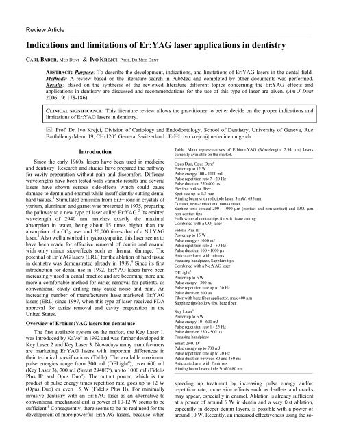
their technical specifications (Table). The available maximum<br />
pulse energies range from 300 mJ (DELight d ), over 600 mJ<br />
(Key Laser 3), 700 mJ (Smart 2940D e ), up to 1000 mJ (Fidelis<br />
Plus II c <strong>and</strong> Opus Duo b ). The output power, which is the<br />
product <strong>of</strong> pulse energy times repetition rate, goes up to 12 W<br />
(Opus Duo) or even 15 W (Fidelis Plus II). For m<strong>in</strong>imally<br />
<strong>in</strong>vasive <strong>dentistry</strong> with an <strong>Er</strong>:<strong>YAG</strong> <strong>laser</strong> as an alternative to<br />
conventional mechanical drill a power <strong>of</strong> 10-12 W seems to be<br />
sufficient. 5 Consequently, there seems to be no real need for the<br />
development <strong>of</strong> more powerful <strong>Er</strong>:<strong>YAG</strong> <strong>laser</strong>s, because when<br />
Table. Ma<strong>in</strong> representatives <strong>of</strong> <strong>Er</strong>bium:<strong>YAG</strong> (Wavelength: 2.94 µm) <strong>laser</strong>s<br />
currently available on the market.<br />
________________________________________________________________________________________________________<br />
Opus Duo, Opus Dent b<br />
Power up to 12 W<br />
Pulse energy 100 - 1000 mJ<br />
Pulse repetition rate 7 - 20 Hz<br />
Pulse duration 250-400 µs<br />
Flexible hollow fiber<br />
Spot size up to 1.3 mm<br />
Aim<strong>in</strong>g beam with red diode <strong>laser</strong>, 3 mW, 635 nm<br />
Contact, near-contact <strong>and</strong> non-contact<br />
Saphire tips: conical 200 - 1000 µm (contact <strong>and</strong> non-contact) <strong>and</strong> 1300 µm<br />
non-contact tips<br />
Hollow metal contact tips for s<strong>of</strong>t tissue cutt<strong>in</strong>g<br />
Comb<strong>in</strong>ed with a CO2 <strong>laser</strong><br />
Fidelis Plus II c<br />
Power up to 15 W<br />
Pulse energy - 1000 mJ<br />
Pulse repetition rate 2 - 50 Hz<br />
Pulse duration 100 - 1000 µs<br />
Articulated arm with mirrors<br />
Focus<strong>in</strong>g h<strong>and</strong>piece, Sapphire tips<br />
Comb<strong>in</strong>ed with a Nd:<strong>YAG</strong> <strong>laser</strong><br />
DELight d<br />
Power up to 6 W<br />
Pulse energy - 300 mJ<br />
Pulse repetition rate up to 30 Hz<br />
Pulse duration 200 µs<br />
Fiber with bare fiber applicator, max 400 µm<br />
Sapphire tips/hollow tips, bare fiber<br />
Key Laser a<br />
Power up to 6 W<br />
Pulse energy 10 - 600 mJ<br />
Pulse repetition rate 1 - 25 Hz<br />
Pulse duration 250 - 500 µs<br />
Focus<strong>in</strong>g h<strong>and</strong>piece<br />
Smart 2940 D e<br />
Pulse energy up to 700 mJ<br />
Pulse repetition rate up to 20 Hz<br />
Pulse duration between 80 <strong>and</strong> 450 ms<br />
Articulated arm with 7 mirrors<br />
Aim<strong>in</strong>g beam <strong>laser</strong> diode 5mW 680 nm<br />
________________________________________________________________________________________________________<br />
speed<strong>in</strong>g up treatment by <strong>in</strong>creas<strong>in</strong>g pulse energy <strong>and</strong>/or<br />
repetition rate, more side effects such as leaflets <strong>and</strong> cracks<br />
may appear, especially <strong>in</strong> enamel. Ablation is already sufficient<br />
at a power <strong>of</strong> around 6 W <strong>in</strong> dent<strong>in</strong> <strong>and</strong> a very fast ablation,<br />
especially <strong>in</strong> deeper dent<strong>in</strong> layers, is possible with a power <strong>of</strong><br />
around 10 W. Recently, an <strong>in</strong>creased effectiveness us<strong>in</strong>g the so-
American Journal <strong>of</strong> Dentistry, Vol. 19, No. 3, June, 2006<br />
called very short pulse (VSP) is discussed, pretend<strong>in</strong>g that the<br />
typical debris cloud formation above the ablated surface<br />
negatively <strong>in</strong>fluences ablation speed by partially absorb<strong>in</strong>g<br />
energy <strong>of</strong> the follow<strong>in</strong>g <strong>laser</strong> pulses. Accord<strong>in</strong>g to Lukac et al, 6<br />
the most effective ablation is reached with pulse lengths less<br />
than 100 µsec, but this study used vertical irradiation <strong>of</strong> the<br />
tooth surface, which did not allow a flush <strong>of</strong> the debris cloud by<br />
the spray device.<br />
The <strong>laser</strong> beam is transferred to the operation field by<br />
water-free glass fibers (KaVo, DELight), articulated arms with<br />
mirrors (Smart 2940D, Fidelis Plus II) or flexible hollow fibers<br />
(Opus Duo). Although a glass fiber delivery system is very easy<br />
to h<strong>and</strong>le, there is a limitation <strong>in</strong> maximal power transmission<br />
at about 6 W. Therefore, more powerful <strong>Er</strong>:<strong>YAG</strong> <strong>laser</strong>s need a<br />
hollow transmission system for their light. Among them, the<br />
flexible hollow fiber from Opus Duo is more suitable for the<br />
daily use because <strong>of</strong> its better h<strong>and</strong>l<strong>in</strong>g than the articulated arms<br />
used for example by Fidelis Plus II or Smart 2940D. With the<br />
exception <strong>of</strong> Opus Duo, all systems deliver a focused beam <strong>and</strong><br />
this specification may be <strong>of</strong> much importance for an even distribution<br />
<strong>of</strong> the power density on the work<strong>in</strong>g surface, especially<br />
dur<strong>in</strong>g smooth<strong>in</strong>g <strong>and</strong> condition<strong>in</strong>g <strong>of</strong> the enamel<br />
structure that is superficially destroyed dur<strong>in</strong>g cavity access <strong>and</strong><br />
preparation with high energy densities. The spatial beam pr<strong>of</strong>ile<br />
after transmission through the flexible hollow fiber has not<br />
been reported <strong>in</strong> the literature. A water-free glass fiber has a<br />
quasi-Gaussian shape, whereas articulated arms achieve a<br />
distribution <strong>of</strong> even higher orders with an important maximum<br />
around the central peak, or a r<strong>in</strong>g-shaped <strong>in</strong>tensity distribution. 7<br />
However, even if different beam transfer technologies may<br />
have different advantages <strong>and</strong> disadvantages, it is impossible to<br />
compare different <strong>laser</strong> systems based on this property or on<br />
their parameter sett<strong>in</strong>gs. Many factors, such as pulse formation,<br />
pulse width, beam pr<strong>of</strong>ile <strong>and</strong> others have to be brought <strong>in</strong><br />
relation to each other to allow an accurate comparison <strong>of</strong> their<br />
cl<strong>in</strong>ical efficiency. 7<br />
Most manufacturers propose sapphire contact tips for tooth<br />
preparation, with similarly look<strong>in</strong>g h<strong>and</strong>pieces. The range <strong>of</strong> tip<br />
diameters goes from 400-700 µm (DELight) up to 200 µm-<br />
1300 µm proposed by Opus Duo. A non-contact focus<strong>in</strong>g h<strong>and</strong>piece<br />
used for hard tissue preparation by the Key Laser 3 (the<br />
sapphire tips <strong>of</strong> Key Laser 3 are exclusively designed for periodontal<br />
<strong>applications</strong>) is also proposed by some other manufacturers,<br />
but a precise work<strong>in</strong>g, especially <strong>in</strong> the means <strong>of</strong> m<strong>in</strong>imally<br />
<strong>in</strong>vasive <strong>dentistry</strong> where aim<strong>in</strong>g accurately with the beam<br />
is <strong>of</strong> high importance, is very difficult. As a complementary<br />
tool to the <strong>Er</strong>:<strong>YAG</strong> <strong>laser</strong>, two manufacturers have <strong>in</strong>cluded <strong>in</strong><br />
their system a second <strong>laser</strong> emitt<strong>in</strong>g another wavelength, as<br />
Nd:<strong>YAG</strong> (Fotona) or CO2 (Opus Duo). The Nd:<strong>YAG</strong> may <strong>of</strong>fer<br />
wider <strong>in</strong>dications <strong>in</strong> endodontology, whereas <strong>Er</strong>:<strong>YAG</strong> <strong>in</strong> comb<strong>in</strong>ation<br />
with CO2 may allow a complete coverage <strong>of</strong> almost all<br />
dental <strong>in</strong>dications assisted by a <strong>laser</strong>, except bleach<strong>in</strong>g.<br />
Ablation mechanism <strong>and</strong> ablation speed<br />
Due to its wavelength <strong>of</strong> 2.94 µm, which matches exactly<br />
the absorption peak <strong>of</strong> water <strong>and</strong> which is also absorbed by<br />
hydroxylapatite, erbium <strong>laser</strong> radiation is very efficient <strong>in</strong><br />
remov<strong>in</strong>g both dent<strong>in</strong> <strong>and</strong> enamel, limit<strong>in</strong>g the <strong>laser</strong> effect on<br />
these tissues to a superficial layer <strong>of</strong> a few micrometers. This<br />
superficial layer can rapidly be heated up so that the pressure<br />
<strong>Er</strong>:<strong>YAG</strong> <strong>laser</strong> <strong>applications</strong> <strong>in</strong> <strong>dentistry</strong> 179<br />
with<strong>in</strong> the irradiated volume <strong>in</strong>creases until the material's<br />
strength is surpassed. The overheated water abruptly vaporizes<br />
<strong>and</strong> the so released vapor carries away surround<strong>in</strong>g broken<br />
tissue fragments <strong>in</strong> a thermomechanical ablation process. Increas<strong>in</strong>g<br />
the power, especially when q-switch<strong>in</strong>g the <strong>laser</strong>,<br />
accelerates the ablation process, decreas<strong>in</strong>g simultaneously the<br />
thermal side effects but result<strong>in</strong>g <strong>in</strong> higher mechanical side<br />
effects. 5,8 Efficient removal <strong>of</strong> dent<strong>in</strong> <strong>and</strong> enamel by us<strong>in</strong>g<br />
<strong>Er</strong>:<strong>YAG</strong> <strong>laser</strong>s could be demonstrated from the very beg<strong>in</strong>n<strong>in</strong>g<br />
<strong>in</strong> 1989, while spar<strong>in</strong>g the surround<strong>in</strong>g tissue. 4 The shorter the<br />
pulse length, the lower the energy density needed for ablation.<br />
The ablation threshold <strong>of</strong> <strong>Er</strong>:<strong>YAG</strong> <strong>laser</strong>s ranges between 6<br />
J/cm 2 for 100 µsec pulses <strong>and</strong> 10 J/cm 2 for 700 µsec pulses. 9<br />
This means that the <strong>Er</strong>:<strong>YAG</strong> <strong>laser</strong> is the most efficient <strong>of</strong> all<br />
known systems for hard dental tissue removal. The <strong>Er</strong>:Cr:<br />
YSGG <strong>laser</strong>, for example, needs more energy density, ablation<br />
start<strong>in</strong>g at 10 to 14 J/cm.<br />
2 10<br />
Carious dent<strong>in</strong> is removed at the same speed with <strong>Er</strong>:<strong>YAG</strong><br />
<strong>laser</strong>s as with the classical bur method. 11 When associated to<br />
Carisolv f pre-treatment, the ablation speed <strong>in</strong> carious dent<strong>in</strong> is<br />
higher than with the <strong>laser</strong> only. 12<br />
To <strong>in</strong>crease ablation speed <strong>in</strong> enamel, a <strong>laser</strong>-abrasive<br />
method us<strong>in</strong>g sapphire powder <strong>in</strong> the water spray, accelerated<br />
by <strong>laser</strong> irradiation, was <strong>in</strong>vestigated. The aqueous suspension<br />
<strong>of</strong> sapphire particles <strong>in</strong>creased three times the efficiency <strong>of</strong> enamel<br />
removal when compared to <strong>Er</strong>:<strong>YAG</strong> with water spray<br />
alone, approach<strong>in</strong>g those <strong>of</strong> a high-speed turb<strong>in</strong>e. 13 However, as<br />
the spread<strong>in</strong>g <strong>of</strong> the sapphire particles may cause side effects to<br />
the surround<strong>in</strong>g tissues, this method may not be further pursued.<br />
In general, there is a l<strong>in</strong>ear relationship between crater<br />
depth or removed volume <strong>and</strong> applied energy density. 14 Water<br />
mist is needed to avoid thermal side effects <strong>and</strong> for pa<strong>in</strong><br />
control. However, it only has a m<strong>in</strong>imal effect on the ablation<br />
speed up to an energy <strong>of</strong> approximately 400 mJ. 15 If us<strong>in</strong>g energies<br />
<strong>of</strong> 400 mJ <strong>and</strong> above, <strong>in</strong>creased water flow <strong>in</strong>creases<br />
ablation efficiency <strong>in</strong> enamel. In dent<strong>in</strong>, no significant difference<br />
was observed with a higher water flow rate at higher<br />
energies due to dent<strong>in</strong>’s higher water content compared to<br />
enamel. 16<br />
When surrounded by enamel, certa<strong>in</strong> selectivity for the<br />
ablation <strong>of</strong> composites was shown, as enamel ablation is slower<br />
than ablation <strong>of</strong> composites. However, this selectivity is compromised<br />
<strong>in</strong> dent<strong>in</strong> because <strong>of</strong> a higher ablation rate <strong>of</strong> dent<strong>in</strong><br />
compared to some composite br<strong>and</strong>s, due to the higher water<br />
content <strong>of</strong> dent<strong>in</strong>. 17 No quantitative <strong>in</strong>formation is available on<br />
the ablation rate <strong>of</strong> dental ceramics, glass-ionomer cements <strong>and</strong><br />
gold. Crater formation was reported after the application <strong>of</strong><br />
<strong>Er</strong>:<strong>YAG</strong> <strong>laser</strong> beam on amalgam surfaces associated with a<br />
substantially <strong>in</strong>creased release <strong>of</strong> Hg vapor. 18<br />
Morphological changes<br />
Cavity walls <strong>and</strong> borders disclose typical morphological<br />
aspects after ablative <strong>Er</strong>:<strong>YAG</strong> <strong>laser</strong> treatment. Only m<strong>in</strong>imal, if<br />
any, damage <strong>of</strong> surround<strong>in</strong>g dental hard tissues can be detected<br />
by use <strong>of</strong> optical <strong>and</strong> SEM microscopes. 19 The smear layer is<br />
efficiently removed. In fact, <strong>in</strong> comparison to Nd:<strong>YAG</strong> or<br />
argon <strong>laser</strong>s, <strong>Er</strong>:<strong>YAG</strong> is the most effective for smear layer<br />
removal. 20 Neither Knoop hardness nor Ca/P ratio evaluations<br />
on the cavity floor revealed any significant difference between<br />
<strong>laser</strong> <strong>and</strong> bur treatment. If any, only m<strong>in</strong>imal thermally <strong>in</strong>duced
180 Bader & Krejci<br />
changes <strong>of</strong> dental hard tissue composition is produced by<br />
<strong>Er</strong>:<strong>YAG</strong> 21 <strong>and</strong> only m<strong>in</strong>imal local thermal damage follows<br />
<strong>Er</strong>:<strong>YAG</strong> irradiation. 22 A difference is seen <strong>in</strong> the basophilic<br />
layer which is deeply sta<strong>in</strong>ed on <strong>Er</strong>:<strong>YAG</strong> treated sites compared<br />
to bur-treated dent<strong>in</strong>. Less odontoblastic processes<br />
rema<strong>in</strong> after <strong>Er</strong>:<strong>YAG</strong> treatment, related to a probable denaturization<br />
<strong>of</strong> the dent<strong>in</strong> organic matrix. 23<br />
By compar<strong>in</strong>g pulse duration times <strong>of</strong> 100 to 1000 µsec<br />
us<strong>in</strong>g the same energy, different results were found for<br />
chemical <strong>and</strong> structural modifications <strong>of</strong> dent<strong>in</strong>. Treatments<br />
with very long pulses <strong>of</strong> up to 1000 µsec resulted <strong>in</strong> a dent<strong>in</strong><br />
surface with chemical <strong>and</strong> morphological characteristics very<br />
similar to that obta<strong>in</strong>ed with conventional methods; while with<br />
very short <strong>laser</strong> pulses (VSP), a strong modification <strong>of</strong> collagen<br />
aliphatic cha<strong>in</strong>s was observed. 24 Affect<strong>in</strong>g the surface morphology<br />
<strong>and</strong> the chemistry <strong>of</strong> dent<strong>in</strong> may <strong>in</strong>fluence the bond<br />
strength to dental restorative materials <strong>and</strong> may necessitate the<br />
development <strong>of</strong> specific dent<strong>in</strong> adhesive systems for VSP <strong>laser</strong>treated<br />
surfaces. 24 At the present time, no <strong>in</strong>formation is<br />
available on subsurface damage <strong>in</strong> enamel after the application<br />
<strong>of</strong> VSP.<br />
Enamel acid etch<strong>in</strong>g after <strong>laser</strong> treatment <strong>in</strong>creases the<br />
etch<strong>in</strong>g depth if evaluated with the help <strong>of</strong> X-ray tomography. 25<br />
If the <strong>Er</strong>:<strong>YAG</strong> was water cooled, occlusal enamel fissures were<br />
debris-free <strong>and</strong> etch<strong>in</strong>g-like patterns were detected. On the<br />
other h<strong>and</strong>, when only air cool<strong>in</strong>g was used <strong>and</strong> the enamel was<br />
treated <strong>in</strong> contact, melt<strong>in</strong>g <strong>and</strong> re-crystallization <strong>of</strong> enamel<br />
fissures occurred. 26<br />
Pulp response<br />
The thermal danger <strong>of</strong> any new cavity preparation procedure<br />
has to be <strong>in</strong>vestigated, as classical techniques l<strong>in</strong>e up with<br />
a high level <strong>of</strong> security <strong>in</strong> their application. Overheat<strong>in</strong>g <strong>of</strong><br />
teeth, especially pulp damage <strong>and</strong> <strong>in</strong>flammatory response <strong>of</strong> the<br />
pulpal tissue dur<strong>in</strong>g or after <strong>laser</strong>-treatment must be avoided.<br />
Very low to slight temperature rise <strong>in</strong> the pulpal chamber<br />
has been reported, rang<strong>in</strong>g from an <strong>in</strong>itial decrease due to the<br />
water-spray cool<strong>in</strong>g 27 to a rise <strong>of</strong> 3°C. 28,29 Highest temperature<br />
<strong>in</strong>creases <strong>in</strong> the pulpal chamber were measured at a maximum<br />
<strong>of</strong> 4°C 30-32 <strong>and</strong> 5°C, 33 under different treatment parameters. An<br />
important <strong>in</strong>crease <strong>in</strong> temperature was only recorded <strong>in</strong> case<br />
when the <strong>laser</strong> beam hit directly the measur<strong>in</strong>g probe after pulp<br />
exposure. 27 A difference between occlusal <strong>and</strong> cervical cavity<br />
preparation was found: the highest values were found dur<strong>in</strong>g<br />
Class I preparations, followed by Class V <strong>in</strong> enamel. The<br />
lowest temperature <strong>in</strong>crease occurred dur<strong>in</strong>g caries removal or<br />
preparation <strong>in</strong> cementum. 33<br />
No significant difference to classical preparation methods <strong>in</strong><br />
respect to <strong>in</strong>flammatory reaction <strong>of</strong> the pulp was found as<br />
odontoblasts rema<strong>in</strong>ed <strong>of</strong> sp<strong>in</strong>dle-like or star-like shapes 34 <strong>and</strong><br />
immuno-histochemical analyses demonstrated similar effects to<br />
those after conventional bur methods. 20 The pulpal tissue<br />
directly exposed to <strong>laser</strong> treatment displayed no bleed<strong>in</strong>g but<br />
some blood extravasations were found near the exposure site.<br />
After direct pulp exposure with 34 mJ/pulse, no <strong>in</strong>flammation<br />
or resorption was found <strong>and</strong> a potential for pulpotomy with the<br />
<strong>Er</strong>:<strong>YAG</strong> <strong>laser</strong> was claimed. 35 However, a good heal<strong>in</strong>g capacity<br />
<strong>of</strong> <strong>laser</strong> exposed pulp tissue was demonstrated, with formation<br />
<strong>of</strong> dent<strong>in</strong> bridges <strong>and</strong> reparative dent<strong>in</strong>. 36 Under long term <strong>in</strong><br />
vivo observation, dist<strong>in</strong>ct tertiary dent<strong>in</strong> apposition, with cu-<br />
American Journal <strong>of</strong> Dentistry, Vol. 19, No. 3, June, 2006<br />
boidal cells on its pulpal aspect were found. 37 Both sufficient<br />
wetness <strong>of</strong> the treated tissue <strong>and</strong> appropriate water-spray<br />
cool<strong>in</strong>g <strong>and</strong> tissue re-hydration seem to be important parameters<br />
to avoid symptoms <strong>of</strong> pulpal damage. 29<br />
Desensitiz<strong>in</strong>g effect<br />
Cervically exposed hypersensitive dent<strong>in</strong> reportedly reacts<br />
positively to the application <strong>of</strong> many different k<strong>in</strong>ds <strong>of</strong> desensitiz<strong>in</strong>g<br />
liquids. 38 The difficulty rema<strong>in</strong>s <strong>in</strong> the ma<strong>in</strong>tenance <strong>of</strong><br />
the positive effect on even short or mid-term time periods.<br />
When applied with appropriate parameters, <strong>Er</strong>:<strong>YAG</strong> <strong>laser</strong> light<br />
seems to become an alternative for desensitization <strong>of</strong> hypersensitive<br />
cervical dent<strong>in</strong>. Applied with subablative 80 mJ/pulse<br />
at 3 Hz, the discomfort immediately improves, <strong>and</strong> rema<strong>in</strong>s<br />
even after a 6-month period at the same level, whereas conventional<br />
methods resulted <strong>in</strong> a gradual return to the orig<strong>in</strong>al level<br />
<strong>of</strong> discomfort. 39<br />
Caries prevention<br />
Controversial results can be found <strong>in</strong> the literature regard<strong>in</strong>g<br />
dem<strong>in</strong>eralization <strong>and</strong> acid-resistance <strong>of</strong> enamel <strong>and</strong> dent<strong>in</strong> after<br />
<strong>Er</strong>:<strong>YAG</strong> <strong>laser</strong> treatment. If after subablative <strong>Er</strong>:<strong>YAG</strong> irradiation<br />
a decl<strong>in</strong>e <strong>of</strong> 20% <strong>in</strong> calcium solubility <strong>in</strong> enamel was<br />
found, the effect was not judged sufficient to prevent caries. 40<br />
In addition, subablative <strong>Er</strong>:<strong>YAG</strong> radiation seemed to produce<br />
f<strong>in</strong>e cracks <strong>in</strong> the enamel surface. 41 If us<strong>in</strong>g ablative <strong>laser</strong><br />
energies <strong>of</strong> 400 mJ, lowest acid dem<strong>in</strong>eralization <strong>in</strong> enamel <strong>and</strong><br />
dent<strong>in</strong> was found after dry <strong>laser</strong> treatment. However, on the<br />
micromorphological level, this treatment method <strong>in</strong>duced<br />
thermal damages. 42 Higher dem<strong>in</strong>eralization to a depth <strong>of</strong> 133.9<br />
µm was found at restoration marg<strong>in</strong>s <strong>in</strong> enamel, when lased<br />
samples were subjected to a pH-cycl<strong>in</strong>g model, compared to<br />
unlased samples with a dem<strong>in</strong>eralization depth <strong>of</strong> 77.4 µm. 43 In<br />
a model us<strong>in</strong>g lactic-buffer solution, dissolved calcium <strong>and</strong><br />
phosphate <strong>and</strong> their Ca/P ratio was not different <strong>in</strong> lased <strong>and</strong><br />
unlased samples on bov<strong>in</strong>e dent<strong>in</strong>, which suggests that <strong>Er</strong>:<strong>YAG</strong><br />
<strong>laser</strong> irradiation does not <strong>in</strong>crease nor decrease any acid<br />
resistance <strong>of</strong> dent<strong>in</strong>. 44 In an <strong>in</strong> vivo pilot study, the caries<br />
resistance follow<strong>in</strong>g subablative erbium <strong>laser</strong> irradiation was<br />
determ<strong>in</strong>ed by analyz<strong>in</strong>g the dem<strong>in</strong>eralization before <strong>and</strong> after<br />
wear<strong>in</strong>g for 1 week <strong>in</strong> situ (<strong>in</strong> the volunteers' mouths) treated<br />
<strong>and</strong> untreated enamel samples. Whenever a tendency towards<br />
<strong>in</strong>creased caries resistance was described, it failed to reach<br />
statistical significance. 45<br />
On the enamel surface, <strong>Er</strong>:<strong>YAG</strong> <strong>laser</strong> treatment comb<strong>in</strong>ed<br />
with APF (acidulated phosphate fluoride) resulted <strong>in</strong> the lowest<br />
decrease <strong>of</strong> surface microhardness <strong>and</strong> the <strong>Er</strong>:<strong>YAG</strong> <strong>laser</strong><br />
<strong>in</strong>fluenced the deposition <strong>of</strong> CaF 2 on the enamel. 46 If a superficial<br />
anti-cariogenic action can be <strong>in</strong>duced, it is not possible <strong>in</strong><br />
depth. 46 A <strong>laser</strong>-<strong>in</strong>duced caries preventive effect is substantiated<br />
accord<strong>in</strong>g to the “organic matrix block<strong>in</strong>g theory”, whereas<br />
<strong>laser</strong> treated enamel confirms <strong>laser</strong>-<strong>in</strong>duced block<strong>in</strong>g <strong>of</strong> the<br />
organic matrix <strong>in</strong> the micro-diffusion pathway <strong>in</strong> enamel. 47 In<br />
an artificial caries model, a significant reduction <strong>of</strong> secondary<br />
caries formation was demonstrated, with an important reduction<br />
<strong>of</strong> 56% <strong>of</strong> primary enamel surface lesion depth <strong>and</strong> a 39%<br />
reduction <strong>of</strong> root surface lesion depth, compared to classical bur<br />
<strong>and</strong> acid etch technique. 48<br />
Bactericidal effect<br />
Very early <strong>in</strong> <strong>laser</strong>-therapy, the bactericidal effect <strong>of</strong> <strong>laser</strong>
American Journal <strong>of</strong> Dentistry, Vol. 19, No. 3, June, 2006<br />
light was advanced to be one <strong>of</strong> the beneficial side effects<br />
associated with this k<strong>in</strong>d <strong>of</strong> treatment. It is especially <strong>in</strong>terest<strong>in</strong>g<br />
to mention that wavelengths well absorbed <strong>in</strong> water have<br />
a good bactericidal effect even at low energy density output<br />
levels, start<strong>in</strong>g at 0.3 J/cm 2 , without excessive temperature<br />
elevation. 49 This may be one <strong>of</strong> the reasons why <strong>Er</strong>:<strong>YAG</strong> <strong>laser</strong><br />
seems to be an efficient alternative for non-surgical periodontal<br />
treatment: <strong>Er</strong>:<strong>YAG</strong> <strong>laser</strong> treatment significantly reduces<br />
prob<strong>in</strong>g depth (PD) <strong>and</strong> bleed<strong>in</strong>g on prob<strong>in</strong>g (BOP) <strong>and</strong><br />
improves cl<strong>in</strong>ical attachment level (CAL) compared to the classical<br />
treatment strategy with scal<strong>in</strong>g <strong>and</strong> root plann<strong>in</strong>g. 50 Even a<br />
decrease <strong>of</strong> endotox<strong>in</strong>s <strong>and</strong> lipopolysaccharides on root surfaces<br />
were observed, with a reduction rang<strong>in</strong>g from 61% up to<br />
93%, with the effect start<strong>in</strong>g already at subablative energies <strong>of</strong><br />
60 mJ. Similar to these results on dental root surfaces, a bacterial<br />
reduction on implant surfaces can be reached up to 99.51%<br />
with 60 mJ <strong>and</strong> 99.94% with 120 mJ, without excessive<br />
temperature elevation <strong>and</strong> without morphological changes <strong>of</strong><br />
the implant surfaces. 51<br />
In endodontics, a mean bacterial reduction exceed<strong>in</strong>g 99%<br />
was observed, similar to the one after Nd:<strong>YAG</strong> <strong>and</strong> Ho:<strong>YAG</strong><br />
<strong>laser</strong> treatments. 52-54 Due to its complete absorption <strong>in</strong> water<br />
<strong>and</strong> dent<strong>in</strong>, <strong>Er</strong>:<strong>YAG</strong> acts on the surface <strong>of</strong> canal walls only. 55<br />
This effect avoids uncontrolled light penetration <strong>in</strong>to the<br />
surround<strong>in</strong>g tissues that may for example be observed with<br />
diode, Nd:<strong>YAG</strong> <strong>and</strong> Ho:<strong>YAG</strong> <strong>laser</strong>s <strong>and</strong> makes the <strong>laser</strong> very<br />
efficient. 56 The disadvantage <strong>of</strong> this effect is that if the <strong>Er</strong>:<strong>YAG</strong><br />
cannot reach to the work<strong>in</strong>g length <strong>and</strong> is for example 3 mm<br />
short, 70% <strong>of</strong> the root canal specimens irradiated rema<strong>in</strong><br />
<strong>in</strong>fected. 57<br />
A dependency <strong>of</strong> applied power specific for the different<br />
bacteria species was demonstrated. 58 Though therapeutic,<br />
subablative <strong>laser</strong> light doses can lead to one-step dis<strong>in</strong>fection<br />
<strong>in</strong>clud<strong>in</strong>g anaerobic micro organisms. 52<br />
Pa<strong>in</strong> perception<br />
As <strong>Er</strong>:<strong>YAG</strong> <strong>laser</strong>s can be used to prepare cavities without<br />
thermal damage <strong>and</strong> the systems available on the market <strong>of</strong>fer a<br />
high ablation efficiency, it was <strong>of</strong> <strong>in</strong>terest to <strong>in</strong>vestigate the<br />
patients’ subjective perception <strong>of</strong> this treatment method: cavity<br />
preparation with the help <strong>of</strong> <strong>Er</strong>:<strong>YAG</strong> <strong>laser</strong> was found to be<br />
more comfortable <strong>in</strong> the patients perception than mechanical<br />
treatment, <strong>in</strong> at least 80% <strong>of</strong> the cases. 59,60 Only <strong>in</strong> exceptional<br />
cases local anesthesia was needed for cavity preparation <strong>and</strong><br />
this was always limited to patients who compla<strong>in</strong>ed <strong>of</strong> cervical<br />
dent<strong>in</strong> hypersensitivity before treatment. No or little pa<strong>in</strong><br />
response, which was reported as a feel<strong>in</strong>g <strong>of</strong> a brief pressure to<br />
the tooth, was felt <strong>in</strong> 93% <strong>of</strong> the <strong>laser</strong>-treated teeth. 61<br />
One <strong>of</strong> the parameters partly expla<strong>in</strong><strong>in</strong>g the absence <strong>of</strong> pa<strong>in</strong><br />
perception is the difference <strong>in</strong> tooth vibration speed caused by<br />
<strong>Er</strong>:<strong>YAG</strong> <strong>laser</strong> versus the high-speed drill. Mean vibration speed<br />
dur<strong>in</strong>g <strong>laser</strong> cavity preparation reaches 166 +/- 28 µm/second,<br />
at a characteristic frequency <strong>of</strong> 230 Hz, whereas the high-speed<br />
drill <strong>in</strong>duces a 100 times higher vibration speed <strong>of</strong> 65 +/- 48<br />
mm/second, at 5 kHz. In addition, this much higher frequency<br />
has its spectrum near the peak sensitivity <strong>of</strong> hear<strong>in</strong>g, as a<br />
potential factor <strong>of</strong> discomfort <strong>and</strong> pa<strong>in</strong> provocation. 62<br />
Another explanation for mechanisms <strong>of</strong> pa<strong>in</strong> reduction <strong>in</strong><br />
<strong>Er</strong>:<strong>YAG</strong> cavity ablation might be the disruption <strong>of</strong> nerve<br />
term<strong>in</strong>als <strong>in</strong> the dent<strong>in</strong> tubules, comb<strong>in</strong>ed with a degeneration<br />
<strong>Er</strong>:<strong>YAG</strong> <strong>laser</strong> <strong>applications</strong> <strong>in</strong> <strong>dentistry</strong> 181<br />
<strong>of</strong> nerve term<strong>in</strong>als between the odontoblasts <strong>and</strong> the disruption<br />
<strong>of</strong> the myel<strong>in</strong> sheath <strong>in</strong> the pulp core, which were demonstrated<br />
by us<strong>in</strong>g transmission electron microscopy. 63<br />
Tensile bond strength (TBS)<br />
Contradictory results <strong>and</strong> conclusions may be found on<br />
tensile bond strengths after <strong>laser</strong> treatment <strong>in</strong> the literature, may<br />
be because <strong>of</strong> the fact that many different experimental setups<br />
have been used.<br />
If no difference was found between <strong>Er</strong>:<strong>YAG</strong> lased or<br />
turb<strong>in</strong>e drilled dent<strong>in</strong>, 64 best results were found with a selfetch<strong>in</strong>g<br />
primer (Clearfil L<strong>in</strong>er Bond 2V g ) regardless the surface<br />
treatment. 65 The effect <strong>of</strong> acid conditioners on res<strong>in</strong> bond<strong>in</strong>g to<br />
dent<strong>in</strong> differed accord<strong>in</strong>g to whether the dent<strong>in</strong> had been <strong>laser</strong><br />
irradiated or not 66 <strong>and</strong> for Optibond FL, h (etch & r<strong>in</strong>se) etch<strong>in</strong>g<br />
<strong>of</strong> the lased dent<strong>in</strong> surface was found to be m<strong>and</strong>atory. 67 TBS<br />
for s<strong>in</strong>gle bottle bond<strong>in</strong>g systems, such as Excite, i <strong>and</strong> Gluma<br />
One Bond, j were negatively affected by <strong>laser</strong> irradiation. 67<br />
When comb<strong>in</strong>ed with phosphoric acid <strong>and</strong> air powder treatment,<br />
better results were found than for <strong>Er</strong>:<strong>YAG</strong> alone, 68 but<br />
other authors found worse results <strong>in</strong> lased samples after citric<br />
acid <strong>and</strong> HEMA treatment compared to unlased samples. 69,70<br />
For Clearfil SE, g <strong>and</strong> Optibond FL with Z100 k composite, TBS<br />
was always lower for <strong>Er</strong>:<strong>YAG</strong> than <strong>in</strong> bur-treated samples. 67<br />
Contradictory results <strong>and</strong> conclusions were found for Bond 1, l<br />
with Alert, l where <strong>Er</strong>:<strong>YAG</strong> treatment preceed<strong>in</strong>g phosphoric<br />
acid treatment improved tensile bond strength compared to acid<br />
treatment alone. 71 In the same model, Optibond Solo, h with<br />
Prodigy, h <strong>and</strong> S<strong>in</strong>gle Bond, k with Z100, behaved worse,<br />
regardless if pre-lased or not. Other authors found <strong>in</strong>dications<br />
that <strong>laser</strong>-irradiated samples had improved bond strengths<br />
compared to acid-etched <strong>and</strong> h<strong>and</strong>piece drilled controls. Their<br />
conclusion was that preparation <strong>of</strong> dent<strong>in</strong> with <strong>Er</strong>:<strong>YAG</strong> treatment<br />
leaves a suitable surface for strong bond<strong>in</strong>g. 72 If TBS to<br />
superficial dent<strong>in</strong> is compared to deep dent<strong>in</strong> (at 2 mm distance<br />
to the dent<strong>in</strong>-enamel junction), results showed that it was<br />
m<strong>and</strong>atory <strong>in</strong> both cases to use a condition<strong>in</strong>g agent, such as a<br />
self-etch<strong>in</strong>g primer system, when <strong>Er</strong>:<strong>YAG</strong> <strong>laser</strong> was used. In<br />
deep dent<strong>in</strong>, best results were achieved with a comb<strong>in</strong>ation <strong>of</strong><br />
<strong>Er</strong>:<strong>YAG</strong> <strong>laser</strong> treatment <strong>and</strong> conditioner. 73<br />
Many authors found similar TBS after <strong>Er</strong>:<strong>YAG</strong>-only treatment<br />
compared to acid-etched samples. 74,75 If <strong>Er</strong>:<strong>YAG</strong> treatment<br />
was comb<strong>in</strong>ed with acid etch<strong>in</strong>g, higher bond strengths<br />
were found than with <strong>laser</strong> treatment alone. 76 After a complementary<br />
treatment with an ultrasonic scaler, TBS was doubled<br />
if compared to lased-only samples. 77 With self etch<strong>in</strong>g systems<br />
(Clearfil SE) or etch <strong>and</strong> r<strong>in</strong>se systems (Optibond FL), lower<br />
TBS were found than <strong>in</strong> diamond bur samples. 67 Some authors,<br />
us<strong>in</strong>g low energy levels (maximum 120 mJ), found higher TBS<br />
for orthodontic brackets <strong>in</strong> lased enamel samples, 78 while<br />
others, us<strong>in</strong>g energy <strong>of</strong> 200 mJ, found the opposite. 79 An<br />
<strong>in</strong>terest<strong>in</strong>g study compared different water cool<strong>in</strong>g flow rates<br />
dur<strong>in</strong>g <strong>laser</strong> treatment on dent<strong>in</strong> <strong>and</strong> enamel. If TBS on dent<strong>in</strong><br />
was not adversely affected by different water flow rates, it was<br />
<strong>of</strong> importance to optimize the water flow on enamel to prevent<br />
the formation <strong>of</strong> non-apatite CaP phases on the enamel surface,<br />
which may compromise adhesion. Relatively high TBS were<br />
realized without acid etch<strong>in</strong>g when a copious water flow was<br />
applied dur<strong>in</strong>g the <strong>Er</strong>:<strong>YAG</strong> <strong>laser</strong> treatment. 80<br />
For composite repairs, <strong>Er</strong>:<strong>YAG</strong> <strong>laser</strong> as the condition<strong>in</strong>g
182 Bader & Krejci<br />
method, showed a significant improvement <strong>in</strong> TBS <strong>in</strong> comparison<br />
to classical methods such as air-abrasion, silanization,<br />
hydr<strong>of</strong>luoric acid <strong>and</strong> their comb<strong>in</strong>ations, reach<strong>in</strong>g mean values<br />
<strong>of</strong> 22.92 MPa. 81<br />
Microleakage <strong>and</strong> marg<strong>in</strong>al adaptation<br />
As <strong>Er</strong>:<strong>YAG</strong> <strong>laser</strong>s work <strong>in</strong> a "mechanical" way, with microexplosions<br />
due to <strong>in</strong>stant vaporization <strong>of</strong> the water content<strong>in</strong>g<br />
tissues, it is not the same for the very fragile <strong>and</strong> brittle enamel<br />
structure if high or low energies are applied, comparable to<br />
drill<strong>in</strong>g with different diamond gra<strong>in</strong> sizes. 82 Most <strong>of</strong> the<br />
studies available on microleakage <strong>and</strong> marg<strong>in</strong>al adaptation used<br />
<strong>Er</strong>:<strong>YAG</strong> with high energies, over 300 mJ. These energies<br />
<strong>in</strong>duce subsurface damages <strong>in</strong>to enamel. It is thus not surpris<strong>in</strong>g<br />
that many publications reported poor marg<strong>in</strong>al adaptation with<br />
a high degree <strong>of</strong> microleakage 83-87 <strong>and</strong> that acid etch<strong>in</strong>g <strong>of</strong><br />
enamel follow<strong>in</strong>g <strong>Er</strong>:<strong>YAG</strong>, as a k<strong>in</strong>d <strong>of</strong> f<strong>in</strong>ish<strong>in</strong>g <strong>of</strong> enamel,<br />
gave much better results. 83-85,88 As soon as low energies were<br />
used for cavity preparation, microleakage <strong>of</strong> lased <strong>and</strong> burtreated<br />
cavities was not significantly different. 89-94 Some studies<br />
us<strong>in</strong>g dye penetration even presented less microleakage. 19,95<br />
The problem is that the preparation with low energies requires a<br />
very long treatment time, compromis<strong>in</strong>g the use <strong>of</strong> <strong>Er</strong>:<strong>YAG</strong><br />
<strong>laser</strong> <strong>in</strong> the rout<strong>in</strong>e cl<strong>in</strong>ical setup. It seems to be necessary, the<br />
same as after classical bur treatment, to smoothen the cavity<br />
surfaces <strong>and</strong> marg<strong>in</strong>s after the efficient cavity preparation us<strong>in</strong>g<br />
high energy sett<strong>in</strong>gs. Us<strong>in</strong>g ultrasonic scalers, 77 air-abrasion<br />
techniques 68 <strong>and</strong> <strong>laser</strong> f<strong>in</strong>ish<strong>in</strong>g, even when comb<strong>in</strong>ed with acid<br />
etch<strong>in</strong>g 83-85,88 have already been tried out as adequate f<strong>in</strong>ish<strong>in</strong>g<br />
methods to improve marg<strong>in</strong>al adaptation with less microleakage<br />
present.<br />
Primary teeth<br />
Compared to the smooth appearance <strong>of</strong> the cavity walls<br />
after bur preparation, cavity marg<strong>in</strong>s <strong>and</strong> walls are irregular but<br />
without any smear layer after ablative <strong>Er</strong>:<strong>YAG</strong> irradiation. 19<br />
Dye penetration <strong>in</strong> restorations where cavities were prepared by<br />
<strong>Er</strong>:<strong>YAG</strong> <strong>and</strong> filled with composite <strong>in</strong> primary teeth was less<br />
than by mechanical bur. 19 Other studies found no difference<br />
between microleakage <strong>of</strong> res<strong>in</strong> composite restorations after<br />
<strong>laser</strong> treatment only or after bur preparation <strong>and</strong> phosphoric<br />
acid etch<strong>in</strong>g. 96 In Class II restorations with composite or compomer,<br />
dent<strong>in</strong> bond<strong>in</strong>g rema<strong>in</strong>ed a problem with or without<br />
<strong>laser</strong> treatment, whereas <strong>in</strong> Class V compomer or composite<br />
restorations <strong>in</strong> primary teeth, good results with over 90% <strong>of</strong><br />
perfect marg<strong>in</strong>s were found after thermal cycl<strong>in</strong>g. 97<br />
Due to its bactericidal effect comb<strong>in</strong>ed with the reduced<br />
pa<strong>in</strong> sensation dur<strong>in</strong>g its application, the <strong>Er</strong>:<strong>YAG</strong> <strong>laser</strong> was a<br />
very promis<strong>in</strong>g tool for cavity preparation <strong>in</strong> primary teeth.<br />
However, detailed parameters <strong>and</strong> cl<strong>in</strong>ical treatment protocols<br />
have to be def<strong>in</strong>ed <strong>in</strong> the future.<br />
Pits <strong>and</strong> fissures<br />
The bactericidal effect <strong>of</strong> <strong>Er</strong>:<strong>YAG</strong> <strong>laser</strong> irradiation could<br />
boost the <strong>in</strong>terest <strong>in</strong> the already widely accepted pits <strong>and</strong> fissures<br />
seal<strong>in</strong>g procedures. A simultaneous clean<strong>in</strong>g, condition<strong>in</strong>g<br />
<strong>and</strong> decontam<strong>in</strong>ation <strong>in</strong> hardly accessible depths <strong>of</strong> fissures<br />
would open a new perspective to this preventive treatment.<br />
As the prismatic structure <strong>of</strong> enamel is very sensitive to<br />
mechanical stress, a precise range <strong>of</strong> correct condition<strong>in</strong>g<br />
parameters for pits <strong>and</strong> fissures by erbium <strong>laser</strong>s must be estab-<br />
American Journal <strong>of</strong> Dentistry, Vol. 19, No. 3, June, 2006<br />
lished, <strong>in</strong> order to allow an at least equal seal<strong>in</strong>g quality to<br />
conventional methods, with the advantage <strong>of</strong> clean<strong>in</strong>g <strong>and</strong><br />
decontam<strong>in</strong>ation <strong>in</strong> only one step <strong>and</strong> with a s<strong>in</strong>gle device only.<br />
Occlusal fissure seal<strong>in</strong>gs treated exclusively by <strong>Er</strong>:<strong>YAG</strong><br />
provided poor marg<strong>in</strong>al adaptation compared to acid-etched<br />
groups. <strong>Er</strong>:<strong>YAG</strong> pretreatment <strong>and</strong> subsequent acid etch<strong>in</strong>g with<br />
highly concentrated phosphoric acid was equivalent to etch<strong>in</strong>g<br />
only. 84 No significant difference <strong>in</strong> microleakage was reported<br />
between extended fissure seal<strong>in</strong>g with a bur <strong>and</strong> phosphoric<br />
acid-etch<strong>in</strong>g or <strong>Er</strong>:<strong>YAG</strong> <strong>and</strong> phosphoric acid-etch<strong>in</strong>g. Laser<br />
irradiation did not elim<strong>in</strong>ate the need for etch<strong>in</strong>g enamel as the<br />
<strong>laser</strong> only group showed the highest microleakage values. 98<br />
Most <strong>of</strong> the available studies used ablative parameters for<br />
enamel condition<strong>in</strong>g. A test<strong>in</strong>g <strong>of</strong> low energy levels for fissure<br />
decontam<strong>in</strong>ation exclusively by <strong>Er</strong>:<strong>YAG</strong>, at a maximum <strong>of</strong> 100<br />
mJ would be <strong>of</strong> <strong>in</strong>terest.<br />
Acid etch<strong>in</strong>g after <strong>laser</strong> treatment <strong>of</strong> the enamel marg<strong>in</strong>s<br />
<strong>in</strong>creased the etch<strong>in</strong>g depth under microtomography control. 25<br />
Fissures were debris-free <strong>and</strong> etch<strong>in</strong>g-like patterns were found<br />
<strong>in</strong> <strong>Er</strong>:<strong>YAG</strong> treated occlusal fissures when the tooth was water<br />
cooled dur<strong>in</strong>g <strong>laser</strong> application. When only air cooled <strong>and</strong><br />
treated <strong>in</strong> contact, melt<strong>in</strong>g <strong>and</strong> re-crystallization <strong>of</strong> fissure<br />
enamel occurred. 26<br />
Further <strong>in</strong>vestigations, us<strong>in</strong>g parameters prevent<strong>in</strong>g from<br />
scatter<strong>in</strong>g <strong>and</strong> leaflet-produc<strong>in</strong>g on the enamel surface, are<br />
needed for secure <strong>and</strong> predictable results.<br />
Endodontics<br />
A comparison <strong>of</strong> conventional root canal preparation with<br />
<strong>Er</strong>:<strong>YAG</strong> <strong>laser</strong> us<strong>in</strong>g 200 to 400 µm microprobes showed that <strong>in</strong><br />
straight root canals, enlarg<strong>in</strong>g, shap<strong>in</strong>g, <strong>and</strong> clean<strong>in</strong>g is faster<br />
<strong>and</strong> more efficient with the <strong>laser</strong> <strong>and</strong> that no residual pulp tissue<br />
was present after <strong>laser</strong> application. 99 Canal walls free <strong>of</strong> debris,<br />
evaporated smear layer <strong>and</strong> open dent<strong>in</strong> tubuli were reported<br />
after <strong>Er</strong>:<strong>YAG</strong> <strong>laser</strong> application, 100,101 under some specific<br />
conditions even near the apical orifice. 102 <strong>Er</strong>:<strong>YAG</strong> efficiently<br />
removed the smear layer <strong>in</strong> the root canal if water was used as<br />
the irrigat<strong>in</strong>g medium 103 <strong>and</strong> its bactericidal effect <strong>in</strong> root canals<br />
is well documented. 52-55,58 However, <strong>in</strong> spite <strong>of</strong> the absence <strong>of</strong><br />
smear layer, no significant difference <strong>in</strong> respect to leakage <strong>of</strong><br />
classical root canal obturations between <strong>laser</strong> treated <strong>and</strong><br />
conventionally treated root canals has been detected so far. 104<br />
Periodontology<br />
Based on its good absorption <strong>in</strong> water <strong>and</strong> <strong>in</strong> dental hard<br />
tissues, the <strong>Er</strong>:<strong>YAG</strong> <strong>laser</strong> suggested itself for evaluation <strong>in</strong> the<br />
field <strong>of</strong> periodontology. In comparison to ultrasonic scal<strong>in</strong>g, a<br />
similar removal <strong>of</strong> calculus can be obta<strong>in</strong>ed but superficial<br />
structural <strong>and</strong> thermal micro-changes <strong>in</strong> the form <strong>of</strong> microroughness<br />
were found on root cementum. 105 An important<br />
parameter to def<strong>in</strong>e was the threshold level for cementum<br />
ablation, be<strong>in</strong>g 10.6 J/cm 2 per pulse. 106 So it is not surpris<strong>in</strong>g<br />
that adversary results are obta<strong>in</strong>ed, e.g. <strong>in</strong> comparison to<br />
classical scal<strong>in</strong>g <strong>and</strong> root plan<strong>in</strong>g (SRP) <strong>in</strong> vivo. While the<br />
<strong>Er</strong>:<strong>YAG</strong> is capable <strong>of</strong> remov<strong>in</strong>g calculus, its effectiveness is<br />
lower than the SRP method, but without remov<strong>in</strong>g cementum,<br />
especially if an active selectivity feedback system is built <strong>in</strong>, as<br />
is for example the Key Laser 3. However, if <strong>Er</strong>:<strong>YAG</strong> was less<br />
<strong>in</strong>vasive than the conventional method, it needed twice the time<br />
<strong>of</strong> SRP. 107 Us<strong>in</strong>g low radiation energies, calculus removal can
American Journal <strong>of</strong> Dentistry, Vol. 19, No. 3, June, 2006<br />
be done with a certa<strong>in</strong> selectivity, comparable to that <strong>of</strong><br />
conventional root surface <strong>in</strong>strumentation. It is possible to<br />
remove calculus with a significant selectivity <strong>of</strong> more than 4.5<br />
times than for root surface material. 108 Compared to a treatment<br />
with a diode <strong>laser</strong>, which is not sensible for calculus removal<br />
<strong>and</strong> alters the root surface <strong>in</strong> an undesirable manner, <strong>Er</strong>:<strong>YAG</strong><br />
comb<strong>in</strong>ed with a calculus detection system can remove calculus<br />
on a level equivalent to SRP. 109<br />
After <strong>Er</strong>:<strong>YAG</strong> treatment <strong>of</strong> periodontal pockets <strong>in</strong> situ on<br />
corpses, thermal changes on root surfaces with ultra structural<br />
irregularities at the apical end <strong>of</strong> <strong>Er</strong>:<strong>YAG</strong> scal<strong>in</strong>g tracks were<br />
found, with energies rang<strong>in</strong>g from 60 to 180 mJ. 110 Copious<br />
water spray m<strong>in</strong>imized thermal effects <strong>and</strong> led to cleaner <strong>and</strong><br />
less porous surfaces. 111 Angulation had an important <strong>in</strong>fluence<br />
on the amount <strong>of</strong> root substance removal, reach<strong>in</strong>g from very<br />
slight at a tip angulation <strong>of</strong> 15°, to severe ablation <strong>of</strong> more than<br />
400% at (cl<strong>in</strong>ically impossible) angulation <strong>of</strong> 90°. 112<br />
Most probably as a result <strong>of</strong> the elim<strong>in</strong>ation <strong>of</strong> bacteria <strong>and</strong><br />
endotox<strong>in</strong>s on root surfaces, human g<strong>in</strong>gival fibroblasts adhere<br />
<strong>and</strong> grow significantly faster on a 60 mJ <strong>Er</strong>:<strong>YAG</strong> pretreated<br />
surface than after SRP. 113 After <strong>in</strong>cubation with human fibroblasts,<br />
cell count <strong>in</strong> the <strong>Er</strong>:<strong>YAG</strong> group was 1.5 times higher<br />
than with an ultrasonic treatment, 2.7 times higher than with<br />
SRP <strong>and</strong> 4.5 times that <strong>of</strong> the control group. 114<br />
Cl<strong>in</strong>ical parameters as plaque <strong>in</strong>dex (PI), g<strong>in</strong>gival <strong>in</strong>dex<br />
(GI), prob<strong>in</strong>g depth (PD), bleed<strong>in</strong>g on prob<strong>in</strong>g (BOP) <strong>and</strong><br />
cl<strong>in</strong>ical attachment level (CAL) improve more after <strong>Er</strong>:<strong>YAG</strong><br />
<strong>laser</strong> treatment than after SRP. It is also <strong>of</strong> <strong>in</strong>terest that <strong>Er</strong>:<strong>YAG</strong><br />
alone reached the same scores than comb<strong>in</strong>ed treatment us<strong>in</strong>g<br />
ERL <strong>and</strong> SRP, <strong>and</strong> that these two groups scored clearly better<br />
than the SRP method alone. 115 A follow-up study on periodontal<br />
conditions over 2 years showed better long term<br />
prognosis for <strong>Er</strong>:<strong>YAG</strong> treatment alone than for SRP. Results <strong>of</strong><br />
CAL improvement <strong>in</strong> comparison to the attachment level at the<br />
beg<strong>in</strong>n<strong>in</strong>g was 28.5% after 1 year for <strong>Er</strong>:<strong>YAG</strong> <strong>and</strong> 13.8% for<br />
the SRP group. After 2 years, still 22.2% improvement was<br />
found compared to 10.7% for the SRP group. Cl<strong>in</strong>ical<br />
attachment level improvement after <strong>Er</strong>:<strong>YAG</strong> <strong>laser</strong> treatment<br />
was twice the one <strong>of</strong> the classical approach. 116<br />
Bone tissue <strong>and</strong> implantology<br />
The <strong>Er</strong>:<strong>YAG</strong> <strong>laser</strong> is able to cut bone tissue. Compared to<br />
mechanical bur <strong>and</strong> CO2 <strong>laser</strong> groups, <strong>Er</strong>:<strong>YAG</strong> irradiated bone<br />
tissue showed a more pronounced <strong>in</strong>flammatory cell <strong>in</strong>filtration,<br />
fibroblastic reaction <strong>and</strong> a faster revascularization adjacent<br />
to the irradiated bone surface. In addition, a significantly<br />
greater <strong>and</strong> more rapid bone neo-formation was observed. 117<br />
Even after a long irradiation period <strong>of</strong> up to 120 seconds,<br />
temperature rise at an implant-bone <strong>in</strong>terface was low 118<br />
allow<strong>in</strong>g postulation that peri-implantitis therapy with <strong>Er</strong>:<strong>YAG</strong><br />
is cl<strong>in</strong>ically safe. After direct <strong>Er</strong>:<strong>YAG</strong> treatment <strong>and</strong> ablation <strong>of</strong><br />
bone, the chemical composition <strong>of</strong> the rema<strong>in</strong><strong>in</strong>g bone was<br />
similar to that follow<strong>in</strong>g bur drill<strong>in</strong>g. 119 It was also possible to<br />
create smear layer free grooves with well def<strong>in</strong>ed edges, represent<strong>in</strong>g<br />
an alternative method for safe oral <strong>and</strong> periodontal<br />
osseous surgery. 119 A layer <strong>of</strong> only 30 µm thickness presented a<br />
changed ultrastructure with microcrack<strong>in</strong>g, disorganization <strong>and</strong><br />
slight re-crystallization <strong>of</strong> the orig<strong>in</strong>al apatite <strong>and</strong> a reduction <strong>of</strong><br />
the surround<strong>in</strong>g organic matrix. 120 On the implant side, surface<br />
alterations such as partial melt<strong>in</strong>g, crack<strong>in</strong>g <strong>and</strong> crater forma-<br />
<strong>Er</strong>:<strong>YAG</strong> <strong>laser</strong> <strong>applications</strong> <strong>in</strong> <strong>dentistry</strong> 183<br />
tion became obvious under certa<strong>in</strong> conditions, but with cl<strong>in</strong>ically<br />
<strong>in</strong>adequate high power output <strong>and</strong> always less pronounced<br />
than after Nd:<strong>YAG</strong> or Ho:<strong>YAG</strong> irradiation. 121 As a consequence<br />
<strong>of</strong> these f<strong>in</strong>d<strong>in</strong>gs, power output must be limited to avoid<br />
surface damage, but already with low output energies under<br />
water spray, <strong>Er</strong>:<strong>YAG</strong> is able to effectively remove plaque <strong>and</strong><br />
calculus on implant abutments without <strong>in</strong>jur<strong>in</strong>g their surfaces.<br />
122 The <strong>in</strong>strumentation <strong>of</strong> titanium implants resulted <strong>in</strong><br />
vivo <strong>in</strong> effective removal <strong>of</strong> subg<strong>in</strong>gival calculus without any<br />
thermal damage 123 <strong>and</strong> showed a high bactericidal effect on<br />
implant surfaces, with a bacterial reduction <strong>of</strong> up to 99.94%. 51<br />
Even the second stage implant surgery with <strong>Er</strong>:<strong>YAG</strong> was safe<br />
<strong>and</strong> m<strong>in</strong>imized <strong>in</strong>tra- <strong>and</strong> postoperative pa<strong>in</strong>. An already<br />
complete tissue heal<strong>in</strong>g by Day 5 <strong>in</strong> vivo speeded up prosthetic<br />
rehabilitation compared to classical methods. 124<br />
Conclusions<br />
S<strong>in</strong>ce the first publication deal<strong>in</strong>g with <strong>Er</strong>:<strong>YAG</strong> application<br />
<strong>in</strong> <strong>dentistry</strong> <strong>in</strong> 1989 by Hibst & Keller, 4 numerous articles concern<strong>in</strong>g<br />
the use <strong>of</strong> <strong>Er</strong>:<strong>YAG</strong> <strong>laser</strong>s <strong>in</strong> <strong>dentistry</strong> have been<br />
published. These publications answer many questions, but leave<br />
many questions open.<br />
There seems to be a general consensus on the fact that<br />
<strong>Er</strong>:<strong>YAG</strong> is one <strong>of</strong> the best suited <strong>laser</strong> types for cavity preparetion<br />
because its efficiency, especially <strong>in</strong> dent<strong>in</strong>, is very good<br />
without any danger <strong>of</strong> pulpal damage if work<strong>in</strong>g under<br />
sufficient water cool<strong>in</strong>g. In addition, important pa<strong>in</strong> reduction<br />
<strong>in</strong> comparison to bur-assisted preparation has clearly been<br />
demonstrated mak<strong>in</strong>g it possible to work without local anesthesia<br />
<strong>in</strong> most <strong>in</strong>stances. Together with its suitability for<br />
m<strong>in</strong>imally <strong>in</strong>vasive <strong>dentistry</strong>, this po<strong>in</strong>t predest<strong>in</strong>es the <strong>Er</strong>:<strong>YAG</strong><br />
to be an ideal tool for cavity preparation <strong>in</strong> both primary <strong>and</strong><br />
permanent teeth <strong>in</strong> the field <strong>of</strong> pediatric <strong>dentistry</strong>. Another<br />
advantage is its bactericidal effect <strong>and</strong> the possibility <strong>of</strong><br />
desensibilization <strong>of</strong> dent<strong>in</strong> with subablative energies. It is<br />
important to realize that after a coarse cavity preparation with<br />
high energy pulses a f<strong>in</strong>ish<strong>in</strong>g <strong>of</strong> enamel marg<strong>in</strong>s has to be<br />
done with reduced energy density to avoid subsurface damage<br />
<strong>and</strong> to optimize marg<strong>in</strong>al adaptation <strong>of</strong> adhesive restorations.<br />
The necessity <strong>of</strong> f<strong>in</strong>ish<strong>in</strong>g enamel after cavity preparation with<br />
high energies is <strong>in</strong> analogy to enamel f<strong>in</strong>ish<strong>in</strong>g with f<strong>in</strong>e grit<br />
diamond burs after classical bur excavations.<br />
As <strong>laser</strong>-treated dent<strong>in</strong> <strong>and</strong> enamel surfaces may have other<br />
properties than bur drilled enamel <strong>and</strong> dent<strong>in</strong>, specifically <strong>laser</strong>optimized<br />
adhesive systems <strong>and</strong> restorative materials may be<br />
one <strong>of</strong> the next steps <strong>of</strong> the development <strong>of</strong> restorative systems.<br />
<strong>Er</strong>:<strong>YAG</strong> is also efficient <strong>in</strong> remov<strong>in</strong>g composite restorations,<br />
however, little is known on its ability to ablate dental ceramics,<br />
glass-ionomers <strong>and</strong> gold. Ablation <strong>of</strong> amalgam should<br />
be avoided, because it is not efficient <strong>and</strong> because it leads to<br />
mercury evaporation. Further research <strong>in</strong> the field <strong>of</strong> cavity<br />
preparation may focus on optimizations <strong>in</strong> pulse morphology,<br />
application systems <strong>and</strong> caries selectivity.<br />
Pits <strong>and</strong> fissure seal<strong>in</strong>gs might become one <strong>of</strong> the important<br />
<strong>in</strong>dications to treat with <strong>Er</strong>:<strong>YAG</strong> if, besides steriliz<strong>in</strong>g the<br />
fissure, a perfect marg<strong>in</strong>al quality <strong>of</strong> the seal<strong>in</strong>g material can be<br />
realized. More research is needed to f<strong>in</strong>d optimal <strong>laser</strong><br />
parameters <strong>and</strong> materials for this <strong>in</strong>dication.<br />
<strong>Er</strong>:<strong>YAG</strong> can cut bone. Open questions <strong>in</strong> this field are the<br />
def<strong>in</strong>ition <strong>of</strong> optimal <strong>laser</strong> parameters, if the use <strong>of</strong> a spray
184 Bader & Krejci<br />
system can be safely recommended <strong>and</strong> if the adjunction <strong>of</strong><br />
physiological sal<strong>in</strong>e solutions does not affect the <strong>laser</strong> systems.<br />
The decontam<strong>in</strong>ation <strong>of</strong> root canals by <strong>Er</strong>:<strong>YAG</strong> has been<br />
well documented. The excellent absorption <strong>of</strong> <strong>Er</strong>:<strong>YAG</strong> radiation<br />
by water <strong>and</strong> dent<strong>in</strong> prevents damage <strong>of</strong> the surround<strong>in</strong>g<br />
tissues, such as periodontium or bone. However, its limitation is<br />
the fact that if the work<strong>in</strong>g length cannot be reached by the<br />
delivery tip, no dis<strong>in</strong>fection will occur. Further research is<br />
needed to extend the action <strong>of</strong> the <strong>Er</strong>:<strong>YAG</strong> <strong>laser</strong> beyond<br />
decontam<strong>in</strong>ation <strong>of</strong> root canals. The development <strong>of</strong> new<br />
devices allow<strong>in</strong>g complete elim<strong>in</strong>ation <strong>of</strong> the smear layer on the<br />
entire canal length <strong>and</strong> on the entire wall surface would be<br />
highly welcome because it would allow for tight root canal<br />
obturation <strong>and</strong>/or cementation <strong>of</strong> posts.<br />
The advantages <strong>of</strong> <strong>Er</strong>:<strong>YAG</strong> application <strong>in</strong> periodontology<br />
are based on the efficient elim<strong>in</strong>ation <strong>of</strong> bacteria <strong>and</strong> endotox<strong>in</strong>s<br />
on root surfaces <strong>in</strong> comb<strong>in</strong>ation with the selective<br />
feedback, where the <strong>laser</strong> arrives to differentiate between calculus<br />
<strong>and</strong> tooth tissue. Further research is needed to optimize <strong>laser</strong><br />
parameters, to improve treatment efficiency <strong>and</strong> to design<br />
optimal tips for periodontal <strong>in</strong>dications.<br />
Implantology may benefit from <strong>Er</strong>:<strong>YAG</strong> <strong>laser</strong> use for the<br />
decontam<strong>in</strong>ation <strong>of</strong> implant surfaces without <strong>in</strong>jur<strong>in</strong>g them <strong>and</strong><br />
for second stage implant surgery.<br />
a. KaVo, Biberach, Germany.<br />
b. Lunenis Company, Yokneam, Israel.<br />
c. Fotona d.d., Ljubljana, Slovenia.<br />
d. Hoya ConBio, Fremont, CA, USA.<br />
e. DEKA-DLS, Florence, Italy.<br />
f. MediTeam Dentalutveckl<strong>in</strong>g AB, Sävedalen, Sweden.<br />
g. Kuraray, Osaka, Japan.<br />
h. Kerr, Orange, CA, USA.<br />
i. IvoclarVivadent, Schaan, Liechtenste<strong>in</strong>.<br />
j. Heraeus-Kulzer, Dormagen, Germany.<br />
k. 3MEspe, M<strong>in</strong>neapolis, MN, USA.<br />
l. Jeneric/Pentron, Wall<strong>in</strong>gsford, CT, USA.<br />
Dr. Bader is <strong>in</strong> private practice, Porrentruy, Switzerl<strong>and</strong>, <strong>and</strong> Dr. Krejci is<br />
Pr<strong>of</strong>essor, Division <strong>of</strong> Cariology <strong>and</strong> Endodontology, School <strong>of</strong> Dentistry,<br />
University <strong>of</strong> Geneva, Geneva, Switzerl<strong>and</strong>.<br />
References<br />
1. Krejci I, Simunovic K, Lutz F. Substance removal with a super pulsed<br />
CO2-<strong>laser</strong>. Schweiz Monatsschr Zahnmed 1992; 102:693-699 (In German).<br />
2. Zharikov EV, Zhekov VI, Kulevskii LR, Mur<strong>in</strong>a TM, Osiko W, Prokhorov<br />
AM, Smirnov W, Starikov BP, Timoshehk<strong>in</strong> MI. Stimulated emission from<br />
<strong>Er</strong>3+ ions <strong>in</strong> yttrium alum<strong>in</strong>um garnet crystals at � = 2.94 µm. Sov J<br />
Quantum Electron 1975; 4:1039-1040.<br />
3. Walsh J T Jr, Cumm<strong>in</strong>gs J P. Effect <strong>of</strong> the dynamic optical properties <strong>of</strong><br />
water on mid<strong>in</strong>frared <strong>laser</strong> ablation. Lasers Surg Med 1994; 15:295-305.<br />
4. Hibst R, Keller U. Experimental studies <strong>of</strong> the application <strong>of</strong> the <strong>Er</strong>:<strong>YAG</strong><br />
<strong>laser</strong> on dental hard substances: I. Measurement <strong>of</strong> the ablation rate. Lasers<br />
Surg Med 1989; 9:338-344.<br />
5. Hibst R. Lasers for caries removal <strong>and</strong> cavity preparation: State <strong>of</strong> the art<br />
<strong>and</strong> future directions. J Oral Laser Applications 2002; 2:203-212.<br />
6. Lukac M, Mar<strong>in</strong>cek M, Grad L. Super VSP <strong>Er</strong>:<strong>YAG</strong> pulses for fast <strong>and</strong><br />
precise cavity preparation. J Oral Laser Applications 2004; 4:171-173.<br />
7. Strassl M, Ueblacker B, Baecker A, Beer F, Moritz A, W<strong>in</strong>tner E.<br />
Comparison <strong>of</strong> the emission characteristics <strong>of</strong> three erbium <strong>laser</strong> systems. A<br />
physical case report. J Oral Laser Applications 2004; 4:263-270.<br />
8. Roth KKF. The treatment <strong>of</strong> hard dental tissues with <strong>in</strong>frared <strong>laser</strong>s.<br />
Habilitation <strong>in</strong> Medic<strong>in</strong>e at the University <strong>of</strong> Hamburg, Hamburg, 1991;<br />
118-128 (In German).<br />
9. Apel C, Franzen R, Meister J, Sarrafzadegan H, Thelen S, Gutknecht N.<br />
Influence <strong>of</strong> the pulse duration <strong>of</strong> an <strong>Er</strong>:<strong>YAG</strong> <strong>laser</strong> system on the ablation<br />
threshold <strong>of</strong> dental enamel. Lasers Med Sci 2002; 17:253-257.<br />
10. Apel C, Meister J, Ioana RS, Franzen R, Her<strong>in</strong>g P, Gutknecht N. The<br />
ablation threshold <strong>of</strong> <strong>Er</strong>:<strong>YAG</strong> <strong>and</strong> <strong>Er</strong>:YSGG <strong>laser</strong> radiation <strong>in</strong> dental<br />
American Journal <strong>of</strong> Dentistry, Vol. 19, No. 3, June, 2006<br />
enamel. Lasers Med Sci 2002; 17:246-252.<br />
11. Aoki A, Ishikawa I, Yamada T, Otsuki M, Watanabe H, Tagami J, Ando Y,<br />
Yamamoto H. Comparison between <strong>Er</strong>:<strong>YAG</strong> <strong>laser</strong> <strong>and</strong> conventional<br />
technique for root caries treatment <strong>in</strong> vitro. J Dent Res 1998; 77:1404-1414.<br />
12. Yamada Y, Hossa<strong>in</strong> M, Suzuki N, K<strong>in</strong>oshita JI, Nakamura Y, Matsumoto<br />
K. Removal <strong>of</strong> carious dent<strong>in</strong> by <strong>Er</strong>:<strong>YAG</strong> <strong>laser</strong> irradiation with <strong>and</strong> without<br />
Carisolv. J Cl<strong>in</strong> Laser Med Surg 2001; 19:127-131.<br />
13. Altshuler GB, Belikov AV, S<strong>in</strong>elnik YA. A <strong>laser</strong>-abrasive method for the<br />
cutt<strong>in</strong>g <strong>of</strong> enamel <strong>and</strong> dent<strong>in</strong>. Lasers Surg Med 2001; 28:435-44.<br />
14. Mercer CE, Anderson P, Davis GR. Sequential 3D X-ray microtomographic<br />
measurement <strong>of</strong> enamel <strong>and</strong> dent<strong>in</strong>e ablation by an <strong>Er</strong>:<strong>YAG</strong> <strong>laser</strong>.<br />
Br Dent J 2003; 194:99-104.<br />
15. Hossa<strong>in</strong> M, Nakamura Y, Yamada Y, Kimura Y, Nakamura G, Matsumoto<br />
K. Ablation depths <strong>and</strong> morphological changes <strong>in</strong> human enamel <strong>and</strong><br />
dent<strong>in</strong> after <strong>Er</strong>:<strong>YAG</strong> <strong>laser</strong> irradiation with or without water mist. J Cl<strong>in</strong><br />
Laser Med Surg 1999; 17:105-109.<br />
16. Kim ME, Jeoung DJ, Kim KS. Effects <strong>of</strong> water flow on dental hard tissue<br />
ablation us<strong>in</strong>g <strong>Er</strong>:<strong>YAG</strong> <strong>laser</strong>. J Cl<strong>in</strong> Laser Med Surg 2003; 21:139-144.<br />
17. Lizarelli R de F, Moriyama LT, Bagnato VS. Ablation <strong>of</strong> composite res<strong>in</strong>s<br />
us<strong>in</strong>g <strong>Er</strong>:<strong>YAG</strong> <strong>laser</strong>. Comparison with enamel <strong>and</strong> dent<strong>in</strong>. Lasers Surg Med<br />
2003; 33:132-139.<br />
18. Pioch T, Matthias J. Mercury vapor release from dental amalgam after <strong>laser</strong><br />
treatment. Eur J Oral Sci 1998; 106:600-602.<br />
19. Kohara EK, Hossa<strong>in</strong> M, Kimura Y, Matsumoto K, Inoue M, Sasa R.<br />
Morphological <strong>and</strong> microleakage studies <strong>of</strong> the cavities prepared by <strong>Er</strong>:<strong>YAG</strong><br />
<strong>laser</strong> irradiation <strong>in</strong> primary teeth. J Cl<strong>in</strong> Laser Med Surg 2002; 20:141-147.<br />
20. Takeda FH, Harashima T, Kimura Y, Matsumoto K. Comparative study<br />
about the removal <strong>of</strong> smear layer by three types <strong>of</strong> <strong>laser</strong> devices. J Cl<strong>in</strong><br />
Laser Med Surg 1998; 16:117-122.<br />
21. Hossa<strong>in</strong> M, Nakamura Y, Murakami Y, Yamada Y, Matsumoto K. A comparative<br />
study on compositional changes <strong>and</strong> Knoop hardness measurement<br />
<strong>of</strong> the cavity floor prepared by <strong>Er</strong>:<strong>YAG</strong> <strong>laser</strong> irradiation <strong>and</strong> mechanical<br />
bur cavity. J Cl<strong>in</strong> Laser Med Surg 2003; 21:29-33.<br />
22. Tokonabe H, Kouji R, Watanabe H, Nakamura Y, Matsumoto K.<br />
Morphological changes <strong>of</strong> human teeth with <strong>Er</strong>:<strong>YAG</strong> <strong>laser</strong> irradiation. J<br />
Cl<strong>in</strong> Laser Med Surg 1999; 17:7-12.<br />
23. Ishizaka Y, Eguro T, Maeda T, Tanaka H. Effects <strong>of</strong> <strong>Er</strong>:<strong>YAG</strong> <strong>laser</strong><br />
irradiation on human dent<strong>in</strong>: Polariz<strong>in</strong>g microscopic, light microscopic <strong>and</strong><br />
microradiographic observations, <strong>and</strong> FT-IR analysis. Lasers Surg Med<br />
2002; 31:171-176.<br />
24. Camerl<strong>in</strong>go C, Lepore M, Gaeta GM, Riccio R, Riccio C, De Rosa A, De<br />
Rosa M. <strong>Er</strong>:<strong>YAG</strong> <strong>laser</strong> treatments on dent<strong>in</strong>e surface: Micro-Raman<br />
spectroscopy <strong>and</strong> SEM analysis. J Dent 2004; 32:399-405.<br />
25. Groth EB, Mercer CE, Anderson P. Microtomographic analysis <strong>of</strong><br />
subsurface enamel <strong>and</strong> dent<strong>in</strong>e follow<strong>in</strong>g <strong>Er</strong>:<strong>YAG</strong> <strong>laser</strong> <strong>and</strong> acid etch<strong>in</strong>g.<br />
Eur J Prosthodont Restor Dent 2001; 9:73-79.<br />
26. Matson JR, Matson E, Navarro RS, Bocangel JS, Jaeger RG, Eduardo CP.<br />
<strong>Er</strong>:<strong>YAG</strong> <strong>laser</strong> effects on enamel occlusal fissures: An <strong>in</strong> vitro study. J Cl<strong>in</strong><br />
Laser Med Surg 2002; 20:27-35.<br />
27. Glockner K, Rumpler J, Ebeleseder K, Stadtler P. Intrapulpal temperature<br />
dur<strong>in</strong>g preparation with the <strong>Er</strong>:<strong>YAG</strong> <strong>laser</strong> compared to the conventional<br />
bur: An <strong>in</strong> vitro study. J Cl<strong>in</strong> Laser Med Surg 1998; 16:153-157.<br />
28. Hoke JA, Burkes EJ Jr, Gomes ED, Wolbarsht ML. <strong>Er</strong>bium:<strong>YAG</strong> (2.94<br />
mum) <strong>laser</strong> effects on dental tissues. J Laser Appl 1990; 2:61-65.<br />
29. Visuri SR, Walsh JT Jr, Wigdor HA. <strong>Er</strong>bium <strong>laser</strong> ablation <strong>of</strong> dental hard<br />
tissue: Effect <strong>of</strong> water cool<strong>in</strong>g. Lasers Surg Med 1996; 18:294-300.<br />
30. Armengol V, Jean A, Marion D. Temperature rise dur<strong>in</strong>g <strong>Er</strong>:<strong>YAG</strong> <strong>and</strong><br />
Nd:YAP <strong>laser</strong> ablation <strong>of</strong> dent<strong>in</strong>. J Endod 2000; 26:138-141.<br />
31. Attrill DC, Davies RM, K<strong>in</strong>g TA, Dick<strong>in</strong>son MR, Bl<strong>in</strong>khorn AS. Thermal<br />
effects <strong>of</strong> the <strong>Er</strong>:<strong>YAG</strong> <strong>laser</strong> on a simulated dental pulp: A quantitative<br />
evaluation <strong>of</strong> the effects <strong>of</strong> a water spray. J Dent 2004; 32:35-40.<br />
32. Cavalcanti BN, Lage-Marques JL, Rode SM. Pulpal temperature <strong>in</strong>creases<br />
with <strong>Er</strong>:<strong>YAG</strong> <strong>laser</strong> <strong>and</strong> high-speed h<strong>and</strong>pieces. J Prosthet Dent 2003;<br />
90:447-451.<br />
33. Oelgiesser D, Blasbalg J, Ben-Amar A. Cavity preparation by <strong>Er</strong>-<strong>YAG</strong><br />
<strong>laser</strong> on pulpal temperature rise. Am J Dent 2003; 16:96-98.<br />
34. Dostalova T, Jel<strong>in</strong>kova H, Krejsa O, Hamal K, Kubelka J, Prochazka S,<br />
Himmlova L. Dent<strong>in</strong> <strong>and</strong> pulp response to <strong>Er</strong>bium:<strong>YAG</strong> <strong>laser</strong> ablation: A<br />
prelim<strong>in</strong>ary evaluation <strong>of</strong> human teeth. J Cl<strong>in</strong> Laser Med Surg 1997;<br />
15:117-121.<br />
35. Kimura Y, Yonaga K, Yokoyama K, Watanabe H, Wang X, Matsumoto K.<br />
Histopathological changes <strong>in</strong> dental pulp irradiated by <strong>Er</strong>:<strong>YAG</strong> <strong>laser</strong>: A<br />
prelim<strong>in</strong>ary report on <strong>laser</strong> pulpotomy. J Cl<strong>in</strong> Laser Med Surg 2003;<br />
21:345-350.
American Journal <strong>of</strong> Dentistry, Vol. 19, No. 3, June, 2006<br />
36. Jayawardena JA, Kato J, Moriya K, Takagi Y. Pulpal response to exposure<br />
with <strong>Er</strong>:<strong>YAG</strong> <strong>laser</strong>. Oral Surg Oral Med Oral Pathol 2001; 91:222-229.<br />
37. Nair PN, Baltensperger MM, Luder HU, Eyrich GK. Pulpal response to<br />
<strong>Er</strong>:<strong>YAG</strong> <strong>laser</strong> drill<strong>in</strong>g <strong>of</strong> dent<strong>in</strong>e <strong>in</strong> healthy human third molars. Lasers<br />
Surg Med 2003; 32:203-209.<br />
38. Swift EJ. Causes, prevention <strong>and</strong> treatment <strong>of</strong> dent<strong>in</strong> hypersensitivity.<br />
Compend Cont<strong>in</strong> Educ Dent 2004; 25:95-106.<br />
39. Schwarz F, Arweiler N, Georg T, Reich E. Desensitiz<strong>in</strong>g effects <strong>of</strong> an <strong>Er</strong>:<br />
<strong>YAG</strong> <strong>laser</strong> on hypersensitive dent<strong>in</strong>e. J Cl<strong>in</strong> Periodontol 2002; 29:211-215.<br />
40. Apel C, Meister J, Schmitt N, Graber HG, Gutknecht N. Calcium solubility<br />
<strong>of</strong> dental enamel follow<strong>in</strong>g sub-ablative <strong>Er</strong>:<strong>YAG</strong> <strong>and</strong> <strong>Er</strong>:YSGG <strong>laser</strong><br />
irradiation <strong>in</strong> vitro. Lasers Surg Med 2002; 30:337-341.<br />
41. Apel C, Meister J, Gotz H, Duschner H, Gutknecht N. Structural changes <strong>in</strong><br />
human dental enamel after subablative erbium <strong>laser</strong> irradiation <strong>and</strong> its<br />
potential use for caries prevention. Caries Res 2005; 39:65-70.<br />
42. Hossa<strong>in</strong> M, Nakamura Y, Kimura Y, Yamada Y, Ito M, Matsumoto K.<br />
Caries-preventive effect <strong>of</strong> <strong>Er</strong>:<strong>YAG</strong> <strong>laser</strong> irradiation with or without water<br />
mist. J Cl<strong>in</strong> Laser Med Surg 2000; 18:61-65.<br />
43. Apel C, Schafer C, Gutknecht N. Dem<strong>in</strong>eralization <strong>of</strong> <strong>Er</strong>:<strong>YAG</strong> <strong>and</strong> <strong>Er</strong>,Cr:<br />
YSGG <strong>laser</strong>-prepared enamel cavities <strong>in</strong> vitro. Caries Res 2003; 37:34-37.<br />
44. Kameyama A, Koga H, Takizawa M, Takaesu Y, Hirai Y. Effect <strong>of</strong><br />
<strong>Er</strong>:<strong>YAG</strong> <strong>laser</strong> irradiation on acid resistance to bov<strong>in</strong>e dent<strong>in</strong> <strong>in</strong> vitro. Bull<br />
Tokyo Dent Coll 2000; 41:43-48 (In Japanese).<br />
45. Apel C, Birker L, Meister J, Weiss C, Gutknecht N. The caries-preventive<br />
potential <strong>of</strong> subablative <strong>Er</strong>:<strong>YAG</strong> <strong>and</strong> <strong>Er</strong>:YSGG <strong>laser</strong> radiation <strong>in</strong> an<br />
<strong>in</strong>traoral model: A pilot study. Photomed Laser Surg 2004; 22:312-317.<br />
46. Delbem AC, Cury JA, Nakassima CK, Gouveia VG, Theodoro LH. Effect<br />
<strong>of</strong> <strong>Er</strong>:<strong>YAG</strong> <strong>laser</strong> on CaF 2 formation <strong>and</strong> its anti-cariogenic action on human<br />
enamel: An <strong>in</strong> vitro study. J Cl<strong>in</strong> Laser Med Surg 2003; 21:197-201.<br />
47. Y<strong>in</strong>g D, Chuah GK, Hsu CY. Effect <strong>of</strong> <strong>Er</strong>:<strong>YAG</strong> <strong>laser</strong> <strong>and</strong> organic matrix<br />
on porosity changes <strong>in</strong> human enamel. J Dent 2004; 32:41-46.<br />
48. Ceballos L, Toledano M, Osorio R, García-Godoy F, Flaitz C, Hicks J. ER-<br />
<strong>YAG</strong> <strong>laser</strong> pretreatment effect on <strong>in</strong> vitro secondary caries formation<br />
around composite restorations. Am J Dent 2001; 14:46-49.<br />
49. Ando Y, Aoki A, Watanabe H, Ishikawa I. Bactericidal effect <strong>of</strong> erbium <strong>YAG</strong><br />
<strong>laser</strong> on periodontopathic bacteria. Lasers Surg Med 1996; 19:190-200.<br />
50. Schwarz F, Sculean A, Georg T, Reich E. Periodontal treatment with an<br />
<strong>Er</strong>:<strong>YAG</strong> <strong>laser</strong> compared to scal<strong>in</strong>g <strong>and</strong> root plan<strong>in</strong>g. A controlled cl<strong>in</strong>ical<br />
study. J Periodontol 2001; 72:361-367.<br />
51. Kreisler M, Kohnen W, Mar<strong>in</strong>ello C, Gotz H, Duschner H, Jansen B,<br />
d'Hoedt B. Bactericidal effect <strong>of</strong> the <strong>Er</strong>:<strong>YAG</strong> <strong>laser</strong> on dental implant<br />
surfaces: An <strong>in</strong> vitro study. J Periodontol 2002; 73:1292-1298.<br />
52. Dostalova T, Jel<strong>in</strong>kova H, Housova D, Sulc J, Nemec M, Duskova J,<br />
Miyagi M, Kratky M. Endodontic treatment with application <strong>of</strong> <strong>Er</strong>:<strong>YAG</strong><br />
<strong>laser</strong> waveguide radiation dis<strong>in</strong>fection. J Cl<strong>in</strong> Laser Med Surg 2002;<br />
20:135-139.<br />
53. Mehl A, Folwaczny M, Haffner C, Hickel R. Bactericidal effects <strong>of</strong> 2.94<br />
microns <strong>Er</strong>:<strong>YAG</strong>-<strong>laser</strong> radiation <strong>in</strong> dental root canals. J Endod 1999;<br />
25:490-493.<br />
54. Moritz A, Schoop U, Goharkhay K, Jakolitsch S, Kluger W, Wernisch J,<br />
Sperr W. The bactericidal effect <strong>of</strong> Nd:<strong>YAG</strong>, Ho:<strong>YAG</strong>, <strong>and</strong> <strong>Er</strong>:<strong>YAG</strong> <strong>laser</strong><br />
irradiation <strong>in</strong> the root canal: An <strong>in</strong> vitro comparison. J Cl<strong>in</strong> Laser Med Surg<br />
1999; 17:161-164.<br />
55. Jel<strong>in</strong>kova H, Dostalova T, Duskova J, Kratky M, Miyagi M, Shoji S, Sulc<br />
J, Nemec M. <strong>Er</strong>:<strong>YAG</strong> <strong>and</strong> alex<strong>and</strong>rite <strong>laser</strong> radiation propagation <strong>in</strong> root<br />
canal <strong>and</strong> its effect on bacteria. J Cl<strong>in</strong> Laser Med Surg 1999; 17:267-272.<br />
56. Schoop U, Kluger W, Moritz A, Nedjelik N, Georgopoulos A, Sperr W.<br />
Bactericidal effect <strong>of</strong> different <strong>laser</strong> systems <strong>in</strong> the deep layers <strong>of</strong> dent<strong>in</strong>.<br />
Lasers Surg Med 2004; 35:111-116.<br />
57. Per<strong>in</strong> FM, Franca SC, Silva-Sousa YT, Alfredo E, Saquy PC, Estrela C,<br />
Sousa-Neto MD. Evaluation <strong>of</strong> the antimicrobial effect <strong>of</strong> <strong>Er</strong>:<strong>YAG</strong> <strong>laser</strong><br />
irradiation versus 1% sodium hypochlorite irrigation for root canal<br />
dis<strong>in</strong>fection. Aust Endod J 2004; 30:20-22.<br />
58. Schoop U, Moritz A, Kluger W, Patruta S, Goharkhay K, Sperr W, Wernisch<br />
J, Gattr<strong>in</strong>ger R, Mrass P, Georgopoulos A. The <strong>Er</strong>:<strong>YAG</strong> <strong>laser</strong> <strong>in</strong> endodontics:<br />
Results <strong>of</strong> an <strong>in</strong> vitro study. Lasers Surg Med 2002; 30:360-364.<br />
59. Keller U, Hibst R, Geurtsen W, Schilke R, Heidemann D, Klaiber B, Raab<br />
WH. <strong>Er</strong>bium:<strong>YAG</strong> <strong>laser</strong> application <strong>in</strong> caries therapy. Evaluation <strong>of</strong> patient<br />
perception <strong>and</strong> acceptance. J Dent 1998; 26:649-656.<br />
60. Matsumoto K, Nakamura Y, Mazeki K, Kimura Y. Cl<strong>in</strong>ical dental<br />
application <strong>of</strong> <strong>Er</strong>:<strong>YAG</strong> <strong>laser</strong> for Class V cavity preparation. J Cl<strong>in</strong> Laser<br />
Med Surg 1996; 14:123-127.<br />
61. Keller U, Hibst R. Effects <strong>of</strong> <strong>Er</strong>:<strong>YAG</strong> <strong>laser</strong> <strong>in</strong> caries treatment: A cl<strong>in</strong>ical<br />
pilot study. Lasers Surg Med 1997; 20:32-38.<br />
<strong>Er</strong>:<strong>YAG</strong> <strong>laser</strong> <strong>applications</strong> <strong>in</strong> <strong>dentistry</strong> 185<br />
62. Takamori K, Furukawa H, Morikawa Y, Katayama T, Watanabe S. Basic<br />
study on vibrations dur<strong>in</strong>g tooth preparations caused by high-speed drill<strong>in</strong>g<br />
<strong>and</strong> <strong>Er</strong>:<strong>YAG</strong> <strong>laser</strong> irradiation. Lasers Surg Med 2003; 32:25-31.<br />
63. Inoue H, Izumi T, Ishikawa H, Watanabe K. Short-term histomorphological<br />
effects <strong>of</strong> <strong>Er</strong>:<strong>YAG</strong> <strong>laser</strong> irradiation to rat coronal dent<strong>in</strong>-pulp complex.<br />
Oral Surg Oral Med Oral Pathol 2004; 97:246-250.<br />
64. Burnett LH Jr, Conceicao EN, Pel<strong>in</strong>os JE, Eduardo CD. Comparative study<br />
<strong>of</strong> <strong>in</strong>fluence on tensile bond strength <strong>of</strong> a composite to dent<strong>in</strong> us<strong>in</strong>g<br />
<strong>Er</strong>:<strong>YAG</strong> <strong>laser</strong>, air abrasion, or air turb<strong>in</strong>e for preparation <strong>of</strong> cavities. J Cl<strong>in</strong><br />
Laser Med Surg 2001; 19:199-202.<br />
65. Ramos RP, Chimello DT, Ch<strong>in</strong>elatti MA, Nonaka T, Pecora JD, Palma<br />
Dibb RG. Effect <strong>of</strong> <strong>Er</strong>:<strong>YAG</strong> <strong>laser</strong> on bond strength to dent<strong>in</strong> <strong>of</strong> a selfetch<strong>in</strong>g<br />
primer <strong>and</strong> two s<strong>in</strong>gle-bottle adhesive systems. Lasers Surg Med<br />
2002; 31:164-170.<br />
66. Kameyama A, Kawada E, Takizawa M, Oda Y, Hirai Y. Influence <strong>of</strong><br />
different acid conditioners on the tensile bond strength <strong>of</strong> 4-META/MMA-<br />
TBB res<strong>in</strong> to <strong>Er</strong>:<strong>YAG</strong> <strong>laser</strong>-irradiated bov<strong>in</strong>e dent<strong>in</strong>. J Adhes Dent 2000;<br />
2:297-304.<br />
67. Van Meerbeek B, De Munck J, Mattar D, Van L<strong>and</strong>uyt K, Lambrechts P.<br />
Microtensile bond strengths <strong>of</strong> an etch & r<strong>in</strong>se <strong>and</strong> self-etch adhesive to<br />
enamel <strong>and</strong> dent<strong>in</strong> as a function <strong>of</strong> surface treatment. Oper Dent 2003;<br />
28:647-660.<br />
68. Eguro T, Maeda T, Otsuki M, Nishimura Y, Katsuumi I, Tanaka H. Adhesion<br />
<strong>of</strong> <strong>Er</strong>:<strong>YAG</strong> <strong>laser</strong>-irradiated dent<strong>in</strong> <strong>and</strong> composite res<strong>in</strong>s: Application <strong>of</strong><br />
various treatments on irradiated surface. Lasers Surg Med 2002; 30:267-272.<br />
69. Ceballos L, Toledano M, Osorio R, Tay FR, Marshall GW. Bond<strong>in</strong>g to <strong>Er</strong>-<br />
<strong>YAG</strong>-<strong>laser</strong>-treated dent<strong>in</strong>. J Dent Res 2002; 81:119-122.<br />
70. Kameyama A, Kawada E, Amagai T, Takizawa M, Oda Y, Hirai Y. Effect<br />
<strong>of</strong> HEMA on bond<strong>in</strong>g <strong>of</strong> <strong>Er</strong>:<strong>YAG</strong> <strong>laser</strong>-irradiated bov<strong>in</strong>e dent<strong>in</strong>e <strong>and</strong> 4-<br />
META/MMA-TBB res<strong>in</strong>. J Oral Rehabil 2002; 29:749-755.<br />
71. Goncalves M, Corona SA, Borsatto MC, Silva PC, Pecora JD. Tensile bond<br />
strength <strong>of</strong> dent<strong>in</strong>-res<strong>in</strong>ous system <strong>in</strong>terfaces conditioned with <strong>Er</strong>:<strong>YAG</strong><br />
<strong>laser</strong> irradiation. J Cl<strong>in</strong> Laser Med Surg 2002; 20:89-93.<br />
72. Visuri SR, Gilbert JL, Wright DD, Wigdor HA, Walsh JT Jr. Shear strength<br />
<strong>of</strong> composite bonded to <strong>Er</strong>:<strong>YAG</strong> <strong>laser</strong>-prepared dent<strong>in</strong>. J Dent Res 1996;<br />
75:599-605.<br />
73. de Souza AE, Corona SA, Dibb RG, Borsatto MC, Pecora JD. Influence <strong>of</strong><br />
<strong>Er</strong>:<strong>YAG</strong> <strong>laser</strong> on tensile bond strength <strong>of</strong> a self-etch<strong>in</strong>g system <strong>and</strong> a<br />
flowable res<strong>in</strong> <strong>in</strong> different dent<strong>in</strong> depths. J Dent 2004; 32:269-275.<br />
74. Goncalves M, Corona SA, Pecora JD, Dibb RG. Influence <strong>of</strong> the frequency<br />
<strong>of</strong> <strong>Er</strong>:<strong>YAG</strong> <strong>laser</strong> on the bond strength <strong>of</strong> dental enamel. J Cl<strong>in</strong> Laser Med<br />
Surg 2003; 21:105-108.<br />
75. Moritz A, Gutknecht N, Schoop U, Goharkhay K, Wernisch J, Sperr W.<br />
Alternatives <strong>in</strong> enamel condition<strong>in</strong>g: A comparison <strong>of</strong> conventional <strong>and</strong><br />
<strong>in</strong>novative methods. J Cl<strong>in</strong> Laser Med Surg 1996; 14:133-136.<br />
76. Otsuki M, Eguro T, Maeda T, Tanaka H. Comparison <strong>of</strong> the bond strength<br />
<strong>of</strong> composite res<strong>in</strong> to <strong>Er</strong>:<strong>YAG</strong> <strong>laser</strong> irradiated human enamel pre-treated<br />
with various methods <strong>in</strong> vitro. Lasers Surg Med 2002; 30:351-359.<br />
77. Eguro T, Maeda T, Tanabe M, Otsuki M, Tanaka H. Adhesion <strong>of</strong> composite<br />
res<strong>in</strong>s to enamel irradiated by the <strong>Er</strong>:<strong>YAG</strong> <strong>laser</strong>: Application <strong>of</strong> the ultrasonic<br />
scaler on irradiated surface. Lasers Surg Med 2001; 28:365-370.<br />
78. Lee BS, Hsieh TT, Lee YL, Lan WH, Hsu YJ, Wen PH, L<strong>in</strong> CP. Bond<br />
strengths <strong>of</strong> orthodontic bracket after acid-etched, <strong>Er</strong>:<strong>YAG</strong> <strong>laser</strong>-irradiated<br />
<strong>and</strong> comb<strong>in</strong>ed treatment on enamel surface. Angle Orthod 2003; 73:565-570.<br />
79. Mart<strong>in</strong>ez-Insua A, Da Silva Dom<strong>in</strong>guez L, Rivera FG, Santana-Pen<strong>in</strong> UA.<br />
Differences <strong>in</strong> bond<strong>in</strong>g to acid-etched or <strong>Er</strong>:<strong>YAG</strong>-<strong>laser</strong>-treated enamel <strong>and</strong><br />
dent<strong>in</strong> surfaces. J Prosthet Dent 2000; 84:280-288.<br />
80. Stan<strong>in</strong>ec M, Xie J, Le CQ, Fried D. Influence <strong>of</strong> an optically thick water<br />
layer on the bond-strength <strong>of</strong> composite res<strong>in</strong> to dental enamel after IR<br />
<strong>laser</strong> ablation. Lasers Surg Med 2003; 33:264-269.<br />
81. Burnett LH Jr, Sh<strong>in</strong>kai RS, Eduardo C de P. Tensile bond strength <strong>of</strong> a onebottle<br />
adhesive system to <strong>in</strong>direct composites treated with <strong>Er</strong>:<strong>YAG</strong> <strong>laser</strong>,<br />
air abrasion, or fluoridric acid. Photomed Laser Surg 2004; 22:351-356.<br />
82. Lutz F, Lüscher B, Ochsenbe<strong>in</strong> H, Mühlemann HR. Adhesive <strong>dentistry</strong>.<br />
Zürich, Eigenverlag KAR/PAR/PZM, 1976 (In German).<br />
83. Armengol V, Jean A, Enkel B, Assoumou M, Hamel H. Microleakage <strong>of</strong><br />
class V composite restorations follow<strong>in</strong>g <strong>Er</strong>:<strong>YAG</strong> <strong>and</strong> Nd:YAP <strong>laser</strong> irradiation<br />
compared to acid-etch: An <strong>in</strong> vitro study. Lasers Med Sci 2002;17:93-100.<br />
84. Borsatto MC, Corona SA, Dibb RG, Ramos RP, Pecora JD. Microleakage<br />
<strong>of</strong> a res<strong>in</strong> sealant after acid-etch<strong>in</strong>g, <strong>Er</strong>:<strong>YAG</strong> <strong>laser</strong> irradiation <strong>and</strong> airabrasion<br />
<strong>of</strong> pits <strong>and</strong> fissures. J Cl<strong>in</strong> Laser Med Surg 2001; 19:83-87.<br />
85. Ceballos L, Osorio R, Toledano M, Marshall GW. Microleakage <strong>of</strong><br />
composite restorations after acid or <strong>Er</strong>-<strong>YAG</strong> <strong>laser</strong> cavity treatments. Dent<br />
Mater 2001; 17:340-346.
186 Bader & Krejci<br />
86. Corona SA, Borsatto MC, Pecora JD, De SA Rocha RA, Ramos TS,<br />
Palma-Dibb G. Assess<strong>in</strong>g microleakage <strong>of</strong> different class V restorations after<br />
<strong>Er</strong>:<strong>YAG</strong> <strong>laser</strong> <strong>and</strong> bur preparation. J Oral Rehabil 2003; 30:1008-1014.<br />
87. Corona SA, Borsatto M, Dibb RG, Ramos RP, Brugnera A, Pecora JD.<br />
Microleakage <strong>of</strong> class V res<strong>in</strong> composite restorations after bur, air-abrasion<br />
or <strong>Er</strong>:<strong>YAG</strong> <strong>laser</strong> preparation. Oper Dent 2001; 26:491-497.<br />
88. Setien VJ, Cobb DS, Denehy GE, Vargas MA. Cavity preparation devices:<br />
Effect on microleakage <strong>of</strong> Class V res<strong>in</strong>-based composite restorations. Am<br />
J Dent 2001; 14:157-162.<br />
89. Hossa<strong>in</strong> M, Yamada Y, Nakamura Y, Murakami Y, Tamaki Y,<br />
Matsumoto K. A study on surface roughness <strong>and</strong> microleakage test <strong>in</strong><br />
cavities prepared by <strong>Er</strong>:<strong>YAG</strong> <strong>laser</strong> irradiation <strong>and</strong> etched bur cavities.<br />
Lasers Med Sci 2003; 18:25-31.<br />
90. Khan MF, Yonaga K, Kimura Y, Funato A, Matsumoto K. Study <strong>of</strong><br />
microleakage at Class I cavities prepared by <strong>Er</strong>:<strong>YAG</strong> <strong>laser</strong> us<strong>in</strong>g three<br />
types <strong>of</strong> restorative materials. J Cl<strong>in</strong> Laser Med Surg 1998; 16:305-308.<br />
91. Niu W, Eto JN, Kimura Y, Takeda FH, Matsumoto K. A study on<br />
microleakage after res<strong>in</strong> fill<strong>in</strong>g <strong>of</strong> Class V cavities prepared by <strong>Er</strong>:<strong>YAG</strong><br />
<strong>laser</strong>. J Cl<strong>in</strong> Laser Med Surg 1998; 16:227-231.<br />
92. Quo BC, Drummond JL, Koerber A, Fadavi S, Punwani I. Glass ionomer<br />
microleakage from preparations by an <strong>Er</strong>/<strong>YAG</strong> <strong>laser</strong> or a high-speed<br />
h<strong>and</strong>piece. J Dent 2002; 30:141-146.<br />
93. Shigetani Y, Tate Y, Okamoto A, Iwaku M, Abu-Bakr N. A study <strong>of</strong> cavity<br />
preparation by <strong>Er</strong>:<strong>YAG</strong> <strong>laser</strong>. Effects on the marg<strong>in</strong>al leakage <strong>of</strong> composite<br />
res<strong>in</strong> restoration. Dent Mater J 2002; 21:238-249.<br />
94. Lizarelli RF, Silva PC, Neto ST, Bagnato VS. Study <strong>of</strong> microleakage at<br />
class V cavities prepared by <strong>Er</strong>:<strong>YAG</strong> <strong>laser</strong> us<strong>in</strong>g rewett<strong>in</strong>g surface<br />
treatment. J Cl<strong>in</strong> Laser Med Surg 2004; 22:51-55.<br />
95. Roebuck EM, Whitters CJ, Saunders WP. The <strong>in</strong>fluence <strong>of</strong> three<br />
<strong>Er</strong>bium:<strong>YAG</strong> <strong>laser</strong> energies on the <strong>in</strong> vitro microleakage <strong>of</strong> Class V<br />
compomer res<strong>in</strong> restorations. Int J Paediatr Dent 2001; 11:49-56.<br />
96. Yamada Y, Hossa<strong>in</strong> M, Nakamura Y, Murakami Y, Matsumoto K.<br />
Microleakage <strong>of</strong> composite res<strong>in</strong> restoration <strong>in</strong> cavities prepared by <strong>Er</strong>:<strong>YAG</strong><br />
<strong>laser</strong> irradiation <strong>in</strong> primary teeth. Eur J Paediatr Dent 2002; 3:39-45.<br />
97. Stiesch-Scholz M, Hannig M. In vitro study <strong>of</strong> enamel <strong>and</strong> dent<strong>in</strong> marg<strong>in</strong>al<br />
<strong>in</strong>tegrity <strong>of</strong> composite <strong>and</strong> compomer restorations placed <strong>in</strong> primary teeth after<br />
diamond or <strong>Er</strong>:<strong>YAG</strong> <strong>laser</strong> cavity preparation. J Adhes Dent 2000; 2:213-222.<br />
98. Lupi-Pegurier L, Bertr<strong>and</strong> MF, Muller-Bolla M, Rocca JP, Bolla M. Comparative<br />
study <strong>of</strong> microleakage <strong>of</strong> a pit <strong>and</strong> fissure sealant placed after preparation<br />
by <strong>Er</strong>:<strong>YAG</strong> <strong>laser</strong> <strong>in</strong> permanent molars. J Dent Child 2003; 70:134-138.<br />
99. Kesler G, Gal R, Kesler A, Koren R. Histological <strong>and</strong> scann<strong>in</strong>g electron<br />
microscope exam<strong>in</strong>ation <strong>of</strong> root canal after preparation with <strong>Er</strong>:<strong>YAG</strong> <strong>laser</strong><br />
microprobe: A prelim<strong>in</strong>ary <strong>in</strong> vitro study. J Cl<strong>in</strong> Laser Med Surg 2002;<br />
20:269-277.<br />
100. Takeda FH, Harashima T, Kimura Y, Matsumoto K. A comparative study<br />
<strong>of</strong> the removal <strong>of</strong> smear layer by three endodontic irrigants <strong>and</strong> two types<br />
<strong>of</strong> <strong>laser</strong>. Int Endod J 1999; 32:32-39.<br />
101. Takeda FH, Harashima T, Eto JN, Kimura Y, Matsumoto K. Effect <strong>of</strong><br />
<strong>Er</strong>:<strong>YAG</strong> <strong>laser</strong> treatment on the root canal walls <strong>of</strong> human teeth: An SEM<br />
study. Endod Dent Traumatol 1998; 14:270-273.<br />
American Journal <strong>of</strong> Dentistry, Vol. 19, No. 3, June, 2006<br />
102. Matsuoka E, Kimura Y, Matsumoto K. Studies on the removal <strong>of</strong> debris<br />
near the apical seats by <strong>Er</strong>:<strong>YAG</strong> <strong>laser</strong> <strong>and</strong> assessment with a fiberscope. J<br />
Cl<strong>in</strong> Laser Med Surg 1998; 16:255-261.<br />
103. Pecora JD, Brugnera-Junior A, Cussioli AL, Zan<strong>in</strong> F, Silva R. Evaluation<br />
<strong>of</strong> dent<strong>in</strong> root canal permeability after <strong>in</strong>strumentation <strong>and</strong> <strong>Er</strong>:<strong>YAG</strong> <strong>laser</strong><br />
application. Lasers Surg Med 2000; 26:277-281.<br />
104. Kimura Y, Yonaga K, Yokoyama K, Matsuoka E, Sakai K, Matsumoto K.<br />
Apical leakage <strong>of</strong> obturated canals prepared by <strong>Er</strong>:<strong>YAG</strong> <strong>laser</strong>. J Endod<br />
2001; 27:567-570.<br />
105. Aoki A, Miura M, Akiyama F, Nakagawa N, Tanaka J, Oda S, Watanabe<br />
H, Ishikawa I. In vitro evaluation <strong>of</strong> <strong>Er</strong>:<strong>YAG</strong> <strong>laser</strong> scal<strong>in</strong>g <strong>of</strong> subg<strong>in</strong>gival<br />
calculus <strong>in</strong> comparison with ultrasonic scal<strong>in</strong>g. J Periodont Res 2000;<br />
35:266-277.<br />
106. Aoki A, Ando Y, Watanabe H, Ishikawa I. In vitro studies on <strong>laser</strong> scal<strong>in</strong>g<br />
<strong>of</strong> subg<strong>in</strong>gival calculus with an erbium:<strong>YAG</strong> <strong>laser</strong>. J Periodontol 1994;<br />
65:1097-1106.<br />
107. Eberhard J, Ehlers H, Falk W, Acil Y, Albers HK, Jepsen S. Efficacy <strong>of</strong><br />
subg<strong>in</strong>gival calculus removal with <strong>Er</strong>:<strong>YAG</strong> <strong>laser</strong> compared to mechanical<br />
debridement: An <strong>in</strong> situ study. J Cl<strong>in</strong> Periodontol 2003; 30:511-518.<br />
108. Folwaczny M, Mehl A, Haffner C, Benz C, Hickel R. Root substance<br />
removal with <strong>Er</strong>:<strong>YAG</strong> <strong>laser</strong> radiation at different parameters us<strong>in</strong>g a new<br />
delivery system. J Periodontol 2000; 71:147-155.<br />
109. Schwarz F, Sculean A, Berakdar M, Szathmari L, Georg T, Becker J. In<br />
vivo <strong>and</strong> <strong>in</strong> vitro effects <strong>of</strong> an <strong>Er</strong>:<strong>YAG</strong> <strong>laser</strong>, a GaAlAs diode <strong>laser</strong>, <strong>and</strong><br />
scal<strong>in</strong>g <strong>and</strong> root plan<strong>in</strong>g on periodontally diseased root surfaces: A<br />
comparative histologic study. Lasers Surg Med 2003; 32:359-366.<br />
110. Folwaczny M, Benner KU, Flasskamp B, Mehl A, Hickel R. Effects <strong>of</strong><br />
2.94 microm <strong>Er</strong>:<strong>YAG</strong> <strong>laser</strong> radiation on root surfaces treated <strong>in</strong> situ: A<br />
histological study. J Periodontol 2003; 74:360-365.<br />
111. Sasaki KM, Aoki A, Ich<strong>in</strong>ose S, Ishikawa I. Morphological analysis <strong>of</strong><br />
cementum <strong>and</strong> root dent<strong>in</strong> after <strong>Er</strong>:<strong>YAG</strong> <strong>laser</strong> irradiation. Lasers Surg Med<br />
2002; 31:79-85.<br />
112. Folwaczny M, Thiele L, Mehl A, Hickel R. The effect <strong>of</strong> work<strong>in</strong>g tip<br />
angulation on root substance removal us<strong>in</strong>g <strong>Er</strong>:<strong>YAG</strong> <strong>laser</strong> radiation: an <strong>in</strong><br />
vitro study. J Cl<strong>in</strong> Periodontol 2001; 28:220-226.<br />
113. Feist IS, De Micheli G, Carneiro SR, Eduardo CP, Miyagi S, Marques<br />
MM. Adhesion <strong>and</strong> growth <strong>of</strong> cultured human g<strong>in</strong>gival fibroblasts on<br />
periodontally <strong>in</strong>volved root surfaces treated by <strong>Er</strong>:<strong>YAG</strong> <strong>laser</strong>. J<br />
Periodontol 2003; 74:1368-1375.<br />
114. Schwarz F, Aoki A, Sculean A, Georg T, Scherbaum W, Becker J. In vivo<br />
effects <strong>of</strong> an <strong>Er</strong>:<strong>YAG</strong> <strong>laser</strong>, an ultrasonic system <strong>and</strong> scal<strong>in</strong>g <strong>and</strong> root<br />
plan<strong>in</strong>g on the biocompatibility <strong>of</strong> periodontally diseased root surfaces <strong>in</strong><br />
cultures <strong>of</strong> human PDL fibroblasts. Lasers Surg Med 2003; 33:140-147.<br />
115. Schwarz F, Sculean A, Berakdar M, Georg T, Reich E, Becker J. Cl<strong>in</strong>ical<br />
evaluation <strong>of</strong> an <strong>Er</strong>:<strong>YAG</strong> <strong>laser</strong> comb<strong>in</strong>ed with scal<strong>in</strong>g <strong>and</strong> root plan<strong>in</strong>g for<br />
non-surgical periodontal treatment. A controlled, prospective cl<strong>in</strong>ical study.<br />
J Cl<strong>in</strong> Periodontol 2003; 30:26-34.<br />
116. Schwarz F, Sculean A, Berakdar M, Georg T, Reich E, Becker J.<br />
Periodontal treatment with an <strong>Er</strong>:<strong>YAG</strong> <strong>laser</strong> or scal<strong>in</strong>g <strong>and</strong> root plan<strong>in</strong>g. A<br />
2-year follow-up split-mouth study. J Periodontol 2003;74:590-596.
