Maxillary Molar Distalization with a Bone-Anchored Pendulum ...
Maxillary Molar Distalization with a Bone-Anchored Pendulum ...
Maxillary Molar Distalization with a Bone-Anchored Pendulum ...

You also want an ePaper? Increase the reach of your titles
YUMPU automatically turns print PDFs into web optimized ePapers that Google loves.
Angle Orthodontist, Vol 76, No 4, 2006<br />
Original Article<br />
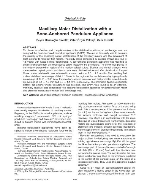
<strong>Maxillary</strong> <strong>Molar</strong> <strong>Distalization</strong> <strong>with</strong> a<br />
<strong>Bone</strong>-<strong>Anchored</strong> <strong>Pendulum</strong> Appliance<br />
Beyza Hancıog˘ lu Kircelli a ; Zafer Özgür Pektas¸ b ; Cem Kircelli c<br />
ABSTRACT<br />
To obtain an effective and compliance-free molar distalization <strong>with</strong>out an anchorage loss, we<br />
designed the bone-anchored pendulum appliance (BAPA). The aim of this study was to evaluate<br />
the stability of the anchoring screw, distalization of the maxillary molars, and the movement of<br />
teeth anterior to maxillary first molars. The study group comprised 10 patients (mean age 13.5 �<br />
1.8 years) <strong>with</strong> Class II molar relationship. A conventional pendulum appliance was modified to<br />
obtain anchorage from an intraosseous screw instead of the premolars. The screw was placed in<br />
the anterior paramedian region of the median palatal suture. Skeletal and dental changes were<br />
measured on cephalograms, and dental casts were obtained before and after distalization. A super<br />
Class I molar relationship was achieved in a mean period of 7.0 � 1.8 months. The maxillary first<br />
molars distalized an average of 6.4 � 1.3 mm in the region of the dental crown by tipping distally<br />
an average of 10.9� �2.8�. Also, the maxillary second premolar and first premolar moved distally<br />
an average of 5.4 � 1.3 mm and 3.8 � 1.1 mm, respectively. The premolars tipped significantly<br />
distally. No anterior incisor movement was detected. The BAPA was found to be an effective,<br />
minimally invasive, and compliance-free intraoral distalization appliance for achieving both molar<br />
and premolar distalization <strong>with</strong>out any anchorage loss.<br />
KEY WORDS: <strong>Molar</strong> distalization; <strong>Pendulum</strong> appliance; Intraosseous screw; Anchorage<br />
INTRODUCTION<br />
Nonextraction treatment of Angle Class II malocclusion<br />
usually requires distalization of maxillary molars.<br />
Beginning in the 1980s, intraoral appliances, such as<br />
repelling magnets, 1 superelastic NiTi coil springs, 2<br />
pendulum, 3 Jones-jig, 4 and distal-jet, 5 have been introduced<br />
to distalize molars <strong>with</strong> minimal patient compliance.<br />
Intraoral distalization appliances have been designed<br />
to deliver a continuous reciprocal force on the<br />
a Assistant Professor, Department of Orthodontics, Adana<br />
Medical Research and Teaching Center, Baskent University,<br />
Adana, Turkey<br />
b Assistant Professor, Oral and Maxillofacial Surgery, Adana<br />
Medical Research and Teaching Center, Baskent University,<br />
Adana, Turkey.<br />
c Instructor, Department of Prosthodontics, Adana Medical Research<br />
and Teaching Center, Baskent University, Adana, Turkey.<br />
Corresponding author: Beyza Hancıog˘lu Kircelli, DDS, PhD,<br />
Department of Orthodontics, Adana Medical Research and<br />
Teaching Center, Baskent University, Dadalog˘lu mah, 39. sok.<br />
No. 6 Yuregır, Adana 01250, Turkey<br />
(e-mail: beyzakircelli@hotmail.com)<br />
Accepted: July 2005. Submitted: April 2005.<br />
� 2006 by The EH Angle Education and Research Foundation,<br />
Inc.<br />
650<br />
maxillary first molars. Any action to move molars distally<br />
produces a mesial reaction force on the anchoring<br />
teeth. 6 As a consequence, if the premolars or incisors<br />
(or both) are the anchoring teeth, they move mesially,<br />
the incisors protrude, and overjet increases. 1–5,7–14<br />
However, this effect is in contradiction <strong>with</strong> the main<br />
objective of Class II treatment. Furthermore, distalized<br />
molars are questionable anchors for the retraction of<br />
premolars and incisors, despite attempts (headgears,<br />
Nance appliance etc) that have been made to maintain<br />
them in their new positions. 7,8<br />
Recently, researchers have tried to overcome this<br />
major problem by designing new intraoral systems involving<br />
rigid skeletal anchorage. Byloff et al 15 designed<br />
the Graz implant-supported pendulum appliance. The<br />
anchorage part of this appliance consisted of a surgical<br />
plate (15 � 10 mm) fixed <strong>with</strong> four titanium miniscrews<br />
to the palatal bone. The acrylic part of the pendulum<br />
appliance was fitted to the cylinders, soldered<br />
to the center of the surgical plate, on the basis of a<br />
telescopic principle. They used this appliance in adult<br />
patients. 16<br />
Keles¸ et al 17 used an osseointegrated palatal implant<br />
instead of a Nance button in the Keles slider appliance.<br />
Carano et al 18 introduced the distal-jet in con-
BONE-ANCHORED PENDULUM APPLIANCE<br />
FIGURE 1. The intraosseous screw.<br />
junction <strong>with</strong> a miniscrew anchorage system. Karaman<br />
et al 19 and Gelgör et al 20 used an intraosseous screw<br />
<strong>with</strong> their distalization mechanics containing compressed<br />
coil springs. Both the authors used this screw<br />
as an indirect rigid anchorage; the first premolars were<br />
connected to the intraoral neck of the screw via a<br />
heavy archwire, using light-cured composite resin.<br />
To obtain an effective and compliance-free molar<br />
distalization <strong>with</strong>out anchorage loss, we designed the<br />
bone-anchored pendulum appliance (BAPA). The aim<br />
of this study was to evaluate the stability of the anchoring<br />
screw, distalization of the maxillary molars,<br />
and the movement of the anchoring teeth anterior to<br />
the maxillary first molars.<br />
MATERIALS AND METHODS<br />
The study group comprised 10 patients (nine female<br />
and one male; mean age 13.5 � 1.8 years) requiring<br />
intraoral molar distalization. The selection criteria for<br />
patients included good oral hygiene, permanent dentition,<br />
Class II molar relationship <strong>with</strong> a moderate<br />
space deficiency in the maxillary dental arch, and minimal<br />
or no crowding in the mandibular arch. No other<br />
appliance was used other than BAPA during the distalization<br />
phase. All patients and parents were informed<br />
about the surgical procedure and signed a<br />
consent form.<br />
Intraosseous screw and surgical procedure<br />
A titanium intraosseous screw (2.0 mm diameter �<br />
8 mm length) (IMF intermaxillary fixation screw, Stryker,<br />
Leibinger, Germany) was used as a rigid bone<br />
anchor (Figure 1). The screw was inserted in the anterior<br />
paramedian region of the median palatal suture,<br />
7–8 mm posterior to the incisive foramen and 3–4 mm<br />
FIGURE 2. Intraoral view of screw in place.<br />
651<br />
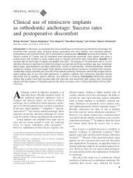
FIGURE 3. Lateral cephalometric radiograph showing screw position.<br />
TABLE 1. Screw Inclination Relative to Palatal Plane (Angle Between<br />
the Long Axis of the Screw and the Palatal Plane)<br />
N Min Max Mean SD<br />
Screw-PP (�) 10 50 76 65 77,655<br />
lateral to the median line (Figure 2). A 1.3-mm-diameter<br />
drill was used to maintain primary stability of the<br />
screw. In two patients, the screws were inserted bilaterally<br />
to the median palatal suture. The screw position<br />
was checked on a lateral cephalometric radiograph<br />
(Figure 3). Minimum, maximum, and mean values for<br />
the screw angulation relative to the palatal plane are<br />
shown in Table 1.<br />
Construction of the BAPA<br />
After soft tissue healing, impressions and stone<br />
casts were obtained <strong>with</strong> the IMF screws in place. On<br />
the stone model, the screw head was blocked out <strong>with</strong><br />
wax, and the pendulum appliance was constructed according<br />
to Hilgers 3 descriptions, excluding the auxiliary<br />
wires that extend to the occlusal surface of the first<br />
and second premolars.<br />
Angle Orthodontist, Vol 76, No 4, 2006
652 B. H. KIRCELLI, PEKTAS¸ , C. KIRCELLI<br />
FIGURE 4. Intraoral application of bone-anchored pendulum appliance.<br />
The appliance adaptation was checked clinically,<br />
and the springs were activated parallel to the median<br />
palatal suture. The acrylic plate was connected to the<br />
screw head using cold-curing, methyl methacrylatefree<br />
acrylic resin (Ufi Gel hard, Voco GmbH, Cuxhaven,<br />
Germany). Finally, activated 0.032-inch titanium<br />
molybdenum alloy (TMA) springs (Ormco Corp, Glendora,<br />
Calif) were inserted into the lingual sheaths on<br />
the first molar bands (Figure 4).<br />
Patients were specially educated to maintain their<br />
oral hygiene and were asked to use a mouthwash regularly.<br />
At every appointment, the soft tissue around the<br />
acrylic plate was checked. Also, the springs were reactivated<br />
if necessary.<br />
Cephalometric and dental cast analysis<br />
After achieving a super Class I molar relationship, a<br />
lateral cephalometric radiograph was obtained to assess<br />
dentoalveolar, skeletal, and soft tissue changes.<br />
The pterygoid vertical plane and the Frankfort horizontal<br />
plane were used as reference planes for measuring<br />
the cephalometric changes (Figures 5 and 6).<br />
The degree of rotation of the maxillary first molars<br />
and the amount of expansion between the maxillary<br />
right and left first molars were measured on photocopies21<br />
of the maxillary plaster casts. On the photocopies,<br />
movements of the maxillary first molars were assessed<br />
according to the median palatal plane22 (Figure<br />
7).<br />
Acquired arch lengths were also calculated from<br />
cast photocopies. The total arch length was measured<br />
as the arch perimeter from one upper first molar to the<br />
other. This measurement was over the contact points<br />
of the posterior teeth and the incisal edges of the incisors,<br />
both before treatment and after distalization. In<br />
addition, the acquired arch length in the anterior segment<br />
was calculated by measuring the arch perimeter<br />
between the mesial contact points of the first premolars.<br />
A piece of ligature wire was contoured to the line<br />
Angle Orthodontist, Vol 76, No 4, 2006<br />
FIGURE 5. Skeletal and soft tissue measurements. (1) SNA. (2)<br />
SNB. (3) ANB. (4) FMA. (5) GoGnSn. (6) PTV-A point. (7) Palatal<br />
plane angle. (8) Upper lip to E-plane. (9) Lower lip to E-plane. PTV<br />
indicates pterygoid vertical plane.<br />
FIGURE 6. Dental linear measurements. (1) U6-PTV. (2) U5-PTV.<br />
(3) U4-PTV. (4) U1-PTV. (5) L6-PTV. (6) U6-FH. (7) U5-FH. (8) U4-<br />
FH. (9) U1-FH. Dental angular measurements. (10) U6-FH. (11) U5-<br />
FH. (12) U4-FH. (13) U1-FH. PTV indicates pterygoid vertical plane;<br />
FH, Frankfort horizontal plane.<br />
of occlusion and straightened out for the measurement.<br />
Statistical analysis<br />
The initial measurements were repeated after 1<br />
week. Spearman’s rho coefficients were calculated to<br />
analyze repeatability. Coefficients were found to be<br />
close to 1.00. Nonparametric Wilcoxon sign rank test
BONE-ANCHORED PENDULUM APPLIANCE<br />
FIGURE 7. <strong>Maxillary</strong> model photocopy measurements. 1. Intermolar<br />
distance. 2. Length of total arch perimeter. 3. Length of anterior arch<br />
perimeter. 4. <strong>Maxillary</strong> first molar-Median palatal plane (�).<br />
TABLE 2. Changes in Cephalometric Skeletal, Soft Tissue, and Dental Measurements From Pretreatment to After <strong>Distalization</strong> a<br />
Measurements<br />
Pretreatment<br />
Mean � SD<br />
After <strong>Distalization</strong><br />
Mean � SD Difference Mean � SD Significance<br />
Skeletal<br />
SNA (�) 78.3 � 3.7 79.0 � 3.9 0.7 � 0.8* .03<br />
SNB (�) 74.8 � 2.3 74.8 � 2.3 0 � 0.6 NS<br />
ANB (�) 3.6 � 2.1 4.2 � 2.9 0.6 � 0.9 NS<br />
FMA (�) 29.9 � 2.8 30.8 � 2.5 0.9 � 1.1* .03<br />
GoGnSn (�) 38.5 � 3.1 39.4 � 2.9 0.9 � 1.1* .03<br />
PTV-A point (mm) 51.4 � 2.9 52.0 � 3.0 0.6 � 0.6* .03<br />
PTV-B point (mm) 42.8 � 3.8 42.6 � 3.6 �0.2 � 1.3 NS<br />
PTV-palatal plane (�)<br />
Soft tissue<br />
1.1 � 2.7 1.1 � 2.4 0 � 1.4 NS<br />
Upper lip to E-plane (mm) �2.7 � 2.3 �2.4 � 2.4 0.3 � 1.1 NS<br />
Lower lip to E-plane (mm)<br />
Dental-linear sagital (mm)<br />
�1.5 � 1.4 �1.2 � 1.1 0.3 � 1.0 NS<br />
<strong>Maxillary</strong> first molar-PTV 26.7 � 2.9 20.3 � 2.6 �6.4 � 1.3** .005<br />
<strong>Maxillary</strong> second premolar-PTV 30.1 � 3.6 24.7 � 3.5 �5.4 � 1.3** .005<br />
<strong>Maxillary</strong> first premolar-PTV 37.6 � 3.7 33.8 � 3.8 �3.8 � 1.1** .005<br />
<strong>Maxillary</strong> incisor-PTV 54.9 � 4.0 54.7 � 4.1 �0.2 � 0.7 NS<br />
Mandibular first molar-PTV 24.0 � 2.6 24.3 � 2.5 0.3 � 0.6 NS<br />
Overjet<br />
Dental-linear vertical (mm)<br />
4.1 � 1.1 4.4 � 1.3 0.3 � 0.6 NS<br />
<strong>Maxillary</strong> first molar-FH 46.5 � 4.0 46.6 � 4.1 0.1 � 0.5 NS<br />
<strong>Maxillary</strong> second premolar-FH 49.0 � 3.0 49.1 � 1 0.1 � 0.6 NS<br />
<strong>Maxillary</strong> first premolar-FH 50.2 � 3.3 50.6 � 3.1 0.4 � 0.7 NS<br />
<strong>Maxillary</strong> incisor-FH 54.5 � 3.7 54.5 � 3.8 0 � 0.6 NS<br />
Overbite<br />
Dental-angular (�)<br />
4.0 � 2.0 3.45 � 2.0 �0.5 � 0.5* .03<br />
<strong>Maxillary</strong> first molar-FH 73.0 � 4.3 62.1 � 5.1 �10.9 � 2.8** .005<br />
<strong>Maxillary</strong> second premolar-FH 83.1 � 5.1 66.8 � 5.5 �16.3 � 6.5** .005<br />
<strong>Maxillary</strong> first premolar-FH 91.2 � 4.8 80.2 � 4.4 �3.8 � 1.1** .005<br />
<strong>Maxillary</strong> incisor-FH 106.6 � 7.6 106.0 � 8.6 �0.6 � 1.8 NS<br />
a PTV indicates pterygoid vertical plane; FH, Frankfort horizontal plane; and NS, non significant.<br />
* P � .05.<br />
** P � .01.<br />
653<br />
was used for comparison of paired values of the measurements.<br />
A probability of .05 was accepted as critical<br />
significance.<br />
RESULTS<br />
A super Class I molar relationship was achieved in<br />
a mean period of 7.0 � 1.8 months. In the region of<br />
the dental crown, the maxillary first molars distalized<br />
an average of 6.4 � 1.3 mm tipping distally, an average<br />
of 10.9� �2.8�. Also, the maxillary second premolar<br />
and first premolar moved distally an average of<br />
5.4 � 1.3 mm and 3.8 � 1.1 mm, respectively. No<br />
anterior movement of the incisors was detected.<br />
The mean, standard deviation, and statistical significance<br />
of the skeletal, dental, and soft tissue cephalometric<br />
changes from pretreatment to after achieving<br />
a super Class I molar relationship <strong>with</strong> BAPA are summarized<br />
in Table 2. The dental cast measurements are<br />
shown in Table 3. Figures 8 through 13 demonstrate<br />
a sample case treated <strong>with</strong> BAPA.<br />
Angle Orthodontist, Vol 76, No 4, 2006
654 B. H. KIRCELLI, PEKTAS¸ , C. KIRCELLI<br />
TABLE 3. Changes in Dental Cast Measurements From Pretreatment to After <strong>Distalization</strong><br />
Measurements<br />
Angle Orthodontist, Vol 76, No 4, 2006<br />
Pretreatment<br />
Mean � SD<br />
After <strong>Distalization</strong><br />
Mean � SD<br />
Difference<br />
Mean � SD Significance a<br />
Intermolar distance (mm) 46.6 � 2.0 49.6 � 2.7 3.0 � 3.0** .01<br />
Length of total arch perimeter (mm) 74.9 � 2.8 88.8 � 5.1 13.9 � 4.1** .008<br />
Length of anterior arch perimeter (mm) 46.1 � 3.5 52.3 � 3.8 6.2 � 3.2*** .000<br />
<strong>Maxillary</strong> first molar-MPP (�) 28.3 � 6.5 32.4 � 10.8 4.1 � 9.0 NS<br />
a NS indicates non significant.<br />
** P � .01.<br />
*** P � .001.<br />
In all the patients, the 2 � 8–mm intraosseous<br />
screw remained stable during the distalization period.<br />
However, we observed a minimal rotational movement<br />
of the acrylic plate during spring reactivation, especially<br />
in patients presenting a shallow palatal vault. After<br />
experiencing this effect, we placed two screws bilaterally<br />
to mid-palatal suture in two patients.<br />
After removing the acrylic plate, mild to moderate<br />
soft tissue irritation was detected on the palatal mucosa,<br />
but this was resolved in a few days (Figure 12).<br />
Relatively less irritation was observed in patients<br />
whose pendulums were supported <strong>with</strong> bilateral<br />
screws.<br />
DISCUSSION<br />
The pendulum appliance has experienced widespread<br />
clinical use, 23 and various studies have demonstrated<br />
its skeletal and dentoalveolar effects. 8–12 Invariably,<br />
the pendulum was found to be an effective<br />
appliance for distalizing maxillary molars. However,<br />
associated anterior anchorage loss, which represented<br />
30–43% of the space created between molars and<br />
premolars, was a constant finding of these studies.<br />
Today, rigid bone anchors including osseointegrated<br />
implants, 17,24–26 titanium miniscrews, 18–20,27–30 and miniplates<br />
15,16 are powerful candidates to solve the anchorage<br />
concern. Elimination of the osseointegration<br />
period (2–6 months), wider range of application sites,<br />
simple surgical procedures during the insertion and removal<br />
processes, and decreased cost make intraosseous<br />
screws preferable rigid bone anchors.<br />
Screws are attached to the bone by mechanical retention.<br />
Osseointegration is not a goal when screws<br />
are placed. However, primary stability is a prerequisite<br />
for future stability. 30,31<br />
In a recent study, Deguchi et al 28 placed 96 small<br />
titanium screws in eight dogs and demonstrated a successful<br />
rigid osseous fixation (97%). Huga et al 30<br />
claimed that the bone supporting monocortical screws<br />
would most likely <strong>with</strong>stand immediate loading and<br />
support tooth-moving forces; they tested the pull-out<br />
strength of monocortical miniscrews <strong>with</strong> mechanical<br />
testing.<br />
On the other hand, Fritz et al 29 investigated the clinical<br />
suitability of the titanium miniscrews for orthodontic<br />
anchorage purposes (predominantly used for premolar<br />
distalization, molar uprighting, and mesial movement<br />
of the molar) and reported a failure rate of 30%.<br />
In our study, a suitable area to insert the screw was<br />
localized <strong>with</strong> respect to a computerized tomographic<br />
study in which Bernhart et al 32 indicated the area for<br />
implant placement was 6 to 9 mm posterior to the incisive<br />
foramen and 3 to 6 mm lateral to the mid-palatal<br />
suture. Moreover, Costa et al 33 reported a mean 10.57mm<br />
bone depth in the paramedian area in the premaxilla<br />
region of the palate.<br />
At first sight, one can assume that severe mucosal<br />
irritation might occur <strong>with</strong> the BAPA; however, the<br />
screw head in the palatal acrylic acts as a stop so that<br />
the palatal mucosa cannot be compressed. In this<br />
study, unilateral screws were applied in eight and bilateral<br />
screws in two patients. Although none of the<br />
unilateral screws had failed to <strong>with</strong>stand reciprocal<br />
forces during the distalization period, we suggest bilateral<br />
screws for eliminating both rotational movements<br />
during spring activations and diminishing soft<br />
tissue irritations. Also, bilateral screws might present<br />
more predictable results for the clinicians when using<br />
this system.<br />
Despite the fact that all patients were strictly encouraged<br />
to maintain their oral hygiene, some plaque<br />
accumulation was evident under the acrylic plate.<br />
However, this condition did not affect the screw stability.<br />
This might be attributed to the dense, thick, and<br />
keratinized structure of the attached palatal mucosa.<br />
The only difficulty experienced <strong>with</strong> the BAPA was<br />
detaching the acrylic plate from the screw head when<br />
removing the appliance. We used a carbide bur <strong>with</strong><br />
an aerator under copious irrigation. As a suggestion,<br />
if the acrylic plate is made no thicker than 2 mm over<br />
the screw head and the grooves at the top of the screw<br />
are filled <strong>with</strong> a thin layer of wax, it will facilitate detaching<br />
the acrylic plate.<br />
When the BAPA is compared <strong>with</strong> other systems,<br />
the Graz implant-supported pendulum appliance 15,16<br />
seems convenient regarding its removable property,
BONE-ANCHORED PENDULUM APPLIANCE<br />
FIGURE 8. Pretreatment photographs of a 13-year-old patient.<br />
655<br />
Angle Orthodontist, Vol 76, No 4, 2006
656 B. H. KIRCELLI, PEKTAS¸ , C. KIRCELLI<br />
FIGURE 9. Postdistalization intraoral photographs. Note maxillary molar and premolar distalization providing adequate space for maxillary<br />
canines spontaneously.<br />
FIGURE 10. Lateral cephalometric radiograph after distalization.<br />
thereby enabling activations extraorally. The pendulum<br />
appliance fixed to an osseointegrated implant 26<br />
could present a more reliable anchorage when an<br />
uprighting bend is added to the springs. However, both<br />
systems are more invasive than applying a screw.<br />
<strong>Maxillary</strong> molar and premolar distalization<br />
In all patients, a super Class I molar relationship<br />
was achieved in approximately 7 months. The maxil-<br />
Angle Orthodontist, Vol 76, No 4, 2006<br />
FIGURE 11. BAPA can be held in place during the full fixed therapy;<br />
thus minor activation of the springs supports the molar anchorage.<br />
BAPA indicates bone-anchored pendulum appliance.<br />
lary molars moved distally a mean of 6.4 mm and the<br />
second and first premolars drifted distally a mean of<br />
5.4 and 3.8 mm, respectively. Because reactive forces<br />
arising from the pendulum springs were directly resisted<br />
by an intraosseous screw, the premolars were<br />
free from any attachment, and they drifted distally via<br />
transeptal fibers during the distalization period. As a<br />
result, in the study group, a Class I relationship was<br />
simultaneously achieved in the second premolars.<br />
The mesial movement of the premolars <strong>with</strong> the con-
BONE-ANCHORED PENDULUM APPLIANCE<br />
FIGURE 12. Soft tissue status immediately after the removal of<br />
BAPA. BAPA indicates bone-anchored pendulum appliance.<br />
ventional pendulum or other intraoral distalizing appliances<br />
is a well-established finding in the literature. 1–<br />
5,7–14 However, this can also occur when distalizing molars<br />
using indirect skeletal anchorage in which reactive<br />
forces are initially resisted by premolars stabilized by<br />
a rigid anchor. Gelgör et al 20 demonstrated a slight anchorage<br />
loss in the anterior segment and attributed<br />
this effect to mesial tipping of first premolars during<br />
molar distalization. In the same manner, a 4.1 mm anterior<br />
incisor movement was reported in a study 34<br />
where premolars were indirectly anchored to an osseointegrated<br />
palatal implant <strong>with</strong> a special abutment.<br />
The most beneficial finding of BAPA is the simultaneous<br />
distal movement of premolars <strong>with</strong> molars. This<br />
is advantageous in several aspects. The new molar<br />
position has not been jeopardized because there is no<br />
need to retract the anterior teeth. An unnecessary anterior<br />
movement has been avoided at the premolars<br />
and incisors. Moreover, anterior crowding has been<br />
spontaneously solved out because of the stretched<br />
transeptal fibers. As a consequence, the total treatment<br />
time is shortened. In this study, after molar distalization,<br />
an average of 13.9 and 6.2 mm of space<br />
was gained in the total maxillary arch and in the anterior<br />
segment, respectively.<br />
Distal tipping<br />
A significant distal tipping was observed <strong>with</strong> both<br />
premolar and molar distalization. Mean tipping values<br />
for the crowns of the maxillary first molars, second premolars,<br />
and first premolars were 10.9�, 16.3�, and 3.8�,<br />
respectively.<br />
<strong>Distalization</strong> <strong>with</strong> a tipping movement can be questioned<br />
because a quantity of space can be lost <strong>with</strong><br />
molar uprighting when full fixed therapy is initiated. 8,14<br />
However, in this system, there is no need to remove<br />
the appliance immediately after distalization. Thus, minor<br />
activation of the pendulum springs (terminal double<br />
back bend is eliminated) helps maintain molar po-<br />
657<br />
sition when a continuous archwire is applied to upright<br />
the molars. Also, the molar position is maintained<br />
when leveling and retracting first premolars and canines.<br />
In the present study, this so-called active anchorage<br />
concept, defined by Byloff et al, 15 coped <strong>with</strong><br />
the anchorage concern of the distalized molars.<br />
On the other hand, overcorrection of the molar relationship<br />
is another goal that taxes molar anchorage 20<br />
during full fixed therapy, especially if distal tipping exists.<br />
Again, a problem appears for the conventional intraoral<br />
distalization appliances because the amount of<br />
distal movement correlates <strong>with</strong> the amount of anchorage<br />
loss. 8 In this context, one can use the BAPA<br />
to freely move the molars to a super Class I relationship<br />
<strong>with</strong>out considering anchorage loss.<br />
Skeletal and soft tissue effects<br />
• In this study, the cant of the palatal plane remained<br />
unchanged and the mandibular plane rotated by 0.9�<br />
in a clockwise direction. This agrees <strong>with</strong> the results<br />
demonstrated by other studies <strong>with</strong> the conventional<br />
pendulum. 8,10 This clockwise rotation can be attributed<br />
to the maxillary molars moving distally into the<br />
wedge of occlusion and to the cusp interferences.<br />
Although it is clinically negligible, point A moved anteriorly<br />
by 0.6 mm. This effect might be remodeling<br />
because of reciprocal forces affecting the anterior<br />
palate. No significant difference was observed regarding<br />
the upper and lower lip positions relative to<br />
the esthetic line. Conversely, protrusion is a result of<br />
the action of a conventional pendulum. 8,10 However,<br />
future studies <strong>with</strong> greater sample size would permit<br />
more reliable statements to be made regarding the<br />
clinical success of the BAPA.<br />
CONCLUSIONS<br />
• <strong>Molar</strong> distalization, as well as premolar distalization,<br />
was achieved <strong>with</strong> BAPA <strong>with</strong>out any anchorage<br />
loss.<br />
• Besides the space gained in the posterior segment,<br />
a quantity of space was also gained in anterior segment,<br />
and spontaneous alignment of anterior crowding<br />
was achieved during molar distalization.<br />
• In this study, the BAPA presented an effective and<br />
minimally invasive, compliance-free alternative for<br />
intraoral molar distalization in nonextraction Class II<br />
treatment.<br />
ACKNOWLEDGEMENT<br />
Special thanks to Mrs. Fahrunnisa Dınkcıog˘lu and Mr. Mustafa<br />
Obaz for their contributions.<br />
Angle Orthodontist, Vol 76, No 4, 2006
658 B. H. KIRCELLI, PEKTAS¸ , C. KIRCELLI<br />
FIGURE 13. Posttreatment photographs.<br />
Angle Orthodontist, Vol 76, No 4, 2006
BONE-ANCHORED PENDULUM APPLIANCE<br />
REFERENCES<br />
1. Gianelly AA, Viatas AS, Thomas WM. The use of magnets<br />
to move molars distally. Am J Orthod. 1989;6:161–167.<br />
2. Gianelly AA, Bednar J, Dietz VS. Japanese NiTi coils to<br />
move molars distally. Am J Orthod. 1991;99:564–566.<br />
3. Hilgers JJ. The pendulum appliance for Class II non-compliance<br />
therapy. J Clin Orthod. 1992;6:700–713.<br />
4. Jones R, White J. Rapid Class II correction <strong>with</strong> an open<br />
coil jig. J Clin Orthod. 1992;26:661–664.<br />
5. Carano A, Testa M. The distal jet appliance for upper molar<br />
distalization. J Clin Orthod. 1996;30:374–380.<br />
6. Proffitt WR. Contemporary Orthodontics. 2nd ed. St Louis,<br />
Mo: CV Mosby; 1993:306–307.<br />
7. Gianelly AA. Distal movement of the maxillary molars. Am<br />
J Orthod. 1998;114:66–72.<br />
8. Ghosh J, Nanda RS. Evaluation of an intraoral maxillary<br />
molar distalization technique. Am J Orthod. 1996;110:639–<br />
646.<br />
9. Byloff FK, Darendeliler MA. Distal molar movement using<br />
the pendulum appliance. Part 1: clinical and radiological<br />
evaluation. Angle Orthod. 1997;67:249–260.<br />
10. Bussick TJ, McNamara JA. Dentoalveolar and skeletal<br />
changes associated <strong>with</strong> the pendulum appliance. Am J Orthod.<br />
2000;117:333–343.<br />
11. Joseph AA, Butchard CJ. An evaluation of the pendulum<br />
distalizing appliance. Semin Orthod. 2000;6:129–135.<br />
12. Torog˘lu MS, Uzel I˙, Çam OY, Hancıog˘lu ZB. Cephalometric<br />
evaluation of pendulum appliance on various vertical growth<br />
patterns and of the changes during short- term stabilization.<br />
Clin Orthod Res. 2001;4:15–27.<br />
13. Haydar S, Üner O. Comparison of Jones Jig molar distalization<br />
appliance <strong>with</strong> extraoral traction. Am J Orthod. 2000;<br />
117:49–53.<br />
14. Bolla E, Muratore F, Carano A, Bowman SJ. Evaluation of<br />
maxillary molar distalization <strong>with</strong> the distal jet: a comparison<br />
<strong>with</strong> other contemporary methods. Angle Orthod. 2002;72:<br />
481–494.<br />
15. Byloff FK, Karcher H, Clar E, Stoff F. An implant to eliminate<br />
anchorage loss during molar distalization: a case report involving<br />
the Graz implant-supported pendulum. Int J Adult<br />
Orthod Orthognath Surg. 2000;15:129–137.<br />
16. Kärcher H, Byloff FK, Clar E. The Graz implant supported<br />
pendulum, a technical note. J Craniomaxillofac Surg. 2002;<br />
30:87–90.<br />
17. Keles¸ A, Erverdi N, Sezen S. Bodily distalization of molars<br />
<strong>with</strong> absolute anchorage. Angle Orthod. 2003;73:471–482.<br />
18. Carano A, Velo S, Leone P, Siciliani G. Clinical applications<br />
of the miniscrew anchorage system. J Clin Orthod. 2005;<br />
39:9–24.<br />
659<br />
19. Karaman AI, Bas¸çiftçi FA, Polat O. Unilateral distal molar<br />
movement <strong>with</strong> an implant-supported distal jet appliance.<br />
Angle Orthod. 2002;72:167–174.<br />
20. Gelgör IE, Büyükyılmaz T, Karaman AI, Dolanmaz D, Kalaycı<br />
A. Intraosseous screw-supported upper molar distalization.<br />
Angle Orthod. 2004;74:836–848.<br />
21. Champagne M. Reliability of measurements from photocopies<br />
of study models. J Clin Orthod. 1992;26:648–650.<br />
22. Keles¸ A, Sayınsu K. A new approach in maxillary molar distalization:<br />
intra molar bodily molar distalizer. Am J Orthod.<br />
2000;117:39–48.<br />
23. Keim RG, Gottlieb EL, Nelson AH, Vogels DS. 2002 JCO<br />
study of orthodontic diagnosis and treatment procedures.<br />
Part 1. Results and trends. J Clin Orthod. 2002;36:553–568.<br />
24. Roberts WE, Helm FR, Marshall KJ, Gongloff RK. Rigid endosseous<br />
implants for orthodontic and orthopedic anchorage.<br />
Angle Orthod. 1989;59:331–338.<br />
25. Wehrbein H, Feifel H, Diedrich P. Palatal implant anchorage<br />
reinforcement of posterior teeth: prospective study. Am J<br />
Orthod. 1999;116:678–686.<br />
26. Kinzinger G, Wehrbein H, Diedrich P. <strong>Pendulum</strong> appliances<br />
<strong>with</strong> different anchorage modalities for non-compliance molar<br />
distal movement in adults. Kieferorthop. 2004;18:11–24.<br />
27. Creekmore TD, Eklund MK. The possibility of skeletal anchorage.<br />
J Clin Orthod. 1983;17:266–269.<br />
28. Deguchi T, Takano-Yamamoto T, Kanomi R, Hartsfield JK<br />
Jr, Roberts WE, Garetto LP. The use of small titanium<br />
screws for orthodontic anchorage. J Dent Res. 2003;82(5):<br />
377–381.<br />
29. Fritz U, Ehmer A, Dietrich P. Clinical suitability of titanium<br />
microscrews for orthodontic anchorage-preliminary experiences.<br />
J Orofac Orthop. 2004;65:410–418.<br />
30. Huga SS, Litsky AS, Beck FM, Johnson KA, Larsen PE.<br />
Pull-out strength of monocortical screws placed in the maxillae<br />
and mandibles of dogs. Am J Orthod. 2005;127:307–<br />
313.<br />
31. Cope JB. Temporary anchorage devices in orthodontics: a<br />
paradigm shift. Semin Orthod. 2005;11:3–9.<br />
32. Bernhart T, Vollgruber A, Gahleitner A, Dörtbudak O, Haas<br />
R. Alternative to the median region of the palate for placement<br />
of an orthodontic implant. Clin Oral Impl Res. 2000;<br />
11:595–601.<br />
33. Costa A, Pasta G, Bergamaschi G. Intraoral hard and soft<br />
tissue depths for temporary anchorage devices. Semin Orthod.<br />
2005;11:10–15.<br />
34. Sezen S, Erverdi N. <strong>Molar</strong> distalzation <strong>with</strong> palatal implant.<br />
Paper presented at: 9th International Congress of the Turkish<br />
Orthodontic Society; October 3–6, 2004; Antalya, Turkey.<br />
Angle Orthodontist, Vol 76, No 4, 2006