Infliximab infusions for persistent back pain in two patients with ...
Infliximab infusions for persistent back pain in two patients with ...
Infliximab infusions for persistent back pain in two patients with ...

Create successful ePaper yourself
Turn your PDF publications into a flip-book with our unique Google optimized e-Paper software.
1588 Letters to the Editor<br />
6. LaSp<strong>in</strong>a M, Russo G. Pneumococcal sepsis <strong>in</strong> a girl <strong>with</strong> systemic<br />
lupus erythematosus. Br J Haematol 2003;122:172.<br />
7. Kang I, Park SH. Infectious complications <strong>in</strong> SLE after<br />
immunosuppressive therapies. Curr Op<strong>in</strong> Rheumatol 2003;5:528–34.<br />
8. Pettersson T, Julkunen H. Asplenia <strong>in</strong> a patient <strong>with</strong> systemic<br />
lupus erythematosus and antiphospholipid antibodies. J Rheumatol<br />
1992;19:1159.<br />
9. Obarski TP, Stoller JK, We<strong>in</strong>ste<strong>in</strong> C, Hayden S. Splenic <strong>in</strong>farction.<br />
A new thrombotic manifestation of the circulat<strong>in</strong>g lupus anticoagulant.<br />
Cleve Cl<strong>in</strong> J Med 1989;56:174–6.<br />
Rheumatology 2005;44:1588<br />
doi:10.1093/rheumatology/kei144<br />
Advance Access publication 18 October 2005<br />
Functional asplenia <strong>in</strong> a patient <strong>with</strong> Rothmann–Makai<br />
syndrome: a pathogenetic relationship?<br />
SIR, A 27-yr-old woman was referred <strong>for</strong> evaluation of mild<br />
anaemia, leucocytosis and thrombocytosis. History revealed a<br />
diagnosis of Rothmann–Makai syndrome (lipogranulomatosis<br />
subcutanea), a rare variant of Weber–Christian disease, at the<br />
age of 13 yr. At that time the patient had presented <strong>with</strong> a m<strong>in</strong>or<br />
fracture of the left ankle, and when the plaster was removed she<br />
was found to have irregular red discoloration on the outer part of<br />
the ankle. This subsequently spread to <strong>in</strong>volve both sh<strong>in</strong>s. A biopsy<br />
specimen was obta<strong>in</strong>ed from one of the erythematous nodules.<br />
In the histopathological sections there was a moderate lymphocytic<br />
<strong>in</strong>filtration <strong>in</strong> the lobules of the panniculus and fibrocytes <strong>in</strong>vad<strong>in</strong>g<br />
the lobules from the fibrous septae. Work-up at that time also<br />
<strong>in</strong>cluded ant<strong>in</strong>uclear antibodies (ANA), rheumatoid factor (RF),<br />
C-reactive prote<strong>in</strong> (CRP), anti-double-stranded DNA, anticardiolip<strong>in</strong>,<br />
anti-Scl-70 and cryoglobul<strong>in</strong>s, which were all negative.<br />
A complete blood count was normal. With these f<strong>in</strong>d<strong>in</strong>gs and<br />
lack of any systemic manifestations, a diagnosis of Rothmann–<br />
Makai syndrome was established. The patient received an 8-month<br />
course of steroids. The erythema resolved and the patient was<br />
left <strong>with</strong> <strong>two</strong> slightly atrophic areas <strong>in</strong> both calves. No relapse has<br />
occurred. History also revealed Hashimoto’s thyroiditis diagnosed<br />
at the age of 14 yr and the onset of alopecia areata at the age<br />
of 20 yr.<br />
The patient has developed progressive <strong>in</strong>crease <strong>in</strong> her leucocyte<br />
count [(8–12) 10 9 /l] <strong>with</strong> a normal distribution and platelet count<br />
[(450–600) 10 9 /l] over the past 8 yr. When seen <strong>in</strong> our cl<strong>in</strong>ic<br />
she was feel<strong>in</strong>g well. Except <strong>for</strong> obesity, the physical exam<strong>in</strong>ation<br />
was unremarkable. Haemoglob<strong>in</strong> was 14 g/dl <strong>with</strong> normal red<br />
cell <strong>in</strong>dices, the platelet count was 668 10 9 /l and the leucocyte<br />
count was 16.2 10 9 /l <strong>with</strong> a normal distribution. A peripheral<br />
blood smear revealed anisopoikilocytosis and numerous<br />
Howell–Jolly bodies, <strong>in</strong>dicat<strong>in</strong>g functional hyposplenism.<br />
Computed tomography revealed a small spleen and no flow signals<br />
could be derived by colour Doppler measurements from the spleen.<br />
Rothmann–Makai syndrome is a rare <strong>for</strong>m of Weber–Christian<br />
disease, usually seen <strong>in</strong> adolescent and middle-aged women [1].<br />
Subcutaneous nodules develop dur<strong>in</strong>g the course of the disease<br />
whereas systemic manifestations are absent. Multiple <strong>pa<strong>in</strong></strong>less<br />
nodules appear throughout the muscle and subcutaneous tissues.<br />
The primary pathological process is lobular panniculitis, which<br />
passes through the same three stages as Weber–Christian disease.<br />
In the first stage, there is acute <strong>in</strong>flammation of the fat lobules <strong>with</strong><br />
fat cell degeneration accompanied by an <strong>in</strong>filtrate of neutrophils,<br />
lymphocytes and macrophages. The second stage is characterized<br />
by many foamy histiocytes, and the <strong>in</strong>filtrate is discretely<br />
localized to the fat lobules. F<strong>in</strong>ally, the foam cells are replaced<br />
by fibroblasts. The first <strong>two</strong> histological stages correspond to<br />
cl<strong>in</strong>ically apparent <strong>in</strong>duration, while <strong>in</strong> the third stage atrophy of<br />
the sk<strong>in</strong> may develop. There are no diagnostic laboratory f<strong>in</strong>d<strong>in</strong>gs.<br />
The diagnosis is based upon cl<strong>in</strong>ical and pathological f<strong>in</strong>d<strong>in</strong>gs.<br />
Corticosteroid treatment does not prevent new lesions or seem to<br />
alter the course of the self-limited disease.<br />
Infiltrative, <strong>in</strong>flammatory or thromboembolic processes <strong>in</strong> the<br />
parenchyma of the spleen can cause a functional loss of the organ.<br />
This phenomenon is called functional asplenia and occurs as a<br />
complication, especially <strong>in</strong> sickle cell disease, lupus erythematosus<br />
(SLE) and after bone marrow transplantation [2–4]. Functional<br />
asplenia has complicated the course of autoimmune diseases other<br />
than SLE, such as candidiasis endocr<strong>in</strong>opathy syndrome and<br />
alopecia areata [5].<br />
This is the first report of functional asplenia occurr<strong>in</strong>g <strong>in</strong><br />
the sett<strong>in</strong>g of Rothmann–Makai syndrome. The aetiology and<br />
pathogenesis of Rothmann–Makai syndrome is still unknown.<br />
Christian–Weber disease often occurs <strong>in</strong> <strong>patients</strong> <strong>with</strong> autoimmune<br />
disorders [6–8], as is the case <strong>with</strong> our patient, re<strong>in</strong><strong>for</strong>c<strong>in</strong>g the view<br />
that Rothmann–Makai syndrome, a variant of Christian–Weber,<br />
may also have an autoimmune basis.<br />
The authors have declared no conflict of <strong>in</strong>terest.<br />
A. PSYRRI and T. ECONOMOPOULOS<br />
2nd Department of Internal Medic<strong>in</strong>e, Attikon University<br />
Hospital, Athens, Greece<br />
Accepted 2 September 2005<br />
Correspondence to: A. Psyrri, Attikon Hospital, Rim<strong>in</strong>i 1,<br />
Haidari, Athens, Greece. E-mail: Diamando.Psyrri@yale.edu<br />
1. Bondi EE, Margolis DJ, Lazarus GS. Panniculitis. In: Freedberg IM,<br />
Eisen AZ, Wolff K et al., ed. Fitzpatrick’s Dermatology <strong>in</strong> General<br />
Medic<strong>in</strong>e, 4th edn. New York: McGraw-Hill, 1999:1275–89.<br />
2. Santilli D, Govoni M, Prand<strong>in</strong>i N, Rizzo N, Trotta F.<br />
Autosplenectomy and antiphospholipid antibodies <strong>in</strong> systemic<br />
lupus erythematosus: a pathogenetic relationship? Sem<strong>in</strong> Arthritis<br />
Rheum 2003;33:125–33.<br />
3. Wong WY. Prevention and management of <strong>in</strong>fection <strong>in</strong> children <strong>with</strong><br />
sickle cell anaemia. Paediatr Drugs 2001;3:793–801.<br />
4. Kalhs P, Panzer S, Kletter K et al. Functional asplenia after bone<br />
marrow transplantation. A late complication related to extensive<br />
chronic graft-versus-host disease. Ann Intern Med 1988;109:461–4.<br />
5. Boni R, Trueb RM, Wuthrich B. Alopecia areata <strong>in</strong> a patient <strong>with</strong><br />
candidiasis-endocr<strong>in</strong>opathy syndrome: unsuccessful treatment trial<br />
<strong>with</strong> diphenylcyclopropenone. Dermatology 1995;191:68–71.<br />
6. Bill<strong>in</strong>gs JK, Milgraum SS, Gupta AK, Head<strong>in</strong>gton JT,<br />
Rasmussen JE. Lipoatrophic panniculitis: a possible autoimmune<br />
<strong>in</strong>flammatory disease of fat. Report of three cases. Arch Dermatol<br />
1987;123:1662–6.<br />
7. Yoshimura A, Itoyama S, Mori S, Mori W, Inoue G. An autopsy<br />
case of Weber-Christian <strong>with</strong> immune complex glomerulonephritis.<br />
Nippon J<strong>in</strong>zo Gakkai Shi 1988;30:291–6.<br />
8. Herr W, Lohse AW, Spahn TW et al. Nodular nonsuppurative<br />
panniculitis <strong>in</strong> association <strong>with</strong> primary biliary cirrhosis and<br />
Hashimoto’s thyroiditis. Z Rheumatol 1996;55:122–6.<br />
Rheumatology 2005;44:1588–1590<br />
doi:10.1093/rheumatology/kei155<br />
Advance Access publication 18 October 2005<br />
<strong>Infliximab</strong> <strong><strong>in</strong>fusions</strong> <strong>for</strong> <strong>persistent</strong> <strong>back</strong> <strong>pa<strong>in</strong></strong><br />
<strong>in</strong> <strong>two</strong> <strong>patients</strong> <strong>with</strong> Schmorl’s nodes<br />
SIR, Schmorl’s nodes (SNs) are herniations of the nucleus<br />
pulposus (NP) material through the vertebral endplates <strong>in</strong>to the<br />
trabecular bone. SNs are the most common non-<strong>in</strong>tervertebral disc<br />
Downloaded from<br />
http://rheumatology.ox<strong>for</strong>djournals.org/ by guest on January 24, 2013
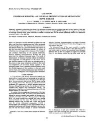
FIG. 1. Post-contrast T1-weighted image of lumbar sp<strong>in</strong>e <strong>in</strong> the<br />
first patient prior to (A) and 6 months after (B) <strong>in</strong>fliximab<br />
<strong><strong>in</strong>fusions</strong>, show<strong>in</strong>g disappearance of enhancement of Schmorl’s<br />
node and of oedema of adjacent vertebral body marrow.<br />
abnormalities <strong>in</strong> magnetic resonance imag<strong>in</strong>g (MRI) <strong>in</strong> persons<br />
<strong>with</strong>out <strong>back</strong> <strong>pa<strong>in</strong></strong> [1]. However, SNs are seldom symptomatic, and<br />
treatment <strong>with</strong> analgesics, non-steroidal anti-<strong>in</strong>flammatory<br />
drugs (NSAIDs) or corticosteroids usually improves <strong>back</strong> <strong>pa<strong>in</strong></strong>.<br />
In contrast to SNs, herniated <strong>in</strong>tervertebral discs (HIDs) are a<br />
common cause of <strong>back</strong> and radicular <strong>pa<strong>in</strong></strong>. TNF- from the NP<br />
does appear to be pathogenic to nerve roots <strong>in</strong> animal models<br />
of sciatica [2]. Furthermore, TNF- blockade is effective <strong>in</strong><br />
prevent<strong>in</strong>g NP-<strong>in</strong>duced functional and structural nerve root <strong>in</strong>jury<br />
<strong>in</strong> animal models [3]. The efficacy of the TNF- <strong>in</strong>hibitors<br />
<strong>in</strong>fliximab and etanercept <strong>in</strong> leg and <strong>back</strong> <strong>pa<strong>in</strong></strong> has recently been<br />
shown <strong>in</strong> <strong>patients</strong> <strong>with</strong> HID-<strong>in</strong>duced sciatica [4, 5]. These results<br />
of TNF- blockade prompted us to test <strong>in</strong>fliximab <strong><strong>in</strong>fusions</strong><br />
<strong>for</strong> refractory symptoms to conventional conservative treatment <strong>in</strong><br />
<strong>two</strong> <strong>patients</strong> <strong>with</strong> <strong>pa<strong>in</strong></strong>ful SN.<br />
Two women aged 52 and 49 yr <strong>with</strong> SNs received <strong>in</strong>fliximab<br />
because of severe, long-stand<strong>in</strong>g <strong>back</strong> <strong>pa<strong>in</strong></strong> (duration of <strong>pa<strong>in</strong></strong> 24<br />
and 18 months, respectively) <strong>with</strong>out radicular symptoms. There<br />
was a partial response of <strong>pa<strong>in</strong></strong> to treatment <strong>with</strong> NSAIDs and<br />
corticosteroids <strong>in</strong> both <strong>patients</strong>, but severe sp<strong>in</strong>al <strong>pa<strong>in</strong></strong> recurred<br />
after discont<strong>in</strong>uation of the above agents. SNs of the L3 and L5<br />
vertebrae <strong>in</strong> the first patient and of the T12, L1 and L2 vertebrae<br />
<strong>in</strong> the second patient were depicted by MRI. There were MRI<br />
f<strong>in</strong>d<strong>in</strong>gs of active SN (oedema of adjacent bone marrow and<br />
contrast enhancement of SN) <strong>in</strong> the L5 vertebra <strong>for</strong> the first patient<br />
and <strong>in</strong> the L2 vertebra <strong>for</strong> the second patient. After obta<strong>in</strong><strong>in</strong>g<br />
ethics committee approval and written consent from the <strong>patients</strong>,<br />
the <strong>patients</strong> scheduled to receive <strong>in</strong>fliximab <strong><strong>in</strong>fusions</strong> at a dose<br />
of 3 mg/kg at weeks 0, 2, 6 and 14. Response was evaluated<br />
us<strong>in</strong>g a 0–100 mm visual analogue scale (VAS) <strong>for</strong> <strong>back</strong> <strong>pa<strong>in</strong></strong>.<br />
Furthermore, the first patient was evaluated <strong>for</strong> radiological<br />
progression of the lesions by sequential MRI exam<strong>in</strong>ation.<br />
Prior to <strong>in</strong>fliximab treatment, the VAS score <strong>for</strong> <strong>back</strong> <strong>pa<strong>in</strong></strong> <strong>for</strong><br />
the <strong>two</strong> <strong>patients</strong> was 90 and 85, respectively. The first patient<br />
responded promptly to <strong>in</strong>fliximab <strong>with</strong> a decrease <strong>in</strong> VAS <strong>for</strong> <strong>back</strong><br />
<strong>pa<strong>in</strong></strong> to 7, and <strong>back</strong> <strong>pa<strong>in</strong></strong> completely resolved after the second<br />
<strong>in</strong>fusion. Due to a severe allergic reaction just after the second<br />
<strong>in</strong>fusion, she discont<strong>in</strong>ued the <strong>in</strong>fliximab regimen. Six months<br />
later, MRI exam<strong>in</strong>ation revealed disappearance of contrast<br />
enhancement of SN of the L5 vertebra and of bone marrow<br />
oedema (Fig. 1). She has rema<strong>in</strong>ed asymptomatic <strong>for</strong> 30 months.<br />
Letters to the Editor 1589<br />
The second patient completed four <strong>in</strong>fliximab <strong><strong>in</strong>fusions</strong>. She<br />
also responded promptly to treatment. VAS <strong>for</strong> <strong>back</strong> <strong>pa<strong>in</strong></strong> ranged<br />
from 15 to 20 at weeks 2, 6 and 14. She was missed 5 months<br />
after the last <strong>in</strong>fliximab <strong>in</strong>fusion, and had VAS <strong>for</strong> <strong>back</strong> <strong>pa<strong>in</strong></strong> 20<br />
over the follow-up period.<br />
MRI is useful <strong>in</strong> differentiat<strong>in</strong>g between symptomatic and<br />
asymptomatic SNs. In an analysis of MRI f<strong>in</strong>d<strong>in</strong>gs of SNs [6],<br />
the vertebral body marrow surround<strong>in</strong>g the SN was seen as low<br />
signal <strong>in</strong>tensity on T1-weighted images and as high signal <strong>in</strong>tensity<br />
on T2-weighted images <strong>in</strong> all symptomatic cases. These MRI<br />
f<strong>in</strong>d<strong>in</strong>gs were not seen <strong>in</strong> asymptomatic <strong>in</strong>dividuals. Histological<br />
exam<strong>in</strong>ation of <strong>two</strong> symptomatic cases demonstrated bone<br />
marrow oedema and <strong>in</strong>flammatory cell <strong>in</strong>filtration <strong>in</strong> the affected<br />
vertebral body. In a small number of cases of acute SNs [7], there<br />
had been improvement <strong>in</strong> <strong>back</strong> <strong>pa<strong>in</strong></strong> by the time the follow-up<br />
MRI (between 6 weeks and 7 months after the <strong>in</strong>itial MRI)<br />
showed reduction <strong>in</strong> bone marrow oedema. Furthermore, there<br />
was resolution of oedema <strong>with</strong> fatty marrow change <strong>in</strong> <strong>two</strong><br />
cases <strong>with</strong> longer follow-up at MRI (10 and 18 months after<br />
the <strong>in</strong>itial MRI, respectively). In addition to surround<strong>in</strong>g bone<br />
marrow oedema, MRI can reveal vascularized tissue <strong>in</strong> SNs<br />
after the <strong>in</strong>travenous adm<strong>in</strong>istration of contrast material [8].<br />
Contrast-enhanc<strong>in</strong>g SNs are larger and more frequently associated<br />
<strong>with</strong> bone marrow oedema <strong>in</strong> <strong>patients</strong> <strong>with</strong> <strong>back</strong> <strong>pa<strong>in</strong></strong> than <strong>in</strong><br />
asymptomatic <strong>patients</strong>.<br />
Our <strong>patients</strong> had not only severe <strong>back</strong> <strong>pa<strong>in</strong></strong>, which was <strong>in</strong>duced<br />
by active SNs (L5 vertebra <strong>in</strong> the first patient and L2 vertebra <strong>in</strong><br />
the second patient), but also long-stand<strong>in</strong>g symptoms, which<br />
could be expla<strong>in</strong>ed by previous active SNs that were <strong>in</strong> the heal<strong>in</strong>g<br />
stage at the time of MRI exam<strong>in</strong>ation (L3 vertebra <strong>in</strong> the first<br />
patient and T12 and L1 vertebra <strong>in</strong> the second patient). A s<strong>in</strong>gle<br />
<strong>in</strong>fliximab <strong>in</strong>fusion <strong>for</strong> HID-<strong>in</strong>duced sciatica produced not only a<br />
susta<strong>in</strong>ed but also an immediate improvement (<strong>with</strong><strong>in</strong> hours) [4].<br />
The first <strong>in</strong>fliximab <strong>in</strong>fusion led to prompt and significant<br />
improvement <strong>in</strong> <strong>back</strong> <strong>pa<strong>in</strong></strong> <strong>in</strong> our <strong>patients</strong> <strong>with</strong><strong>in</strong> the first 24 h.<br />
There was a complete response to the second <strong>in</strong>fliximab <strong>in</strong>fusion<br />
<strong>in</strong> the first patient, but the second patient did not have a<br />
complete improvement of <strong>back</strong> <strong>pa<strong>in</strong></strong> <strong>with</strong> the four-dose <strong>in</strong>fliximab<br />
regimen. A pilot study is required to validate the results of our <strong>two</strong><br />
case reports.<br />
Rheumatology<br />
Key message<br />
TNF- blockade seems to be effective <strong>for</strong><br />
<strong>persistent</strong>ly <strong>pa<strong>in</strong></strong>ful Schmorl’s nodes.<br />
The authors have declared no conflicts of <strong>in</strong>terest.<br />
G. T. SAKELLARIOU, I.CHATZIGIANNIS and I. TSITOURIDIS 1<br />
Department of Rheumatology, St Paul’s Hospital and<br />
1<br />
Department of Radiology, Papageorgiou Hospital,<br />
Thessaloniki, Greece<br />
Accepted 9 September 2005<br />
Correspondence to: G. T. Sakellariou, Department of<br />
Rheumatology, St Paul’s Hospital, 161 Ethnikis Andistaseos<br />
Street, 551 34, Thessaloniki, Greece. E-mail: sakelgr@otenet.gr<br />
or sakellariou.doc@mycosmos.gr<br />
1. Jensen MC, Brant-Zawadzki MN, Obuchowski N, Modic MT,<br />
Malkasian D, Ross JS. Magnetic resonance imag<strong>in</strong>g of the lumbar sp<strong>in</strong>e<br />
<strong>in</strong> people <strong>with</strong>out <strong>back</strong> <strong>pa<strong>in</strong></strong>. N Engl J Med 1994;331:69–73.<br />
Downloaded from<br />
http://rheumatology.ox<strong>for</strong>djournals.org/ by guest on January 24, 2013
1590 Letters to the Editor<br />
2. Igarashi T, Kikuchi S, Shubayev V, Myers RR. 2000 Volvo<br />
Award w<strong>in</strong>ner <strong>in</strong> basic science studies: Exogenous<br />
tumor necrosis factor-alpha mimics nucleus pulposus-<strong>in</strong>duced<br />
neuropathology. Molecular, histologic, and behavioral comparisons<br />
<strong>in</strong> rats. Sp<strong>in</strong>e 2000;25:2975–80.<br />
3. Olmarker K, Rydevik B. Selective <strong>in</strong>hibition of tumor necrosis<br />
factor-alpha prevents nucleus pulposus-<strong>in</strong>duced thrombus <strong>for</strong>mation,<br />
<strong>in</strong>traneural edema, and reduction of nerve conduction velocity:<br />
possible implications <strong>for</strong> future pharmacologic treatment strategies<br />
of sciatica. Sp<strong>in</strong>e 2001;26:863–9.<br />
4. Karpp<strong>in</strong>en J, Korhonen T, Malmivaara A et al. Tumor necrosis<br />
factor-alpha monoclonal antibody, <strong>in</strong>fliximab, used to manage severe<br />
sciatica. Sp<strong>in</strong>e 2003;28:750–4.<br />
5. Genevay S, St<strong>in</strong>gel<strong>in</strong> S, Gabay C. Efficacy of etanercept <strong>in</strong> the<br />
treatment of acute, severe sciatica: a pilot study. Ann Rheum Dis<br />
2004;63:1120–3.<br />
6. Takahashi K, Miyazaki T, Ohnari H, Tak<strong>in</strong>o T, Tomita K.<br />
Schmorl’s nodes and low-<strong>back</strong> <strong>pa<strong>in</strong></strong>. Analysis of magnetic resonance<br />
imag<strong>in</strong>g f<strong>in</strong>d<strong>in</strong>gs <strong>in</strong> symptomatic and asymptomatic <strong>in</strong>dividuals.<br />
Eur Sp<strong>in</strong>e J 1995;4:56–9.<br />
7. Seymour R, Williams LA, Rees JI, Lyons K, Lloyd DC. Magnetic<br />
resonance imag<strong>in</strong>g of acute <strong>in</strong>traosseous disc herniation. Cl<strong>in</strong> Radiol<br />
1998;53:363–8.<br />
8. Stabler A, Bellan M, Weiss M, Gartner C, Brossmann J, Reiser MF.<br />
MR imag<strong>in</strong>g of enhanc<strong>in</strong>g <strong>in</strong>traosseous disc herniation (Schmorl’s<br />
nodes). AJR Am J Roentgenol 1997;168:933–8.<br />
Downloaded from<br />
http://rheumatology.ox<strong>for</strong>djournals.org/ by guest on January 24, 2013