MicroCaster - A Tool for Building Protein Microarrays - Whatman
MicroCaster - A Tool for Building Protein Microarrays - Whatman
MicroCaster - A Tool for Building Protein Microarrays - Whatman

You also want an ePaper? Increase the reach of your titles
YUMPU automatically turns print PDFs into web optimized ePapers that Google loves.
tion since this method may produce spot images on film of 1.5 mm<br />
in diameter as observed in Figure 6. The <strong>MicroCaster</strong> can place up<br />
to 768 spots on the FAST Slide by using a pitch of 1.25 mm by 0.75<br />
mm. Each pin delivers approximately 40 nl of fluid to the surface.<br />
Fluid delivery will vary depending on composition of buffer system,<br />
salt, detergent, viscosity, etc.<br />
Figure 5. Typical fluorescent image of cytokine array<br />
(actual image is IL-8 array from figure 2a, LPS-treated<br />
cell lysate detected.)<br />
DISCUSSION<br />
The reproducibility of data points per antibody concentration set<br />
is illustrated by low spot to spot CV values and the strong positive<br />
correlation of linearity. We have found that one of the most consistent<br />
methods <strong>for</strong> producing arrays is through piezo-electric deposition,<br />
which requires expensive robotics. Our data show that the<br />
<strong>MicroCaster</strong> is capable of arraying a titration of capture antibodies<br />
with linearity that is close to that generated using piezo-electric<br />
deposition technologies.<br />
Other methods of detection were demonstrated successfully using the<br />
<strong>MicroCaster</strong> as a deposition tool. Chemiluminescent and colorimetric<br />
detection assays were per<strong>for</strong>med as described above using treated<br />
and non-treated cell lysates bound to complementary biotinylated<br />
primary antibodies and appropriate secondary streptavidin labeled<br />
antibodies. Each method utilized their recommended substrates<br />
<strong>for</strong> detection. Both of these methods resulted in easily detectable<br />
signals, with titration gradients that display linearity of binding,<br />
similar to that described by the fluorescence assay reported in this<br />
application note.<br />
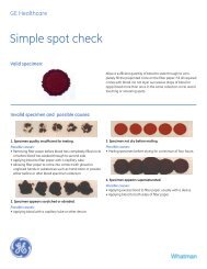
Figure 6. Digitally<br />
scanned images of<br />
the chemiluminescent<br />
(left) and colorimetric<br />
(right)<br />
detection<br />
systems <strong>for</strong> the<br />
targeted cytokines.<br />
The layout of the<br />
detected cytokines<br />
is the same as in<br />
figure 2.<br />
Further In<strong>for</strong>mation<br />
The concept of protein screening is an invaluable tool <strong>for</strong> researchers<br />
and clinicians. Miniaturization reduces the amount of precious<br />
sample required to build an array, and it also opens the door to<br />
the use of multiple probes, i.e., multiplexing. The system allows<br />
researchers to do comparative expression studies <strong>for</strong> drug target<br />
analysis as well as providing a plat<strong>for</strong>m <strong>for</strong> clinical applications,<br />
where complex solutions can be screened <strong>for</strong> specific antibodies<br />
and proteins. 18 There are protein arrays currently on the market<br />
that can be purchased “off-the-shelf ”, containing sub-sets of easily<br />
distinguishable capture antibodies commonly screened in the field<br />
(i.e., cytokines). 18,19 Frequent applications of protein screening<br />
technologies may require the researcher to personalize the array to<br />
his or her needs.<br />
In conclusion, it has been found that current protein microarraying<br />
protocols <strong>for</strong> high throughput objectives have conveniently<br />
con<strong>for</strong>med to the equipment used in generating DNA microarrays,<br />
specifically robotic arrayers. These arraying robots are very expensive,<br />
though necessary nonetheless <strong>for</strong> high-throughput processes. For<br />
low-throughput experiments, the need <strong>for</strong> this type of technology is<br />
not absolutely required since the use of manual type array tools will<br />
provide adequate per<strong>for</strong>mance. The <strong>MicroCaster</strong> generates reproducible,<br />
consistent arrays <strong>for</strong> low throughput research at a fraction<br />
of the cost of an automated arrayer. Similarly to high-throughput<br />
technologies, this nucleic acid-designed arrayer con<strong>for</strong>ms to protein<br />
applications with little manipulation of the current protocol <strong>for</strong> ease<br />
in producing protein arrays.<br />
1 Gillespie, D. and Spiegelman, S.; J. Mol. Biol. 12; 829-842 (1965).<br />
2 Schleicher & Schuell BioScience, Inc., Application Note #710, Methods <strong>for</strong> Preparing<br />
PCR Based Arrays on Nytran N and SuPerCharge Nylon Membranes. Debbie English<br />
and Breck Parker.<br />
3 Stillman, B.A., and Tonkinson, J.L., FASTSlides: A novel surface <strong>for</strong> microarrays.,<br />
BioTechniques (2000), 29: 630-35<br />
4 De Wildt, R. et. al; Antibody arrays <strong>for</strong> high-throughput screening of antibody-antigen<br />
interactions, Nature Biotechnology 18:989-994 (2000).<br />
5 Stillman, B.A. and Tonkinson, J.L., Expressionmicroarray hybridization kinetics are<br />
dependant on the length of immobilized species, but independant of surface chemistry.<br />
Analytical Biochemistry (2001), 295: 149-157.<br />
6 Knezevic, V., et. al; Proteomic profiling of the cancer microenvironmentby antibody<br />
arrays, Proteomics 1:1271-1278 (2001).<br />
7 Paweletz, C. P., et. al; Reverse phase protein microarrays which capture disease progression<br />
show activation of pro-survival pathways at the cancer invasion front, Oncogene<br />
20:1981-1989 (2001).<br />
8 Tonkinson, J.L., Osborne, D.S., Zhao, W.W. and Stillman, B.A., Expression microarray<br />
hybridization kinetics are dependant on the lengthof immobilized species, but independant<br />
of surface chemistry., AnalyticalBiochemistry (2001), 295:149-157<br />
9 Tonkinson, J.L., Parker, B.O., and Harvey, M.A., Chemluminescent detection of immobilized<br />
DNA from Southern blots to microarrays., Luminesence Biotechnology,<br />
(2002) CRC Press, pp189-201<br />
10 Wang, D., et. al; Carbohydrate microarrays <strong>for</strong> the recognition of cross-reactive molecular<br />
markers of microbes and host cells, Nature Biotechnology 20:275-281 (2002)<br />
11 Tonkinson, J.L., Osborne, D.S., Zhao, W.W. and Stillman, B.A., Development of micro<br />
scale immunoassays <strong>for</strong> parallel analysis of multiple analytes.<br />
12 Schena, M., Shalen, D., Davis, R.W., and Brown, P.O.; Science 270; 467-469 (1995).<br />
13 Duggan, D.J., Bittner, M., Chen, Y., Meltzer, P., and Trent, J. M.; Nature Genetics<br />
21;10-14 (1999).<br />
14 Venter, J. C. et.al; Science 291:1304-1351 (2001).<br />
15 MacBeath, G. and Schreiber, S.L.; Science 289;1760-1763 (2000)<br />
16 Walter, G. et. al; Current Opinion in Microbiology 2000; 3:298-302<br />
17 Moody, M.D.; S.W. Van Arsdell, K.P. Murphy, S.F. Orencole, and C.Burns,<br />
BioTechniques 31: 186-194 (July 2001)<br />
18 Schleicher & Schuell BioScience ProVision Microarray Kit, request ordering in<strong>for</strong>mation<br />
from S&S.<br />
19 Haab, B.B. , Dunham, M.J. and Brown, P.O.; Genome Biology<br />
2(2);RESEARCH0004.1 (2001).<br />
Note: <strong>MicroCaster</strong> protocol can be found on www.arraying.com<br />
Cy is a trademark of Amersham Biosciences. BioMax is a trademark of the Eastman<br />
Kodak Company. Super Signal is a registered trademark of Pierce Chemical Company.<br />
FAST ® , <strong>MicroCaster</strong> and <strong>Whatman</strong> ® are trademarks of the <strong>Whatman</strong> Group<br />
<strong>Whatman</strong> Inc., 200 Park Avenue, Suite 210, Florham Park, NJ 07932 USA, Tel: 1-800-<strong>Whatman</strong> (US and Canada), Fax: 1-973-245-8329, Email: info@whatman.com<br />
<strong>Whatman</strong> International Ltd., Springfield Mill, James <strong>Whatman</strong> Way, Maidstone, Kent ME14 2LE UK, Tel: +44 (0) 1622 676670, Fax: +44 (0) 1622 677011, Email: in<strong>for</strong>mation@whatman.com<br />
<strong>Whatman</strong> GmbH, Hahnestrasse 3, D-37586 Dassel, Germany, Tel: +49 (0) 5564 204 100, Fax: +49 80) 5564 204 533, Email: in<strong>for</strong>mation@whatman.com<br />
©2006 <strong>Whatman</strong> E2/11/06