MicroCaster - A Tool for Building Protein Microarrays - Whatman
MicroCaster - A Tool for Building Protein Microarrays - Whatman MicroCaster - A Tool for Building Protein Microarrays - Whatman
Arraying Methods Two FAST Slides (single pad, 20 mm x 51 mm) were placed on the stage of the slide holder and the indexing unit was lowered into position above the slides. The entire unit was oriented with the X-axis motion pin distally located and set in the first position (farthest right), and the Y-axis motion pin distally located in the first position as well (farthest left). Proteins were transferred onto FAST Slides following the MicroCaster protocol. Briefly, the pin tool was lowered into the protein sample making sure the pin was centered within the source plate well and did not touch the well’s bottom. With a steady motion, the pin tool was removed from the well and placed in the indexing unit. The pin tool was depressed with an even, steady motion until the second stop position was reached on the indexing unit. The hand tool was then placed back into the same set of source plate wells and a duplicate array was made on the second FAST Slide. The Y-axis motion pin was moved two places every cycle until a total of six replicates were transferred. The preceding steps were repeated for each subsequent concentration set of capture antibodies. Between each concentration set, the x-axis motion pin was moved two places to the left. The pins were washed and dried between concentration sets by dipping them into the three consecutive water baths with a swirling motion followed by an ethanol wash as described in the “MicroCaster Preparation and Cleaning Protocol” section of this application note. Immediately after the ethanol wash step, the pins were put through the same series of washes a second time and hot air-dried using a hair dryer. The arrayed FAST Slides were removed from the holder, air-dried for 5 minutes, and stored at room temperature prior to processing. Cell Lysate Preparation Human THP-1 leukemia cells (ATCC, Manassas, VA) were cultured to log phase in flasks containing 50 ml of RPMI 1640/10% fetal bovine serum. One flask was treated with 5 µg/ml lipopolysaccharide (LPS; Sigma-Aldrich) in the media to induce a cytokine response, while the other flask only contained media. Both flasks were incubated for an additional 6 hours. The cells were pelleted by centrifugation. The cell pellet was washed 2 times with ice cold 1X PBS, pH 7.4 and spun in a centrifuge to pellet the cells. The cells were resuspended in 1 ml of lysis buffer (1X PBS pH 7.4, 0.2% NP-40, 1mM EDTA, protease inhibitor cocktail) and subsequently vortexed. After a 15-minute incubation on ice, the lysed cells were centrifuged and the supernatants, containing the targeted cytokines, were removed and stored at -70°C until use. Detection Protocol Fluorescence: The arrayed FAST Slides were placed in sufficient Protein Array Blocking Solution cover the slides and gently agitated for 15 minutes. Following the blocking step, Incubation Chambers were attached to the slides and 725 µl of a 1:10 dilution of treated or untreated cell lysates with Protein Array Blocking Solution as the diluent was added to the appropriate slide. After completion of a 4-hour incubation at room temperature, the chambers were removed and the cell lysate solutions were discarded. The slides were washed 3 times with Protein Array Wash/Block Buffer for 5 minutes each and new chambers were added to the slides. A cocktail solution containing 200 ng/ml each of biotinylated IL-4, IL-8, IL-1β and ICAM-1 polyclonal antibodies in Protein Array Wash/Block Buffer was prepared and 725 µl of the cocktail was added to each of the slides. The slides were mixed and incubated for one hour at room temperature. The slides were washed three times with Protein Array Wash/Block Buffer for 5 minutes each. From this point on, the slides were protected from light to prevent photobleaching. Streptavidin-Cy 5 (Amersham Biosciences) was diluted 1:5000 in Protein Array Wash/Block Buffer and added to the slides, which were gently agitated for 1 hour at room temperature. The slides were subsequently washed 3 times at room temperature with Protein Array Wash/Block Buffer for 5 minutes each, and then rinsed for 2 seconds in dH 20, dried and scanned with the Perkin Elmer Scanarray 4000. ScanArray and QuantArray software from Perkin Elmer were used to capture images and to calculate specific intensities and spot morphologies. Chemiluminescence and Colorimetric Detection: The arrayed slides were placed into a sufficient volume of 1X TBS/1% casein to cover the slides and gently agitated for 15 minutes. Following the blocking step, Incubation Chambers were attached to the slides and 725 µl of a 1:10 dilution of treated or untreated cell lysates with 1X TBS/1% casein as the diluent was added to the appropriate slide. After completion of a 4 hour incubation at room temperature, the chambers were removed and the cell lysate solutions were discarded. The slides were washed 3 times with 1X TBS for 5 minutes each and new chambers were added to the slides. A cocktail solution containing 100 ng/ml each of biotinylated IL-4, IL-8, IL-1β and ICAM-1 polyclonal antibodies in 1X TBS/1% casein was prepared. The cocktail solution (725 µl) was added to each of the slide chambers, mixed and incubated for 1 hour at room temperature. They were washed 3 times with 1X TBS for 5 minutes each. Chemiluminescence: Streptavidin-HRP (horse radish peroxidase) was diluted 1:5000 in 1X TBS/1% casein and added to the Incubation Chamber. The slides were gently agitated for 1 hour at room temperature. The slides were subsequently washed 3 times at room temperature with 1X TBS for 5 minutes each. Detection utilized Super Signal ® Substrate (Pierce Endogen) treatment for 5 minutes followed by a 5 minute exposure to Kodak’s BioMax film. Colorimetric: Streptavidin-AP (alkaline phosphatase) was diluted 1:1650 in 1X TBS/1% casein and added to the Incubation Chamber. The slides were gently agitated for 1 hour at room temperature, and subsequently washed 3 times at room temperature with 1X TBS for 5 minutes each. The slides were placed into 25 mls of BCIP substrate (Sigma-Aldrich) and gently rocked to generate a colorimetric reaction. Figure 1. Diagram detailing the components involved in standard “sandwich” format. For colorimetric and chemiluminescent detection substitute SA-Cy 5 with SA-HRP and SA-AP respectively.
RESULTS The cytokine antibody microarrays used in this work were printed on FAST Slides using the MicroCaster manual arrayer, screened with cell lysates, and detected with a labeled antibody in a standard “sandwich” format (Figure 1). The titration of each cytokine antibody was printed as a row of 12 replicates, while the control rows consisted of a combination of six positive and six negative replicates (Figure 2a). The immobilized monoclonal antibodies to IL-4, IL-8, IL-1β and ICAM-1 were used to screen untreated and LPS-stimulated THP-1 cell lysates. IL-4 cytokine was not LPS-induced and therefore, was not detected, while IL-8, IL-1β and ICAM-1 cytokines generated signals representing the induction of each cytokine (Figure 2a). The cytokine antibodies arrayed on the FAST Slide supported linear binding of antibody to its cytokine target as shown for IL-8 cytokine in Figure 3 (IL-1β and ICAM-1 also display similar linearity [data not shown]). IL-4 IL-8 IL-1β ICAM-1 Pos. Control Neg. Control Controls 0.5 mg/ml 0.25 mg/ml 0.125 mg/ml Figure 2a. Image generated from the LPS-stimulated cell lysate. Image was scanned at Laser Power 85 and PMT 41. IL-4 IL-8 IL-1β ICAM-1 Pos. Control Neg. Control Controls 0.5 mg/ml 0.25 mg/ml 0.125 mg/ml Figure 2b. Image generated from the untreated cell lysate. Image was scanned at Laser Power 85 and PMT 41. The spot-to-spot CV’s ranged from 3.66-10.78%. The non-treated cell lysates resulted in low to non-detectable levels of all specific targeted cytokines (Figure 2b). The level of induction of cytokines in LPS-treated cells was determined by calculating the average specific intensities of the 0.5 mg/ml antibody rows in the LPS stimulated THP-1 cells and dividing it by the corresponding average specific intensities of the untreated cells. Figure 4 shows the fold-induction of cytokine protein expression when THP-1 cells are stimulated with LPS as judged by a protein microarray experiment (Figure 4). Figure 3. The filled diamonds represent the average specific intensity of the array per concentration of IL-8 capture antibody spotted. The calculated R2=0.9936 for the The MicroCaster pins are 457 µm in diameter, and each solid pin contains a small slot to assist with the sample transfer. These pins produce an average spot diameter of 973 µm at a center-to-center pitch of 2.5 x 1.5 mm resulting in 192 spots on the FAST Slide surface (20 mm x 51 mm). The evaluation of spot uniformity was based on a normality of 1.0 being perfect, and the mean uniformity was calculated to be 0.922. The fluorescent signal intensity was very consistent throughout the spot itself, which resulted in near perfect uniformity across an individual spot of the array. Spot morphology was not expected to be as robust and circular as robotically controlled deposition, however the spots produced by the solid pins of the MicroCaster proved to be consistently circular and of excellent morphology (Figure 5). In addition to fluorescent detection, arrays produced with the MicroCaster may be processed using chemiluminescent, colorimetric, and isotopic detection techniques. Examples of chemiluminescent and colorimetric detection of a cytokine array are shown in Figure 6. The arrays were incubated with LPS-stimulated THP-1 cell extracts and detected as described above. Only the treated slides are presented; the arrays probed with untreated extracts did not indicate stimulation of cytokines (data not shown) and are in agreement with the data collected using fluorescent detection methods. We used a pitch of 2.5 mm by 1.5 mm to provide extra room for chemiluminescent detec- Figure 4. Shows the fold-increase of four cytokines in LPS-treated versus untreated THP-1 cells.
Arraying Methods<br />
Two FAST Slides (single pad, 20 mm x 51 mm) were placed on the<br />
stage of the slide holder and the indexing unit was lowered into position<br />
above the slides. The entire unit was oriented with the X-axis<br />
motion pin distally located and set in the first position (farthest<br />
right), and the Y-axis motion pin distally located in the first position<br />
as well (farthest left).<br />
<strong>Protein</strong>s were transferred onto FAST Slides following the<br />
<strong>MicroCaster</strong> protocol. Briefly, the pin tool was lowered into the protein<br />
sample making sure the pin was centered within the source plate<br />
well and did not touch the well’s bottom. With a steady motion, the<br />
pin tool was removed from the well and placed in the indexing unit.<br />
The pin tool was depressed with an even, steady motion until the<br />
second stop position was reached on the indexing unit. The hand tool<br />
was then placed back into the same set of source plate wells and a<br />
duplicate array was made on the second FAST Slide. The Y-axis<br />
motion pin was moved two places every cycle until a total of six<br />
replicates were transferred.<br />
The preceding steps were repeated <strong>for</strong> each subsequent concentration<br />
set of capture antibodies. Between each concentration set, the x-axis<br />
motion pin was moved two places to the left. The pins were washed<br />
and dried between concentration sets by dipping them into the three<br />
consecutive water baths with a swirling motion followed by an<br />
ethanol wash as described in the “<strong>MicroCaster</strong> Preparation and<br />
Cleaning Protocol” section of this application note. Immediately<br />
after the ethanol wash step, the pins were put through the same<br />
series of washes a second time and hot air-dried using a hair dryer.<br />
The arrayed FAST Slides were removed from the holder, air-dried<br />
<strong>for</strong> 5 minutes, and stored at room temperature prior to processing.<br />
Cell Lysate Preparation<br />
Human THP-1 leukemia cells (ATCC, Manassas, VA) were cultured<br />
to log phase in flasks containing 50 ml of RPMI 1640/10% fetal<br />
bovine serum. One flask was treated with 5 µg/ml lipopolysaccharide<br />
(LPS; Sigma-Aldrich) in the media to induce a cytokine response,<br />
while the other flask only contained media. Both flasks were incubated<br />
<strong>for</strong> an additional 6 hours. The cells were pelleted by centrifugation.<br />
The cell pellet was washed 2 times with ice cold 1X PBS, pH<br />
7.4 and spun in a centrifuge to pellet the cells. The cells were resuspended<br />
in 1 ml of lysis buffer (1X PBS pH 7.4, 0.2%<br />
NP-40, 1mM EDTA, protease inhibitor cocktail) and subsequently<br />
vortexed. After a 15-minute incubation on ice, the lysed cells were<br />
centrifuged and the supernatants, containing the targeted cytokines,<br />
were removed and stored at -70°C until use.<br />
Detection Protocol<br />
Fluorescence: The arrayed FAST Slides were placed in sufficient<br />
<strong>Protein</strong> Array Blocking Solution cover the slides and gently agitated<br />
<strong>for</strong> 15 minutes. Following the blocking step, Incubation Chambers<br />
were attached to the slides and 725 µl of a 1:10 dilution of treated or<br />
untreated cell lysates with <strong>Protein</strong> Array Blocking Solution as the<br />
diluent was added to the appropriate slide. After completion of a<br />
4-hour incubation at room temperature, the chambers were removed<br />
and the cell lysate solutions were discarded. The slides were washed<br />
3 times with <strong>Protein</strong> Array Wash/Block Buffer <strong>for</strong> 5 minutes each<br />
and new chambers were added to the slides. A cocktail solution<br />
containing 200 ng/ml each of biotinylated IL-4, IL-8, IL-1β and<br />
ICAM-1 polyclonal antibodies in <strong>Protein</strong> Array Wash/Block Buffer<br />
was prepared and 725 µl of the cocktail was added to each of the<br />
slides. The slides were mixed and incubated <strong>for</strong> one hour at room<br />
temperature. The slides were washed three times with <strong>Protein</strong> Array<br />
Wash/Block Buffer <strong>for</strong> 5 minutes each. From this point on, the slides<br />
were protected from light to prevent photobleaching. Streptavidin-Cy 5<br />
(Amersham Biosciences) was diluted 1:5000 in <strong>Protein</strong> Array<br />
Wash/Block Buffer and added to the slides, which were gently agitated<br />
<strong>for</strong> 1 hour at room temperature. The slides were subsequently washed<br />
3 times at room temperature with <strong>Protein</strong> Array Wash/Block Buffer<br />
<strong>for</strong> 5 minutes each, and then rinsed <strong>for</strong> 2 seconds in dH 20, dried and<br />
scanned with the Perkin Elmer Scanarray 4000. ScanArray and<br />
QuantArray software from Perkin Elmer were used to capture images<br />
and to calculate specific intensities and spot morphologies.<br />
Chemiluminescence and Colorimetric Detection: The arrayed<br />
slides were placed into a sufficient volume of 1X TBS/1% casein to<br />
cover the slides and gently agitated <strong>for</strong> 15 minutes. Following the<br />
blocking step, Incubation Chambers were attached to the slides and<br />
725 µl of a 1:10 dilution of treated or untreated cell lysates with 1X<br />
TBS/1% casein as the diluent was added to the appropriate slide.<br />
After completion of a 4 hour incubation at room temperature, the<br />
chambers were removed and the cell lysate solutions were discarded.<br />
The slides were washed 3 times with 1X TBS <strong>for</strong> 5 minutes each and<br />
new chambers were added to the slides. A cocktail solution<br />
containing 100 ng/ml each of biotinylated IL-4, IL-8, IL-1β and<br />
ICAM-1 polyclonal antibodies in 1X TBS/1% casein was prepared.<br />
The cocktail solution (725 µl) was added to each of the slide chambers,<br />
mixed and incubated <strong>for</strong> 1 hour at room temperature. They<br />
were washed 3 times with 1X TBS <strong>for</strong> 5 minutes each.<br />
Chemiluminescence: Streptavidin-HRP (horse radish peroxidase)<br />
was diluted 1:5000 in 1X TBS/1% casein and added to the<br />
Incubation Chamber. The slides were gently agitated <strong>for</strong> 1 hour at<br />
room temperature. The slides were subsequently washed 3 times at<br />
room temperature with 1X TBS <strong>for</strong> 5 minutes each. Detection<br />
utilized Super Signal ® Substrate (Pierce Endogen) treatment <strong>for</strong> 5<br />
minutes followed by a 5 minute exposure to Kodak’s BioMax film.<br />
Colorimetric: Streptavidin-AP (alkaline phosphatase) was diluted<br />
1:1650 in 1X TBS/1% casein and added to the Incubation Chamber.<br />
The slides were gently agitated <strong>for</strong> 1 hour at room temperature, and<br />
subsequently washed 3 times at room temperature with 1X TBS<br />
<strong>for</strong> 5 minutes each. The slides were placed into 25 mls of BCIP<br />
substrate (Sigma-Aldrich) and gently rocked to generate a<br />
colorimetric reaction.<br />
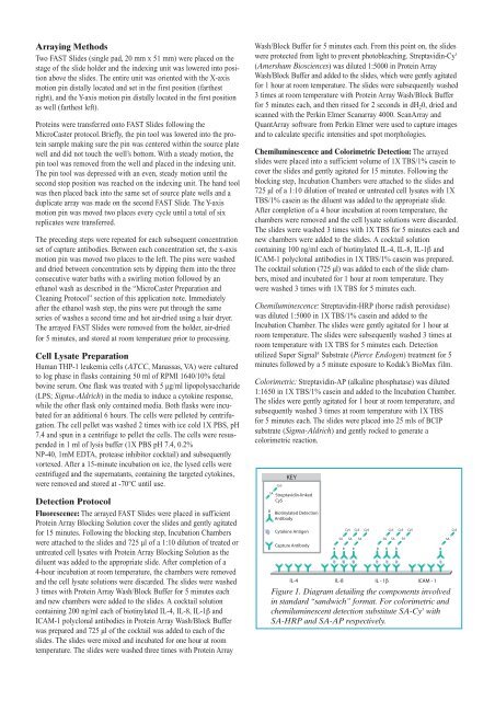
Figure 1. Diagram detailing the components involved<br />
in standard “sandwich” <strong>for</strong>mat. For colorimetric and<br />
chemiluminescent detection substitute SA-Cy 5 with<br />
SA-HRP and SA-AP respectively.


