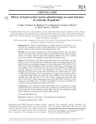
Tissue deposits of hydroxyethyl starch (HES): dose-dependent ... - BJA
Tissue deposits of hydroxyethyl starch (HES): dose-dependent ... - BJA
Tissue deposits of hydroxyethyl starch (HES): dose-dependent ... - BJA

You also want an ePaper? Increase the reach of your titles
YUMPU automatically turns print PDFs into web optimized ePapers that Google loves.
British Journal <strong>of</strong> Anaesthesia 82 (4): 510–15 (1999)<br />
<strong>Tissue</strong> <strong>deposits</strong> <strong>of</strong> <strong>hydroxyethyl</strong> <strong>starch</strong> (<strong>HES</strong>): <strong>dose</strong>-<strong>dependent</strong> and<br />
time-related<br />
C. Sirtl 1 *, H. Laubenthal 1 , V. Zumtobel 2 , D. Kraft 3 and W. Jurecka 4<br />
1 Department <strong>of</strong> Anaesthesiology and 2 Department <strong>of</strong> Surgery, St Josef-Hospital, Ruhr-Universität Bochum,<br />
Gudrunstr. 56, D-44791 Bochum, Germany. 3 Institute <strong>of</strong> General and Experimental Pathology and<br />
4 Department <strong>of</strong> Dermatology, Division <strong>of</strong> General Dermatology, University <strong>of</strong> Vienna, Vienna, Austria<br />
*To whom correspondence should be addressed<br />
<strong>Tissue</strong> <strong>deposits</strong> occur after administration <strong>of</strong> plasma substitutes. After <strong>hydroxyethyl</strong> <strong>starch</strong><br />
(<strong>HES</strong>), <strong>deposits</strong> may last for months, causing pruritus and impairment <strong>of</strong> function. Because<br />
elimination <strong>of</strong> <strong>HES</strong> <strong>deposits</strong> has not been demonstrated in humans, we studied 26 patients, for<br />
up to 7 yr after <strong>HES</strong> administration, to assess <strong>HES</strong> storage. <strong>HES</strong> <strong>dose</strong> ranged from 0.34 to<br />
15.00 g kg –1 body weight, and administration intervals from 1 day to 7 yr. Biopsies <strong>of</strong> the liver,<br />
muscle, spleen, intestine or skin were studied using light and electron microscopy and<br />
immunohistochemistry. <strong>HES</strong> storage was <strong>dose</strong>-<strong>dependent</strong>, decreased in all organs with time<br />
and was greater in patients suffering from pruritus. We conclude that tissue deposition <strong>of</strong> <strong>HES</strong><br />
is transitory and <strong>dose</strong>-<strong>dependent</strong>, with differences between subjects in severity and duration.<br />
Br J Anaesth 1999; 82: 510–15<br />
Keywords: blood, substitutes; pharmacology, <strong>hydroxyethyl</strong> <strong>starch</strong>; blood, colloids; fluid therapy<br />
Accepted for publication: November 23, 1998<br />
<strong>Tissue</strong> deposition <strong>of</strong> <strong>hydroxyethyl</strong> <strong>starch</strong> (<strong>HES</strong>) (used widely<br />
as a plasma substitute) is common, as with other colloidal<br />
plasma substitutes. After <strong>HES</strong> administration, deposition in<br />
the cells <strong>of</strong> the monocyte–macrophage system <strong>of</strong> various<br />
organs in addition to slow elimination <strong>of</strong> these <strong>deposits</strong> has<br />
been shown by several indirect techniques. 1–8 <strong>HES</strong> was<br />
assumed to be present in mainly empty-appearing intracytoplasmic<br />
vacuoles on light and electron microscopy.<br />
Using a polyclonal anti-<strong>HES</strong> antiserum, these ‘empty’<br />
vacuoles can be shown to contain <strong>HES</strong> at their margins. 910<br />
In some patients, storage <strong>of</strong> <strong>HES</strong> can be shown by immunohistochemistry<br />
and immuno-electron microscopy more than<br />
2 yr after administration, 10 raising the question <strong>of</strong> whether<br />
<strong>deposits</strong> may persist.<br />
Severe untreatable pruritus is a major side effect <strong>of</strong> high<br />
<strong>dose</strong>s <strong>of</strong> <strong>HES</strong>. It has been assumed that this itching<br />
phenomenon is caused by large amounts <strong>of</strong> tissue deposition<br />
<strong>of</strong> <strong>HES</strong>. 10–15 At present, it is not known if the duration <strong>of</strong><br />
tissue storage <strong>of</strong> <strong>HES</strong> is <strong>dose</strong>-<strong>dependent</strong> or if elimination<br />
<strong>of</strong> <strong>HES</strong> from the tissues occurs. Therefore, we biopsied<br />
tissues from patients after <strong>HES</strong> administration over a range<br />
<strong>of</strong> <strong>dose</strong>s, after periods <strong>of</strong> up to 7 yr, using light and electron<br />
microscopy, and immunohistochemical methods.<br />
Patients and methods<br />
We studied 26 patients retrospectively. They had previously<br />
received <strong>HES</strong> infusions and were studied during re-admission<br />
© British Journal <strong>of</strong> Anaesthesia<br />
to hospital. A complete history was obtained, including the<br />
date and duration <strong>of</strong> <strong>HES</strong> infusion, <strong>dose</strong> and preparation<br />
<strong>of</strong> <strong>HES</strong> (molecular weight, degree <strong>of</strong> substitution and<br />
trademark). No other agents had been given. The amount<br />
<strong>of</strong> <strong>HES</strong> given varied between 0.34 and 15.00 g kg –1 body<br />
weight, given over several days. According to the <strong>dose</strong> <strong>of</strong><br />
<strong>HES</strong>, patients were allocated to one <strong>of</strong> two groups. Patients<br />
in group A (n�16) received a maximum <strong>dose</strong> <strong>of</strong> 2 g kg –1 ,<br />
which is <strong>of</strong>ten used for volume replacement. Patients in<br />
group B (n�10) received greater <strong>dose</strong>s <strong>of</strong> <strong>HES</strong>, ranging<br />
from more than 3 up to 15 g kg –1 , given for microcirculatory<br />
disorders (Table 1).<br />
All 16 patients in group A were undergoing surgery for<br />
malignancy, other surgical indications or for chronic or<br />
bilateral orthopaedic disorders. They received a mean <strong>dose</strong><br />
<strong>of</strong> 0.8 (range 0.3–1.9) g kg –1 within 24 h (Table 1). None<br />
<strong>of</strong> the patients developed pruritus. Patients were comparable<br />
in blood loss, organ function and clinical laboratory findings.<br />
Informed consent was obtained for taking pea-sized tissue<br />
samples from the operating site. Biopsies were obtained<br />
from one or more sites (skin, liver, small intestine, striated<br />
muscle or spleen) between 1 day and 84 months after <strong>HES</strong><br />
infusion (Table 1). This procedure was approved by the<br />
Ethics Committee <strong>of</strong> the Faculty <strong>of</strong> Medicine at the Ruhr-<br />
Universität Bochum.<br />
Patients in group B received greater amounts <strong>of</strong> <strong>HES</strong>,<br />
with a mean <strong>dose</strong> <strong>of</strong> 7.8 (3.1–15.0) g kg –1 . Nine patients<br />
(Nos B1–8, B10) received <strong>HES</strong> infusions for vascular disease,<br />
Downloaded from<br />
http://bja.oxfordjournals.org/ by guest on January 23, 2013
<strong>Tissue</strong> <strong>deposits</strong> <strong>of</strong> <strong>HES</strong><br />
Table 1 Patient details (<strong>dose</strong> group, sex, age), type (molecular weight (Mw)/molar substitution (MS)) and amount <strong>of</strong> <strong>HES</strong> administered, time interval between<br />
<strong>HES</strong> infusion and biopsies, organ biopsied (skin, liver, small (S.) intestine, striated (St.) muscle, spleen), and findings regarding <strong>HES</strong> <strong>deposits</strong> in organ tissues<br />
(–negative, � weak, �� moderate, ��� marked signs <strong>of</strong> storage, i.e. vacuoles)<br />
Type <strong>of</strong> <strong>HES</strong> infused Amount <strong>of</strong> <strong>HES</strong> infused Organ investigated and Time <strong>of</strong> biopsy after <strong>HES</strong> administration<br />
Patient No. Sex Age (yr) (Mw/MS) (g kg –1 ) <strong>HES</strong> storage (d � days, m � months)<br />
A1 F 45 200/0.5 1.69 Skin � 1d<br />
Liver �<br />
S. intestine �<br />
St. muscle �<br />
Spleen �<br />
A2 M 42 200/0.5 0.52 Skin �� 3.5 m<br />
St. muscle �<br />
A3 M 65 200/0.5 1.19 Skin – 3.5 m<br />
S. intestine –<br />
St. muscle –<br />
A4 F 40 200/0.5 1.00 Skin �� 5m<br />
A5 M 62 200/0.5 0.65 Skin � 9m<br />
St. muscle –<br />
A6 M 65 450/0.7 0.34 Skin � 10 m<br />
St. muscle �<br />
S. intestine �<br />
A7 M 54 200/0.5 0.68 Skin � 12 m<br />
St. muscle �<br />
A8 F 61 200/0.5 0.86 Skin � 13 m<br />
A9 F 52 200/0.5 0.53 Skin – 15 m<br />
St. muscle –<br />
A10 F 54 450/0.7 0.50 Skin � 15 m<br />
St. muscle �<br />
S. intestine �<br />
A11 F 63 200/0.5 1.00 Skin � 15 m<br />
Liver �<br />
St. muscle �<br />
A12 M 24 200/0.5 0.69 Skin – 26 m<br />
St. muscle -<br />
A13 M 55 200/0.5 0.60 Skin � 34 m<br />
St. muscle –<br />
A14 F 71 (a) 200/0.5 1.86 Skin � (a) 16 m<br />
(b) 450/0.7 Liver – (b) 48 m<br />
St. muscle –<br />
A15 F 72 450/0.7 0.43 Skin � 53 m<br />
St. muscle –<br />
A16 F 67 450/0.7 0.56 Skin – 84 m<br />
Liver –<br />
St. muscle –<br />
B1 F 26 200/0.5 15.00 Skin ��� 4m<br />
B2 M 50 200/0.5 6.80 Skin ��� 6m<br />
B3 F 68 200/0.5 3.22 Skin ��� 12 m<br />
B4 M 25 200/0.5 4.40 Skin �� 12 m<br />
B5 F 42 200/0.5 6.92 Skin ��� 14 m<br />
St. muscle ���<br />
B6 M 58 200/0.5 7.24 Skin ��(�) 19 m<br />
B7 F 54 200/0.5 8.33 Skin ��(�) 25 m<br />
B8 M 65 200/0.5 13.88 Skin ���/�� 6 m/41 m<br />
B9 M 56 200/0.6 3.10 Skin ���/– 6 m/45 m<br />
B10 M 27 200/0.5 7.80 Skin ���/� 17 m/54 m<br />
one patient (No. B5) to improve tissue perfusion after plastic<br />
surgery and another (No. B9) for treatment <strong>of</strong> a chronic leg<br />
ulcer. Seven <strong>of</strong> these patients (Nos B2, B3, B6–10) have<br />
been reported on previously, 10 but biopsy data have not been<br />
published. <strong>HES</strong> patients B1–8 and B10 developed severe<br />
pruritus, leading to consultation with dermatologists and/or<br />
anaesthetists. After informed consent, one or several diagnostic<br />
and follow-up skin biopsies were performed using<br />
local anaesthesia, between 6 and 25 months after <strong>HES</strong> infusion.<br />
In patients B8, B9 and B10, additional follow-up<br />
biopsies were obtained approximately 3 yr later (Table 1).<br />
511<br />
<strong>Tissue</strong> processing<br />
All biopsies, regardless <strong>of</strong> tissue source, were processed<br />
similarly. Biopsies were divided into several pieces and<br />
fixed in 7% buffered paraformaldehyde or Karnovsky’s<br />
fixative. Some were post-fixed in osmium tetroxide<br />
and embedded in paraplast, Epon 812 or LR White.<br />
Haematoxylin–eosin (HE) or periodic acid–Schiff (PAS)<br />
stained paraffin sections, or semi-thin toluidine blue<br />
stained Epon sections were used for light microscopy,<br />
and unstained paraffin sections for immunohistochemistry.<br />
Ultra-thin uranyl acetate and lead citrate stained Epon<br />
Downloaded from<br />
http://bja.oxfordjournals.org/ by guest on January 23, 2013
sections and ultra-thin LR White sections were used for<br />
electron microscopy or immuno-electron microscopy,<br />
respectively.<br />
Immunohistochemistry<br />
For light microscopy, after blocking endogenous peroxidases,<br />
paraffin sections were incubated with a well defined<br />
polyclonal rabbit antiserum against <strong>HES</strong> (kindly given by<br />
Dr Richter, Helsingborg, Sweden), 16 followed by swine antirabbit<br />
immunoglobulin, horseradish peroxidase conjugated<br />
with rabbit anti-peroxidase (peroxidase anti-peroxidase<br />
(PAP) technique) and 3-amino-9-ethyl-carbazol. Sections<br />
were counterstained with haematoxylin. For immunoelectron<br />
microscopy, ultra-thin LR White sections were<br />
incubated with the anti-<strong>HES</strong> antiserum followed by protein<br />
A gold using 15-nm gold particles and counterstained with<br />
uranyl acetate.<br />
Controls included dilution series, omission <strong>of</strong> primary<br />
antibody, replacement <strong>of</strong> the primary antibody by a nonimmune<br />
serum or an unrelated antibody, incubation with<br />
protein A gold alone or incubation with non-labelled protein<br />
A before protein A gold application.<br />
Quantification<br />
The amount <strong>of</strong> <strong>HES</strong> deposition in each biopsy was quantified<br />
by combining the results <strong>of</strong> all staining procedures. Results<br />
were categorized as follows: ����a large number <strong>of</strong><br />
storage vacuoles present on light microscopy (semi-thin<br />
section, paraffin section) confirmed by immunohistochemistry<br />
or immuno-electron microscopy; ���moderate<br />
numbers <strong>of</strong> storage vacuoles present on light microscopy<br />
(mainly semi-thin section) confirmed by immunohistochemistry<br />
or immuno-electron microscopy; ��scanty storage<br />
vacuoles either in semi-thin sections or immunohistochemistry;<br />
and – �no evidence <strong>of</strong> deposition with any<br />
method used.<br />
The methods <strong>of</strong> tissue processing, staining, immunohistochemistry<br />
and controls for light and electron microscopy<br />
have been described previously. 10<br />
Results<br />
We considered either a positive immunohistochemistry/<br />
immuno-electron microscopy or a definite vacuolization <strong>of</strong><br />
cells to indicate <strong>HES</strong> deposition. Paraffin section vacuolated<br />
cells, which were partly PAS-positive, could only weakly<br />
be seen in the skin <strong>of</strong> patients in group B. However, in<br />
toluidine blue stained semi-thin sections, apparently empty<br />
intra-cytoplasmic vacuoles could be detected easily in<br />
various cells <strong>of</strong> positive organs. In addition to vascular<br />
endothelial cells, <strong>HES</strong> deposition occurred mainly in cells <strong>of</strong><br />
the monocyte–macrophage system. In the liver, parenchymal<br />
cells or sinusoidal spindle-shaped cells showed vacuolization.<br />
In striated muscle, muscle cells showed no signs<br />
<strong>of</strong> <strong>HES</strong> deposition, but interstitial histiocytes or macrophages<br />
had intra-cytoplasmic vacuoles. In the spleen,<br />
Sirtl et al.<br />
512<br />
Fig 1 Positivity or negativity <strong>of</strong> biopsies <strong>of</strong> several organs for <strong>HES</strong><br />
deposition at different time intervals after <strong>HES</strong> infusion (Note <strong>HES</strong> persists<br />
longest in skin).<br />
vacuoli were prominent in reticular cells in the red pulp<br />
and at the border between red and white pulp. In the<br />
intestine, mucosal epithelium did not have <strong>HES</strong> deposition,<br />
but vascular endothelial cells and stromal macrophages had<br />
some vacuoli. In the skin, vascular endothelial cells and<br />
dermal macrophages had vacuoli. In patients in group B,<br />
perineural cells, endoneural connective tissue cells and<br />
Langerhans’ cells also had vacuoli. Light microscopy findings<br />
were confirmed by electron microscopy, showing intracytoplasmic<br />
empty membrane-bound vacuoles with some<br />
electron-dense material at their margins. In each patient,<br />
<strong>HES</strong> deposition was also shown either by positive immunohistochemical<br />
or immuno-electron microscopy staining <strong>of</strong><br />
vacuolated cells with the anti-<strong>HES</strong> antiserum. Control<br />
stainings were negative.<br />
<strong>HES</strong> <strong>deposits</strong> were found in all biopsied organs (liver,<br />
muscle, spleen, intestine, skin) on the first day after <strong>HES</strong><br />
infusion. The amount <strong>of</strong> <strong>HES</strong> found in all patients at<br />
different times after <strong>HES</strong> administration is shown in Table 1.<br />
In group A (low <strong>dose</strong> group, 0.3–1.9 g kg –1 ), in liver,<br />
muscle and skin biopsies, <strong>HES</strong> <strong>deposits</strong> decreased with<br />
time, and organs became negative for <strong>HES</strong>. In the spleen<br />
and intestine, not enough patients were studied to show the<br />
time course. In liver and muscle, <strong>deposits</strong> were found up<br />
to 15 months, and in skin up to 54 months, after <strong>HES</strong><br />
infusion (Table 1, Fig. 1).<br />
In all 10 patients in group B (high <strong>dose</strong> group, 3.1–15.00<br />
gkg –1 ) massive <strong>deposits</strong> <strong>of</strong> <strong>HES</strong> were demonstrated in the<br />
Downloaded from<br />
http://bja.oxfordjournals.org/ by guest on January 23, 2013
Fig 2 Skin biopsies <strong>of</strong> patient No. B8, who received <strong>HES</strong> 200/0.5<br />
13.88 g kg –1 . (A) Six months after infusion, numerous large ‘empty’<br />
vacuoles can be seen in the dermis in cells <strong>of</strong> the monocyte–macrophage<br />
system; (B) 45 months after infusion only few small vacuoles can be seen<br />
in a macrophage (Y) and endothelial cell (I) Toluidine blue stained semithin<br />
sections; A�420, B�340.<br />
skin and in one patient in muscle at 4–25 months after <strong>HES</strong><br />
administration (Table 1). Nine <strong>of</strong> 10 patients (Nos B1–4,<br />
B6–8, B10) had severe pruritus at the time <strong>of</strong> the first<br />
biopsy. In three patients, biopsies repeated approximately<br />
3 yr later showed remarkable reduction <strong>of</strong> <strong>HES</strong> deposition<br />
in the skin (Fig. 2A, B) and one patient (No. B9) was<br />
<strong>Tissue</strong> <strong>deposits</strong> <strong>of</strong> <strong>HES</strong><br />
513<br />
completely negative by that time (Table 1). Pruritus had<br />
resolved in two <strong>of</strong> these patients (Nos B8, B10).<br />
Discussion<br />
Our results showed clearly that tissue deposition <strong>of</strong> <strong>HES</strong><br />
was <strong>dose</strong>- and time-<strong>dependent</strong>. In particular, patients in<br />
group B who received 3.1–15.00 g kg –1 within a few days,<br />
showed massive tissue deposition <strong>of</strong> <strong>HES</strong>. In the low <strong>dose</strong><br />
group (0.3–1.9 g kg –1 ) only moderate or slight tissue<br />
<strong>deposits</strong> were found. With greater time between <strong>HES</strong><br />
administration and biopsy, a decrease in <strong>HES</strong> <strong>deposits</strong><br />
occurred in both groups, with complete loss <strong>of</strong> <strong>HES</strong> <strong>deposits</strong><br />
in some patients in group A and in one patient in group B.<br />
<strong>Tissue</strong> deposition occurs with all colloidal plasma substitutes.<br />
For gelatins and albumin there is only indirect<br />
evidence <strong>of</strong> delayed elimination from the circulation and<br />
<strong>of</strong> some <strong>deposits</strong> in the cells <strong>of</strong> the monocyte–macrophage<br />
system in vitro. 17–20 For dextrans, storage has been described<br />
in the monocyte–macrophage system in vivo and in vitro, 21<br />
which may be important in patients undergoing chronic<br />
haemodialysis. 522<strong>HES</strong> has been demonstrated mainly in<br />
the cells <strong>of</strong> the monocyte–macrophage system <strong>of</strong> various<br />
tissues by indirect methods (i.e. empty vacuoles2 4–8 ),<br />
14C-<strong>HES</strong> uptake, 3 7 photometric measurements3 7 and<br />
specifically by immunohistochemical and immuno-electron<br />
microscopy techniques in several rat tissues and in human<br />
skin by our group. 910Using the same techniques, we have<br />
shown <strong>HES</strong> <strong>deposits</strong> in human liver, striated muscle, spleen<br />
and intestine. We also showed that <strong>HES</strong> <strong>deposits</strong> in skin<br />
increased with the amount <strong>of</strong> <strong>HES</strong> administered and<br />
decreased with time after <strong>HES</strong> administration.<br />
Until now, complete elimination <strong>of</strong> <strong>HES</strong> <strong>deposits</strong> from<br />
human tissue was uncertain. We found no <strong>HES</strong> in all organs<br />
investigated, including skin, in five <strong>of</strong> our 26 patients (Nos<br />
A3, A9, A12, A16, B9) and a decrease (i.e. negative<br />
findings) in some organs, except skin, in four other patients<br />
(Nos A5, A13, A14, A15) over several years. This loss <strong>of</strong><br />
<strong>deposits</strong> may reflect the different specific kinetics <strong>of</strong> <strong>HES</strong><br />
in human organs, as is the case for animal organs, except<br />
skin. 23 Hitherto, slow elimination <strong>of</strong> <strong>HES</strong> from plasma and<br />
tissues has been inferred from indirect investigations in<br />
animals and humans1 3–6 8 24over relatively short time<br />
intervals, but complete elimination has never been demonstrated.<br />
Normally, persistent tissue <strong>deposits</strong> <strong>of</strong> <strong>HES</strong> do not<br />
appear to be clinically relevant to the function <strong>of</strong> organs or<br />
<strong>of</strong> the monocyte–macrophage system. 810252627However, in patients with renal failure started on chronic dialysis,<br />
even small quantities <strong>of</strong> penta<strong>starch</strong>, such as <strong>HES</strong> 200/0.5<br />
or <strong>HES</strong> 70/0.55, may cause hepatosplenomegaly, portal<br />
hypertension and ascites. This is thought to be caused<br />
by hepatocytes ballooning with <strong>HES</strong> vacuoli and thus<br />
narrowing hepatic sinusoids. This can resolve after months<br />
or years. 228<br />
In our 26 patients, in addition to the amount <strong>of</strong> <strong>HES</strong><br />
Downloaded from<br />
http://bja.oxfordjournals.org/ by guest on January 23, 2013
administered, other factors appeared to be responsible for<br />
the duration <strong>of</strong> <strong>HES</strong> deposition. It is not surprising that<br />
patients who received the largest amounts <strong>of</strong> <strong>HES</strong> had<br />
persistence <strong>of</strong> this substance in their tissues. However,<br />
patient No. A3, who received a relatively large amount <strong>of</strong><br />
<strong>HES</strong> (1.19 g kg –1 ) over a short time showed no tissue<br />
<strong>deposits</strong> in skin, intestine and muscle after 3.5 months.<br />
Comparing patients A9 and A10, who received two different<br />
types (A9 penta<strong>starch</strong>-like vs A10 heta<strong>starch</strong>-like with a<br />
different degree <strong>of</strong> substitution) but comparable amounts<br />
<strong>of</strong> <strong>HES</strong>, patient No. A9 had no <strong>deposits</strong> in skin and muscle<br />
while patient A10 had <strong>deposits</strong> in skin, muscle and intestine<br />
after 15 months. Comparing patients A12 and A13, who<br />
received <strong>HES</strong> 200/0.5 in comparable quantities, patient<br />
No. A12 had no <strong>deposits</strong> in skin and muscle at 26 months<br />
while patient No. A13 had none in muscle but still had<br />
<strong>deposits</strong> in skin 34 months after <strong>HES</strong> administration. This<br />
could be caused by differences in patient hydration status<br />
(amount <strong>of</strong> fluid ingested by or crystalloid infusions given<br />
to patients during or shortly after <strong>HES</strong> administration) or<br />
kidney function, resulting in differences in elimination<br />
<strong>of</strong> <strong>HES</strong> from the circulation in the early phase after<br />
administration.<br />
2 28–31<br />
Another explanation is that increased blood loss may<br />
eliminate <strong>HES</strong>, as shown in patient No. A3. Finally,<br />
metabolic differences may exist. <strong>HES</strong> is metabolized by<br />
hydrolysis <strong>of</strong> glycosidic bonds within the glucose chains<br />
by various plasma and lysosomal glycosidases. Differences<br />
in metabolic capacity could be responsible for the development<br />
<strong>of</strong> pruritus, 32 which could also explain differences in<br />
duration <strong>of</strong> <strong>HES</strong> <strong>deposits</strong> in patients receiving similar<br />
amounts <strong>of</strong> the same substance.<br />
<strong>HES</strong> <strong>deposits</strong> have been described in several organs, and<br />
probably all organs except the central nervous system are<br />
involved. In 16 patients in group A it is obvious that the<br />
skin was the organ most involved, retaining <strong>HES</strong> <strong>deposits</strong><br />
longer than other organs (Fig. 1), but the number <strong>of</strong> patients<br />
investigated was small. No clear explanation can be given<br />
for this phenomenon. Differences in the density <strong>of</strong> cells <strong>of</strong><br />
the monocyte–macrophage system and/or differences in<br />
their mobility, or metabolic capacity in various organs could<br />
be responsible.<br />
Deposits may not be found if they are in very coarsely<br />
or inhomogeneously distributed vacuoles. Previous investigations,<br />
taking parallel biopsies at different sites in the<br />
same organ, did not find inhomogeneity <strong>of</strong> the <strong>deposits</strong> in<br />
various tissues. 8 Very isolated vacuoles (e.g. 1 cm –3 ) may<br />
be <strong>of</strong> no clinical importance.<br />
Hepatosplenomegaly in chronic terminal renal failure and<br />
persistent pruritus are the only major side effects attributed<br />
to tissue deposition after <strong>HES</strong> infusion. Even after large<br />
<strong>dose</strong>s during prolonged ICU therapy, patients do not complain<br />
<strong>of</strong> pruritus. Previous studies concluded that tissue<br />
deposition in general and pruritus in particular are <strong>dose</strong><strong>dependent</strong><br />
and time-limited phenomena. 10–13 15 33 Our study<br />
supports this. Only patients in the high <strong>dose</strong> group (except<br />
Sirtl et al.<br />
514<br />
patient No. B9) suffered from pruritus, although the latter<br />
received comparable amounts <strong>of</strong> <strong>HES</strong> and showed similar<br />
massive deposition <strong>of</strong> <strong>HES</strong> in the skin. At follow-up<br />
approximately 3 yr later, patients B8 and B10 had recovered<br />
from pruritus, and their skin biopsies still had <strong>HES</strong> <strong>deposits</strong>,<br />
but to a lesser content. This supports the belief that <strong>HES</strong><br />
<strong>deposits</strong> and pruritus are <strong>dose</strong>-<strong>dependent</strong> and transitory.<br />
Previous recommendations to reduce the <strong>dose</strong> <strong>of</strong> <strong>HES</strong>,<br />
especially for long-term treatment, reduced the frequency<br />
<strong>of</strong> <strong>HES</strong>-induced pruritus.<br />
In summary, tissue deposition <strong>of</strong> <strong>HES</strong> and its major<br />
clinical effect (pruritus) are mainly <strong>dose</strong>-<strong>dependent</strong> transitory<br />
phenomena. Complete elimination <strong>of</strong> <strong>HES</strong> <strong>deposits</strong><br />
occurs over different times after administration.<br />
References<br />
1 Boon JC, Jesch F, Ring J, Messmer K. Intravascular persistence <strong>of</strong><br />
<strong>hydroxyethyl</strong> <strong>starch</strong> in man. Eur Surg Res 1976; 8: 497–503<br />
2 Dienes HP, Gerharz CD, Wagner R, Weber M, John HD.<br />
Accumulation <strong>of</strong> <strong>hydroxyethyl</strong> <strong>starch</strong> (<strong>HES</strong>) in the liver <strong>of</strong> patients<br />
with renal failure and portal hypertension. J Hepatol 1986; 3: 223–7<br />
3 Hulse JD, Stoll RG, Yacobi A, Gupta SD, Lai CM. Elimination <strong>of</strong><br />
high molecular weight <strong>hydroxyethyl</strong> <strong>starch</strong> in rats. Res Commun<br />
Clin Pathol Pharmacol 1980; 29: 149–58<br />
4 Jesch F, Hübner G, Zumtobel V, Zimmermann M, Messmer K.<br />
Hydroxyäthylstärke (HÄS 450/0,7) in Plasma und Leber.<br />
Konzentrationsverlauf und histologische Veränderungen beim<br />
Menschen. Infusionstherapie 1979; 6: 112–17<br />
5 Lindblad G, Falk J. Konzentrationsverlauf von Hydroxyäthylstärke<br />
und Dextran im Serum und Lebergewebe von Kaninchen und die<br />
histopathologischen Folgen der Speicherung von Hydroxyäthylstärke.<br />
Infusionstherapie 1976; 3: 301–3<br />
6 Paulini K, Sonntag W. Veränderungen des RHS der Ratte nach<br />
parenteraler Gabe von Dextran (Mw 40 000) und Hydroxyäthylstärke<br />
(Mw 40 000). Infusionstherapie 1976; 3: 294–9<br />
7 Thompson WL, Fukushima T, Rutherford RB, Walton RP.<br />
Intravascular persistence, tissue storage and excretion <strong>of</strong><br />
<strong>hydroxyethyl</strong> <strong>starch</strong>. Surg Gynecol Obstet 1970; 131: 965–72<br />
8 Sirtl C, Hübner G, Jesch F. Zur Speicherung von<br />
Hydroxyäthylstärke (HÄS) im menschlichen Gewebe. Beitr<br />
Anaesthesiol Intensivmed 1988; 26: 74–97<br />
9 Parth E, Jurecka W, Szépfalusi Z, et al. Histological and<br />
immunohistochemical investigations <strong>of</strong> <strong>hydroxyethyl</strong><strong>starch</strong><br />
<strong>deposits</strong> in rat tissues. Eur Surg Res 1992; 24: 13–21<br />
10 Jurecka W, Szépfalusi Z, Parth E, et al. Hydroxyethyl<strong>starch</strong> <strong>deposits</strong><br />
in human skin—a model for pruritus? Arch Dermatol Res 1993;<br />
285: 13–19<br />
11 Hong B, Zhang T. Studies on the side-effect <strong>of</strong> <strong>hydroxyethyl</strong><br />
<strong>starch</strong>. J Pharmacol 1988; 108: 43<br />
12 Parker NE, Porter JB, Williams HJM, Leftley N. Pruritus after<br />
administration <strong>of</strong> heta<strong>starch</strong>. BMJ 1982; 284: 385–6<br />
13 Hermann J, Gall H. Diagnose und Therapie des persistierenden<br />
Pruritus nach Infusion von Hydroxyäthylstärke (HÄS). Aktuelle<br />
Dermatol 1990; 16: 166–7<br />
14 Schneeberger R, Albegger K, Oberascher G, Miller K. Juckreiz—<br />
Eine Nebenwirkung von Hydroxyäthylstärke (<strong>HES</strong>)? HNO 1990;<br />
38: 298–303<br />
15 Gall H, Kaufmann R, von Ehr M, Schumann K, Sterry W.<br />
Persistierender Pruritus nach Hydroxyäthylstärke-Infusionen.<br />
Hautarzt 1993; 44: 713–16<br />
Downloaded from<br />
http://bja.oxfordjournals.org/ by guest on January 23, 2013
16 Richter W, de Belder AN. Antibodies against <strong>hydroxyethyl</strong> <strong>starch</strong><br />
produced in rabbits by immunization with a protein–<strong>HES</strong>conjugate.<br />
Int Arch Allergy Appl Immunol 1976; 52: 307–14<br />
17 Frank G. Zur Frage der Speicherung von Haemaccel in<br />
Lymphknotenmakrophagen, Inauguraldissertation. Mainz: Johannes-<br />
Gutenberg-Universitaet, 1978<br />
18 Zekorn D. Intravascular retention, dispersal, excretion and breakdown<br />
<strong>of</strong> gelatin plasma substitutes. Bibl Haemat 1969; 33: 131–40<br />
19 Kief H. Morphological findings following single or multiple<br />
administration <strong>of</strong> gelatin plasma substitutes. Bibl Haemat 1969; 33:<br />
380–97<br />
20 Muchmore E, Bonhard K, Kothe N. Distribution and clearance<br />
from the body <strong>of</strong> an oxypolygelatin plasma substitute determined<br />
by radioactive tracer study in chimpanzees. Arzneimittelforschung<br />
1983; 11: 1552–4<br />
21 Sonntag W, Paulini K. Speicherung von Plasmaexpandern im RHS.<br />
Verh Dtsch Ges Pathol 1978; 62: 346<br />
22 Bergonzi G, Paties C, Vassallo G, et al. Dextran <strong>deposits</strong> in tissues<br />
<strong>of</strong> patients undergoing haemodialysis. Nephrol Dial Transplant 1990;<br />
5: 54–8<br />
23 Elliger J. Experimentelle Untersuchung zur Elimination und<br />
Gewebespeicherung mittel- und niedermolekularer Hydroxyäthylstärke<br />
(‘HAES-steril’ und ‘Expafusin’), Inauguraldissertation. Frankfurt<br />
(Main): Johann Wolfgang Goethe Universität, 1984<br />
24 Thompson WL, Fukushima T, Rutherford RB, Walton RP.<br />
Intravasale Persistenz, Gewebespeicherung und Ausscheidung von<br />
Hydroxyäthylstärke. Infusionstherapie 1979; 6: 151–5<br />
25 Lenz G, Hempel V, Junger H, Werle H, Buckenmaier P.<br />
Auswirkungen von Hydroxyäthylstärke, Oxypolygelytine und<br />
<strong>Tissue</strong> <strong>deposits</strong> <strong>of</strong> <strong>HES</strong><br />
515<br />
Humanalbumin auf die Phygozytosefunktion des RES gesunder<br />
Probanden. Anaesthesist 1986; 35: 423–8<br />
26 Heilmann L, Lorch E, Hojnacki B, Müntefering H, Förster H.<br />
Die Speicherung von zwei unterschiedlichen Hydroxyäthylstärke-<br />
Präparaten in der Plazenta nach Hämodilution bei Patientinnen<br />
mit fetaler Mangelentwicklung oder Schwangerschaftshochdruck.<br />
Infusionstherapie 1991; 18: 236–43<br />
27 Szépfalusi Z, Parth E, Jurecka W, Luger TA, Kraft D. Human<br />
monocytes and keratinocytes in culture ingest <strong>hydroxyethyl</strong><strong>starch</strong>.<br />
Arch Dermatol Res 1993; 283: 144–50<br />
28 Pfeifer U, Kult J, Förster H. Ascites als Komplikation hepatischer<br />
Speicherung von Hydroxyethylstärke (<strong>HES</strong>) bei Langzeitdialyse.<br />
Klin Wochenschr 1984; 62: 862–6<br />
29 Kiesewetter H, Waldhausen P, Schimetta W, Wilhelm HJ,<br />
Koscielny J. Mögliche Nebenwirkungen einer HAES-Infusion und<br />
deren Behandlung. In: Koscielny J, Kiesewetter H, Jung F, Haass<br />
A, eds. Hämodilution. Berlin: Springer Verlag, 1991; 207–14<br />
30 Messmer K. The use <strong>of</strong> plasma substitutes with special attention<br />
to their side effects. World J Surg 1987; 11: 69–74<br />
31 Sirtl C, Laubenthal H, Dieterich HJ, Hügler P, Peter K.<br />
Nebenwirkungen von kolloidalen Plasmaersatzmitteln unter<br />
besonderer Berücksichtigung von Hydroxyethylstärke (<strong>HES</strong>). Beitr<br />
Anaesthesiol Intensivmed 1990; 31: 35–56<br />
32 Cox NH, Poppel AW. Persistent erythema and pruritus, with<br />
a confluent histiocytic skin infiltrate, following the use <strong>of</strong> a<br />
<strong>hydroxyethyl</strong><strong>starch</strong> plasma expander. Br J Dermatol 1996; 134:<br />
353–7<br />
33 Albegger K, Schneeberger R, Franke V, Oberascher G, Miller<br />
K. Juckreiz nach Therapie mit Hydroxyäthylstärke (<strong>HES</strong>) bei<br />
otoneurologischen Erkrankungen. Wien Med Wochenschr 1992;<br />
142: 1–7<br />
Downloaded from<br />
http://bja.oxfordjournals.org/ by guest on January 23, 2013