

Rapid prototyping in tissue engineering: challenges and potential
Rapid prototyping in tissue engineering: challenges and potential
Rapid prototyping in tissue engineering: challenges and potential

You also want an ePaper? Increase the reach of your titles
YUMPU automatically turns print PDFs into web optimized ePapers that Google loves.
Review TRENDS <strong>in</strong> Biotechnology Vol.22 No.12 December 2004<br />
<strong>Rapid</strong> <strong>prototyp<strong>in</strong>g</strong> <strong>in</strong> <strong>tissue</strong><br />
eng<strong>in</strong>eer<strong>in</strong>g: <strong>challenges</strong> <strong>and</strong> <strong>potential</strong><br />
Wai-Yee Yeong 1 , Chee-Kai Chua 1 , Kah-Fai Leong 1 <strong>and</strong> Margam Ch<strong>and</strong>rasekaran 2<br />
1<br />
<strong>Rapid</strong> Prototyp<strong>in</strong>g Research Laboratory, Design Research Centre, School of Mechanical <strong>and</strong> Production Eng<strong>in</strong>eer<strong>in</strong>g,<br />
Nanyang Technological University, S<strong>in</strong>gapore 639798<br />
2<br />
Form<strong>in</strong>g Technology Group, S<strong>in</strong>gapore Institute of Manufactur<strong>in</strong>g Technology, S<strong>in</strong>gapore 638075<br />
Tissue eng<strong>in</strong>eer<strong>in</strong>g aims to produce patient-specific<br />
biological substitutes <strong>in</strong> an attempt to circumvent the<br />
limitations of exist<strong>in</strong>g cl<strong>in</strong>ical treatments for damaged<br />
<strong>tissue</strong> or organs. The ma<strong>in</strong> regenerative <strong>tissue</strong> eng<strong>in</strong>eer<strong>in</strong>g<br />
approach <strong>in</strong>volves transplantation of cells onto<br />
scaffolds. The scaffold attempts to mimic the function of<br />
the natural extracellular matrix, provid<strong>in</strong>g a temporary<br />
template for the growth of target <strong>tissue</strong>s. Scaffolds<br />
should have suitable architecture <strong>and</strong> strength to serve<br />
their <strong>in</strong>tended function. This paper presents a comprehensive<br />
review of the fabrication methods, <strong>in</strong>clud<strong>in</strong>g<br />
conventional, ma<strong>in</strong>ly manual, techniques <strong>and</strong> advanced<br />
process<strong>in</strong>g methods such as rapid <strong>prototyp<strong>in</strong>g</strong> (RP)<br />
techniques. The <strong>potential</strong> <strong>and</strong> <strong>challenges</strong> of scaffoldbased<br />
technology are discussed from the perspective of<br />
RP technology.<br />
Tissue eng<strong>in</strong>eer<strong>in</strong>g has ga<strong>in</strong>ed more attention <strong>in</strong> the past<br />
decade, ow<strong>in</strong>g to its success <strong>in</strong> enabl<strong>in</strong>g <strong>tissue</strong> regeneration<br />
for therapeutic purposes. It is an <strong>in</strong>terdiscipl<strong>in</strong>ary<br />
field which applies the pr<strong>in</strong>ciples of eng<strong>in</strong>eer<strong>in</strong>g <strong>and</strong> life<br />
sciences to the development of biological substitutes that<br />
restore, ma<strong>in</strong>ta<strong>in</strong> or improve <strong>tissue</strong> function [1].<br />
Tissue eng<strong>in</strong>eer<strong>in</strong>g aims to produce patient-specific<br />
biological substitutes <strong>in</strong> an attempt to circumvent the<br />
limitations of exist<strong>in</strong>g cl<strong>in</strong>ical treatments for damaged<br />
<strong>tissue</strong> or organs. These limitations <strong>in</strong>clude shortage of<br />
donor organs, chronic rejection <strong>and</strong> cell morbidity. The<br />
ma<strong>in</strong> regenerative <strong>tissue</strong> eng<strong>in</strong>eer<strong>in</strong>g approaches <strong>in</strong>clude<br />
<strong>in</strong>jection of cells alone, development of encapsulated<br />
systems <strong>and</strong> transplantation of cells onto scaffolds [2].<br />
The latter approach appears to be the dom<strong>in</strong>ant method<br />
used <strong>in</strong> research on <strong>tissue</strong> eng<strong>in</strong>eer<strong>in</strong>g because it permits<br />
experimental manipulation at three levels to achieve<br />
optimal construct: the cells, the polymer scaffolds <strong>and</strong><br />
the construction method [3].<br />
The scaffold attempts to mimic the function of the natural<br />
extracellular matrix. The primary roles of scaffold are: (i) to<br />
serve as an adhesion substrate for the cell, facilitat<strong>in</strong>g the<br />
localization <strong>and</strong> delivery of cells when they are implanted;<br />
(ii) to provide temporary mechanical support to the newly<br />
grown <strong>tissue</strong> by def<strong>in</strong><strong>in</strong>g <strong>and</strong> ma<strong>in</strong>ta<strong>in</strong><strong>in</strong>g a 3D structure<br />
<strong>and</strong> (iii) to guide the development of new <strong>tissue</strong>s with the<br />
appropriate function [4].<br />
Correspond<strong>in</strong>g author: Chee-Kai Chua (mckchua@ntu.edu.sg).<br />
Available onl<strong>in</strong>e 2 November 2004<br />
www.sciencedirect.com 0167-7799/$ - see front matter Q 2004 Elsevier Ltd. All rights reserved. doi:10.1016/j.tibtech.2004.10.004<br />
A successful scaffold should possess the follow<strong>in</strong>g<br />
characteristics [5]: (i) a suitable macrostructure to<br />
promote cell proliferation <strong>and</strong> cell-specific matrix production;<br />
(ii) an open-pore geometry with a highly porous<br />
surface <strong>and</strong> microstructure that enables cell <strong>in</strong>growth;<br />
(iii) optimal pore size employed to encourage <strong>tissue</strong><br />
regeneration <strong>and</strong> to avoid pore occlusion; (iv) suitable<br />
surface morphology <strong>and</strong> physiochemical properties to<br />
encourage <strong>in</strong>tracellular signal<strong>in</strong>g <strong>and</strong> recruitment of<br />
cells <strong>and</strong> (v) be<strong>in</strong>g made from a material with a predictable<br />
rate of degradation, with a nontoxic degraded material.<br />
Scaffolds can be produced <strong>in</strong> a variety of ways, us<strong>in</strong>g<br />
conventional techniques or advanced process<strong>in</strong>g methods.<br />
Conventional scaffold fabrication methods<br />
Conventional methods for manufactur<strong>in</strong>g scaffolds<br />
<strong>in</strong>clude solvent cast<strong>in</strong>g <strong>and</strong> particulate leach<strong>in</strong>g [6], gas<br />
foam<strong>in</strong>g [7], fiber meshes <strong>and</strong> fiber bond<strong>in</strong>g [8], phase<br />
separation [9], melt mold<strong>in</strong>g [10], emulsion freeze dry<strong>in</strong>g<br />
[11], solution cast<strong>in</strong>g <strong>and</strong> freeze dry<strong>in</strong>g [12]. However,<br />
there are <strong>in</strong>herent limitations <strong>in</strong> these process<strong>in</strong>g<br />
methods, which offer little capability precisely to control<br />
pore size, pore geometry, pore <strong>in</strong>terconnectivity, spatial<br />
distribution of pores <strong>and</strong> construction of <strong>in</strong>ternal channels<br />
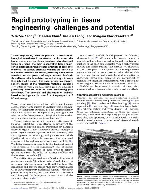
with<strong>in</strong> the scaffold (Figure 1).<br />
Figure 1. Scaffold produced us<strong>in</strong>g conventional freeze-dry<strong>in</strong>g method. The pores<br />
formed are <strong>in</strong>terconnected but <strong>in</strong>homogeneous ow<strong>in</strong>g to the r<strong>and</strong>om freez<strong>in</strong>g<br />
k<strong>in</strong>etics. The architecture of the pores can be adjusted by controll<strong>in</strong>g the freez<strong>in</strong>g<br />
k<strong>in</strong>etics of the solution. A dense layer of ‘sk<strong>in</strong>’ is formed at the open surface, which<br />
greatly affects the diffusion efficiency of the scaffold.
644<br />
Review TRENDS <strong>in</strong> Biotechnology Vol.22 No.12 December 2004<br />
Consequently, researchers try to modify the conventional<br />
techniques to overcome these <strong>in</strong>herent process<br />
limitations. Kim <strong>and</strong> Mooney [13] produced polyglycolic<br />
acid (PGA) fibers bonded with poly-L-lactide (PLLA) to<br />
enhance the mechanical strength <strong>and</strong> the degradation<br />
rate of the unbonded PGA fiber meshes. As a variant to the<br />
freeze-dry<strong>in</strong>g process, Ho et al. [14] prepared scaffolds<br />
us<strong>in</strong>g a freeze-extraction method, which was relatively<br />
more time- <strong>and</strong> energy efficient. Murphy et al. [15]<br />
enhanced pore <strong>in</strong>terconnectivity by fus<strong>in</strong>g the porogen<br />
together to form a template, <strong>in</strong>stead of us<strong>in</strong>g unbounded<br />
particles <strong>in</strong> so1vent cast<strong>in</strong>g/particulate leach<strong>in</strong>g process.<br />
The result showed that holes were formed <strong>in</strong> pore walls,<br />
guarantee<strong>in</strong>g pore <strong>in</strong>terconnectivity. Chen <strong>and</strong> Ma [16]<br />
created nanofibrous PLLA scaffolds which <strong>in</strong>corporated<br />
<strong>in</strong>terconnected spherical macropores. The macropores<br />
were voids left by paraff<strong>in</strong> spheres, which were thermally<br />
bonded before the cast<strong>in</strong>g of the polymer solution. In place<br />
of paraff<strong>in</strong> spheres, Gross <strong>and</strong> Rodríguez-Lorenzo [17]<br />
used a s<strong>in</strong>tered salt template to produce PLLA-re<strong>in</strong>forced<br />
apatite scaffolds.<br />
Notwithst<strong>and</strong><strong>in</strong>g the improvements that have been<br />
atta<strong>in</strong>ed, the control over scaffold architecture us<strong>in</strong>g these<br />
conventional techniques is highly process dependent<br />
rather than design dependent. As a result, RP is seen to<br />
be a viable alternative for achiev<strong>in</strong>g extensive <strong>and</strong><br />
detailed control over scaffold architecture [18,19].<br />
Advanced scaffold-fabrication methods<br />
RP is a common name for a group of techniques that can<br />
generate a physical model directly from computer-aided<br />
design data. It is an additive process <strong>in</strong> which each part is<br />
constructed <strong>in</strong> a layer-by-layer manner. Table 1 presents<br />
<strong>and</strong> compares the RP techniques that can be used to<br />
fabricate scaffolds directly or <strong>in</strong>directly.<br />
Direct RP fabrication method<br />
RP systems such as fused deposition model<strong>in</strong>g (FDM), 3D<br />
pr<strong>in</strong>t<strong>in</strong>ge (3-DP) <strong>and</strong> selective laser s<strong>in</strong>ter<strong>in</strong>g (SLS) have<br />
been shown to be feasible for produc<strong>in</strong>g porous structures<br />
for use <strong>in</strong> <strong>tissue</strong> eng<strong>in</strong>eer<strong>in</strong>g. In this review, the <strong>tissue</strong><br />
scaffold fabrication techniques are categorized by virtue of<br />
their mode of assembly <strong>in</strong>to one of two processes: the melt–<br />
dissolution deposition technique <strong>and</strong> the particle bond<strong>in</strong>g<br />
technique.<br />
Melt–dissolution deposition technique<br />
In a typical melt–dissolution deposition system, each layer<br />
is created by extrusion of a str<strong>and</strong> of material through an<br />
orifice while it moves across the plane of the layer crosssection.<br />
The material cools, solidify<strong>in</strong>g itself <strong>and</strong> fix<strong>in</strong>g to<br />
the previous layer [20]. Successive layer formation, one<br />
atop another, forms a complex 3D solid object.<br />
Porosity <strong>in</strong> the horizontal XY plane is created by<br />
controll<strong>in</strong>g the spac<strong>in</strong>g between adjacent filaments<br />
(Figure 2). The vertical Z gap is formed by deposit<strong>in</strong>g the<br />
subsequent layer of filaments at an angle with respect to<br />
the previous layer. Repetitive pattern draw<strong>in</strong>g will<br />
produce a porous structure ready to be used as a scaffold.<br />
A representative system us<strong>in</strong>g melt–dissolution<br />
www.sciencedirect.com<br />
deposition is FDM. This method sp<strong>in</strong>s off several new<br />
systems that operate under similar pr<strong>in</strong>ciples.<br />
FDM: In this method, a filament of a suitable material<br />
is fed <strong>and</strong> melted <strong>in</strong>side a heated liquefier before be<strong>in</strong>g<br />
extruded through a nozzle. The system operates <strong>in</strong> a<br />
temperature-controlled environment to ma<strong>in</strong>ta<strong>in</strong> sufficient<br />
fusion energy between each layer.<br />
Researchers have demonstrated the feasibility of<br />
utiliz<strong>in</strong>g FDM to fabricate a functional scaffold directly.<br />
Ze<strong>in</strong> et al. [21] fabricated polycaprolactone (PCL) scaffolds<br />
with a honeycomb structure <strong>and</strong> a channel size of 160–<br />
770 mm. Samar et al. [22] have successfully produced a<br />
polymer-ceramic composite scaffold made of polypropylene-tricalcium<br />
phosphate (PP-TCP). The scaffolds were<br />
reported to have a pore size of 160 mm <strong>and</strong> a mechanical<br />
strength of 12.7 MPa, which is comparable to the tensile<br />
strength of natural cancellous bone, which has a value of<br />
7.4MPa [23]. In a recent study, human mesenchymal<br />
progenitor cells were seeded on PCL <strong>and</strong> PCL-hydroxyapatite<br />
(HA) scaffolds fabricated by FDM [24]. Proliferation<br />
of cells toward <strong>and</strong> onto the scaffold surfaces was detected.<br />
Drawbacks of the FDM technique <strong>in</strong>clude the need for<br />
<strong>in</strong>put material of a specific diametric size <strong>and</strong> material<br />
properties to feed through the rollers <strong>and</strong> nozzle. Any<br />
changes <strong>in</strong> the properties of the material require effort to<br />
recalibrate the sett<strong>in</strong>g of the feed<strong>in</strong>g parameters. As a<br />
result, FDM has a narrow process<strong>in</strong>g w<strong>in</strong>dow. The<br />
resolution of FDM is relatively low, at 250 mm. In FDM,<br />
a limited range of material s can be used, with almost<br />
complete exclusion of natural polymers, as the material<br />
used must be made <strong>in</strong>to filaments <strong>and</strong> melted <strong>in</strong>to a semiliquid<br />
phase before extrusion. The operat<strong>in</strong>g temperature<br />
of the system is too high to <strong>in</strong>corporate biomolecules <strong>in</strong>to<br />
the scaffold, hence limit<strong>in</strong>g the biomimetic aspects of the<br />
scaffold produced. Moreover, the material deposited<br />
solidifies <strong>in</strong>to dense filaments, block<strong>in</strong>g the formation of<br />
microporosity. Microporosity is an important factor <strong>in</strong><br />
encourag<strong>in</strong>g neovascularization <strong>and</strong> cell attachment [25].<br />
Modifications of FDM to circumvent these limitations<br />
have encouraged the emergence of several new techniques.<br />
These <strong>in</strong>clude techniques that elim<strong>in</strong>ate the<br />
requirement of precursor filaments or a system with<br />
reduced operat<strong>in</strong>g temperatures. Some variants of the<br />
FDM process <strong>in</strong>clude the 3D fiber-deposition technique<br />
[26], precision extrud<strong>in</strong>g deposition (PED) [27] <strong>and</strong> precise<br />
extrusion manufactur<strong>in</strong>g (PEM) [28].<br />
3D fiber-deposition technique: In this method, the<br />
feedstock material is <strong>in</strong> a pellet or granule form that can<br />
be poured <strong>in</strong>to the heated liquefier directly. Poly(ethylene<br />
glycol)-terephthalate-poly(butylenes terephthalate)<br />
(PEGT–PBT) block copolymer scaffolds have been<br />
fabricated for articulate <strong>tissue</strong> eng<strong>in</strong>eer<strong>in</strong>g applications<br />
[26]. Material flow is regulated by apply<strong>in</strong>g<br />
pressure to the syr<strong>in</strong>ge.<br />
PED: The extruder <strong>in</strong> this system is equipped with a<br />
built-<strong>in</strong> heat<strong>in</strong>g unit to melt the feedstock material, hence<br />
elim<strong>in</strong>at<strong>in</strong>g the need to produce precursor filaments. PCL<br />
scaffolds with a pore size of 250 mm were fabricated [27].<br />
PEM: PLLA scaffolds with controllable porous<br />
architectures from 200 to 500 mm <strong>in</strong> size have been<br />
produced [28].
Table 1. Comparison of different rapid <strong>prototyp<strong>in</strong>g</strong> (RP) technologies applied <strong>in</strong> <strong>tissue</strong> eng<strong>in</strong>eer<strong>in</strong>g<br />
RP system Resolution (mm) Material Strength Weakness Refs<br />
Melt–dissolution deposition technique<br />
Fused deposition model<strong>in</strong>g 250 PCL a , PP-TCP,<br />
PCL-HA, PCL-<br />
TCP<br />
The melt process is generally undesirable from the<br />
perspective of scaffold bioactivity because of the elevated<br />
temperatures <strong>in</strong>volved. This limitation drives researchers<br />
to replace the melt<strong>in</strong>g process with that of dissolution of<br />
materials. Systems developed <strong>in</strong>clude low-temperature<br />
deposition manufactur<strong>in</strong>g (LDM) [29], mult<strong>in</strong>ozzle<br />
Good mechanical strength;<br />
versatile <strong>in</strong> lay-down pattern<br />
design<br />
High temperature; need to<br />
produce filament material;<br />
narrow process<strong>in</strong>g w<strong>in</strong>dow;<br />
rigid filament<br />
High temperature;<br />
rigid filament<br />
3D fiber-deposition technique 250 PEGT-PBT Input material <strong>in</strong> pellet form;<br />
preparation time is reduced<br />
[26]<br />
Precision extrud<strong>in</strong>g depo- 250 PCL Input material <strong>in</strong> pellet form High temperature;<br />
[27]<br />
sition<br />
rigid filament<br />
Precise extrusion<br />
200–500 PLLA-TCP Input material <strong>in</strong> pellet form High temperature;<br />
[28]<br />
manufactur<strong>in</strong>g<br />
rigid filament<br />
Low-temperature deposition 400 PLLA-TCP Can <strong>in</strong>corporate biomolecule Solvent is used;<br />
[29]<br />
manufactur<strong>in</strong>g<br />
requires freeze dry<strong>in</strong>g<br />
Multi-nozzle deposition 400 PLLA-TCP Enhanced range of materials Solvent is used;<br />
[30]<br />
manufactur<strong>in</strong>g<br />
can be used; can <strong>in</strong>corporate<br />
biomolecule<br />
requires freeze dry<strong>in</strong>g<br />
Pressure-assisted<br />
10–600 PCL, PLLA Enhanced range of materials Small nozzle <strong>in</strong>hibits<br />
[31]<br />
microsyr<strong>in</strong>ge<br />
can be used; can <strong>in</strong>corporate <strong>in</strong>corporation of particle;<br />
biomolecule; very f<strong>in</strong>e resol- narrow range of pr<strong>in</strong>table<br />
ution can be achieved<br />
viscosities; solvent is used<br />
Robocast<strong>in</strong>g 100–1000 Organic <strong>in</strong>k Enhanced range of materials Precise control of <strong>in</strong>k properties [34]<br />
can be used<br />
is crucial<br />
3D bioplotter 250 Hydrogel Enhanced range of materials Low mechanical strength; [35]<br />
can be used; can <strong>in</strong>corporate smooth surface; low accuracy;<br />
biomolecule<br />
slow process<strong>in</strong>g; precise control<br />
of properties of material<br />
<strong>and</strong> medium; calibration for<br />
new material<br />
<strong>Rapid</strong> <strong>prototyp<strong>in</strong>g</strong> robotic 400–1000 Chitosanchito- Enhanced range of materials Precise control properties of [36]<br />
dispens<strong>in</strong>g system<br />
san-HA<br />
can be used; can <strong>in</strong>corporate material <strong>and</strong> medium; requires<br />
Particle bond<strong>in</strong>g techniques<br />
biomolecule<br />
freeze dry<strong>in</strong>g<br />
3-dimensional pr<strong>in</strong>t<strong>in</strong>ge 200 PLGA, starch- Microposity <strong>in</strong>duced <strong>in</strong> the Material must be <strong>in</strong> powder [41]<br />
based polymer scaffold; enhanced range of form; limited mechanical<br />
materials can be used; water strength; Powdery surface<br />
used as b<strong>in</strong>der; no support f<strong>in</strong>ish; trapped powder issue;<br />
structure needed; fast<br />
process<strong>in</strong>g<br />
might require postprocess<strong>in</strong>g<br />
TheriForme 300 PLLA Microposity <strong>in</strong>duced <strong>in</strong> the Material must be <strong>in</strong> powder [45]<br />
scaffold; enhanced range of form; powdery surface f<strong>in</strong>ish;<br />
materials can be used; nonorganic<br />
b<strong>in</strong>der is possible; no<br />
support structure is needed;<br />
fast process<strong>in</strong>g<br />
trapped powder<br />
Selective laser s<strong>in</strong>ter<strong>in</strong>g 500 PEEK-HA, PCL Microposity <strong>in</strong>duced <strong>in</strong> the Material must be <strong>in</strong> powder [47]<br />
scaffold enhanced range of form; high temperature;<br />
materials can be used;<br />
powdery surface f<strong>in</strong>ish;<br />
Indirect RP fabrication method<br />
no support structure needed;<br />
fast process<strong>in</strong>g<br />
trapped powder<br />
Melt deposition 250 Thermoplastic Enhanced range of materials Multisteps <strong>in</strong>volved [48]<br />
polymer<br />
can be used; control of external<br />
<strong>and</strong> <strong>in</strong>ternal morphology<br />
Droplet deposition 180 Wax Enhanced range of materials<br />
can be used; control of external<br />
<strong>and</strong> <strong>in</strong>ternal morphology<br />
Multisteps <strong>in</strong>volved [49]<br />
Photo-polymerization 366 Res<strong>in</strong> Enhanced range of materials<br />
can be used; control of external<br />
<strong>and</strong> <strong>in</strong>ternal morphology<br />
Multisteps <strong>in</strong>volved [50]<br />
a<br />
Abbreviations: HA, hydroxyapatite; PCL, polycaprolactone; PEEK-HA, polyetheretherketone-hydroxyapatite; PEGT-PBT, poly(ethylene glycol)-terephthalate-poly(butylenes<br />
terephthalate; PLLA, poly-L-lactide; PP-TCP, polypropylene-tricalcium phosphate.<br />
www.sciencedirect.com<br />
Review TRENDS <strong>in</strong> Biotechnology Vol.22 No.12 December 2004 645<br />
[21]<br />
deposition manufactur<strong>in</strong>g (MDM) [30], pressure-assisted<br />
microsyr<strong>in</strong>ges (PAM) [31] <strong>and</strong> robocast<strong>in</strong>g [32].<br />
LDM: The scaffold-build<strong>in</strong>g cycle is performed <strong>in</strong> a lowtemperature<br />
environment under 08C [29]. A PLLA–TCP<br />
pipe scaffold has been produced.<br />
MDM: This is an improved version of LDM, with the
646<br />
Figure 2. structure produced us<strong>in</strong>g fused deposition model<strong>in</strong>g. Uniformly<br />
<strong>in</strong>terconnected square channels are obta<strong>in</strong>ed with a pattern sett<strong>in</strong>g of 08 or 908.<br />
The scaffold structure is highly regular <strong>and</strong> reproducible. The portion of the<br />
filament that spans across two support<strong>in</strong>g po<strong>in</strong>ts is subjected to gravity-<strong>in</strong>duced<br />
deformation dur<strong>in</strong>g the solidification phase. Therefore, the sett<strong>in</strong>g of the mach<strong>in</strong>e<br />
parameters, as well as the material properties, must be precisely controlled to<br />
ensure m<strong>in</strong>imum filament deflection. Filaments are aligned orthogonally, with<br />
grooves at the <strong>in</strong>tersection po<strong>in</strong>t between consecutive layers. Cells on the scaffold<br />
must span across these grooves to cellularize the entire structure.<br />
advantage of be<strong>in</strong>g able to use a greater range of<br />
materials. The enhancement is achieved by <strong>in</strong>corporat<strong>in</strong>g<br />
more jett<strong>in</strong>g nozzles <strong>in</strong>to the system [29]. Support<br />
structures can be built us<strong>in</strong>g water, which is nontoxic<br />
<strong>and</strong> easy to remove. In the work by Zhuo et al. [30],<br />
biomolecules <strong>in</strong> the form of bone morphogenic prote<strong>in</strong><br />
were embedded <strong>in</strong> the bulk material <strong>and</strong> then released<br />
slowly as the scaffold degraded.<br />
PAM: A microsyr<strong>in</strong>ge is used to expel the dissolved<br />
polymer under low <strong>and</strong> constant pressure to form the<br />
desired pattern. The resolution of this method is on a<br />
cellular scale, which is remarkably high compared with<br />
the techniques described previously. Vozzi et al. [31]<br />
developed PCL <strong>and</strong> PLLA scaffolds with l<strong>in</strong>e width of<br />
20 mm. It has been demonstrated that the performance of<br />
this method is comparable to that of soft lithography [33].<br />
However, capillaries with a very small diameter require<br />
careful h<strong>and</strong>l<strong>in</strong>g to avoid any tip breakage. Higher<br />
pressure is also needed to expel the material from a<br />
small orifice.<br />
Robocast<strong>in</strong>g: This patented system is able to lay down<br />
a highly concentrated, pseudoplastic-like colloidal suspension<br />
[32]. Therriault et al. [34] fabricated a 3D microvascular<br />
network by robocast<strong>in</strong>g fugitive organic <strong>in</strong>k,<br />
followed by scaffold <strong>in</strong>filtration with epoxy res<strong>in</strong> <strong>and</strong><br />
further postprocess<strong>in</strong>g.<br />
In general, the scaffolds fabricated us<strong>in</strong>g the melt or<br />
solution deposition techniques described are usually<br />
meant to serve as hard-<strong>tissue</strong> scaffolds. L<strong>and</strong>ers <strong>and</strong><br />
Mülhaupt [35] have developed an aqueous system, the 3D<br />
bioplotter, to meet the dem<strong>and</strong> for fabrication of hydrogel<br />
scaffolds useful <strong>in</strong> soft-<strong>tissue</strong> eng<strong>in</strong>eer<strong>in</strong>g. Hydrogels are<br />
becom<strong>in</strong>g <strong>in</strong>creas<strong>in</strong>gly popular as a material for <strong>tissue</strong><br />
eng<strong>in</strong>eer<strong>in</strong>g because of their high water content <strong>and</strong> the<br />
fact that they have similar mechanical properties to those<br />
of many soft <strong>tissue</strong>s <strong>in</strong> the human body. Ang et al. [36]<br />
www.sciencedirect.com<br />
Review TRENDS <strong>in</strong> Biotechnology Vol.22 No.12 December 2004<br />
adopted a similar concept to develop a robotic dispenser,<br />
the rapid <strong>prototyp<strong>in</strong>g</strong> robotic dispens<strong>in</strong>g system(RPBOD),<br />
for the fabrication of a chitosan scaffold.<br />
3D bioplotter: The key feature of this method is the<br />
3D dispens<strong>in</strong>g of liquids <strong>and</strong> pastes <strong>in</strong>to a liquid medium<br />
with matched density. The plott<strong>in</strong>g material leaves the<br />
nozzle <strong>and</strong> solidifies <strong>in</strong> the plott<strong>in</strong>g medium after bond<strong>in</strong>g<br />
to the previous layer. The liquid medium compensates for<br />
gravity <strong>and</strong> hence no support structure is needed.<br />
Hydrogel scaffolds with well-def<strong>in</strong>ed <strong>in</strong>ternal pore<br />
structure were prepared by L<strong>and</strong>ers et al. [37]. The<br />
hydrogel scaffolds had <strong>in</strong>terconnected pores, 200–400 mm<br />
<strong>in</strong> diameter. However, the hydrogel presented a smooth<br />
surface, which might be nonadherent to cells [38]. Therefore,<br />
further surface coat<strong>in</strong>g was required to render the<br />
surface favorable for cell-adhesion. Fibroblasts seeded on<br />
the scaffolds showed almost complete coverage of cells.<br />
However, the scaffolds had limited resolution <strong>and</strong> mechanical<br />
strength. Material rigidity was shown to <strong>in</strong>fluence<br />
cell spread<strong>in</strong>g <strong>and</strong> migration speed, as demonstrated by<br />
Wong et al. [39]. Cells displayed a preference for stiffer<br />
regions, <strong>and</strong> tended to migrate faster on surfaces with<br />
lower compliance.<br />
RPBOD: This system, developed by Ang et al. [36],<br />
consists of a computer-guided desktop robot <strong>and</strong> a onecomponent<br />
pneumatic dispenser. Material <strong>in</strong> liquid form<br />
was dispensed <strong>in</strong>to a dispens<strong>in</strong>g medium through a small<br />
Teflon-l<strong>in</strong>ed nozzle. Chitosan scaffolds with pore size of<br />
400–1000 mm were produced <strong>in</strong> the prelim<strong>in</strong>ary study.<br />
Particle-bond<strong>in</strong>g techniques<br />
In particle-bond<strong>in</strong>g techniques, particles are selectively<br />
bonded <strong>in</strong> a th<strong>in</strong> layer of powder material. The th<strong>in</strong> 2D<br />
layers are bonded one upon another to form a complex 3D<br />
solid object. Dur<strong>in</strong>g fabrication, the object is supported by<br />
<strong>and</strong> embedded <strong>in</strong> unprocessed powder. Therefore, this<br />
technique enables the fabrication of through channels <strong>and</strong><br />
overhang<strong>in</strong>g features. After completion of all layers, the<br />
object is removed from the bed of unbonded powder [20].<br />
The powder utilized can be a pure powder or surfacecoated<br />
powder, depend<strong>in</strong>g on the application of the scaffold.<br />
It is possible to use a s<strong>in</strong>gle one-component powder or a<br />
mixture of different powders, blended together.<br />
These techniques are capable of produc<strong>in</strong>g a porous<br />
structure with controllable macroporosity as well as<br />
microporosity. The microporosity arises from the space<br />
between the <strong>in</strong>dividual granules of powder. These techniques<br />
offer control over pore architecture by manipulat<strong>in</strong>g<br />
the region of bond<strong>in</strong>g. However, the pore size is limited<br />
by the powder size of the stock material. Larger pores can<br />
be generated by mix<strong>in</strong>g porogen <strong>in</strong>to the powder bed<br />
before the bond<strong>in</strong>g process.<br />
The powder-based materials provide a rough surface to<br />
the scaffold. It has been suggested that topographical cues<br />
might have a significant effect upon cellular behavior [40].<br />
As a cell attaches to the scaffold, stretch receptors are<br />
activated. Receptors on the scaffold surface might be<br />
subjected to vary<strong>in</strong>g degrees of deformation, lead<strong>in</strong>g to<br />
activation of cell signal transduction pathways. Therefore,<br />
scaffolds fabricated via a particle-bond<strong>in</strong>g technique
might be more advantageous <strong>in</strong> the context of cell<br />
attachment.<br />
Typical systems <strong>in</strong> this category <strong>in</strong>clude 3-DP [41],<br />
TheriForme [42] <strong>and</strong> SLS [43].<br />
3DP: In this process, a stream of adhesive droplets is<br />
expelled through an <strong>in</strong>kjet pr<strong>in</strong>thead, selectively bond<strong>in</strong>g<br />
a th<strong>in</strong> layer of powder particles to form a solid shape [20].<br />
The resolution achieved is w300 mm. Kim et al. [44]<br />
employed 3DP us<strong>in</strong>g a particulate leach<strong>in</strong>g technique to<br />
create porous scaffolds us<strong>in</strong>g polylactic-co-glycolic acid<br />
(PLGA) mix with salt particles <strong>and</strong> a suitable organic<br />
solvent. Pores formed were of the scale 45–150 mm with<br />
60% porosity. In vitro cell culture with hepatocytes showed<br />
<strong>in</strong>growth of these cells <strong>in</strong>to the pore space.<br />
In an effort to render the system more biocompatible,<br />
Lam et al. [43] have formulated a blend of starch-based<br />
polymer powders that can be bonded together us<strong>in</strong>g<br />
distilled water. The group also tried to enhance the<br />
mechanical property of the scaffold produced by <strong>in</strong>filtrat<strong>in</strong>g<br />
the structure with PLLA <strong>and</strong> PCL copolymer solution.<br />
TheriForm: This system is similar to 3DP, <strong>in</strong> that a<br />
pr<strong>in</strong>thead assembly deposits b<strong>in</strong>der droplets onto selected<br />
regions of the powder, swell<strong>in</strong>g <strong>and</strong> dissolv<strong>in</strong>g the polymer<br />
powder <strong>in</strong> the pr<strong>in</strong>ted regions. Zelt<strong>in</strong>ger et al. [45]<br />
performed a study on a TheriForm-built PLLA scaffold<br />
with different pore sizes us<strong>in</strong>g can<strong>in</strong>e dermal fibroblasts<br />
(DmFb), vascular smooth muscle cells (VSMC) <strong>and</strong><br />
microvascular epithelial cells (MVEC). They found that<br />
DmFb is <strong>in</strong>different to pore size, whereas MVEC <strong>and</strong><br />
VSMC favored a pore size of 90 mm <strong>and</strong> 107 mm, respectively,<br />
suggest<strong>in</strong>g the existence of an optimal pore size for<br />
different cell types.<br />
SLS: This technique uses a deflected laser beam<br />
selectively to scan over the powder surface follow<strong>in</strong>g the<br />
cross-sectional profiles carried by the slice data. The<br />
<strong>in</strong>teraction of the laser beam with the powder elevates the<br />
powder temperature to reach the glass-transition<br />
temperature, caus<strong>in</strong>g surfaces <strong>in</strong> contact to deform<br />
<strong>and</strong> fuse together [20]. Thisisthepreferredfabrication<br />
process for produc<strong>in</strong>g complex porous ceramic matrices<br />
suitable for implantation <strong>in</strong> a bone defect, as demonstrated<br />
by Vail et al. [46].<br />
The authors’ group has successfully s<strong>in</strong>tered polyetheretherketone-hydroxyapatite<br />
(PEEK-HA) powder blends<br />
on a commercial SLS mach<strong>in</strong>e [47]. In the research,<br />
different weight percentage compositions of physically<br />
mixed PEEK-HA powder blends were s<strong>in</strong>tered by vary<strong>in</strong>g<br />
the laser power <strong>and</strong> temperature sett<strong>in</strong>gs (Figure 3).<br />
Indirect RP fabrication methods<br />
RP systems can also be utilized to produce a sacrificial<br />
mould to fabricate <strong>tissue</strong>-eng<strong>in</strong>eer<strong>in</strong>g scaffolds. These<br />
multistep methods usually <strong>in</strong>volve cast<strong>in</strong>g of material <strong>in</strong><br />
a mould <strong>and</strong> then remov<strong>in</strong>g or sacrific<strong>in</strong>g the mould to<br />
obta<strong>in</strong> the f<strong>in</strong>al scaffold. Such techniques enable the user<br />
to control both the external <strong>and</strong> the <strong>in</strong>ternal morphology of<br />
the f<strong>in</strong>al construct. In addition, <strong>in</strong>direct methods also<br />
require less raw scaffold material while <strong>in</strong>creas<strong>in</strong>g the<br />
range of materials that can be used <strong>and</strong> mak<strong>in</strong>g it possible<br />
to use composite blends that might require conflict<strong>in</strong>g<br />
process<strong>in</strong>g parameters. The orig<strong>in</strong>al properties of the<br />
www.sciencedirect.com<br />
Review TRENDS <strong>in</strong> Biotechnology Vol.22 No.12 December 2004 647<br />
Figure 3. Micrographs of 90% polyetheretherketone by weight <strong>and</strong> 10% hydroxyapatite<br />
by weight s<strong>in</strong>tered us<strong>in</strong>g selective laser s<strong>in</strong>ter<strong>in</strong>g. The bond<strong>in</strong>g between<br />
granules of particles is clearly observed, <strong>in</strong>dicat<strong>in</strong>g the formation of microporosity<br />
with<strong>in</strong> the structure. The surface topography of the powder is irregular <strong>and</strong> rough.<br />
biomaterial are well conserved because no heat<strong>in</strong>g process<br />
is imposed on the scaffold material. Common RP techniques<br />
employed <strong>in</strong>clude melt deposition [48], droplet<br />
deposition [49] <strong>and</strong> photopolymerization [50].<br />
Melt deposition techniques<br />
The operat<strong>in</strong>g pr<strong>in</strong>ciple of these techniques, of which FDM<br />
is an example, is presented above.<br />
FDM: Bose et al. [48] produced alum<strong>in</strong>a <strong>and</strong> b-TCP<br />
ceramic scaffolds with pore sizes <strong>in</strong> the range of 300–500 mm<br />
<strong>and</strong> porosity of 25–45%. The moulds were made us<strong>in</strong>g a<br />
st<strong>and</strong>ard thermoplastic polymer. Their research aimed to<br />
<strong>in</strong>vestigate the effect of pore size <strong>and</strong> porosity on the<br />
mechanical <strong>and</strong> biological responses.<br />
Droplet deposition technique<br />
This technique is based on <strong>in</strong>kjet pr<strong>in</strong>t<strong>in</strong>g technology. A<br />
stream of molten thermoplastic droplets is deposited on a<br />
work<strong>in</strong>g surface. Thermal energy <strong>in</strong> the deposited droplet<br />
causes local melt<strong>in</strong>g on the previous layer <strong>and</strong> solidifies as<br />
one piece.<br />
Droplet deposition is a complex <strong>in</strong>teraction of droplet<br />
spread<strong>in</strong>g upon impact, settl<strong>in</strong>g as a result of viscous <strong>and</strong><br />
surface-tension forces, <strong>and</strong> a solidification process. A<br />
representative system is the ModelMakerIIe (MMII) [20].<br />
MMII: This mach<strong>in</strong>e uses a s<strong>in</strong>gle jet each for a plastic<br />
build<strong>in</strong>g material <strong>and</strong> a wax-like support material, which<br />
are held <strong>in</strong> a melted liquid state <strong>in</strong> reservoirs. The pr<strong>in</strong>ter<br />
head ejects droplets of the materials as they are moved <strong>in</strong><br />
X-Y fashion. After an entire layer of the object has<br />
hardened, a mill<strong>in</strong>g head is passed over the layer to<br />
ensure that a uniform thickness has been achieved.<br />
Taboas et al. [51] produced PLLA scaffolds with micro<strong>and</strong><br />
macroporosity for bone <strong>tissue</strong> eng<strong>in</strong>eer<strong>in</strong>g, <strong>in</strong> this case<br />
specifically trabecular bone. The global pores (500 mm<br />
wide channels) were computationally designed, whereas<br />
the local pores (50–100 mm wide voids) were formed by the<br />
porogen. PLA–PGA discrete composites were made us<strong>in</strong>g<br />
melt process<strong>in</strong>g.
648<br />
MMII is selected by researchers to fabricate calcium<br />
phosphate scaffolds because the build<strong>in</strong>g material has a<br />
very low coefficient of thermal expansion. There will be<br />
m<strong>in</strong>imal risk of fracture due to coefficient of thermal<br />
expansion mismatch with the ceramic dur<strong>in</strong>g pyrolysis.<br />
Limpanuphap <strong>and</strong> Derby [52] fabricated TCP scaffolds<br />
with controlled <strong>in</strong>ternal porosity us<strong>in</strong>g a suspension of<br />
TCP <strong>in</strong> an acrylate b<strong>in</strong>der. A similar route was used to<br />
produce a polymer–TCP scaffold, which is believed to show<br />
more <strong>potential</strong> for cell adhesion. Wilson et al. [53]<br />
fabricated HA scaffolds with a def<strong>in</strong>ed macroarchitecture.<br />
Channels achieved are w350–400 mm wide. These authors<br />
suggested that the <strong>in</strong>herent surface texture obta<strong>in</strong>ed from<br />
an MMII-built mould <strong>in</strong>creased the surface area of the<br />
scaffold <strong>and</strong> led to a higher degree of calcium <strong>and</strong> phosphate<br />
release, which is advantageous to bone formation.<br />
Sachlos et al. [54] have successfully produced collagen<br />
scaffolds with predef<strong>in</strong>ed <strong>and</strong> reproducible <strong>in</strong>ternal channels.<br />
The smallest channel width achieved was reported to<br />
be as low as 135 mm. In a variation to Sachlos’ work, the<br />
authors have produced chitosan-collagen scaffolds. MMII<br />
moulds with <strong>in</strong>tricate channels were built to conta<strong>in</strong> a<br />
chitosan–collagen solution. The resultant gel-like scaffold<br />
was capable of atta<strong>in</strong><strong>in</strong>g the designed morphology<br />
(Figure 4). These channels can serve as flow channels<br />
when coupled with a customized bioreactor, achiev<strong>in</strong>g<br />
more efficient perfusion of the culture medium.<br />
Photopolymerization techniques<br />
For photopolymerization techniques, optical energy is<br />
applied to irradiate the th<strong>in</strong> layer at the surface of a<br />
liquid photopolymer res<strong>in</strong>. The irradiation areas of<br />
res<strong>in</strong> react chemically <strong>and</strong> transform <strong>in</strong>to a solid<br />
phase. A well-known photopolymerization technique is<br />
stereolithography (SLA) [20].<br />
SLA: A UV laser traces out the first layer of photocurable<br />
res<strong>in</strong>, solidify<strong>in</strong>g the model’s cross-section while<br />
leav<strong>in</strong>g the rema<strong>in</strong><strong>in</strong>g areas <strong>in</strong> liquid form. The elevator<br />
then drops by a sufficient amount to cover the solid<br />
polymer with another layer of liquid res<strong>in</strong>. A sweeper<br />
recoats the solidified layer with liquid res<strong>in</strong> <strong>and</strong> the laser<br />
traces the second layer atop the first.<br />
Chu et al. [50] have produced HA-based porous implants<br />
us<strong>in</strong>g SLA-built epoxy moulds. A thermal curable HA–<br />
acrylate suspension was cast <strong>in</strong>to the mould to obta<strong>in</strong> a<br />
scaffold with <strong>in</strong>terconnected channels. The resolution of<br />
channel width achieved was as low as 366 mm.<br />
In another study, <strong>in</strong>vestigators carried out an <strong>in</strong> vivo<br />
study us<strong>in</strong>g two different architecture designs, orthogonal<br />
Figure 4. Scaffold (right) produced us<strong>in</strong>g ModelMakerIIe-built mould (left). The<br />
scaffold is able to reproduce the predeterm<strong>in</strong>ed morphology of the mould. The<br />
channels ensure high diffusion efficiency across the scaffold.<br />
www.sciencedirect.com<br />
Review TRENDS <strong>in</strong> Biotechnology Vol.22 No.12 December 2004<br />
<strong>and</strong> radial channels [55]. The prelim<strong>in</strong>ary results showed<br />
that controll<strong>in</strong>g the overall geometry of the regenerated<br />
bone <strong>tissue</strong> was possible through the <strong>in</strong>ternal architectural<br />
design of the scaffolds.<br />
Challenges of RP <strong>in</strong> <strong>tissue</strong> eng<strong>in</strong>eer<strong>in</strong>g<br />
In spite of the <strong>in</strong>creas<strong>in</strong>g <strong>in</strong>terest of <strong>tissue</strong> eng<strong>in</strong>eers <strong>in</strong> the<br />
use of RP, there are several <strong>challenges</strong> that need to be<br />
addressed, namely the limited range of materials, the<br />
optimal scaffold design, the bioactivity of the scaffold, as<br />
well as the issues of cell seed<strong>in</strong>g <strong>and</strong> vascularization. Each<br />
of the issues will be discussed <strong>in</strong> detail.<br />
Range of materials<br />
Material processability: RP techniques are very<br />
specialized technologies <strong>in</strong> terms of material processability.<br />
Each technique requires a specific form of <strong>in</strong>put<br />
material such as filament, powder, solid pellet or solution.<br />
Therefore, it must be ensured that the choice of materials<br />
for the scaffold is compatible with the selected RP process<br />
<strong>and</strong> that it can be efficiently produced <strong>in</strong> the form<br />
required. Other considerations dur<strong>in</strong>g the selection of<br />
materials <strong>in</strong>clude the degradation profile <strong>and</strong> the mechanical<br />
strength of the scaffold.<br />
Degradation rate: In an ideal case, the scaffold should<br />
be remodeled <strong>and</strong> resorbed by grow<strong>in</strong>g cells <strong>and</strong> gradually<br />
replaced by the newly formed extracellular matrix <strong>and</strong><br />
differentiated cells. A desirable feature would be synchronization<br />
of the polymer degradation rate with the rate of<br />
<strong>tissue</strong> <strong>in</strong>growth. Therefore, the degradation properties of a<br />
scaffold are of crucial importance for the success of the<br />
scaffold-based approach.<br />
The degradation–absorption mechanism is the result of<br />
many <strong>in</strong>terrelated factors, <strong>in</strong>clud<strong>in</strong>g the hydrophilicity of<br />
the polymer backbone, degree of crystall<strong>in</strong>ity, presence<br />
of catalysts, volume of porosity <strong>and</strong> the surface area.<br />
Balanc<strong>in</strong>g each of these factors will enable an implant to<br />
degrade slowly <strong>and</strong> transfer stress at an appropriate rate<br />
to the surround<strong>in</strong>g <strong>tissue</strong>s as they heal. This is one of the<br />
major <strong>challenges</strong> fac<strong>in</strong>g <strong>tissue</strong> eng<strong>in</strong>eer<strong>in</strong>g research today.<br />
Degradation product: Even though degradation<br />
products of biodegradable polymers are known to be<br />
largely non-cytotoxic, little <strong>in</strong>formation is available<br />
regard<strong>in</strong>g the degradation rate-dependent acidic byproduct<br />
effect of the scaffold. Sunga et al. [56] found that fast<br />
degradation of the polymer negatively affects cell viability<br />
<strong>and</strong> migration <strong>in</strong>to the scaffold, both <strong>in</strong> vitro <strong>and</strong> <strong>in</strong> vivo.<br />
This can be expla<strong>in</strong>ed by the rapid local acidification due<br />
to polymer degradation. Therefore, a more systematic<br />
<strong>in</strong>vestigative approach is needed to classify the material<br />
degradation profile.<br />
Mechanical strength of scaffolds: Cells are able to<br />
detect with high sensitivity the mechanical properties of<br />
the adhesion substrate, <strong>and</strong> to regulate <strong>in</strong>tegr<strong>in</strong> b<strong>in</strong>d<strong>in</strong>g<br />
<strong>and</strong> the assembly of focal adhesion plaques <strong>and</strong> the<br />
cytoskeleton accord<strong>in</strong>gly [57]. If the adhesion substrate<br />
is too rigid <strong>and</strong> nondeformable, the cells are not able to<br />
reorganize <strong>and</strong> recruit the receptors <strong>in</strong>to focal adhesion<br />
plaques, which is a prerequisite for the delivery of signals,<br />
ensur<strong>in</strong>g the viability of anchorage-dependent cells.<br />
Similarly, if the material is too compliant, it does not
enable the anchorage of cells, ow<strong>in</strong>g to the <strong>in</strong>ability to<br />
resist the tractional forces generated by the assembl<strong>in</strong>g<br />
cytoskeleton.<br />
Scaffold architecture design<br />
Pore size: The diverse nature of <strong>tissue</strong>s requires<br />
different optimal pore size for different types of <strong>tissue</strong>.<br />
Despite numerous proof-of-concept studies exhibit<strong>in</strong>g the<br />
existence of an optimal range of pore size for different cell<br />
types [58–60], little is known about specific optimal pore<br />
sizes for particular types of cell. Therefore, the pore size<br />
selected is governed by general empirical guidel<strong>in</strong>es.<br />
Scaffold morphology: RP-fabricated scaffolds generally<br />
present many edges <strong>and</strong> grooves. The effect of these<br />
discont<strong>in</strong>uities <strong>in</strong> topography might affect the adhesion<br />
<strong>and</strong> migration of cells, as shown by Y<strong>in</strong>’s work. Y<strong>in</strong> et al.<br />
[61] grew cardiac cells on microgrooved elastic scaffolds to<br />
<strong>in</strong>vestigate the topography-driven changes <strong>in</strong> cardiac<br />
electromechanics. The grooves are 50 mm <strong>in</strong> depth <strong>and</strong><br />
120 mm <strong>in</strong> width. These authors demonstrated a direct<br />
<strong>in</strong>fluence of the microstructure on cardiac function <strong>and</strong><br />
susceptibility to arrhythmias via calcium-dependent<br />
mechanisms.<br />
Surface topography: The surface roughness of the<br />
scaffold is important <strong>in</strong> cell–matrix <strong>in</strong>teractions. The<br />
rough powder surface produced from powder-based RP<br />
techniques might enhance cell adhesion. However, if the<br />
surface is too rough, the cells adher<strong>in</strong>g to these materials<br />
might not be able to develop dist<strong>in</strong>ct focal adhesion<br />
plaques or bridge the irregularities. Moreover, the sharpness<br />
of the surface could damage the cell physically. In<br />
certa<strong>in</strong> RP systems, such as FDM <strong>and</strong> bioplotter, the<br />
smooth surface of solidified materials cannot ensure firm<br />
cell adhesion <strong>and</strong> therefore require further surface<br />
modification or coat<strong>in</strong>g.<br />
Bioactivity of RP-fabricated scaffolds<br />
The <strong>in</strong>teraction of cells with the scaffold is governed by<br />
both structural <strong>and</strong> chemical signal<strong>in</strong>g molecules that<br />
have a decisive role for cell adhesion <strong>and</strong> the further<br />
behavior of cells after <strong>in</strong>itial contact [62].<br />
The extent of <strong>in</strong>itial cell adhesion decides the number,<br />
size, shape <strong>and</strong> distribution of focal adhesion plaques<br />
formed on the cell membrane, which subsequently<br />
describes the size <strong>and</strong> shape of the cell-spread<strong>in</strong>g area.<br />
The extent of spread<strong>in</strong>g is crucial for further migratory,<br />
proliferation <strong>and</strong> differentiation behavior of anchoragedependent<br />
cells.<br />
Current strategies to control the proliferation <strong>and</strong> other<br />
behaviors of cells on advanced biospecific materials<br />
<strong>in</strong>volve pattern<strong>in</strong>g the material surfaces with adhesive<br />
molecules or by <strong>in</strong>corporat<strong>in</strong>g a controlled release of<br />
biomolecules, such as natural growth factors, hormones,<br />
enzymes or synthetic cell cycle regulators.<br />
Some RP systems that have excluded high-temperature<br />
operation, such as MDM <strong>and</strong> bioplotter, offer the opportunity<br />
of <strong>in</strong>corporat<strong>in</strong>g the biomolecule dur<strong>in</strong>g the build<strong>in</strong>g<br />
cycle. However, further <strong>in</strong>formation, such as the type<br />
of biomolecule, the optimal concentration <strong>and</strong> spatial<br />
control of these biomolecules, is needed to produce the<br />
most favorable scaffold.<br />
www.sciencedirect.com<br />
Review TRENDS <strong>in</strong> Biotechnology Vol.22 No.12 December 2004 649<br />
Cell seed<strong>in</strong>g <strong>and</strong> vascularization<br />
One significant challenge <strong>in</strong> the scaffold-based approach<br />
<strong>in</strong> <strong>tissue</strong> eng<strong>in</strong>eer<strong>in</strong>g is to distribute a high density of cells<br />
efficiently <strong>and</strong> uniformly throughout the scaffold volume.<br />
The only Food <strong>and</strong> Drug Adm<strong>in</strong>istration-approved cell<br />
seed<strong>in</strong>g process <strong>in</strong>volves the use of a Petri dish. However,<br />
this method has been shown to fail to deliver cells deep<br />
<strong>in</strong>side the scaffold with uniform distribution [63–65].<br />
Therefore, the cellularization of a 3D scaffold is closely<br />
related to the advances of bioreactor technologies.<br />
Bioreactors are generally def<strong>in</strong>ed as devices <strong>in</strong> which<br />
biological <strong>and</strong>/or biochemical processes develop under<br />
closely monitored <strong>and</strong> tightly controlled operat<strong>in</strong>g conditions<br />
[66]. Types of bioreactor <strong>in</strong>clude the sp<strong>in</strong>ner flask,<br />
perfusion cartridge <strong>and</strong> rotary cell culture system [67].<br />
Each of the systems utilizes different physical pr<strong>in</strong>ciples<br />
<strong>and</strong> might necessitate specific design considerations <strong>in</strong><br />
terms of scaffold shape <strong>and</strong> strength.<br />
RP systems present great flexibility <strong>in</strong> scaffold design<br />
<strong>and</strong> development. RP-fabricated scaffolds can be designed<br />
to have <strong>in</strong>terconnected flow channels to fit <strong>in</strong>to the<br />
operation of the bioreactor, as displayed by the work of<br />
Sakai et al. [68].<br />
Sufficient vascularization of the scaffold, to ma<strong>in</strong>ta<strong>in</strong><br />
adequate perfusion, is a primary consideration <strong>in</strong> the<br />
eng<strong>in</strong>eer<strong>in</strong>g of large <strong>tissue</strong> constructs. <strong>Rapid</strong> <strong>and</strong> high<br />
levels of vascularization of the cell-seeded scaffold are<br />
essential to meet the challenge. One possible approach to<br />
achieve vascularization is by <strong>in</strong>corporat<strong>in</strong>g a growth<br />
factor <strong>in</strong>to the scaffold. Several angiogenic factors, such<br />
as vascular endothelial growth factor, fibroblast growth<br />
factor, epidermal growth factor, platelet-derived growth<br />
factors <strong>and</strong> transform<strong>in</strong>g growth factors, have been<br />
identified, <strong>and</strong> these promote the formation of new<br />
vascular beds from endothelial cells present with<strong>in</strong> <strong>tissue</strong>s<br />
[69]. Sheridan et al. [70] considered that the localization<br />
<strong>and</strong> controlled release of these factors from a matrix might<br />
br<strong>in</strong>g about enhanced vascularization of eng<strong>in</strong>eered<br />
<strong>tissue</strong>s.<br />
An alternative approach to enhance the rate of<br />
vascularization is to transplant endothelial cells onto the<br />
scaffold [71]. Experimental studies with rats have confirmed<br />
that the vascularization of matrices is accelerated<br />
with endothelial cell transplantation. The bioactivity of<br />
the scaffold has a crucial role <strong>in</strong> this approach.<br />
The RP fabrication method offers the flexibility <strong>and</strong><br />
capability to couple the design <strong>and</strong> development of a<br />
bioactive scaffold with the advances of cell-seed<strong>in</strong>g<br />
technologies, to enhance the success of scaffold-based<br />
<strong>tissue</strong> eng<strong>in</strong>eer<strong>in</strong>g.<br />
New development: automation <strong>and</strong> direct organ<br />
fabrication<br />
Automated design, development <strong>and</strong> characterization:<br />
RP has the <strong>potential</strong> of automat<strong>in</strong>g the design <strong>and</strong><br />
fabrication of patient-specific scaffolds. In the work of<br />
Cheah et al. [72], computer-aided design (CAD) data<br />
manipulation techniques were utilized to develop a<br />
program algorithm that can be used to design scaffold<br />
<strong>in</strong>ternal architectures from a selection of open-celled<br />
polyhedral shapes. The automated scaffold assembly
650<br />
algorithm can be <strong>in</strong>terfaced with various RP technologies,<br />
to achieve automated production of scaffolds.<br />
Because RP processes offer complete user control <strong>in</strong><br />
terms of the structural features of the scaffold, it is<br />
therefore possible to characterize the scaffold us<strong>in</strong>g an<br />
automated algorithm. A computer-aided characterization<br />
approach can be applied to predict the effective mechanical<br />
properties of scaffolds <strong>and</strong> also to <strong>in</strong>vestigate the effect<br />
of design <strong>and</strong> process parameters on the structural<br />
properties of the scaffolds. Fang et al. [73] characterized<br />
the effective mechanical properties of porous PCL scaffolds<br />
manufactured by PED us<strong>in</strong>g a computational algorithm<br />
for f<strong>in</strong>ite element implementation <strong>and</strong> numerical solution<br />
of asymptotic homogenization theory.<br />
The ease of scaffold fabrication us<strong>in</strong>g RP provides a<br />
straightforward way to study the cell–matrix <strong>in</strong>teraction.<br />
The effects of material rigidity, surface topography <strong>and</strong><br />
roughness, pore size <strong>and</strong> architecture can be <strong>in</strong>vestigated<br />
<strong>in</strong>dependently to ga<strong>in</strong> more <strong>in</strong>sight <strong>in</strong>to cell behavior.<br />
Recent studies have displayed a new school of thought,<br />
us<strong>in</strong>g the concept of layered manufactur<strong>in</strong>g techniques to<br />
produce an organ directly. These new technologies <strong>in</strong>clude<br />
organ pr<strong>in</strong>t<strong>in</strong>g [74–76], laser pr<strong>in</strong>t<strong>in</strong>g of cells [77],<br />
photopattern<strong>in</strong>g of hydrogel [78] <strong>and</strong> microfluidics technology<br />
[79].<br />
Organ pr<strong>in</strong>t<strong>in</strong>g: Bol<strong>and</strong> et al. [76] developed a cell<br />
pr<strong>in</strong>ter to implement the technology. The device is capable<br />
of pr<strong>in</strong>t<strong>in</strong>g s<strong>in</strong>gle cells, cell aggregates <strong>and</strong> the supportive<br />
thermoreversible gel that serves as ‘pr<strong>in</strong>t<strong>in</strong>g paper’. These<br />
authors demonstrated the feasibility of this technique by<br />
pr<strong>in</strong>t<strong>in</strong>g a tubular collagen gel with bov<strong>in</strong>e aortal<br />
endothelial cells.<br />
Laser pr<strong>in</strong>t<strong>in</strong>g of cells: A laser-based pr<strong>in</strong>ter, termed<br />
matrix-assisted pulsed laser evaporation direct write<br />
(MAPLE DW), was used to deposit micron-scale patterns<br />
of pluripotent embryonic carc<strong>in</strong>oma cells onto th<strong>in</strong> layers<br />
of hydrogel [77]. A cell viability of 95% was reported.<br />
Photopattern<strong>in</strong>g of hydrogels: Valerie <strong>and</strong> Sangeeta<br />
[78] adapted photolithographic techniques from the silicon<br />
chip <strong>in</strong>dustry. The process starts with fill<strong>in</strong>g a Teflon base<br />
with a th<strong>in</strong> layer of polymer solution loaded with cells. UV<br />
light is shone through a patterned template atop the th<strong>in</strong><br />
film, cur<strong>in</strong>g the exposed polymer that sets with cells<br />
<strong>in</strong>side. Complex 3D structures, conta<strong>in</strong><strong>in</strong>g regions of<br />
different cells, can be built by us<strong>in</strong>g different templates<br />
<strong>and</strong> add<strong>in</strong>g layers atop each other.<br />
Microfluidics technology: Tan <strong>and</strong> Desai [79]<br />
reported a layer-by-layer microfluidic method to build a<br />
3D heterogeneous multiplayer <strong>tissue</strong>-like structure <strong>in</strong>side<br />
microchannels. This approach extends the 2D cell pattern<strong>in</strong>g<br />
technique <strong>in</strong>to the vertical axis, <strong>in</strong>volv<strong>in</strong>g immobilization<br />
of a cell–matrix assembly, cell–matrix<br />
contraction <strong>and</strong> pressure-driven microfluidic delivery<br />
processes.<br />
Conclusion<br />
The emergence of various different approaches <strong>in</strong> <strong>tissue</strong><br />
eng<strong>in</strong>eer<strong>in</strong>g, rang<strong>in</strong>g from a scaffold-based approach to<br />
scaffold-free layer-by-layer manufactur<strong>in</strong>g technique, has<br />
highlighted the fact that the field of <strong>tissue</strong> eng<strong>in</strong>eer<strong>in</strong>g is<br />
still grow<strong>in</strong>g. Look<strong>in</strong>g towards the future, RP technologies<br />
www.sciencedirect.com<br />
Review TRENDS <strong>in</strong> Biotechnology Vol.22 No.12 December 2004<br />
hold great <strong>potential</strong> <strong>in</strong> the context of scaffold fabrication.<br />
This technology enables the <strong>tissue</strong> eng<strong>in</strong>eer to have full<br />
control over the design, fabrication <strong>and</strong> model<strong>in</strong>g of the<br />
scaffold be<strong>in</strong>g constructed, provid<strong>in</strong>g a systematic learn<strong>in</strong>g<br />
channel for <strong>in</strong>vestigat<strong>in</strong>g cell–matrix <strong>in</strong>teractions.<br />
Additionally, <strong>in</strong>direct RP methods, coupled with conventional<br />
pore-form<strong>in</strong>g techniques, further exp<strong>and</strong> the range<br />
of materials that can be used <strong>in</strong> <strong>tissue</strong> eng<strong>in</strong>eer<strong>in</strong>g.<br />
Inspired by the additive nature of layered manufactur<strong>in</strong>g,<br />
the layer-by-layer fabrication method underl<strong>in</strong>es the<br />
future development of <strong>tissue</strong> eng<strong>in</strong>eer<strong>in</strong>g. Further development<br />
<strong>and</strong> advances <strong>in</strong> RP <strong>in</strong> <strong>tissue</strong> eng<strong>in</strong>eer<strong>in</strong>g require<br />
the design of new materials, optimal scaffold design <strong>and</strong><br />
the <strong>in</strong>put of enhanced knowledge of cell physiology,<br />
<strong>in</strong>clud<strong>in</strong>g optimal cell seed<strong>in</strong>g <strong>and</strong> vascularization, so as<br />
to enable the <strong>tissue</strong> eng<strong>in</strong>eer to lay down more specific<br />
design requirements. Nevertheless, RP is a promis<strong>in</strong>g<br />
c<strong>and</strong>idate, serv<strong>in</strong>g as a methodical <strong>in</strong>terface between<br />
<strong>tissue</strong> <strong>and</strong> eng<strong>in</strong>eer<strong>in</strong>g.<br />
References<br />
1 Langer, R. <strong>and</strong> Vacanti, J. (1993) Tissue eng<strong>in</strong>eer<strong>in</strong>g. Science 260,<br />
920–926<br />
2 Sonal, L. et al. (2001) Tissue eng<strong>in</strong>eer<strong>in</strong>g <strong>and</strong> its <strong>potential</strong> impact on<br />
surgery. World J. Surg. 25, 1458–1466<br />
3 Jennifer, J.M. et al. (1998) Transplantation of cells <strong>in</strong> matrices for<br />
<strong>tissue</strong> regeneration. Adv. Drug Deliv. Rev. 33, 165–182<br />
4 Kim, B.S. et al. (2001) Development of biocompatible synthetic<br />
extracellular matrices for <strong>tissue</strong> eng<strong>in</strong>eer<strong>in</strong>g. Trends Biotechnol. 16,<br />
224–230<br />
5 Leong, K.F. et al. (2003) Solid freeform fabrication of three-dimensional<br />
scaffolds for eng<strong>in</strong>eer<strong>in</strong>g replacement <strong>tissue</strong>s <strong>and</strong> organs.<br />
Biomaterials 24, 2363–2378<br />
6 Mikos, A.G. et al. (1994) Preparation <strong>and</strong> characterization of poly<br />
(L-lactic acid) foam. Polymer 35, 1068–1077<br />
7 Mooney, D.J. et al. (1996) Novel approach to fabricate porous sponge of<br />
poly(D,L-lactic-co-glycolic acid) without the use of organic solvents.<br />
Biomaterials 17, 1417–1422<br />
8 Freed, L.E. et al. (1994) Biodegradable polymer scaffolds for <strong>tissue</strong><br />
eng<strong>in</strong>eer<strong>in</strong>g. Biotechnology (N. Y.) 12, 689–693<br />
9 Lo, H. et al. (1995) Fabrication of controlled release biodegradable<br />
foams by phase separation. Tissue Eng. 1, 15–28<br />
10 Thomson, J.I. et al. (1995) Fabrication of biodegradable polymer<br />
scaffolds to eng<strong>in</strong>eer<strong>in</strong>g trabecular bone. J. Biomater. Sci. Polym. Ed.<br />
7, 23–28<br />
11 Whang, K. et al. (1995) A novel method to fabricate bioabsorbable<br />
scaffolds. Polym. 36, 837<br />
12 Hsu, Y.Y. et al. (1997) Effect of polymer foam morphology <strong>and</strong> density<br />
on k<strong>in</strong>etics of <strong>in</strong> vitro controlled release of ionized from compressed<br />
foam matrices. J. Biomed Mater Sci 35, 107–116<br />
13 Kim, B.S. <strong>and</strong> Mooney, D.J. (1998) Eng<strong>in</strong>eer<strong>in</strong>g smooth muscle <strong>tissue</strong><br />
with a predef<strong>in</strong>ed structure. J. Biomed. Mater. Res. 41, 322–332<br />
14 Ho, M.H. et al. (2004) Preparation of porous scaffolds by us<strong>in</strong>g freezeextraction<br />
<strong>and</strong> freeze-gelation methods. Biomaterials 25, 129–138<br />
15 Murphy, W.L. et al. (2002) Salt fusion: an approach to improve pore<br />
<strong>in</strong>terconnectivity with<strong>in</strong> <strong>tissue</strong> eng<strong>in</strong>eer<strong>in</strong>g scaffold. Tissue Eng. 8,<br />
43–52<br />
16 Chen, V.J. <strong>and</strong> Ma, P.X. (2004) Nano-fibrous poly(L-lactic acid)<br />
scaffolds with <strong>in</strong>terconnected spherical macropores. Biomaterials 25,<br />
2065–2073<br />
17 Gross, K.A. <strong>and</strong> Rodríguez-Lorenzo, L.M. (2004) Biodegradable<br />
composite scaffolds with an <strong>in</strong>terconnected spherical network for<br />
bone <strong>tissue</strong> eng<strong>in</strong>eer<strong>in</strong>g. Biomaterials 25, 4955–4962<br />
18 Yang, S. et al. (2001) The design of scaffolds for use <strong>in</strong> <strong>tissue</strong><br />
eng<strong>in</strong>eer<strong>in</strong>g. Part I. Traditional factors. Tissue Eng. 7, 679–689<br />
19 Yang, S. et al. (2002) The design of scaffolds for use <strong>in</strong> <strong>tissue</strong><br />
eng<strong>in</strong>eer<strong>in</strong>g. Part II. <strong>Rapid</strong> <strong>prototyp<strong>in</strong>g</strong> techniques. Tissue Eng. 8,<br />
1–11
20 Chua, C.K. et al. (2003) <strong>Rapid</strong> Prototyp<strong>in</strong>g Pr<strong>in</strong>ciples <strong>and</strong> Applications,<br />
World Scientific<br />
21 Ze<strong>in</strong>, I. et al. (2002) Fused deposition model<strong>in</strong>g of novel scaffold<br />
architectures for <strong>tissue</strong> eng<strong>in</strong>eer<strong>in</strong>g applications. Biomaterials 23,<br />
1169–1185<br />
22 Samar, J.K. et al. (2003) Development of controlled porosity polymerceramic<br />
composite scaffolds via fused deposition model<strong>in</strong>g. Mater Sci<br />
Eng C23, 611–620<br />
23 Ramakrishma, S. et al. (2001) Biomedical applications of polymercomposite<br />
materials: a review. Compos. Sci. Technol. 61, 1189–1224<br />
24 Endres, M. et al. (2003) Osteogenic <strong>in</strong>duction of human bone marrowderived<br />
mesenchymal progenitor cells <strong>in</strong> novel synthetic polymerhydrogel<br />
matrices. Tissue Eng. 9, 689–702<br />
25 Tienen, T.G.V. et al. (2002) Tissue <strong>in</strong>growth <strong>and</strong> degradation of two<br />
biodegradable porous polymers with different porosities <strong>and</strong> pore size.<br />
Biomaterials 23, 1731–1738<br />
26 Woodfield, T.B.G. et al. (2004) Design of porous scaffolds for cartilage<br />
<strong>tissue</strong> eng<strong>in</strong>eer<strong>in</strong>g us<strong>in</strong>g a three-dimensional fiber-deposition technique.<br />
Biomaterials 25, 4149–4161<br />
27 Wang, F. et al. (2004) Precision extrud<strong>in</strong>g deposition <strong>and</strong> characterization<br />
of cellular poly-3-Caprolactone <strong>tissue</strong> scaffolds. <strong>Rapid</strong> Prototyp<strong>in</strong>g<br />
J 10, 42–49<br />
28 Zhuo, X. et al. (2001) Fabrication of porous poly(L-lactic acid) scaffolds<br />
for born <strong>tissue</strong> eng<strong>in</strong>eer<strong>in</strong>g via precise extrusion. Scripta Mater. 45,<br />
773–779<br />
29 Zhuo, X. et al. (2002) Fabrication of porous scaffolds for bone <strong>tissue</strong><br />
eng<strong>in</strong>eer<strong>in</strong>g via low-temperature deposition. Scripta Mater. 46,<br />
771–776<br />
30 Zhuo, X. et al. (2003) Layered manufactur<strong>in</strong>g of <strong>tissue</strong> eng<strong>in</strong>eer<strong>in</strong>g<br />
scaffolds via multi-nozzle deposition. Mater. Lett. 57, 2623–2628<br />
31 Vozzi, G. et al. (2002) Microsyr<strong>in</strong>ge-based deposition of two-dimensional<br />
<strong>and</strong> three-dimensional polymer scaffolds with a well-def<strong>in</strong>ed<br />
geometry for application to <strong>tissue</strong> eng<strong>in</strong>eer<strong>in</strong>g. Tissue Eng. 8,<br />
1089–1098<br />
32 Cesarano, J. <strong>and</strong> Calvert, P. (2000) Freeform<strong>in</strong>g objects with lowb<strong>in</strong>der<br />
slurry. US Patent 6,027,326<br />
33 Vozzi, G. et al. (2003) Fabrication of PLGA scaffolds us<strong>in</strong>g soft<br />
lithography <strong>and</strong> microsyr<strong>in</strong>ge deposition. Biomaterials 24, 2533–2540<br />
34 Therriault, D. et al. (2003) Chaotic mix<strong>in</strong>g <strong>in</strong> three-dimensional<br />
microvascular networks fabricated by direct-write assembly. Nat.<br />
Mater. 2, 265–271<br />
35 L<strong>and</strong>ers, R. <strong>and</strong> Mülhaupt, R. (2000) Desktop manufactur<strong>in</strong>g of<br />
complex objects, prototypes <strong>and</strong> biomedical scaffolds by means of<br />
computer-assisted design comb<strong>in</strong>ed with computer-guided 3D plott<strong>in</strong>g<br />
of polymers <strong>and</strong> reactive oligomers. Macromol. Mater. Eng. 282, 17–21<br />
36 Ang, T.H. et al. (2002) Fabrication of 3D chitosan-hydroxyapatite<br />
scaffolds us<strong>in</strong>g a robotic dispens<strong>in</strong>g system. Mater. Sci. Eng. C 20,<br />
35–42<br />
37 L<strong>and</strong>ers, R. et al. (2002) Fabrication of soft <strong>tissue</strong> eng<strong>in</strong>eer<strong>in</strong>g<br />
scaffolds by means of rapid <strong>prototyp<strong>in</strong>g</strong> techniques. J. Mater. Sci. 37,<br />
3107–3116<br />
38 L<strong>and</strong>ers, R. et al. (2002) <strong>Rapid</strong> <strong>prototyp<strong>in</strong>g</strong> of scaffolds derived from<br />
thermoreversible hydrogels <strong>and</strong> tailored for applications <strong>in</strong> <strong>tissue</strong><br />
eng<strong>in</strong>eer<strong>in</strong>g. Biomaterials 23, 4437–4447<br />
39 Wong, J.Y. et al. (2003) Directed movement of vascular smooth muscle<br />
cells on gradient-compliant hydrogels. Langmuir 19, 1908–1913<br />
40 Flemm<strong>in</strong>g, R.G. et al. (1999) Effects of synthetic micro- <strong>and</strong> nanostructured<br />
surfaces on cell behavior. Biomaterials 20, 573–588<br />
41 Lam, C.X.F. et al. (2002) Scaffold development us<strong>in</strong>g 3D pr<strong>in</strong>t<strong>in</strong>g with<br />
a starch-based polymer. Mater. Sci. Eng. C 20, 49–56<br />
42 Griffith, L.G. <strong>and</strong> Naughton, G. (2002) Tissue eng<strong>in</strong>eer<strong>in</strong>g – current<br />
<strong>challenges</strong> <strong>and</strong> exp<strong>and</strong><strong>in</strong>g opportunities. Science 298, 1009–1014<br />
43 Calvert, J.W. <strong>and</strong> Weiss, L. (2001) Assembled scaffolds for threedimensional<br />
cell cultur<strong>in</strong>g <strong>and</strong> <strong>tissue</strong> regeneration. US Patent<br />
6,143,293<br />
44 Kim, S.S. et al. (1998) Survival <strong>and</strong> function of hepatocytes on a novel<br />
three-dimensional synthetic biodegradable polymeric scaffold with an<br />
<strong>in</strong>tr<strong>in</strong>sic network of channels. Ann. Surg. 228, 8–13<br />
45 Zelt<strong>in</strong>ger, J. et al. (2001) Effect of pore size <strong>and</strong> void fraction on cellular<br />
adhesion, proliferation <strong>and</strong> matrix deposition. Tissue Eng. 7, 557–572<br />
46 Vail, N.K. et al. (1999) Materials for biomedical applications. Mater.<br />
Des. 20, 123–132<br />
www.sciencedirect.com<br />
Review TRENDS <strong>in</strong> Biotechnology Vol.22 No.12 December 2004 651<br />
47 Tan, K.H. et al. (2003) Scaffold development us<strong>in</strong>g selective laser<br />
s<strong>in</strong>ter<strong>in</strong>g of polyetheretherketone-hydroxyapatite biocomposite<br />
blends. Biomaterials 24, 3115–3123<br />
48 Bose, S. et al. (2003) Pore size <strong>and</strong> pore volume effects on alum<strong>in</strong>a <strong>and</strong><br />
TCP ceramic scaffolds. Mater. Sci. Eng. C 23, 479–486<br />
49 Sachlos, E. <strong>and</strong> Czernuska, J.T. (2003) Mak<strong>in</strong>g <strong>tissue</strong> eng<strong>in</strong>eer<strong>in</strong>g<br />
scaffolds work. Review on the application of solid freeform fabrication<br />
technology to the production of <strong>tissue</strong> eng<strong>in</strong>eer<strong>in</strong>g scaffolds. Eur Cell<br />
Mater 5, 29–40<br />
50 Chu, T.M.G. et al. (2001) Hydroxyapatite implants with designed<br />
<strong>in</strong>ternal architecture. J. Mater. Sci. Mater. Med. 12, 471–478<br />
51 Taboas, J.M. et al. (2003) Indirect SFF fabrication of local <strong>and</strong> global<br />
porous, biomimetic <strong>and</strong> composite 3D polymer-ceramic scaffolds.<br />
Biomaterials 24, 181–194<br />
52 Limpanuphap, S. <strong>and</strong> Derby, B. (2002) Manufacture of biomaterials by<br />
a novel pr<strong>in</strong>t<strong>in</strong>g process. J. Mater. Sci. Mater. Med. 13, 1163–1166<br />
53 Wilson, C.E. et al. (2004) Design <strong>and</strong> fabrication of st<strong>and</strong>ardized<br />
hydroxyapatite scaffolds with a def<strong>in</strong>ed macro-architecture by rapid<br />
<strong>prototyp<strong>in</strong>g</strong> for bone-<strong>tissue</strong>-eng<strong>in</strong>eer<strong>in</strong>g research. J. Biomed. Mater.<br />
Res. 68A, 123–132<br />
54 Sachlos, E. et al. (2003) Novel collagen scaffolds with predef<strong>in</strong>ed<br />
<strong>in</strong>ternal morphology made by solid freeform fabrication. Biomaterials<br />
24, 1487–1497<br />
55 Chu, T.M.G. et al. (2002) Mechanical <strong>and</strong> <strong>in</strong> vivo performance of<br />
hydroxyapatite implants with controlled architectures. Biomaterials<br />
23, 1283–1293<br />
56 Sung, H. et al. (2004) The effect of scaffold degradation rate on threedimensional<br />
cell growth <strong>and</strong> angiogenesis. Biomaterials 25,<br />
5735–5742<br />
57 Wang, N. et al. (2001) Mechanical behavior <strong>in</strong> liv<strong>in</strong>g cells consistent<br />
with the tensegrity model. Proc. Natl. Acad. Sci. U. S. A. 98,<br />
7765–7770<br />
58 Ranucci, C.S. et al. (2000) Control of hepatocyte function on collagen<br />
foams: siz<strong>in</strong>g matrix pores for selective <strong>in</strong>duction of 2-D <strong>and</strong> 3-D<br />
morphogenesis. Biomaterials 21, 783–793<br />
59 Dalton, B.A. et al. (1999) Modulation of corneal epithelial stratification<br />
by polymer surface topography. J. Biomed. Mater. Res. 45,<br />
384–394<br />
60 Bignon, A. et al. (2003) Effect of micro- <strong>and</strong> macroporosity of bone<br />
substitutes on their mechanical properties <strong>and</strong> cellular response.<br />
J. Mater. Sci. Mater. Med. 14, 1089–1097<br />
61 Y<strong>in</strong>, L. et al. (2004) Scaffold topography alters <strong>in</strong>tracellular calcium<br />
dynamics <strong>in</strong> cultured cardiomyocyte networks. Am. J. Physiol. Heart<br />
Circ. Physiol. 287, H1276–H1285<br />
62 Bacakova, L. et al. (2004) Cell adhesion on artificial materials for<br />
<strong>tissue</strong> eng<strong>in</strong>eer<strong>in</strong>g. Physiol. Res. 53, S35–S45<br />
63 Li, Y. et al. (2001) Effects of filtration seed<strong>in</strong>g on cell density, spatial<br />
distribution, <strong>and</strong> proliferation <strong>in</strong> nonwoven fibrous matrices. Biotechnol.<br />
Prog. 17, 935–944<br />
64 Xiao, Y.L. et al. (1999) Static <strong>and</strong> dynamic fibroblast seed<strong>in</strong>g <strong>and</strong><br />
cultivation <strong>in</strong> porous PEO/PBT scaffolds. J. Mater. Sci. Mater. Med. 10,<br />
773–777<br />
65 Kim, B.S. et al. (1998) Optimiz<strong>in</strong>g seed<strong>in</strong>g <strong>and</strong> culture methods to<br />
eng<strong>in</strong>eer smooth muscle <strong>tissue</strong> on biodegradable polymer matrices.<br />
Biotechnol. Bioeng. 57, 46–54<br />
66 Mart<strong>in</strong>, I. et al. (2004) The role of bioreactors <strong>in</strong> <strong>tissue</strong> eng<strong>in</strong>eer<strong>in</strong>g.<br />
Trends Biotechnol. 22, 80–86<br />
67 Goldste<strong>in</strong>, A.S. et al. (2001) Effect of convection on osteoblastic cell<br />
growth <strong>and</strong> function <strong>in</strong> biodegradable polymer foam scaffolds.<br />
Biomaterials 22, 1279–1288<br />
68 Sakai, Y. et al. (2004) A novel poly-L-lactic acid scaffold that possesses<br />
a macroporous structure <strong>and</strong> a branch<strong>in</strong>g/jo<strong>in</strong><strong>in</strong>g three-dimensional<br />
flow channel network: its fabrication <strong>and</strong> application to perfusion<br />
culture of human hepatoma Hep G2 cells. Mater. Sci. Eng. C 24,<br />
379–386<br />
69 Bouhadir, K.H. <strong>and</strong> Mooney, D.J. (2001) Promot<strong>in</strong>g angiogenesis <strong>in</strong><br />
eng<strong>in</strong>eered <strong>tissue</strong>s. J. Drug Target. 9, 397–406<br />
70 Sheridan, M.H. et al. (2000) Bioabsorbable polymer scaffolds for <strong>tissue</strong><br />
eng<strong>in</strong>eer<strong>in</strong>g capable of susta<strong>in</strong>ed growth factor delivery. J. Control.<br />
Release 64, 91–102<br />
71 Holder, W.D. et al. (1997) Increased vascularization <strong>and</strong> heterogeneity<br />
of vascular structures occurr<strong>in</strong>g <strong>in</strong> polyglycolide matrices conta<strong>in</strong><strong>in</strong>g<br />
aortic endothelial cells implanted <strong>in</strong> the rat. Tissue Eng. 3, 149–160
652<br />
Review TRENDS <strong>in</strong> Biotechnology Vol.22 No.12 December 2004<br />
72 Cheah, C.M. et al. (2004) Automatic algorithm for generat<strong>in</strong>g complex<br />
polyhedral scaffold structures for <strong>tissue</strong> eng<strong>in</strong>eer<strong>in</strong>g. Tissue Eng. 10,<br />
595–610<br />
73 Fang, Z. et al. Computer-aided characterization for effective mechanical<br />
properties of porous <strong>tissue</strong> scaffolds. Comput Aided Design Article<br />
(<strong>in</strong> press)<br />
74 Mironov, V. et al. (2003) Organ pr<strong>in</strong>t<strong>in</strong>g: computer-aided jet-based 3D<br />
<strong>tissue</strong> eng<strong>in</strong>eer<strong>in</strong>g. Trends Biotechnol. 21, 157–161<br />
75 Wilson, W.C. <strong>and</strong> Bol<strong>and</strong>, T. (2003) Cell <strong>and</strong> organ pr<strong>in</strong>t<strong>in</strong>g 1: prote<strong>in</strong><br />
<strong>and</strong> cell pr<strong>in</strong>ters. Anat. Rec. 272A, 491–496<br />
Endeavour<br />
the quarterly magaz<strong>in</strong>e for the history<br />
<strong>and</strong> philosophy of science<br />
You can access Endeavour onl<strong>in</strong>e via<br />
ScienceDirect, where you’ll f<strong>in</strong>d a<br />
collection of beautifully illustrated<br />
articles on the history of science, book<br />
reviews <strong>and</strong> editorial comment.<br />
Featur<strong>in</strong>g<br />
76 Bol<strong>and</strong>, T. et al. (2003) Cell <strong>and</strong> organ pr<strong>in</strong>t<strong>in</strong>g 2: fusion of cells<br />
aggregates <strong>in</strong> three-dimensional gels. Anat. Rec. 272A, 497–502<br />
77 R<strong>in</strong>geisen, R.B. et al. (2004) Laser pr<strong>in</strong>t<strong>in</strong>g of pluripotent embryonal<br />
carc<strong>in</strong>oma cells. Tissue Eng. 10, 483–491<br />
78 Valerie, A.L. <strong>and</strong> Sangeeta, N.B. (2002) Three-dimensional photopattern<strong>in</strong>g<br />
of hydrogels conta<strong>in</strong><strong>in</strong>g liv<strong>in</strong>g cells. Biomed Microdevices<br />
4, 257–266<br />
79 Tan, W. <strong>and</strong> Desai, T.A. (2004) Layer-by-layer microfluidics for<br />
biomemitic three-dimensional structures. Biomaterials 25,<br />
1355–1364<br />
Sverre Petterssen <strong>and</strong> the Contentious (<strong>and</strong> Momentous) Weather Forecasts for D-Day, 6 June 1944 by J.R. Flem<strong>in</strong>g<br />
Food of Paradise: Tahitian breadfruit <strong>and</strong> the Autocritique of European Consumption by P. White <strong>and</strong> E.C. Spary<br />
Two Approaches to Etiology: The Debate Over Smok<strong>in</strong>g <strong>and</strong> Lung Cancer <strong>in</strong> the 1950s by M. Parasc<strong>and</strong>ola<br />
Sicily, or sea of tranquility? Mapp<strong>in</strong>g <strong>and</strong> nam<strong>in</strong>g the moon by J. Vertesi<br />
The Prehistory of the Periodic Table by D. Rouvray<br />
Two portraits of Edmond Halley by P. Fara<br />
<strong>and</strong> com<strong>in</strong>g soon<br />
Fight<strong>in</strong>g the ‘microbe of sport<strong>in</strong>g mania’: Australian science <strong>and</strong> Antarctic exploration <strong>in</strong> the early twentieth century<br />
by P. Roberts<br />
Learn<strong>in</strong>g from Education to Communicate Science as a Good Story by A. Negrete <strong>and</strong> C. Lartigue<br />
The Traffic <strong>and</strong> Display of Body Parts <strong>in</strong> the Early-19th Century by S. Alberti <strong>and</strong> S. Chapl<strong>in</strong><br />
The Rise, Fall <strong>and</strong> Resurrection of Group Selection by M. Borrello<br />
Pomet’s great ‘‘Compleat History of Drugs’’ by S. Sherman<br />
Sherlock Holmes: scientific detective by L. Snyder<br />
The Future of Electricity <strong>in</strong> 1892 by G.J.N. Gooday<br />
The First Personal Computer by J. November<br />
Baloonmania: news <strong>in</strong> the air by M.G. Kim<br />
www.sciencedirect.com<br />
<strong>and</strong> much, much more . . .<br />
Locate Endeavour on ScienceDirect (http://www.sciencedirect.com)


