Time-resolved protein nanocrystallography using an X-ray free ...
Time-resolved protein nanocrystallography using an X-ray free ...
Time-resolved protein nanocrystallography using an X-ray free ...

You also want an ePaper? Increase the reach of your titles
YUMPU automatically turns print PDFs into web optimized ePapers that Google loves.
<strong>Time</strong>-<strong>resolved</strong> <strong>protein</strong> <strong>n<strong>an</strong>ocrystallography</strong><br />
<strong>using</strong> <strong>an</strong> X-<strong>ray</strong> <strong>free</strong>-electron laser<br />
Andrew Aquila, 1,2,* Mark S. Hunter, 3 R. Bruce Doak, 4 Richard A. Kiri<strong>an</strong>, 4 Petra<br />
Fromme, 3 Thomas A. White, 1 Jakob Andreasson, 5 David Arnlund, 6 Saša Bajt, 2 Thomas<br />
R. M. Barends, 7,8 Miriam Barthelmess, 2 Michael J. Bog<strong>an</strong>, 9 Christoph Bostedt, 10 Hervé<br />
Bottin, 11 John D. Bozek, 10 Carl Calem<strong>an</strong>, 1 Nicola Coppola, 12 J<strong>an</strong> Davidsson, 5 D<strong>an</strong>iel P.<br />
DePonte, 1 Veit Elser, 13 Sascha W. Epp, 7,14 Benjamin Erk, 7,14 Holger Fleckenstein, 1 Lutz<br />
Foucar, 7,8 Matthias Fr<strong>an</strong>k, 15 Raimund Fromme, 3 Heinz Graafsma, 2 Ingo Grotjoh<strong>an</strong>n, 3<br />
Lars Gumprecht, 1 J<strong>an</strong>os Hajdu, 5 Christina Y. Hampton, 9 Andreas Hartm<strong>an</strong>n, 16 Robert<br />
Hartm<strong>an</strong>n, 16 Stef<strong>an</strong> Hau-Riege, 15 Günter Hauser, 17 Helmut Hirsem<strong>an</strong>n, 2 Peter Holl, 16<br />
James M. Holton, 18 André Hömke, 7,14 Linda Joh<strong>an</strong>sson, 6 Nils Kimmel, 17 Steph<strong>an</strong><br />
Kassemeyer, 8 Faton Krasniqi, 7,8 Kai-Uwe Kühnel, 14 Mengning Li<strong>an</strong>g, 1 Lukas Lomb, 7,8<br />
Erik Malmerberg, 6 Stef<strong>an</strong>o Marchesini, 18 Andrew V. Martin, 1 Filipe R.N.C. Maia, 5<br />
Marc Messerschmidt, 10 Karol Nass, 19 Christi<strong>an</strong> Reich, 16 Richard Neutze, 6 D<strong>an</strong>iel<br />
Rolles, 7,8 Benedikt Rudek, 7,14 Artem Rudenko, 7,14 Ilme Schlichting, 7,8 Carlo Schmidt, 7,8<br />
Kevin E. Schmidt, 4 Joachim Schulz, 1 M. Marvin Seibert, 5 Robert L. Shoem<strong>an</strong>, 8<br />
Raymond Sierra, 9 Heike Soltau, 16 Dmitri Starodub, 9 Fr<strong>an</strong>cesco Stellato, 1 Steph<strong>an</strong> Stern, 1<br />
Lothar Strüder, 7,17 Nicusor Timne<strong>an</strong>u, 5 Joachim Ullrich, 7,14 Xiaoyu W<strong>an</strong>g, 4 Garth J.<br />
Williams, 10 Georg Weidenspointner, 20,17 Uwe Weierstall, 4 Cornelia Wunderer, 2 Anton<br />
Barty, 1 John C. H. Spence, 4 <strong>an</strong>d Henry N. Chapm<strong>an</strong> 1,19<br />
1. Center for Free-Electron Laser Science, DESY, Notkestraße 85, 22607 Hamburg, Germ<strong>an</strong>y.<br />
2. Photon Science, DESY, Notkestraße 85, 22607 Hamburg, Germ<strong>an</strong>y.<br />
3. Department of Chemistry <strong>an</strong>d Biochemistry, Arizona State University, Tempe, Arizona 85287-1604 USA.<br />
4. Department of Physics, Arizona State University, Tempe, Arizona 85287 USA.<br />
5. Laboratory of Molecular Biophysics, Department of Cell <strong>an</strong>d Molecular Biology, Uppsala University, Husargat<strong>an</strong><br />
3 (Box 596), SE-751 24 Uppsala, Sweden.<br />
6. Department of Chemistry, Biochemistry, <strong>an</strong>d Biophysics, Göteborg University, SE-405 30 Göteborg, Sweden.<br />
7. Max Pl<strong>an</strong>ck Adv<strong>an</strong>ced Study Group, Center for Free Electron Laser Science (CFEL), Notkestraße 85, 22607<br />
Hamburg, Germ<strong>an</strong>y<br />
8. Max-Pl<strong>an</strong>ck-Institut für medizinische Forschung, Jahnstr. 29, 69120 Heidelberg, Germ<strong>an</strong>y.<br />
9. PULSE Institute <strong>an</strong>d SLAC National Accelerator Laboratory, 2575 S<strong>an</strong>d Hill Road. Menlo Park, CA 94025, USA.<br />
10. LCLS, SLAC National Accelerator Laboratory, 2575 S<strong>an</strong>d Hill Road. Menlo Park, CA 94025, USA.<br />
11. CEA, Institut de Biologie et de Technologies de Saclay, 91191 Gif-sur-Yvette Cedex, Fr<strong>an</strong>ce<br />
12. Europe<strong>an</strong> XFEL GmbH, Albert-Einstein-Ring 19, 22761 Hamburg, Germ<strong>an</strong>y.<br />
13. Department of Physics, Cornell University, Ithaca, New York 14853 USA.<br />
14. Max-Pl<strong>an</strong>ck-Institut für Kernphysik, Saupfercheckweg 1, 69117 Heidelberg, Germ<strong>an</strong>y.<br />
15. Lawrence Livermore National Laboratory, 7000 East Avenue, Mail Stop L-211, Livermore, CA 94551, USA.<br />
16. PNSensor GmbH, Otto-Hahn-Ring 6, 81739 München, Germ<strong>an</strong>y.<br />
17. Max-Pl<strong>an</strong>ck-Institut Halbleiterlabor, Otto-Hahn-Ring 6, 81739 München, Germ<strong>an</strong>y.<br />
18. Adv<strong>an</strong>ced Light Source, Lawrence Berkeley National Laboratory, Berkeley, California 94720, USA.<br />
19. University of Hamburg, Luruper Chaussee 149, 22761 Hamburg, Germ<strong>an</strong>y.<br />
20. Max-Pl<strong>an</strong>ck-Institut für extraterrestrische Physik, Giessenbachstraße, 85741 Garching, Germ<strong>an</strong>y.<br />
.* Andrew.Aquila@desy.de<br />
Abstract: We demonstrate the use of <strong>an</strong> X-<strong>ray</strong> <strong>free</strong> electron<br />
laser synchronized with <strong>an</strong> optical pump laser to obtain X-<strong>ray</strong><br />
diffraction snapshots from the photoactivated states of large<br />
membr<strong>an</strong>e <strong>protein</strong> complexes in the form of n<strong>an</strong>ocrystals<br />
flowing in a liquid jet. Light-induced ch<strong>an</strong>ges of Photosystem<br />
I-Ferredoxin co-crystals were observed at time delays of 5 to<br />
10 µs after excitation. The result correlates with the<br />
microsecond kinetics of electron tr<strong>an</strong>sfer from Photosystem I to
ferredoxin. The undocking process that follows the electron<br />
tr<strong>an</strong>sfer leads to large rearr<strong>an</strong>gements in the crystals that will<br />
terminally lead to the disintegration of the crystals. We<br />
describe the experimental setup <strong>an</strong>d obtain the first time<strong>resolved</strong><br />
femtosecond serial X-<strong>ray</strong> crystallography results from<br />
<strong>an</strong> irreversible photo-chemical reaction at the Linac Coherent<br />
Light Source. This technique opens the door to time-<strong>resolved</strong><br />
structural studies of reaction dynamics in biological systems.<br />
©2011 Optical Society of America<br />
OCIS codes: (170.7160) Ultrafast technology; (170.7440) X-<strong>ray</strong> imaging; (140.3450) Laserinduced<br />
chemistry; (140.7090) Ultrafast lasers; (170.0170) Medical optics <strong>an</strong>d biotechnology.<br />
References <strong>an</strong>d links<br />
1. F. Schotte, J. Som<strong>an</strong>, J.S. Olson, M. Wulff, <strong>an</strong>d P.A. Anfinrud, “Picosecond time-<strong>resolved</strong> X-<strong>ray</strong><br />
crystallography: probing <strong>protein</strong> function in real time,” J. Struct. Biol. 147(3), 235-246 (2004).<br />
2. A. B. Wöhri, G. Katona, L. C. Joh<strong>an</strong>sson, E. Fritz, E. Malmerberg, M. Andersson, J. Vincent, M. Eklund,<br />
M. Cammarata, M. Wulff, J. Davidsson, G. Groenhof, <strong>an</strong>d R. Neutze, “Light-Induced Structural Ch<strong>an</strong>ges in<br />
a Photosynthetic Reaction Center Caught by Laue Diffraction,” Science 328, 630-633 (2010).<br />
3. T. Graber, S. Anderson, H. Brewer, Y. S. Chen, H. S. Cho, N. Dashdorj, R. W. Henning, I. Kosheleva, G.<br />
Macha, M. Meron, R. Pahl, Z. Ren, S. Ru<strong>an</strong>, F. Schotte, V. Srajer, P. J. Viccaro, F. Westferro, P. Anfinrud,<br />
<strong>an</strong>d K. Moffat, “BioCARS: a synchrotron resource for time-<strong>resolved</strong> X-<strong>ray</strong> science,” J Synch Rad 18, 658-<br />
670 (2010).<br />
4. K. Moffat, “The frontiers of time-<strong>resolved</strong> macromolecular crystallography: movies <strong>an</strong>d chirped X-<strong>ray</strong><br />
pulses,” Faraday Discuss 122, 65-77; discussion 79-88 (2003).<br />
5. K. Moffat, D. Szebenyi, <strong>an</strong>d D. Bilderback, “X-<strong>ray</strong> Laue Diffraction from Protein Crystals,” Science 223,<br />
1423-1425 (1984).<br />
6. F. Schotte, M. Lim, T. A. Jackson, A. V. Smirnov, J. Som<strong>an</strong>, J. S. Olson, G. N. Phillips Jr., M. Wulff, <strong>an</strong>d<br />
P. A. Anfinrud, “Watching a <strong>protein</strong> as it functions with 150-ps time-<strong>resolved</strong> x-<strong>ray</strong> crystallography,”<br />
Science 300, 1944-1947 (2003).<br />
7. D. Bourgeois, B. Vallone, A. Arcovito, G. Sciara, F. Schotte, P.A. Anfinrud, <strong>an</strong>d M. Brunori, “Extended<br />
subn<strong>an</strong>osecond structural dynamics of myoglobin revealed by Laue crystallography,” Proc Natl Acad Sci<br />
USA 103, 4924-4929 (2006).<br />
8. D. Bourgeois, F. Schotte, M. Brunori <strong>an</strong>d B. Vallone, “<strong>Time</strong>-<strong>resolved</strong> methods in biophysics. 6. <strong>Time</strong><strong>resolved</strong><br />
Laue crystallography as a tool to investigate photo-activated <strong>protein</strong> dynamics,” Photochem.<br />
Photobiol. Sci. 6, 1047-1056 (2007).<br />
9. I. Schlichting, S. C. Almo, G. Rapp, K. Wilson, K. Petratos, A. Lentfer, A. Wittinghofer, W. Kabsch,<br />
E. F. Pai, G. A. Petsko, <strong>an</strong>d R. S. Goody, “<strong>Time</strong>-<strong>resolved</strong> X-<strong>ray</strong> crystallographic study of the<br />
conformational ch<strong>an</strong>ge in Ha-Ras p21 <strong>protein</strong> on GTP hydrolysis,” Nature 345, 309-315 (1990).<br />
10. M. Cammarata, M. Lev<strong>an</strong>tino, F. Schotte, P. A. Anfinrud, F. Ewald, J. Choi, A. Cup<strong>an</strong>e, M. Wulff, <strong>an</strong>d H.<br />
Ihee, “Tracking the structural dynamics of <strong>protein</strong>s in solution <strong>using</strong> time-<strong>resolved</strong> wide-<strong>an</strong>gle x-<strong>ray</strong><br />
scattering,” Nat. Meth. 5, 881-886 (2008).<br />
11. H.S. Cho, N. Dashdorj, F. Schotte, T. Graber, R. Henning, <strong>an</strong>d P. Anfinrud, “Protein structural dynamics in<br />
solution unveiled via 100-ps time-<strong>resolved</strong> x-<strong>ray</strong> scattering,” Proc Natl Acad Sci USA 107, 7281-7286<br />
(2010).<br />
12. S. Westenhoff, E. Nazarenko, E. Malmerberg, J. Davidsson, G. Katona, <strong>an</strong>d R. Neutze, “<strong>Time</strong>-<strong>resolved</strong><br />
structural studies of <strong>protein</strong> reaction dynamics,” Acta Cryst. A 66, 207–219 (2010).<br />
13. D. P. DePonte, U. Weierstall, K. Schmidt, J. Warner, D. Starodub, J. Spence <strong>an</strong>d R. B. Doak, “Gas dynamic<br />
virtual nozzle for generation of microscopic droplet streams,” J. Phys. D: Appl. Phys. 41 195505 (2008).<br />
14. U. Weierstall, R. B. Doak, <strong>an</strong>d J. C. H. Spence, “A pump-probe XFEL particle injector for hydrated<br />
samples,” Rev. Sci. Instr. Submitted (2011), http://arxiv.org/abs/1105.2104<br />
15. H. N. Chapm<strong>an</strong>, P. Fromme, A. Barty, T. A. White, R. A. Kiri<strong>an</strong>, A. Aquila, M. S. Hunter, J. Schulz, D. P.<br />
DePonte, U. Weierstall, R. B. Doak, F. R. N. C. Maia, A. V. Martin, I. Schlichting, L. Lomb, N. Coppola,<br />
R. L. Shoem<strong>an</strong>, S. W. Epp, R. Hartm<strong>an</strong>n, D. Rolles, A Rudenko, L. Foucar, N. Kimmel, G.<br />
Weidenspointner, P. Holl, M. Li<strong>an</strong>g, M. Barthelmess, C. Calem<strong>an</strong>, S. Boutet, M. J. Bog<strong>an</strong>, J. Krzywinski,<br />
C. Bostedt, S. Bajt, L. Gumprecht, B. Rudek, B. Erk, C. Schmidt, A. Hömke, C. Reich, D. Pietschner, L.<br />
Strüder, G. Hauser, H. Gorke, J. Ullrich, S. Herrm<strong>an</strong>n, G. Schaller, F. Schopper, H. Soltau, K. U. Kühnel,<br />
M. Messerschmidt, J. D. Bozek, S. P. Hau-Riege, M. Fr<strong>an</strong>k, C. Y. Hampton, R. G. Sierra, D. Starodub, G.
J. Williams, J. Hajdu, N. Timne<strong>an</strong>u, M. M. Seibert, J. Andreasson, A. Rocker, O. Jönsson, M Svenda, S.<br />
Stern, K. Nass, R. Andritschke, C. D. Schröter, F. Krasniqi, M. Bott, K. E. Schmidt, X. W<strong>an</strong>g, I.<br />
Grotjoh<strong>an</strong>n, J. M. Holton, T. R. M. Barends, R. Neutze, S. Marchesini, R. Fromme, S. Schorb, D. Rupp, M.<br />
Adolph, T. Gorkhover, I. Andersson, H. Hirsem<strong>an</strong>n, G. Potdevin, H. Graafsma, B. Nilsson, <strong>an</strong>d J. C. H.<br />
Spence, “Femtosecond X-<strong>ray</strong> <strong>protein</strong> <strong>n<strong>an</strong>ocrystallography</strong>,” Nature 470, 73–77 (2011).<br />
16. A. Barty, C. Calem<strong>an</strong>, A. Aquila, N. Timne<strong>an</strong>u, L. Lomb, T. A. White, J. Andreasson, D. Arnlund, S. Bajt,<br />
T. R. M. Barends, M. Barthelmess, M. J. Bog<strong>an</strong>, C. Bostedt, J. D. Bozek, R. Coffee, N. Coppola, J.<br />
Davidsson, D. P. DePonte, R. B. Doak, T. Ekeberg, V. Elser, S. W. Epp, B. Erk, H. Fleckenstein, L.<br />
Foucar, P. Fromme, H Graafsma, L. Gumprecht, J. Hajdu, C. Y. Hampton, R. Hartm<strong>an</strong>n, A. Hartm<strong>an</strong>n, G.<br />
Hauser, H. Hirsem<strong>an</strong>n, P. Holl, M. S. Hunter, L. Joh<strong>an</strong>sson, S. Kassemeyer, N. Kimmel, R. A. Kiri<strong>an</strong>, M<br />
Li<strong>an</strong>g, F. R. N. C. Maia, E. Malmerberg, S. Marchesini, A. V. Martin, K. Nass, R. Neutze, C. Reich, D.<br />
Rolles, B. Rudek, A. Rudenko, H. Scott, I. Schlichting, J. Schulz, M. M. Seibert, R. L. Shoem<strong>an</strong>, R. Sierra,<br />
H. Soltau, J. C. H. Spence, F. Stellato, S. Stern, L Strüder, J. Ullrich, X. W<strong>an</strong>g, G. Weidenspointner, U.<br />
Weierstall, C. B. Wunderer, <strong>an</strong>d H. N. Chapm<strong>an</strong>, “Self-terminating diffraction gates femtosecond X-<strong>ray</strong><br />
<strong>n<strong>an</strong>ocrystallography</strong> measurements,” Nat. Phot. (in press).<br />
17. A. Diaz-Quint<strong>an</strong>a, W. Leibl, H. Bottin, <strong>an</strong>d P. Setif, “Electron tr<strong>an</strong>sfer in photosystem I reaction centers<br />
follows a linear pathway in which iron-sulfur cluster FB is the immediate electron donor to soluble<br />
ferredoxin,” Biochem. 37, 3429-3439 (1998).<br />
18. P. Q. Setif <strong>an</strong>d H. Bottin, “Laser flash absorption spectroscopy study of ferredoxin reduction by<br />
photosystem I: spectral <strong>an</strong>d kinetic evidence for the existence of several photosystem I-ferredoxin<br />
complexes,” Biochem. 34, 9059-9070 (1995).<br />
19. P. Setif, “Ferredoxin <strong>an</strong>d flavodoxin reduction by photosystem I,” Biochim Biophys Acta 1507, 161-179<br />
(2001).<br />
20. V. V. Ptushenko, D. A. Cherep<strong>an</strong>ov, L. I. Krishtalik <strong>an</strong>d A.Yu. Semenov, “Semi-continuum electrostatic<br />
calculations of redox potentials in photosystem I,” Photosynth. Res. 97, 55-77 (2008).<br />
21. P. Fromme, H. Bottin, N. Krauss, <strong>an</strong>d P. Setif, “Crystallization <strong>an</strong>d electron paramagnetic reson<strong>an</strong>ce<br />
characterization of the complex of photosystem I with its natural electron acceptor ferredoxin,” Biophys. J.<br />
83(4), 1760–1773 (2002).<br />
22. P. Jord<strong>an</strong>, P. Fromme, H. T. Witt, O. Klukas, W. Saenger, <strong>an</strong>d N. Krauss, “Three-dimensional structure of<br />
cy<strong>an</strong>obacterial photosystem I at 2.5 Å resolution,” Nature 411, 909-917 (2001).<br />
23. P. Fromme <strong>an</strong>d H. T. Witt, “Improved isolation <strong>an</strong>d crystallization of Photosystem I for structural <strong>an</strong>alysis,”<br />
BBA-Bioenergetics 1365, 175-184 (1998).<br />
24. H. Bottin <strong>an</strong>d B. Lagoutte, “Ferredoxin <strong>an</strong>d flavodoxin from the cy<strong>an</strong>obacterium Synechocystis sp pcc-<br />
6803,” Biochim Biophys Acta, 1101(1), 48–56 (1992).<br />
25. P. Emma, R. Akre, J. Arthur, R. Bionta, C. Bostedt, J. Bozek, A. Brachm<strong>an</strong>n, P. Bucksbaum, R. Coffee, F.-<br />
J. Decker, Y. Ding, D. Dowell, S. Edstrom, A. Fisher, J. Frisch, S. Gilevich, J. Hastings, G. Hays, Ph.<br />
Hering, Z. Hu<strong>an</strong>g, R. Iverson, H. Loos, M. Messerschmidt, A. Miahnahri, S. Moeller, H.-D. Nuhn, G. Pile,<br />
D. Ratner, J. Rzepiela, D. Schultz, T. Smith, P. Stef<strong>an</strong>, H. Tompkins, J. Turner, J. Welch, W. White, J. Wu,<br />
G. Yocky, <strong>an</strong>d J. Galayda, “First lasing <strong>an</strong>d operation of <strong>an</strong> Ångstrom wavelength <strong>free</strong>-electron laser,” Nat.<br />
Phot. 4, 641-647 (2010).<br />
26. J. D. Bozek, “AMO instrumentation for the LCLS X-<strong>ray</strong> FEL,” Euro. Phys. J. – Special Topics 169, 129–<br />
132 (2009).<br />
27. L. Strüder, S. Epp, D. Rolles, R. Hartm<strong>an</strong>n, P. Holl, G. Lutz, H. Soltau, R. Eckart, C. Reich, K. Heinzinger,<br />
C. Thamm, A. Rudenko, F. Krasniqi, K. U. Kühnel, C. Bauer, C. D. Schröter, R. Moshammer, S. Techert,<br />
D. Miessner, M. Porro, O. Hälker, N. Meidinger, N. Kimmel, R. Andritschke, F. Schopper, G.<br />
Weidenspointner, A. Ziegler, D. Pietschner, S. Herrm<strong>an</strong>n, U. Pietsch, A. Walenta, W. Leitenberger, C.<br />
Bostedt, T. Möller, D. Rupp, M. Adolph, H. Graafsma, H. Hirsem<strong>an</strong>n, K. Gärtner, R. Richter, L. Foucar, R.<br />
L. Shoem<strong>an</strong>, I. Schlichting, <strong>an</strong>d J. Ullrich, “Large-format, high-speed, X-<strong>ray</strong> pnCCDs combined with<br />
electron <strong>an</strong>d ion imaging spectrometers in a multipurpose chamber for experiments at 4th generation light<br />
sources,” Nucl. Instrum. Meth. Phys. Res. A 614, 483 – 496 (2010).<br />
28. J. M. Glownia, J. Cry<strong>an</strong>, J. Andreasson, A. Belkacem, N. Berrah, C. I. Blaga, C. Bostedt, J. Bozek, L. F.<br />
DiMauro, L. F<strong>an</strong>g, J. Frisch, O. Gessner, M. Guehr, J. Hajdu, M. P. Hertlein, M. Hoener, G. Hu<strong>an</strong>g, O.<br />
Kornilov, J. P. Mar<strong>an</strong>gos, A. M. March, B. K. McFarl<strong>an</strong>d, H. Merdji, V. S. Petrovic, C. Ram<strong>an</strong>, D. Ray, D.<br />
A. Reis, M. Trigo, J. L. White, W. White, R. Wilcox, L. Young, R. N. Coffee, <strong>an</strong>d P. H. Bucksbaum,<br />
“<strong>Time</strong>-<strong>resolved</strong> pump-probe experiments at the LCLS,” Opt Exp, 18(17), 17620–17630 (2010).<br />
29. T. A. White, R. A. Kiri<strong>an</strong>, A. V. Martin, A. Aquila, K. Nass, A. Barty, <strong>an</strong>d H. N. Chapm<strong>an</strong>, “CrystFEL: A<br />
software suite for snapshot serial crystallography,” J. Appl. Cryst. (in press).<br />
30. R. A. Kiri<strong>an</strong>, T. A. White, J. M. Holton, H. N. Chapm<strong>an</strong>, P. Fromme, A. Barty, L. Lomb, A. Aquila, F. R.<br />
N. C. Maia, A. V. Martin, R. Fromme, X. W<strong>an</strong>g, M. S. Hunter, K. E. Schmidt, <strong>an</strong>d J. C. H. Spenc,<br />
“Structure factor <strong>an</strong>alysis of femtosecond microdiffraction patterns from <strong>protein</strong> n<strong>an</strong>ocrystals,”. Acta Cryst<br />
A 67, 131-140 (2011).
1. Introduction<br />
A full underst<strong>an</strong>ding of the function <strong>an</strong>d interactions of biological<br />
macromolecules requires a determination of their three-dimensional (3D)<br />
atomic structures <strong>an</strong>d the dynamics of the rearr<strong>an</strong>gement of these structures as<br />
reactions occur. <strong>Time</strong>-<strong>resolved</strong> Laue Crystallography has been developed to<br />
determine ch<strong>an</strong>ges in the electron densities of molecules, which c<strong>an</strong> be related<br />
to the trajectories of atoms in <strong>protein</strong>s over timescales from picoseconds to<br />
seconds [1-6]. In the Laue method, diffraction patterns are recorded from a<br />
single crystal in a selected set of <strong>an</strong>gular orientations <strong>using</strong> broad b<strong>an</strong>dwidth<br />
X-<strong>ray</strong> pulses. <strong>Time</strong>-<strong>resolved</strong> measurements require a fast trigger for initiating<br />
the reaction in the crystal, which is then probed at different time points along<br />
the reaction coordinate, with the crystal in different orientations, until<br />
complete data sets are obtained. Both reversible [e.g. 7,8] <strong>an</strong>d irreversible [e.g.<br />
9] reactions have been studied. The latter are complicated by the fact that for<br />
short-lived intermediates or reactions resulting in destruction of the crystal<br />
lattice, fresh crystals need to be used for each time-point <strong>an</strong>d orientation,<br />
which c<strong>an</strong> make the experiments impractical. Therefore, ultra-fast Laue<br />
methods are usually carried out on reversible photo-reactions <strong>an</strong>d has been<br />
very successfully applied to <strong>protein</strong> crystals with small unit cells, for which<br />
large, highly ordered <strong>an</strong>d radiation resist<strong>an</strong>t single <strong>protein</strong> crystals could be<br />
grown. The <strong>an</strong>alysis of conformational ch<strong>an</strong>ges in large <strong>protein</strong> complexes by<br />
time-<strong>resolved</strong> Laue crystallography is, however, hampered by the inherent<br />
problem of overlapping reflections, <strong>an</strong>d is normally used only to study<br />
reproducible processes that c<strong>an</strong> be repeatedly excited by <strong>an</strong> external trigger.<br />
A complementary approach for studying <strong>protein</strong> dynamics with structural<br />
sensitivity is wide-<strong>an</strong>gle X-<strong>ray</strong> scattering (WAXS) [10-12], where data are<br />
collected from <strong>protein</strong>s in a solution that flows across the pulsed X-<strong>ray</strong> source.<br />
Irreversible reactions c<strong>an</strong> be studied since the sample is const<strong>an</strong>tly refreshed.<br />
However, the orientationally averaged scattering pattern gives less structural<br />
information th<strong>an</strong> achievable by crystallography.<br />
We describe here a new approach that combines elements of time-<strong>resolved</strong><br />
crystallography <strong>an</strong>d WAXS, in which diffraction patterns from diffracting<br />
microcrystals flowing in liquid suspension [13, 14] are recorded <strong>using</strong><br />
femtosecond pulses from <strong>an</strong> X-<strong>ray</strong> <strong>free</strong>-electron laser (FEL). Each 70 fs X-<strong>ray</strong><br />
pulse generates a diffraction pattern <strong>an</strong>d then terminates before the subsequent<br />
photoelectron cascade destroys the microcrystal [15]. Since the sample is<br />
replenished after every shot, irreversible conformational ch<strong>an</strong>ges of crystals<br />
c<strong>an</strong> be studied. The advent of X-<strong>ray</strong> FELs has enabled new approaches to<br />
molecular structure determination, <strong>using</strong> crystals that are too small or too<br />
radiation sensitive for <strong>an</strong>alysis at conventional synchrotron facilities [15]. The<br />
coherent combination of scattered intensities to form Bragg peaks provides<br />
much higher resolution data th<strong>an</strong> c<strong>an</strong> be achieved by WAXS. Individual<br />
microcrystal diffraction patterns c<strong>an</strong> either be assembled into time-<strong>resolved</strong>
“virtual” powder diffraction patterns [16] or, after indexing, combined into a<br />
time-<strong>resolved</strong> three-dimensional set of structure factors.<br />
In this experiment we induced structural ch<strong>an</strong>ges in Photosystem I-<br />
Ferredoxin co-crystals (PSI-fd) <strong>using</strong> <strong>an</strong> optical laser. Photosystem I (PSI)<br />
occurs in all green pl<strong>an</strong>ts, <strong>an</strong>d is responsible for producing the chemical<br />
energy, by driving the reduction from NADP+ to NADPH, needed to<br />
tr<strong>an</strong>sform carbon dioxide into sugar. Light excitation of PSI leads to charge<br />
separation in which its primary donor chlorophyll, P700, is excited <strong>an</strong>d <strong>an</strong><br />
electron tr<strong>an</strong>sferred by a chain of electron carriers across the membr<strong>an</strong>e over a<br />
dist<strong>an</strong>ce of 60 Å to the terminal electron acceptor in PSI the iron-sulfur<br />
cluster. After ferredoxin, the soluble electron tr<strong>an</strong>sfer <strong>protein</strong>, docks to the<br />
acceptor site of PSI it is reduced. The electron tr<strong>an</strong>sfer is multi-phasic with<br />
time const<strong>an</strong>ts of 500 ns <strong>an</strong>d 2 µs [17-20] as determined by time-<strong>resolved</strong><br />
optical spectroscopy <strong>an</strong>d is followed by undocking of reduced ferredoxin from<br />
PSI. Ferredoxin leaves its binding site to convey the electron to FNR<br />
(ferredoxin-NADP+ reductase), which finally reduces NADP+ to NADPH.<br />
This electron tr<strong>an</strong>sfer reaction is one of the key steps in oxygenic<br />
photosynthesis, underst<strong>an</strong>ding the dynamics of the tr<strong>an</strong>sfer <strong>an</strong>d subsequent<br />
undocking of the ferredoxin c<strong>an</strong> help us determine how this molecular<br />
machine operates. It has been shown previously by EPR spectroscopy that the<br />
PSI-fd co-crystals contain a photoactive complex of PSI with ferredoxin [21]<br />
<strong>an</strong>d crystals dissolve in less th<strong>an</strong> a second upon excitation with visible light at<br />
room temperature.<br />
PSI-fd was chosen as a test case because of the irreversible nature of the<br />
conformational ch<strong>an</strong>ges in the crystal <strong>an</strong>d the fast rate of dissolution that<br />
occurs as a consequence of those ch<strong>an</strong>ges. This fast dissolution of the crystals<br />
prevents measurements of photoexcited crystals to be carried out at<br />
conventional synchrotron sources. A schematic of our serial time-<strong>resolved</strong><br />
<strong>n<strong>an</strong>ocrystallography</strong> experiment is shown in Fig. 1, <strong>an</strong>d is similar to previous<br />
femtosecond <strong>n<strong>an</strong>ocrystallography</strong> experiments [15]. The main modification<br />
required to carry out time-<strong>resolved</strong> diffraction is the addition of the pulsed<br />
optical laser that illuminates the liquid jet in <strong>an</strong>d near the interaction region. In<br />
the future, other reaction triggers, such as the mixing of substrate to a<br />
suspension of crystals that perform enzymatic reactions, c<strong>an</strong> be implemented<br />
enabling the extension of this method to the determination of <strong>protein</strong><br />
dynamics in systems that are not triggered by light.
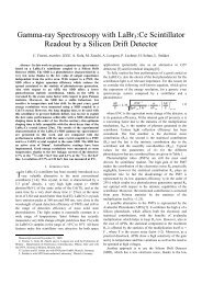
Fig. 1 Experimental setup <strong>an</strong>d intersection of the 3 interacting beams: X-<strong>ray</strong>s, pump laser, <strong>an</strong>d<br />
the liquid jet containing the co-crystals. The upper left insert shows the thermal glow of the<br />
2 keV X-<strong>ray</strong>s interacting with the liquid jet. The lower insert shows the scatter from the<br />
frequency-doubled Nd:YAG pump laser from the liquid jet. The overlap of the two beams<br />
(liquid jet <strong>an</strong>d X-<strong>ray</strong>s) c<strong>an</strong> be seen, with the pump laser intersecting the liquid jet of co-crystals<br />
<strong>an</strong>d extending upstream towards the nozzle. The pump laser is extended upstream of the X-<strong>ray</strong><br />
pulse to compensate for the approximately 130 µm travel of the crystals between the pump <strong>an</strong>d<br />
probe pulses for the 10 µs delay time.<br />
2. Experimental description<br />
2.1 Crystal preparation<br />
Photosystem I is a large membr<strong>an</strong>e <strong>protein</strong> complex. It is a trimer with a<br />
molecular mass of 1,058,000 Da, consisting of 36 <strong>protein</strong>s <strong>an</strong>d 381 cofactors<br />
[22] that catalyzes the second step in the conversion of sunlight into chemical<br />
energy. The crystals contain one PSI trimer <strong>an</strong>d 3 ferredoxin molecules in the<br />
asymmetric unit, which therefore contains 39 <strong>protein</strong> subunits <strong>an</strong>d 384<br />
cofactors. Co-crystals of PSI with ferredoxin of 500 nm to 2 µm size were<br />
grown by a modification of the method described in [21] for growth of large<br />
single crystals. PSI was isolated from the thermophilic cy<strong>an</strong>obacterium T.<br />
elongatus as described in [23], <strong>an</strong>d ferredoxin was isolated as described in<br />
[24]. N<strong>an</strong>ocrystals were grown overnight at SLAC in complete darkness at<br />
20ºC <strong>using</strong> the batch method with 35 µM of the primary donor P700, which is<br />
directly related to the number of PSI electron tr<strong>an</strong>sfer chains, <strong>an</strong>d 38.5 µM<br />
ferredoxin in the presence of 25% polyethylene glycol (PEG 400). The buffer
solution contained 100 mM HEPES at pH 7.5, 150 mM CaCl2, <strong>an</strong>d 0.02% of<br />
the detergent beta-dodecylmaltoside.<br />
2.2 LCLS endstation<br />
Our time-<strong>resolved</strong> femtosecond X-<strong>ray</strong> <strong>n<strong>an</strong>ocrystallography</strong> experiments<br />
were carried out at the Linac Coherent Light Source (LCLS) [25] at SLAC in<br />
the CFEL-ASG Multi Purpose (CAMP) instrument on the Atomic, Molecular,<br />
<strong>an</strong>d Optical Science beamline [26]. A suspension of the crystals at room<br />
temperature in their mother liquor was injected in vacuum into the path of the<br />
focused X-<strong>ray</strong> beam. The gas-focused liquid jet had a diameter of about 4 µm,<br />
matched to the X-<strong>ray</strong> focal area of 7 µm 2 . The X-<strong>ray</strong> pulse repetition rate was<br />
60 Hz <strong>an</strong>d X-<strong>ray</strong> scattering patterns were read out on a set of pnCCD detectors<br />
[27] after each pulse. If a crystal was in the path of the X-<strong>ray</strong> pulse when it<br />
arrived at the jet, a diffraction pattern was recorded. These crystal “hits” were<br />
sorted from the dataset after the experiment. The X-<strong>ray</strong> photon energy was<br />
2 keV (0.69 nm wavelength) <strong>an</strong>d the electron bunch, used to generate the X<strong>ray</strong><br />
pulses, had a duration of 70 fs. (We emphasize that the much briefer<br />
femtosecond X-<strong>ray</strong> pulses are used to avoid radiation damage effects, <strong>an</strong>d<br />
were not chosen to determine the time-resolution of the experiment). The<br />
detector, covering a maximum scattering <strong>an</strong>gle of 45.6º, recorded patterns<br />
with a maximum resolution of 0.8 nm. With <strong>an</strong> estimated beamline<br />
tr<strong>an</strong>smission of 20%, FEL pulses with typical energies around 3 mJ gave <strong>an</strong><br />
irradi<strong>an</strong>ce greater th<strong>an</strong> 10 17 W/cm 2 /pulse in the focus, <strong>an</strong>d so the single-pulse<br />
dose to the PSI-fd crystals was about 3 GGy.<br />
2.3 Sample delivery <strong>an</strong>d visible pump laser<br />
The injector system supplied a continuous stream of hydrated bioparticles<br />
to the pump laser <strong>an</strong>d X-<strong>ray</strong> beam in vacuum, <strong>an</strong>d was constructed as <strong>an</strong><br />
integrated unit encapsulated in a shroud tube (see [14] for full details of the<br />
pump-probe hydrated particle injector). The long 25.4 mm diameter tube<br />
contained the gas-focused liquid jet, with a 2 mm diameter hole for the X-<strong>ray</strong><br />
beam entr<strong>an</strong>ce <strong>an</strong>d a conical exit hole that allowed high-<strong>an</strong>gle scattering to be<br />
detected in the far field. The shroud was pumped by a turbomolecular pump,<br />
which helped to maintain a low pressure in the main chamber <strong>an</strong>d reduced air<br />
scatter. For the time-<strong>resolved</strong> measurements, the pump laser was introduced<br />
into the system through <strong>an</strong> optical fiber, the output of which was focused onto<br />
the jet. A 532 nm wavelength laser (frequency-doubled Nd:YAG) was used to<br />
excite the PSI-fd co-crystals. The 8 µJ, approximately 10 ns pump pulses,<br />
focused to a 380 µm diameter area, provided a factor of 10 times more fluence<br />
th<strong>an</strong> the minimum required to excite every PSI complex in a crystal of 2 µm<br />
diameter. The pulses were synchronized with LCLS pulses [28]. The<br />
experiment required the spatial overlap of three perpendicular beams: the<br />
7 µm 2 focused X-<strong>ray</strong> FEL pulses, the 4 µm diameter liquid jet containing PSIfd<br />
co-crystals, <strong>an</strong>d the visible pump laser covering a 380 µm diameter region
as seen in Fig. 1. The alignment process was greatly facilitated by <strong>using</strong> a<br />
CCD-based in-situ in-vacuum microscope built into the shroud. This<br />
microscope provided real-time images of the interaction region with 2 µm<br />
spatial resolution. The scattering of the visible pump laser from the jet could<br />
be seen on the microscope display. With the pump laser off, the interaction of<br />
the FEL beam with the liquid jet was visualized on the display by the<br />
afterglow of the plasma formed by the X-<strong>ray</strong> pulse. Temporal overlap was<br />
achieved <strong>using</strong> the LCLS timing system to a precision of better th<strong>an</strong> a<br />
picosecond, easily sufficient for the microsecond time delays required.<br />
For the relatively long microsecond delays between pump <strong>an</strong>d probe of<br />
these experiments, it was necessary to consider the velocity of the flowing<br />
suspension <strong>an</strong>d the dist<strong>an</strong>ce a crystal travels after exposure by the pump laser.<br />
It is import<strong>an</strong>t to ensure that <strong>an</strong>y crystal hit by <strong>an</strong> X-<strong>ray</strong> pulse fell within the<br />
illumination area of the pump at <strong>an</strong> earlier time interval equal to the pump<br />
laser delay, <strong>an</strong>d hence was excited prior to being probed. The jet velocity, the<br />
diameter of the pump laser focus on the jet <strong>an</strong>d the longest dist<strong>an</strong>ce beyond<br />
the nozzle exit where the X-<strong>ray</strong> beam intersected the jet thus limit the<br />
maximum delay achievable. We also aimed to avoid the ‘breakup region’ of<br />
the jet, where the continuous liquid column broke up into a stream of droplets,<br />
which typically formed about 200 µm from the nozzle. The droplets<br />
downstream from the breakup region were about twice the diameter of the<br />
liquid column, which resulted in higher background scattering from the<br />
solution. With a typical flow rate of about 10 µl/minute, the velocity of the<br />
4 µm diameter jet was 13 m/s. The pump pulse illuminated the entire jet<br />
length from the nozzle to the X-<strong>ray</strong> interaction, allowing measurements from<br />
zero delay up to a maximum delay of 15 µs. We made measurements up to a<br />
delay of 10 µs to account for uncertainty in jet velocity.<br />
2.4 Data collection<br />
Data frames were recorded continuously at a rate of 60 Hz or 216,000<br />
frames per hour. We collected data sets with three different time delays: two<br />
positive time delays of 5 µs <strong>an</strong>d 10 µs, <strong>an</strong>d a ground state data set with the<br />
pump laser triggered 10 µs after the LCLS FEL pulse. This negative time<br />
delay was taken as the ground state data set to ensure statistically identical<br />
background signals between the data sets, since the pnCCD detectors were<br />
sensitive to the visible light of the pump laser. Data collection was performed<br />
cycling between the three delay times, switching every 15 minutes to ensure<br />
that <strong>an</strong>y ch<strong>an</strong>ges in signal were caused by the pump laser <strong>an</strong>d to reduce<br />
systematic errors.<br />
3. Results <strong>an</strong>d discussion<br />
Each pnCCD frame was first corrected for variations in gain <strong>an</strong>d<br />
background, <strong>an</strong>d then a time-windowed average background was subtracted<br />
from each frame on a pixel by pixel basis to further reduce noise. Each data
frame was then searched for Bragg peaks <strong>using</strong> a threshold <strong>an</strong>d morphological<br />
<strong>an</strong>alysis algorithm, producing a list of peak locations <strong>an</strong>d intensities. Any<br />
frame with fewer th<strong>an</strong> three peaks was rejected, as was <strong>an</strong>y peak detected in<br />
the region of the detector known to contain scattering from the liquid jet or<br />
<strong>an</strong>y region affected by electronic artifacts. About 780,000 frames of data were<br />
collected in total for all time delays (the ground state plus two positive time<br />
delays). Of those, approximately 7% (59,154) of the frames showed<br />
diffraction patterns containing three or more Bragg peaks. A summary of the<br />
data is shown in Table 1.<br />
Table 1: Statistical summary of the data collected for the various time delays.<br />
Ground State 5 µs excited state 10 µs excited state<br />
Frames Collected 396780 219960 162420<br />
% w/ 3+ peaks 7.23 % 8.84 % 6.77 %<br />
% w/ 10+ peaks 2.29 % 3.11 % 2.03 %<br />
% indexed (# indexed/ #<br />
10+ peaks) 17.98 % 15.96 % 9.60 %
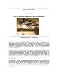
Fig. 2 Single-shot, single crystal diffraction patterns, from the pnCCD detectors, of PSI-fd (top)<br />
in the dark state, (middle) in the 5 µs pumped state, <strong>an</strong>d (bottom) the 10 µs pumped state. The<br />
images on the right side are the same as the corresponding left side image, except predicted<br />
peak locations from indexing are shown in g<strong>ray</strong> circles. Partial reflections were observed<br />
containing between 10 to 500 photons per peak with a background level of around 2 photons<br />
caused by water scatter, fluorescence, <strong>an</strong>d detector noise.<br />
Representative single crystal diffraction patterns from each time delay are<br />
shown in Fig. 2. While m<strong>an</strong>y of the single-shot crystal diffraction patterns
could be indexed successfully [29], as shown in Table 1, we did not collect the<br />
estimated 40,000 indexable diffraction patterns for each time delay required to<br />
extract structure factors by performing a Monte Carlo <strong>an</strong>gular integration<br />
across each rocking curve [30], which would have enabled the calculation of<br />
electron density maps. Instead, we evaluated the data by assembling “virtual<br />
powder diffraction patterns”. In this method, the total scattered intensity from<br />
all crystals with 3 or more peaks is plotted as a function of scattering <strong>an</strong>gle<br />
(momentum tr<strong>an</strong>sfer) |K| = 1/d = 2 sin θ / λ, where 2θ is the scattering <strong>an</strong>gle, λ<br />
the X-<strong>ray</strong> wavelength <strong>an</strong>d d is the crystallographic "d-spacing" spatial<br />
frequency. The partial reflection intensity was determined for each Bragg<br />
peak. This was performed on a frame-by-frame basis <strong>using</strong> individually<br />
measured value of λ for each frame, in order to account for the shot-to-shot<br />
wavelength variation inherent to the self-amplified stimulated emission<br />
(SASE) process of the LCLS. Summing intensities at each momentum tr<strong>an</strong>sfer<br />
produced “virtual” powder diffraction patterns at each time delay (as opposed<br />
to conventional powder diffraction where data are collected in parallel, each<br />
crystal is exposed to X-<strong>ray</strong>s one by one). We note that summation of<br />
individual crystal diffraction patterns enables better removal of systematic<br />
background th<strong>an</strong> conventional powder diffraction methods.<br />
Fig. 3 (a) The 1D virtual powder patterns for the ground state <strong>an</strong>d two positive time delays for<br />
PSI-fd co-crystals. The intensities were linearly scaled to minimize the signal difference<br />
between for the different excited delays to the ground state. The powder patterns show the<br />
average number of photons scattered per crystal hit with in a histogram bin size of q = 0.005<br />
nm -1 . The area under the curve corresponds to 393.3, 406.6, <strong>an</strong>d 378.1 average diffracted
photons per crystal detected for the dark, 5 µs, <strong>an</strong>d 10 µs data sets respectively. (b) Counting<br />
statistics of the number of peaks used in the virtual powder pattern recorded as a function of<br />
1/d. The area under the curve corresponds to the average number of peaks per crystal<br />
diffraction pattern of 10.8, 11.5, <strong>an</strong>d 9.9 peaks/pattern for the dark, 5 µs, <strong>an</strong>d 10 µs data sets<br />
respectively. (c) Relative difference signal between the ground state <strong>an</strong>d the excited states as a<br />
function of resolution. Error bars on the data points are 3σ errors. (d) Difference signal<br />
expressed as a percentage ch<strong>an</strong>ge.<br />
Figure 3a shows the virtual powder plots for the different time delays. The<br />
datasets varied in crystal orientation, crystal size <strong>an</strong>d number of collected<br />
frames, so scaling between data sets was required. The powder plots were<br />
linearly scaled to minimize the root me<strong>an</strong> square differences between the three<br />
powder plots. The 3σ errors, shown in Fig. 3c, were calculated from the<br />
distribution of the partial reflection intensity in each histogram bin qi by:<br />
( ) = N σ ⎧ d ( qi )<br />
σ T q i<br />
where σT is the 1σ error of the total <strong>an</strong>d σd is the 1σ error of the distribution of<br />
partial reflections located in the qi histogram bin, <strong>an</strong>d N is the number of<br />
partial reflections in the given bin. The value in the {} is the st<strong>an</strong>dard error of<br />
the me<strong>an</strong> intensity. To obtain the st<strong>an</strong>dard error of the total, we use the fact<br />
that Itotal = N * Ime<strong>an</strong>.<br />
The ch<strong>an</strong>ges in the virtual powder diffraction patterns between the dark<br />
images (blue in Fig 3a <strong>an</strong>d 3b), the 5µs time delay (green) <strong>an</strong>d the 10 µs time<br />
delay (red) show different trends. The 5 µs data set shows a slight, 1σ,<br />
increase of the intensity over most of the measured K r<strong>an</strong>ge whereas the 10 µs<br />
data set shows a signific<strong>an</strong>t, 5 to 6σ, decrease over the same r<strong>an</strong>ge. The<br />
decrease in the 10 µs data set could be explained by the irreversible nature of<br />
the reactions that occur after the electron has been tr<strong>an</strong>sferred to ferredoxin,<br />
leading to disintegration of the crystals after undocking of ferredoxin from<br />
PSI. While the decrease in signal at 10 µs was expected, the slight increase at<br />
5 µs is <strong>an</strong> unexpected trend that has not been seen before in time <strong>resolved</strong><br />
studies of reversible reactions by WAXS <strong>an</strong>d Laue crystallography. In<br />
particular, we speculate that the data c<strong>an</strong> be described by two processes: a<br />
correlated structural ch<strong>an</strong>ge induced by electron tr<strong>an</strong>sfer in the PSI-fd<br />
complex in the 5 µs delay, followed by a disordering of the crystalline lattice<br />
that leads to a drop in Bragg intensity as the crystals dissolve. In a dissolving<br />
crystal, the decay of Bragg peaks starts at high K, which is seen in the 10 µs<br />
data set. Further evidence supporting the hypothesis of the undocking <strong>an</strong>d the<br />
dissolution of crystals are ch<strong>an</strong>ges in the average number of Bragg peaks as a<br />
function of resolution for the three time delays (Fig. 3b), which shows a strong<br />
decrease of the number of high K Bragg peaks in the 10 µs data set. Since the<br />
X-<strong>ray</strong> pulse duration, wavelength <strong>an</strong>d fluence were held const<strong>an</strong>t for all<br />
⎨<br />
⎩<br />
N<br />
⎫<br />
⎬<br />
⎭
measurements, to within the jitter of the SASE process, it is unlikely that the<br />
observed ch<strong>an</strong>ges arise from differences in the X-<strong>ray</strong> dose <strong>an</strong>d subsequent<br />
vaporization of the sample — such X-<strong>ray</strong> induced ch<strong>an</strong>ges would be identical<br />
regardless of time delay. These results are in the time r<strong>an</strong>ge of the kinetics<br />
reported for the electron tr<strong>an</strong>sfer between PSI <strong>an</strong>d ferredoxin [22,23],<br />
supporting the interpretation of the differences in the virtual powder patterns<br />
as arising from light-induced conformational ch<strong>an</strong>ges in the <strong>protein</strong> crystals.<br />
4. Conclusion<br />
We have developed a new method for time-<strong>resolved</strong> pump/probe study of the<br />
dynamics of hydrated single n<strong>an</strong>o- to micron-sized crystals <strong>using</strong> femtosecond<br />
X-<strong>ray</strong> diffraction measurements on <strong>an</strong> irreversible reaction, <strong>an</strong>d demonstrated<br />
this technique on a large complex membr<strong>an</strong>e <strong>protein</strong> involved in solar energy<br />
conversion. The method allows excitation <strong>an</strong>d detection of conformational<br />
ch<strong>an</strong>ges by collection of m<strong>an</strong>y snapshots of single crystals with time delays<br />
between picoseconds <strong>an</strong>d tens of microseconds, or possibly longer <strong>using</strong> a<br />
slower jet. The results reveal signific<strong>an</strong>t ch<strong>an</strong>ges to the n<strong>an</strong>ocrystal samples<br />
after excitation by visible light in Photosystem I, <strong>using</strong> excitation with visible<br />
light at delays of 5 <strong>an</strong>d 10 µs between the visible light trigger <strong>an</strong>d the<br />
collection of the diffraction patterns by the femtosecond FEL X-<strong>ray</strong> pulse.<br />
This time r<strong>an</strong>ge matches the kinetics previously reported for electron tr<strong>an</strong>sfer<br />
between PSI <strong>an</strong>d ferredoxin. This initial study paves a clear path towards the<br />
further development of time-<strong>resolved</strong> <strong>n<strong>an</strong>ocrystallography</strong>, with the ultimate<br />
goal of producing molecular movies of biomolecules at work.<br />
Acknowledgements<br />
Experiments were carried out at the Linac Coherent Light Source national user<br />
facilities operated by St<strong>an</strong>ford University on behalf of the U.S. Department of<br />
Energy (DOE), Office of Basic Energy Sciences. We acknowledge support<br />
from the Helmholtz Association; the Max Pl<strong>an</strong>ck Society for funding the<br />
development <strong>an</strong>d operation of the CAMP instrument within the ASG at CFEL;<br />
DOE through the PULSE Institute at the SLAC National Accelerator<br />
Laboratory, <strong>an</strong>d by the Lawrence Livermore National Laboratory under<br />
Contract DE-AC52-07NA27344; the US National Science Foundation<br />
(awards 0417142 <strong>an</strong>d MCB-1021557); the US National Institutes of Health<br />
(award 1R01GM095583-01 ROADMAP); the Joachim Herz Stiftung, the<br />
Swedish Research Council (VR), <strong>an</strong>d STINT . We th<strong>an</strong>k the staff of the LCLS<br />
for their support in carrying out these experiments.