BY LOU BYERLY AND SUSUMU HAGIWARA University of ...
BY LOU BYERLY AND SUSUMU HAGIWARA University of ...
BY LOU BYERLY AND SUSUMU HAGIWARA University of ...

You also want an ePaper? Increase the reach of your titles
YUMPU automatically turns print PDFs into web optimized ePapers that Google loves.
J. Physiol. (1982), 322, pp. 503-528 503<br />
With 12 text-ftgures<br />
Printed in Great Britain<br />
CALCIUM CURRENTS IN INTERNALLY PERFUSED NERVE CELL<br />
BODIES OF LIMNEA STAGNALIS<br />
<strong>BY</strong> <strong>LOU</strong> <strong>BY</strong>ERLY <strong>AND</strong> <strong>SUSUMU</strong> <strong>HAGIWARA</strong><br />
From the Department <strong>of</strong> Biological Sciences, <strong>University</strong> <strong>of</strong> Southern California,<br />
Los Angeles, CA 90007, U.S.A. and the Department <strong>of</strong> Physiology,<br />
Ahmanson Laboratory-B.R.I., and Jerry Lewis Neuromuscular Research Center,<br />
<strong>University</strong> <strong>of</strong> California, Los Angeles, CA 90024, U.S.A.<br />
(Received 22 May 1981)<br />
SUMMARY<br />
1. When K+ is removed from both sides <strong>of</strong> the somal membrane <strong>of</strong> Limnea<br />
neurones, time-dependent, voltage-dependent outward currents are observed at<br />
positive potentials. These currents can be carried by Tris+ and tetraethylammonium<br />
(TEA+), as well as Cs+, but the Cs currents are several times larger. The Cs currents<br />
are not affected by external or internal TEA, but are strongly reduced by 4aminopyridine<br />
(4-AP) and all Ca blockers tried.<br />
2. The presence <strong>of</strong> these non-specific outward currents and their sensitivity to all<br />
treatments that eliminate the Ca currents prevent the complete isolation <strong>of</strong> Ca<br />
currents. The non-specific outward currents are most prominent at large positive<br />
potentials and as slow tail currents on stepping back to the holding potential.<br />
3. Ca currents are 'washed out' in well perfused cells. Typically the Ca current has<br />
decayed to less than one tenth <strong>of</strong> its original size after I h <strong>of</strong> perfusion. This wash-out<br />
is specific for the Ca current; Na and K currents persist for several hours.<br />
4. Once the Ca current has completely decayed, it is possible to study one type<br />
<strong>of</strong> non-specific current without overlapping inward currents. This current activates<br />
between 0 and + 30 mV and appears to reverse near 0 mV.<br />
5. In spite <strong>of</strong> the probable presence <strong>of</strong> slowly activating outward currents, the net<br />
inward currents measured show little apparent inactivation. In all the cells studied<br />
the inward current evoked at + 20 mV has never decayed by more than 50 % during<br />
a 60 ms pulse. So the true inactivation <strong>of</strong> these Ca currents must be quite slow, with<br />
time constants <strong>of</strong> the order <strong>of</strong> 100 ms and larger.<br />
6. The activation <strong>of</strong> the Ca current agrees with m2 kinetics. The rate <strong>of</strong> activation<br />
is the same for Ba currents as for Ca currents.<br />
7. When the membrane potential is stepped back to the holding level (-50 mV),<br />
the Ca current turns <strong>of</strong>f quite rapidly with a time constant <strong>of</strong> about 100 /Ls (25 0C).<br />
The time constant for turning <strong>of</strong>f the Ca current is not related to the time constant<br />
for turning on the Ca current at the same voltage as expected for m2 kinetics in the<br />
Hodgkin and Huxley model. At -30 mV the Tm for turn-on is eight times larger than<br />
the rm for turn-<strong>of</strong>f.<br />
Downloaded from J Physiol (<br />
jp.physoc.org)<br />
by guest on January 20, 2013
504<br />
L. B YERL Y <strong>AND</strong> S. <strong>HAGIWARA</strong><br />
INTRODUCTION<br />
Biophysical studies <strong>of</strong> voltage-dependent Ca currents have been impeded by the<br />
presence <strong>of</strong> overlapping K currents and the complicated geometries <strong>of</strong> the membranes<br />
involved. Invaginated membranes, attached axons and electrical coupling to other<br />
cells have prevented good control <strong>of</strong> the membrane potential. The overlapping K<br />
currents cannot be completely blocked by any pharmacological means and are <strong>of</strong>ten<br />
dependent on the Ca current, making complete isolation <strong>of</strong> the Ca current impossible.<br />
In the last few years new techniques have been developed which appear to largely<br />
overcome these difficulties for biophysical studies <strong>of</strong> Ca currents in the nerve cell<br />
bodies <strong>of</strong> snails. Krishtal & Pidoplichko (1975) first introduced techniques for voltage<br />
clamping and exchanging intracellular ions <strong>of</strong> isolated nerve cell bodies. The<br />
subsequent development and exploitation <strong>of</strong> this technique for studying Ca currents<br />
was reported in a series <strong>of</strong> papers (Kostyuk, Krishtal & Pidoplichko, 1975, 1977a,<br />
1981; Kostyuk, Krishtal & Shakhovalov, 1977b; Kostyuk & Krishtal, 1977; Doroshenko,<br />
Kostyuk & Tsyndrenko, 1978a, b, 1979; Doroshenko & Tsyndrenko, 1978;<br />
Krishtal, 1978; Kostyuk, 1980). Most <strong>of</strong> these studies were done on neurones from<br />
Helix pomatia. Lee, Akaike & Brown (1977, 1978) developed somewhat different<br />
methods for isolating, voltage clamping and internally perfusing snail nerve cell<br />
bodies and used them to study the Ca currents <strong>of</strong> Helix aspersa neurones (Akaike,<br />
Lee & Brown, 1978).<br />
When we began our study <strong>of</strong> the Ca current in Limnea neurones, we tried<br />
procedures published by both <strong>of</strong> the above laboratories. Some <strong>of</strong> the techniques never<br />
worked in our hands. The techniques we finally adopted, which are described below,<br />
are a combination <strong>of</strong> those used by the two laboratories, plus a few innovations <strong>of</strong><br />
our own. This paper reports the properties <strong>of</strong> the Ca current <strong>of</strong> Limnea nerve cell<br />
bodies as determined using our technique for internal perfusion and voltage clamping.<br />
In general we find that the Ca currents and the overlapping background currents in<br />
Limnea neurones are much more like those <strong>of</strong> Helix pomatia than those reported for<br />
Helix aspersa. Preliminary reports <strong>of</strong> this work have been published (Byerly,<br />
Hagiwara, Masuda & Yoshii, 1979; Byerly & Hagiwara, 1981).<br />
METHODS<br />
Isolation <strong>of</strong> cells<br />
The circumoesophageal nerve ring is dissected out <strong>of</strong> an adult Limnea stagnalis and then soaked<br />
in a 0-2 % trypsin (Sigma, Type III) solution for 90 min at room temperature. The visceral, parietal<br />
and pedal ganglia are used. Each ganglion is opened and the neuropil with cell bodies attached is<br />
freed from the covering sheath, using sharpened tungsten wires and irredectomy scissors. The<br />
exposed neuropil and cell bodies are then transferred by pipette to a dish containing Limnea saline<br />
with 7 mM-glucose. Using two glass micro-electrodes, the neuropil is then torn apart until a length<br />
<strong>of</strong> the axon connecting a cell body to the neuropil is exposed. This axon is then severed with the<br />
tip <strong>of</strong> one <strong>of</strong> the micro-electrodes, while the axon is lying on the casting resin (Dow Corning, Sylgard<br />
184) covering the bottom <strong>of</strong> the dish. The axon is usually severed within 50,cm <strong>of</strong> the soma. The<br />
isolated cell body is given 2 h to recover, during which time the axon stub rounds into the soma.<br />
Many cells do not survive the isolation procedure; those that do can easily be identified by their<br />
shiny appearance and the absence <strong>of</strong> the white colour which appears in damaged cells. Usually more<br />
than ten healthy isolated cell bodies with diameters from 80 to 120,m are obtained from each<br />
animal. These cells remain in a healthy state for at least 24 h in glucose-containing saline solution.<br />
Downloaded from J Physiol (<br />
jp.physoc.org)<br />
by guest on January 20, 2013
Ca CURRENTS IN LIMNEA NEURONES<br />
505<br />
We find this method <strong>of</strong> isolating the cells before sucking them onto the suction electrode to be<br />
superior to isolating the cells after they are attached to the suction electrode (Lee et al. 1978). With<br />
this method we routinely obtain healthy cells with strong Ca currents. In contrast, only a small<br />
percentage <strong>of</strong> the attached cells sucked into the suction electrode can be isolated without killing<br />
the cell, and those that do survive the isolation usually have too long an axon stub to allow good<br />
voltage control or complete perfusion. Since the membrane currents irreversibly change during the<br />
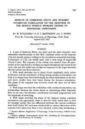
Fig. 1. Experimental apparatus. Drawing is not to scale. The suction electrode (SE) is<br />
3 mm in diameter and 4 cm long, while the cell diameter is 80-120 #sm. The plastic block<br />
holding the suction electrode is mounted to a micromanipulator. The arrows drawn inside<br />
the suction electrode indicate the flow <strong>of</strong> the internal solution. The inputs to the voltage<br />
follower (VF) and virtual ground (VG) amplifiers are connected to Ag/AgCl wires. The R8<br />
potentiometer connected to the summing amplifier (1) provides series resistance compensation.<br />
The three positions on the switch <strong>of</strong> the negative input <strong>of</strong> the clamp amplifier<br />
(A) allow for suction electrode voltage clamp (SEC), current clamp (IC), and hybrid<br />
voltage clamp (HC). The micro-electrodes (uE) are filled with 3 M-KCl. The identified<br />
signals are micro-electrode voltage (TVME), suction electrode voltage (VSE), command<br />
voltage (VI), and membrane current (I).<br />
first I h <strong>of</strong> perfusion (see below), it is important for our studies to have the cell in an otherwise stable<br />
state when it is sucked against the suction electrode. By the time we study the isolated cell the<br />
axon stub has rounded into the soma, so that relatively stable cells with nearly spherical shapes<br />
are obtained. In contrast, when the cell is isolated after being sucked against the suction electrode,<br />
the currents must be studied while the cell is recovering from the isolation procedure.<br />
In these studies no attempt has been made to identify neurones. Cells are only selected to have<br />
a diameter <strong>of</strong> 80-120 ,gm and to have survived the isolation procedure (this may select against cells<br />
with larger axons or stronger ties to connective tissues). However, the technique is suitable for<br />
isolating identified neurones. In preliminary trials a pre-selected neurone could be isolated with<br />
about a 50 % success rate.<br />
It is important to point out that only one type <strong>of</strong> Ca current was found, despite the variation<br />
Downloaded from J Physiol (<br />
jp.physoc.org)<br />
by guest on January 20, 2013
506<br />
L. B YERL Y <strong>AND</strong> S. <strong>HAGIWARA</strong><br />
in size and probable variation in function <strong>of</strong> the cells studied. Although the magnitude <strong>of</strong> the Ca<br />
current varied remarkably from cell to cell, the voltage and time dependencies <strong>of</strong> the Ca current<br />
showed no significant variation. The apparent inactivation <strong>of</strong> Ca currents was somewhat faster in<br />
some cells, but we assume this resulted from the presence <strong>of</strong> a greater amount <strong>of</strong> overlapping<br />
outward current in these cells, rather than a basic difference in the Ca current.<br />
Apparatus<br />
Figure 1 shows a schematic diagram <strong>of</strong> the physical arrangement used in these studies.<br />
Suction electrode. The suction electrode is made from 3 mm (o.d.) Pyrex glass tubing. The glass<br />
tubing is pulled on an electrode puller (Narishige, PE2) in two pulls (the second at a lower<br />
temperature) to give a sharp taper. The tip is next broken at about 80 num (o.d.) and then fire-polished<br />
in a micr<strong>of</strong>orge to have an inner diameter at the tip <strong>of</strong> 25-30 ,sm. These tips form the best seal<br />
(highest resistance) to the somal membrane when they are clean. Coating the tip with various<br />
adhesive or sealant materials (Parafilm, oil, silicon, protamine, polylysine, etc.) does not improve<br />
the seal. The best seals are obtained the first few times an electrode is used. Therefore, we change<br />
suction electrodes frequently. The attachment for mounting the suction electrode in the plastic<br />
block is designed to allow easy replacement <strong>of</strong> the suction electrode.<br />
Internal solution flow system. The internal solution enters the suction electrode through a glass<br />
pipette that enters the plastic block through a vaseline-filled metal sleeve. This allows for position<br />
adjustment so that the tip <strong>of</strong> this inlet tube can be placed just a few hundred microns from the<br />
cell, in spite <strong>of</strong> the unavoidable variation in the length <strong>of</strong> the suction electrodes. With the inlet<br />
so positioned intracellular K+ can be completely replaced in less than 5 min (see below).<br />
The internal solution flows from open bottles sitting at a level about 40 cm above the suction<br />
electrode. The exhaust for the internal solution leads to a trap bottle in which the pressure is<br />
measured by a manometer. The pressure difference (relative to atmospheric) measured by the<br />
manometer is expected to be somewhat greater than the pressure difference at the tip <strong>of</strong> the suction<br />
electrode, due to the drop in pressure along the exhaust line. The trap bottle is connected to a<br />
vacuum pump via a second trap bottle. The pressure at the tip <strong>of</strong> the suction electrode is varied<br />
by means <strong>of</strong> two valves, one on the vacuum line to the trap bottle and the other on a bleeder line<br />
to the trap bottle. Internal solution flows through the suction electrode at a rate <strong>of</strong> about 1 ml every<br />
5 min when the cell is in place.<br />
Current measurement. Membrane current flows from the control amplifier (Biodyne AL- 1) to the<br />
stainless-steel sleeve through which the internal solution inlet enters. The current is then carried<br />
through the internal solution into the cell. In the external solution the current flows into a pipette<br />
filled with a 3 M-KCl solution in agar. This electrode is connected via a Ag/AgCl wire to the virtual<br />
ground input <strong>of</strong> a current-to-voltage converter (5 #s time constant). Even during the large<br />
capacitive current transients associated with voltage steps the potential in the bath varies by less<br />
than one millivolt.<br />
Potential measurement. The potential across the membrane is measured in two ways. One way<br />
is with a conventional micro-electrode (5-10 MCI) inserted through the membrane. The second<br />
measurement is from a flowing-KCl electrode in the internal solution at a considerable distance<br />
downstream from the cell. This is a conventional micro-electrode broken to a resistance <strong>of</strong> less than<br />
1 MC. The open end <strong>of</strong> the micro-electrode is connected to a reservoir <strong>of</strong> 3 M-KC1. Given the lower<br />
pressure in the suction electrode, a constant stream <strong>of</strong> 3 M-KCl flows from this electrode. The<br />
potential measured from a Ag/AgCl wire in the KCl reservoir is then corrected electronically for<br />
the potential drop that occurs at the tip <strong>of</strong> the suction electrode due to the flow <strong>of</strong> current across<br />
the tip resistance (usually about 400 KM) when current is passed through the membrane. The<br />
membrane current is usually less than 30 nA, even during positive potential pulses, but can reach<br />
several microamperes during the capacitive transients accompanying potential steps.<br />
Clamping techiques. When current-clamp experiments are done the output <strong>of</strong> the current-tovoltage<br />
converter is fed back to the control amplifier. This is superior to clamping the current put<br />
out by the control amplifier, since there is considerable capacitance between the tubing carrying<br />
the internal solution and ground.<br />
Voltage-clamp experiments are done using either <strong>of</strong> two feed-back signals. In 'hybrid-clamp'<br />
experiments the potential measured by an intracellular micro-electrode is fed back to the control<br />
amplifier. This type <strong>of</strong> voltage-clamp is faster and superior whenever large currents are involved<br />
or when large liquid junction potentials exist between different internal solutions. In the<br />
Downloaded from J Physiol (<br />
jp.physoc.org)<br />
by guest on January 20, 2013
Ca CURRENTS IN LIMNEA NEURONES 507<br />
suction-electrode clamp' the corrected potential measured by the flowing-KCl electrode is fed back<br />
to the control amplifier. This type <strong>of</strong> voltage-clamp has the advantage <strong>of</strong> convenience, since it is<br />
not necessary to impale the cell with a micro-electrode. Smaller, slower currents measured by the<br />
suction-electrode clamp are the same as when measured by the hybrid clamp; but, without the<br />
micro-electrode penetrating the membrane, the cell is much more stable and external solution<br />
changes are easier. The series-resistance correction to the suction-electrode potential is made by<br />
adjusting the fraction <strong>of</strong> the current signal subtracted so as to give the fastest possible capacitive<br />
current transient. When this procedure is applied to a resistor-capacitor equivalent circuit, it is<br />
found to compensate for about 80 % <strong>of</strong> the series resistance. Therefore, the remaining effective series<br />
resistance is less than 100 kfl; so the true membrane potential will be less than 3 mV from the<br />
recorded value as long as the current is less than 30 nA.<br />
Data storage. All current and voltage records are photographed from the oscilloscope screen and<br />
stored on 35 mm film. When current or voltage records are to be subtracted, fitted or manipulated<br />
in some other manner, they are digitized on a 12 bit A/D converter (Datel DAS-250), sampling<br />
at intervals down to 10 ,us. The digital data is then stored on floppy disk and later manipulated<br />
by a microprocessor (North Star-Horizon).<br />
TABLE 1. Composition <strong>of</strong> solutions (mM)<br />
External solutions<br />
Solution Na+ K+ Cl- Ca2+ Mg2+ Tris<br />
Limnea saline<br />
Tris saline<br />
Internal solutions<br />
50<br />
0<br />
2-5<br />
0<br />
78<br />
74<br />
4<br />
4<br />
4<br />
4<br />
10<br />
65<br />
Solution K+ Cs+ Tris Aspartate HEPES EGTA<br />
K aspartate<br />
Cs aspartate<br />
Tris aspartate<br />
74<br />
0<br />
0<br />
0<br />
74<br />
0<br />
0<br />
0<br />
78<br />
62<br />
62<br />
59<br />
5<br />
5<br />
5<br />
5<br />
5<br />
5<br />
Solutions<br />
Table 1 shows the compositions <strong>of</strong> the main external and internal solutions used. The major anion<br />
in the external solutions is Cl-, while it is aspartate- in the internal solutions. When 7 mM-glucose,<br />
10mM-4-aminopyridine(4-AP) or 1 mM-Cd2+ are used in the external solutions, they are added<br />
without adjustment <strong>of</strong> the concentrations <strong>of</strong> other ions. Extracellular tetraethylammonium (TEA)<br />
is tested by substituting it for all the Na+ and K+ in Limnea saline. Ba currents are studied by<br />
replacing the Ca2+ with Ba2+ in Tris saline. When Co2+ is used as a Ca-current blocker it is<br />
substituted for all the Mg2+ and 3 mM-Ca2+ in Tris saline. The pH <strong>of</strong> all external solutions is 7-4.<br />
The pH <strong>of</strong> internal solutions is 7-3. The concentration <strong>of</strong> free Ca2+ in the internal solutions is assumed<br />
to be less than 10-8 M. All experiments are done at room temperature (23-27 'C).<br />
Procedure<br />
Zero potential is established by removing the flowing-KCl electrode from the plastic block and<br />
placing it in the external bath. When the flowing-KCl electrode is returned to its position in the<br />
plastic block with K aspartate solution inside the suction electrode and Limnea saline outside, a<br />
potential <strong>of</strong> about -15 mV (inside negative with respect to outside) is recorded due to the junction<br />
potential between the two solutions; the junction potential between NaCl and K aspartate solutions<br />
can be calculated from the Henderson equation to be - 14-5 mV. Individual isolated nerve cell<br />
bodies are transferred by pipette from the dish in which they are isolated to the recording chamber.<br />
The cell is gently sucked against the tip <strong>of</strong> the suction electrode and then the pressure in the trap<br />
bottle is reduced to about 50 cm <strong>of</strong> water lower than atmospheric pressure. Constant-current pulses<br />
show a large (about 100-fold) increase in resistance as the cell seals against the glass. When the<br />
internal solution inlet is brought close to the cell, the potential drops and shows attenuated action<br />
potentials as the patch <strong>of</strong> membrane across the tip breaks down, due to the very low concentration<br />
<strong>of</strong> Ca2+ on the extracellular side <strong>of</strong> the membrane. A constant current is applied to hold the<br />
Downloaded from J Physiol (<br />
jp.physoc.org)<br />
by guest on January 20, 2013
508<br />
L. <strong>BY</strong>ERLY <strong>AND</strong> S. <strong>HAGIWARA</strong><br />
membrane potential near -50 mV. If an instantaneous jump in potential occurs at the beginning<br />
<strong>of</strong> the square-current pulse, the rupture <strong>of</strong> the membrane is not complete; so the pressure in the<br />
suction electrode is reduced further for a few seconds to completely break the membrane patch.<br />
Once this membrane is properly broken, the resistance through the tip <strong>of</strong> the suction electrode (the<br />
resistance in series with the membrane) is increased by less than 50% from the value it had before<br />
the cell was present.<br />
Shunt resistance<br />
When isolated nerve cell bodies are studied with a single micro-electrode, they have membrane<br />
resistances <strong>of</strong> 40-400 MCI and time constants from 50 to several hundred milliseconds. When the<br />
same parameters are measured in cells sealed to the suction electrode by passing hyperpolarizing<br />
current pulses through the suction electrode, apparent valves <strong>of</strong> 20-200 MCI and 10-100 ms are<br />
obtained. Since the time constant and resistance measured when a cell is sealed to the suction<br />
electrode are somewhat less than the values measured by the micro-electrode impalement, we<br />
conclude that the shunt resistance (the resistance to flow <strong>of</strong> current between the membrane and<br />
the glass), is, in general, <strong>of</strong> about the same magnitude as the membrane resistance. In some<br />
preparations the shunt resistance is probably considerably less than the membrane resistance, so<br />
the total resistance is roughly equal to the shunt resistance. In other cases the shunt resistance<br />
is considerably larger than the membrane resistance, possibly exceeding a gigaohm, as judged by<br />
the nearly equal resistances (and time constants) measured first by micro-electrode and then by<br />
suction electrode. The size <strong>of</strong> the shunt resistance relative to the resting membrane resistance is<br />
<strong>of</strong> little importance in this study <strong>of</strong> the Ca current. Cells sealed to the suction electrode are only<br />
used if they generate fast, all-or-nothing, overshooting (+ 20 to + 40 mV) action potentials in<br />
response to small pulses <strong>of</strong> outward current. So clearly the inward membrane currents are<br />
considerably larger than shunt currents in all cells studied.<br />
Space clamp<br />
When a negative-voltage pulse is applied to the membrane, the capacitive current transients<br />
(Fig. 2 A and B) settle in less than1 ms for both suction-electrode and hybrid clamps. The membrane<br />
capacitance is given by the ratio <strong>of</strong> the area under the capactive current transient and the<br />
magnitude <strong>of</strong> the voltage step. The specific membrane capacitance is calculated by dividing the<br />
membrane capacitance by the surface area <strong>of</strong> the cell, assuming the cell to be a sphere. (No<br />
correction is made for the area <strong>of</strong> membrane eliminated by the suction electrode; thus, the specific<br />
capacitance is underestimated by about 10 %o.) The specific capacitances calculated for thirty-eight<br />
cells have a mean value <strong>of</strong> 1-5 puF/cm2 (S.D. = 05 #sF/cm2). This value is close to that expected for<br />
a simple membrane, suggesting that there is little invagination <strong>of</strong> this nerve cell body membrane.<br />
Therefore, the space clamp should be good, i.e. there should be little variation <strong>of</strong> membrane<br />
potential from one region to another.<br />
A much smaller, slow displacement-type current is also observed in these cell-suction-electrode<br />
preparations. This current decays over several milliseconds and is symmetrical for negative or<br />
positive voltage pulses. It does not result from error in potential measurement, since it appears<br />
the same in suction-electrode clamp and hybrid clamp <strong>of</strong> the same cell. Since this slow current has<br />
not been seen in the few two-micro-electrode voltage-clamp studies we have done on isolated nerve<br />
cell bodies, we suspect that it is associated with the glass-membrane seal. If portions <strong>of</strong> the<br />
membrane pressed against the glass slowly charge due to current flowing between the membrane<br />
and glass, such a prolonged displacement current would result. The charge carried by this slow<br />
current is always less than one tenth <strong>of</strong> the charge carried by the fast capacitive transient, which<br />
implies an area <strong>of</strong> membrane is involved that is consistent with the area <strong>of</strong> the cell in contact with<br />
the glass. Since this slow current is symmetrical, its presence can be overcome by adding the<br />
currents from equal negative and positive voltage steps. However, if this 'covered' portion <strong>of</strong><br />
membrane is active, the quality <strong>of</strong> the clamp is clearly degraded.<br />
Clamp speed<br />
The most reliable indication <strong>of</strong> the speed <strong>of</strong> a voltage-clamp is the duration <strong>of</strong> the transient<br />
capacitive currents. We adjust the clamp amplifier so that the response is slightly underdamped,<br />
so that the current reverses sign after the main current surge (see Fig. 2A and B). It is convenient<br />
to measure the speed <strong>of</strong> the clamp by the duration <strong>of</strong> this main current surge. After this time the<br />
Downloaded from J Physiol (<br />
jp.physoc.org)<br />
by guest on January 20, 2013
Ca CURRENTS IN LIMNEA NEURONES 509<br />
membrane potential is close to the command value, and the remaining capacitive transient can be<br />
eliminated by adding currents for equal positive and negative voltage steps. When the membrane<br />
potential is clamped using the signal recorded from the flowing-KCl electrode (suction-electrode<br />
clamp), the main surge lasts 150-600 ts (Fig 2A), the slower clamps resulting from larger cell<br />
capacitance and larger suction-electrode resistance. When the same preparation is clamped using<br />
the signal recorded from an intracellular micro-electrode (hybrid clamp) the speed <strong>of</strong> the clamp<br />
is about doubled (Fig. 2B). The main surge <strong>of</strong> capacitive current for hybrid clamps lasts 80-400 Is,<br />
A B<br />
V<br />
1OnALj<br />
SEC HC<br />
20 nAL__<br />
1 ms 1 ms<br />
7 C¢ O~~min Hlo_<br />
1 mnI5 /<br />
V~~~ ~ ~~~~~~~~~ andI4 min<br />
nA<br />
150 mV<br />
10 ms<br />
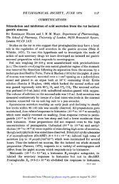
Fig. 2. Speed <strong>of</strong> voltage clamp and intracellular ion exchange. Capacitive current<br />
transients associated with 10 mV hyperpolarizing voltage pulses during suction electrode<br />
(A) and hybrid (B) voltage clamps <strong>of</strong> the same cell. Note change in current scale. Diameter<br />
<strong>of</strong> cell is 90 ,um; opening <strong>of</strong> suction electrode is 24 pm. Holding potential is -50 mV. C,<br />
exchange <strong>of</strong> intracellular K+ with Cs+. Currents are recorded at 1 min intervals following<br />
the transition from K aspartate to Cs aspartate in the suction electrode. External solution<br />
is Tris saline. Holding potenial is -50 mV; pulse is to + 20 mV. Cell diameter is 100 ptm;<br />
suction electrode opening is 26,um. SEC, suction electrode voltage clamp; HC, hybrid<br />
voltage clamp.<br />
depending on micro-electrode resistance as well as suction-etectrode resistance and cell capacitance.<br />
Typically, the main current surge lasted about is 150 for the studies reported in this paper. The<br />
brevity <strong>of</strong> the capacitive current confirms the good space clamp obtained with this preparation.<br />
The hybrid clamp is always used when currents are to be studied within 1 ms <strong>of</strong> a potential step.<br />
Exchange <strong>of</strong> intracellular ions<br />
Using suction electrodes with an inner diameter <strong>of</strong> 25-30 pm and cell bodies about 100 pam in<br />
diameter, the intracellular K+ can be replaced with Cs+ or Na+ in less than 5 min. This conclusion<br />
is based upon the times required for the membrane current to reach new steady-state levels<br />
following changes <strong>of</strong> the internal solution. When the internal solution is switched from K aspartate<br />
Downloaded from J Physiol (<br />
jp.physoc.org)<br />
by guest on January 20, 2013
510 L. B YERL Y <strong>AND</strong> S. <strong>HAGIWARA</strong><br />
to Cs aspartate, the currents measured during steps to + 20 mV reach a new steady state in 3-4 min<br />
(see Fig. 2C). The return <strong>of</strong> the K currents follows approximately the same time course when the<br />
internal solution is switched back to K aspartate. A similar time course <strong>of</strong> exchange is observed<br />
when replacing intracellular K+ with Na+. Five minutes after the internal solution is switched from<br />
K aspartate to Na aspartate (Na+ substituted for K+) the Na current reverses at a potential near<br />
the value expected for complete replacement <strong>of</strong> intracellular K+ with Na+. Thus, it appears that<br />
monovalent cations can be exchanged completely and rapidly. However, we have not yet<br />
determined the extent to which we can control more highly regulated intracellular ions, such as<br />
Ca2+ and H+.<br />
+80 * 60 ms<br />
10 nA<br />
/(nA)<br />
0 Peak 20<br />
10 Ms -100 100<br />
-10 V (mV)<br />
+20<br />
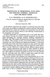
Fig. 3. Currents in absence <strong>of</strong> K+. On the left, tracings <strong>of</strong> the currents elicited by stepping<br />
the membrane potential to values (in MV) indicated on each trace. On the right, I- V plots<br />
<strong>of</strong> the peak current (0) and the current at 60 ms from the pulse beginning (0). Peak<br />
current is measured when the current reaches its most negative (inward) value. External<br />
solution is Tris saline; internal solution is Cs aspartate, Holding potential is -50 mV.<br />
Suction electrode voltage clamp used.<br />
RESULTS<br />
This study <strong>of</strong> the Ca current in Limnea neurones is complicated by two problems.<br />
(1) The Ca current is not stable under internal perfusion. Within half an hour <strong>of</strong> the<br />
beginning <strong>of</strong> the exchange <strong>of</strong> cytoplasm for saline solution the Ca current has almost<br />
completely disappeared. (2) Although the background currents have been greatly<br />
reduced by the replacement <strong>of</strong> intracellular K+ with less permeant ions, non-specific,<br />
time- and voltage-dependent currents are still present. Since these currents are<br />
sensitive to all the treatments that change the Ca current, a complete isolation <strong>of</strong><br />
the Ca current is not possible. However, in spite <strong>of</strong> these problems, a number <strong>of</strong> the<br />
properties <strong>of</strong> the Ca current can be studied. We will first discuss these two problems,<br />
before turning to the properties <strong>of</strong> the Ca current.<br />
I. Labile nature <strong>of</strong> Ca currents<br />
When the neurone is internally perfused with Cs aspartate and bathed externally<br />
in Tris saline, prolonged inward currents are elicited by stepping the membrane<br />
Downloaded from J Physiol (<br />
jp.physoc.org)<br />
by guest on January 20, 2013<br />
-20
-100<br />
s C p L-<br />
IL)<br />
Cu a)<br />
Ca CURRENTS IN LIMNEA NEURONES<br />
-10<br />
-20<br />
A<br />
B<br />
20 60<br />
Before decay<br />
* 60ms<br />
o Peak<br />
After decay<br />
x 60ms<br />
6 Peak<br />
-100<br />
C<br />
Time (min)<br />
V (mV)<br />
Fig. 4. Wash-out <strong>of</strong> Ca current. A, dependence <strong>of</strong> magnitude <strong>of</strong> Ca current on time. The<br />
peak current evoked by a step to + 20 mV is plotted. The size <strong>of</strong> the opening <strong>of</strong> the suction<br />
electrode used in each <strong>of</strong>the four experiments is given near the data; all cells were 90-100 yam<br />
in diameter. Times are measured from the completion <strong>of</strong> the replacement <strong>of</strong> intracellular<br />
K+ with Cs+. B and C, I-V curves for two different cells before (0O peak; @, 60 ms) and<br />
after (A, peak; x, 60 ms) Ca current decay. External solution is Tris saline and internal<br />
solution is Cs aspartate. Suction electrode voltage-clamp used.<br />
potential to values between -20 and + 50 mV (see Fig. 3). These inward currents<br />
are assumed to be carried by Ca2+, since they disappear when external Ca2+ is replaced<br />
by Mg2+ and are blocked by Ca blockers such as Co2+, La3+ and Cd2+. These Ca<br />
currents are not stable. If the membrane potential is stepped to + 20 mV for 60 ms<br />
at intervals <strong>of</strong> 1 or 2 min, the magnitude <strong>of</strong> the inward current decreases each time,<br />
until after 20-30 min there is no longer a net inward current (see Fig. 4A, records<br />
for 24 and 25 ,tm). This is not a general deterioration <strong>of</strong> the cell, because the current<br />
,-10<br />
Downloaded from J Physiol (<br />
jp.physoc.org)<br />
by guest on January 20, 2013<br />
511
512 L. B YERL Y <strong>AND</strong> S. <strong>HAGIWARA</strong><br />
asymptotically approaches a small positive value <strong>of</strong> about the size expected for the<br />
leakage current. The I-V relation measured for cells after the current reaches this<br />
new steady state no longer contains a negative-slope region (Fig. 4B and C), indicating<br />
that the Ca current has disappeared. As illustrated in Fig. 4B and C, the resistance<br />
<strong>of</strong> the cell can increase or decrease during this decay <strong>of</strong> the Ca current. The<br />
capacitance <strong>of</strong> the cell does not change. This is a selective loss <strong>of</strong> the Ca current; the<br />
Na and K currents last for much longer times. If after the Ca current has decayed<br />
the internal solution is changed to K aspartate, the K currents return. After complete<br />
decay <strong>of</strong> the Ca current cell bodies which originally had a combined Na and Ca spike<br />
still have a fast overshooting Na spike.<br />
The loss <strong>of</strong> the Ca current is due to the exchange <strong>of</strong> intracellular solution. We found<br />
that as we improved the intracellular perfusion, increasing the speed with which<br />
intracellular ions could be exchanged, the Ca current decayed more rapidly. When<br />
intracellular exchange is retarded by using suction electrodes with small openings,<br />
the Ca current decays much more slowly. Figure 4A shows that the Ca currents<br />
measured in 100 jzm cells sealed to suction electrodes with 11-12 jam openings<br />
decayed only slightly over a period <strong>of</strong> 1 h. It is not Cs+ that causes the decay <strong>of</strong> the<br />
Ca current. If a cell is perfused with K aspartate for 30 min and then switched to Cs<br />
aspartate to eliminate the outward currents, the Ca current is already very small,<br />
presumably due to decay during the perfusion period.<br />
We use suction electrodes with openings <strong>of</strong> about 25 gm and measure the Ca current<br />
during the first 15-20 min following the beginning <strong>of</strong> intracellular perfusion. Since<br />
the magnitude <strong>of</strong> the Ca current is continually declining during this period, the Ca<br />
current in test solutions is always compared to the Ca currents in control solutions<br />
before and after the test (when possible). We do not use suction electrodes with<br />
smaller openings, which greatly reduces the decline <strong>of</strong> the Ca current, because the<br />
rate <strong>of</strong> intracellular perfusion is so reduced that the ability to control intracellular<br />
ionic concentrations in the presence <strong>of</strong> large membrane currents is questionable. The<br />
exchange <strong>of</strong> intracellular K+ for Cs+ requires 15-30 min using suction electrodes with<br />
10 jtm openings and 80-120 #am cells.<br />
II. Non-specific outward current<br />
The currents measured when internal K+ is replaced with Cs+ and external Na+<br />
and K+ are replaced with Tris+ (Fig. 3) are not carried by Ca2+ alone. Given the very<br />
low concentration <strong>of</strong> free Ca2+ in the cell, Ca2+ can carry almost no outward current.<br />
The large time-dependent outward currents measured at potentials above + 60 mV<br />
can only be carried by an efflux <strong>of</strong> Cs+ or an influx <strong>of</strong> Cl-. These outward currents<br />
show very little change when external Cl- is replaced by methane sulphonate or when<br />
80 % <strong>of</strong> the external Tris+Cl- is replaced with glucose. Therefore, it seems Cs+ must<br />
carry this outward current. The replacement <strong>of</strong> intracellular Cs+ with Tris+, TEA+<br />
or arginine+ reversibly reduced the outward currents at large positive voltages by<br />
factors <strong>of</strong> two to four. The fact that these large cations can carry outward current<br />
demonstrates that this conductance is not very selective. The inward Ca current was<br />
also reversibly reduced by each <strong>of</strong> these replacements. (These experiments were done<br />
using a hybrid clamp, due to the increase in suction-electrode resistance caused by<br />
the low mobility <strong>of</strong> these ions.) Since intracellular Cs+ gives the largest Ca current<br />
Downloaded from J Physiol (<br />
jp.physoc.org)<br />
by guest on January 20, 2013
. ~~~~~~~~~G<br />
Ca CURRENTS IN LIMNEA NEURONES 513<br />
and a low suction-electrode resistance, it is used to replace K+ in all the studies<br />
reported here.<br />
Since the substitution <strong>of</strong> Tris+, TEA+, or arginine+ for Cs+ affected the Ca current as well as the<br />
outward current, it is not clear if the reduction <strong>of</strong> outward currents is due to the lower permeability<br />
<strong>of</strong> these large cations compared to Cs+, or if this is a pharmacological effect <strong>of</strong> certain intracellular<br />
/O -/L<br />
(nA)<br />
80<br />
10<br />
60<br />
20 ..x 50 100<br />
V (MV)<br />
-= -40<br />
-40<br />
~~~~~~~~~~~~(,umho)<br />
--0-2<br />
5 nA<br />
10 ms<br />
/ox~X~ 510<br />
x<br />
100<br />
V (mV)<br />
Fig. 5. Residual currents left after Ca current decay. On the left, tracings <strong>of</strong> currents<br />
evoked by depolarizing pulses. Numbers on traces give potentials (in mV) reached by<br />
pulses. External solution is Tris saline; internal solution is Cs aspartate. Suction electrode<br />
clamp used. On the upper right, plot <strong>of</strong> steady-state outward current (I.) against<br />
membrane potential. A linear leakage current (IL) has been subtracted from the data. Data<br />
are plotted both for currents (0) shown at left and for currents ( x ) from a second cell.<br />
On the lower right, conductance (0) is plotted against voltage for the currents plotted<br />
above. Conductance is calculated from G = (IO- IL)/(V- Vr), where Tr is the potential<br />
at which the straight line drawn through the currents intersects the voltage axis.<br />
cations that affects all currents. The Ca current was similarly reduced when the internal solution<br />
was changed from Cs aspartate to a mixture <strong>of</strong> one part Cs aspartate and four parts isotonic glucose,<br />
suggesting that the inward Ca current may be promoted by intracellular alkaline cations (Cs+, Na+<br />
and K+). (Replacing intracellular Cs+ with Na+ produced little change in the inward Ca current<br />
or the outward current.) Almers & Palade (1981) reported a similar effect on the Ca current <strong>of</strong> the<br />
frog muscle membrane; replacing intracellular K+ with TEA+ reduced the Ca current. The<br />
interaction <strong>of</strong> intracellular cations with the Ca currents is not clear.<br />
Residual current<br />
The time course and voltage dependence <strong>of</strong> the non-specific current in the presence<br />
<strong>of</strong> the Ca current is not known. As will be discussed below, all treatments that block<br />
the Ca current also change (usually reduce) the magnitude <strong>of</strong> the non-specific current<br />
and quite possibly also change its time course and voltage dependence. However, the<br />
17<br />
Downloaded from J Physiol (<br />
jp.physoc.org)<br />
by guest on January 20, 2013<br />
PRHY 322
514 L. BV7ERLY <strong>AND</strong> S. <strong>HAGIWARA</strong><br />
current remaining after the Ca current has completely decayed provides a description<br />
<strong>of</strong> at least one component <strong>of</strong> the non-specific current. The voltage and time<br />
dependencies <strong>of</strong> this residual current are shown in Fig. 5. This conductance is turned<br />
on over a range <strong>of</strong> about 30 mV, somewhere between - 10 and +40 mV. The<br />
activation <strong>of</strong> these currents becomes more and more rapid as the membrane potential<br />
is stepped to more positive levels; there is no inactivation. The linear regions <strong>of</strong> the<br />
Tris saline<br />
o Peak<br />
* 60Oms<br />
Tris saline+ 1 mm-Cd 2<br />
+2 10 mAs j<br />
10msL<br />
/ (nA)<br />
40<br />
Fig. 6. Effects <strong>of</strong> 1 mm-Cd2+ on currents. I-V relations before (0, peak; 0, 60 msec) and<br />
after ( x, 60 ins) the addition <strong>of</strong> 1 mm-Cd2+ to external solution. Inset shows currents<br />
before and after addition <strong>of</strong> Cd2+, at + 20 and + 90 mV. External solution is Tris saline<br />
(with and without Cd2+) ;internal solution is Cs aspartate. Suction electrode clamp is used.<br />
I- V curves for the steady-state values <strong>of</strong> this current project to zero current near<br />
the origin, suggesting that this current would reverse around 0 mV. This reversal<br />
potential is supported by the fact that inward tail currents are seen when the potential<br />
is returned to -50 mV following positive steps which activate this current (see Fig.<br />
12 C). Given the highly asymmetric nature <strong>of</strong> the solutions on the two sides <strong>of</strong> the<br />
membrane (Cs+Asp- inside and Tris+Clh outside), this reversal potential implies that<br />
the conductance mechanism responsible for the residual current is fairly non-selective.<br />
Sensitivity to Ca blockers<br />
The Ca current could be isolated from the background currents that persist with<br />
external Tris saline and internal Cs aspartate if some treatment were available which<br />
selectively reduced or eliminated the Ca current. Unfortunately, we find the outward<br />
currents at large positive voltages are always changed (usually reduced) when the<br />
Ca current is reduced, regardless <strong>of</strong> the treatment used to reduce the Ca current. (1)<br />
The Ca current can be eliminated by replacing Ca2+ with Mg2+. This causes the cells<br />
Downloaded from J Physiol (<br />
jp.physoc.org)<br />
by guest on January 20, 2013
Ca CURRENTS IN LIMNEA NEURONES 515<br />
to rapidly become leaky. (2) The current can be blocked by a number <strong>of</strong> polyvalent<br />
cations and organic blockers. La3+, Cd2+, Co2+, Ni2+ and Verapamil are effective in<br />
blocking the Ca current, but they all also reduced the outward current. Cd2+ was<br />
chosen as the preferred blocker, because it eliminates the Ca current at 1 mm, a<br />
concentration which should have little effect on the surface potential. Figure 6<br />
illustrates the action <strong>of</strong> 1 mM-.Cd2 . It has little effect on the resting resistance,<br />
eliminates the inward Ca current, but also greatly reduces the outward current. (3)<br />
Ca current spontaneously decays with internal perfusion, but the outward current<br />
also changes in the process (Fig. 4), either increasing or decreasing. (4) Ca current<br />
can be quickly eliminated by adding 10 mM-Mg2+ to the internal solution. This always<br />
also reduces the outward currents, leaving a residual current <strong>of</strong> the type characterized<br />
in Fig. 5.<br />
One possible explanation for the reduction in outward currents that usually<br />
accompanies elimination <strong>of</strong> the Ca current is that part <strong>of</strong> the outward current is<br />
passing through the Ca-activated K conductance (Meech, 1974). This does not seem<br />
likely for several reasons. First, there is 5 mM-EGTA inside the cell, which strongly<br />
limits accumulation <strong>of</strong> free Ca2+ at the inner surface <strong>of</strong> the membrane. Secondly,<br />
Woolum & Gorman (1981) have shown that the Ca-activated K conductance is quite<br />
selective, having a permeability for Cs+ that is only 0-03 that for K+. Thirdly, when<br />
the Ca2+ in Tris saline is replaced with Ba2+, the outward currents at large positive<br />
voltages get larger. The increase is probably due to a 15 mV shift <strong>of</strong> the I-V curves<br />
to the left, resulting from the weaker binding <strong>of</strong> Ba2+ to the membrane surface charge.<br />
However, the fact that the outward current is not reduced argues against a<br />
contribution from a Ca-activated K conductance, since Gormon & Hermann (1979)<br />
demonstrated that Ba2+ is much less effective than Ca2+ in activating this<br />
conductance.<br />
Another explanation for the reduction <strong>of</strong> outward current when the Ca current is<br />
blocked is that the Ca conductance is not very selective, so that Cs+ could flow<br />
outward through the Ca conductance. This explanation is not supported by the<br />
details <strong>of</strong> the reversal <strong>of</strong> the current. When the membrane is stepped to potentials<br />
around +60 mV, the current is initially inward and then becomes outward later.<br />
Unless we are willing to accept the idea <strong>of</strong> a conductance with a time-dependent<br />
selectivity, the switching <strong>of</strong> current direction at one voltage must indicate the<br />
involvement <strong>of</strong> more than one type <strong>of</strong> conductance. If only the Ca conductance were<br />
involved, there should be one voltage at which the membrane current is zero (ignoring<br />
leakage) at all times, which is clearly not the case (see Figs. 3 or 6).<br />
Sensitivity to K blockers<br />
Since molluscan neurones are known to have a number <strong>of</strong>K conductances that are<br />
activated by depolarization, it is reasonable to question if these outward Cs currents<br />
might be blocked by K blockers. (1) Ba2+ has been found to be quite effective in<br />
blocking some K currents (Hagiwara, Fukuda & Eaton, 1974; Hagiwara, Miyazaki,<br />
Moody & Patlak, 1978; Gorman & Hermann, 1979). However, as discussed above,<br />
replacing the Ca2+ in Tris saline with Ba2+ does not reduce the outward current. (2)<br />
When the Tris in Tris saline is replaced with TEA, outward K currents are greatly<br />
reduced (but are still larger than the outward currents obtained when intracellular<br />
Downloaded from J Physiol (<br />
jp.physoc.org)<br />
by guest on January 20, 2013<br />
17-2
516 L. B YERL Y <strong>AND</strong> S. <strong>HAGIWARA</strong><br />
K+ is replaced with Cs+). However, when the internal solution is Cs aspartate,<br />
switching from Tris saline to the TEA saline does not reduce the outward Cs currents.<br />
Also, adding 10 mM-TEA+ to the internal solution does not reduce the outward Cs+<br />
current. (3) The K blocker 4-AP is found to have some effect on the outward Cs current.<br />
It does not completely block the outward current, but at a particular membrane<br />
potential it slows down the activation <strong>of</strong> the outward current and reduces its<br />
steady-state magnitude. This action <strong>of</strong> 4-AP could be interpreted as a shifting <strong>of</strong> the<br />
voltage dependence <strong>of</strong> the outward current to the right. Since 4-AP reduces the<br />
magnitude <strong>of</strong> the outward Cs current, the inward current becomes larger and more<br />
prolonged. Therefore, 10 mM-4-AP has been added to all external solutions in the<br />
following experiments.<br />
Cd Co<br />
- ~~~90 2 nA -90<br />
10 ms<br />
+100'<br />
+10 +80<br />
0 |nA 5<br />
f<br />
Fig. 7. Difference between currents measured before and after Ca blockers. Numbers <strong>of</strong><br />
current records indicate the potentials (in mV) reached by the pulses. Holding potential<br />
is -50 mV. External solution is Tris saline with 10 mM-4-AP (with and without Ca<br />
blocker); internal solution is Cs aspartate. Suction electrode clamp is used. On the left,<br />
difference currents using Cd2+ for Ca blocker. 1 mM-Cd2+ is added to the Tris saline. On<br />
the right, difference currents using Co2+ for Ca blocker. 7 mMCo2+ is substituted for<br />
3 mm-Ca2+ and 4 mM-Mg2+ in Tris saline.<br />
III. Ca Current<br />
Isolation<br />
The closest approach we can make to isolating the Ca current is to subtract the<br />
current recorded after a Ca blocker is applied from the current recorded before the<br />
blocker application; all currents are measured (1) with internal Cs aspartate and<br />
external Tris saline, (2) with 10 mM-4-AP in external solutions and (3) within 15 min<br />
after replacing internal K+ with Cs+. These difference currents will show all the<br />
blocker-sensitive currents. Figure 7 shows typical difference currents for Cd2+ and<br />
for Co2+. A higher concentration <strong>of</strong> Co2+ is necessary to block the Ca current. At this<br />
Downloaded from J Physiol (<br />
jp.physoc.org)<br />
by guest on January 20, 2013
Ca CURRENTS IN LIMNEA NEURONES 517<br />
concentration (7 mM) Co2+ clearly reduces the leakage current, as well as outward<br />
currents at large positive potentials. The Cdd2+ has very little effect on the leakage<br />
current, but still blocks some outward current. The amount <strong>of</strong> outward current<br />
appearing in these difference currents varies from cell to cell. Figure 8 shows the best<br />
cell studied with respect to amount <strong>of</strong> blocker-sensitive outward current. For this cell<br />
the difference currents reverse above + 80 mV. Presumably the currents shown in Fig.<br />
8, at least up to + 50 mV, are almost pure Ca currents. Note that the I- V relations<br />
2 nA<br />
-90 0<br />
_ -100 100<br />
X-% ~~V(mV)<br />
10 ms<br />
100 60rns -10<br />
oPeak<br />
-20 x 1 ms tail<br />
InA<br />
T<br />
(nA)<br />
20 -20tS<br />
Fig. 8. Difference currents with smallest non-specific current component. On the left,<br />
difference <strong>of</strong> currents recorded before and after 1 mM-Cd2+. Numbers indicate the<br />
potentials (in mV) reached by the pulses. Holding potential is -50 mV. External solution<br />
is Tris saline with 10 mM-4-AP (with and without Cd2+); internal solution is Cs aspartate.<br />
Suction electrode clamp is used. On the right, I- V relations for the peak current recorded<br />
during the pulse (0), the current 60 ms from beginning <strong>of</strong> pulse (@), and the current 1<br />
ms after stepping back to the holding potential (x).<br />
at large positive potentials have a negative curvature, as should be expected from<br />
the very low concentration <strong>of</strong> Ca2+ inside the membrane. Since there is too little<br />
intracellular Ca2+ to carry appreciable outward current, true Ca I-V relations must<br />
approach the voltage axis at a very small angle. These inward currents show little<br />
decay (inactivation) during the 60 ms pulses used. Similar difference currents for<br />
voltage pulses <strong>of</strong> 1 s duration show that the Ca current decays with two time constants<br />
at + 10 mV, one <strong>of</strong> about 70 ms, the other almost 3 s.<br />
Activation kinetics<br />
It seems reasonable to assume that the background currents that contaminate the<br />
Ca currents are small and activate slowly for potentials below + 30 mV, somewhat<br />
like the residual currents <strong>of</strong> Fig. 5. Therefore, the activation <strong>of</strong> Ca currents at<br />
potentials below + 30 mV can probably be studied without complication from the<br />
Downloaded from J Physiol (<br />
jp.physoc.org)<br />
by guest on January 20, 2013
518<br />
Ca<br />
L. B YERL Y <strong>AND</strong> S. <strong>HAGIWARA</strong><br />
-10<br />
WIIA*~~~~~~~~~~~~<br />
V<br />
(mV)<br />
a_ +10<br />
Ca<br />
+50<br />
+30 \<br />
Fig. 9. Activation <strong>of</strong> Ca and Ba currents. Upper part <strong>of</strong> the Figure, the sum <strong>of</strong> currents<br />
recorded during equal-amplitude positive and negative pulses. The records are identified<br />
by the membrane potential reached by the positive pulse. Holding potential is -50 mV.<br />
Both Ca (left) and Ba (right) currents are recorded from the same cell. Internal solution<br />
is Cs aspartate; external solution is Tris saline with 10 mM-4-AP for Ca currents. For Ba<br />
currents the Ca2+ in Tris saline is replaced by Ba2+. Hybrid voltage clamp is used. Lower<br />
part <strong>of</strong> the figure, peak current and half-activation time (T.) are plotted against membrane<br />
potential. The curve drawn through the Ba half-activation times is the same curve drawn<br />
through the Ca data, but shifted to the left by 15 mV.<br />
Downloaded from J Physiol (<br />
jp.physoc.org)<br />
by guest on January 20, 2013<br />
Ba<br />
2 ms
Ca CURRENTS IN LIMNEA NEURONES<br />
519<br />
background current. Figure 9 shows the activation <strong>of</strong> Ca and Ba currents in the same<br />
cell. Each current record is the sum <strong>of</strong> the currents recorded during equal-amplitude<br />
positive and negative pulses from the holding potential <strong>of</strong> -50 mV; the trace is<br />
labelled with the potential reached by the positive pulse. Summing the current<br />
50<br />
,20<br />
=12 ms1 0=O Ms<br />
2-0 10 1V m<br />
T=12 ms<br />
5-5 ms11 ms<br />
10 'I1n<br />
T ~~~+20- -50 mV<br />
j5 nA IlonA<br />
Fig. 10. Time course <strong>of</strong> tail currents. Lower part <strong>of</strong> Figure, two examples (from different<br />
cells) <strong>of</strong> the current recorded before and after stepping the membrane potential from<br />
+ 20 mV to -50 mV. Current records start with the Ca current already activated; the<br />
duration <strong>of</strong> the pulse to + 20 mV was 60 ms for the cell on the left, 10 ms for the cell on<br />
the right. Capacitive transients have been minimized (on the left) by subtracting the<br />
currents recorded from an identical pulse after application <strong>of</strong> 1 mM-Cd2+ and (on the right)<br />
by addition <strong>of</strong> the current recorded during a step from -120 mV to -50 mV. Suction<br />
electrode voltage-clamp was used for data on left, and a hybrid voltage-clamp was used<br />
for data on right. External solution is Tris saline with 10 mM-4-AP; internal solution is<br />
Cs aspartate. Upper part <strong>of</strong> Figure, semi-log plots <strong>of</strong> currents given below. Filled circles<br />
represent experimental data. Straight lines show the exponential components (<strong>of</strong> time<br />
constants given in the Figure) that add to give the continuous curves drawn through the<br />
data.<br />
_~~~~~~~~~~~~~~~~~~ -<br />
records eliminates most <strong>of</strong> the capacitive and linear leakage currents without having<br />
to apply Cd2+. The outward currents recorded immediately after the capacitive<br />
transient for more positive potentials probably result from the non-linear nature <strong>of</strong><br />
the leakage current. The shape <strong>of</strong> the early Ca current best fits an m2 form where<br />
m = [1-exp (-t/7rm)], as was found by Kostyuk et at. (1977 b). When the fit is<br />
constrained to agree with the data at half-maximum current, the m shape rises too<br />
rapidly at early times and too slowly later, while the m3 shape rises too slowly at early<br />
times and too rapidly after passing the half-maximum value. The time required for<br />
the Ca current to reach one half <strong>of</strong> its maximum value, which is 1-23 Tm, decreases<br />
from about 3 ms at -30 mV to 1 ms at + 30 mV (Fig. 9).<br />
When the external Ca2+ is replaced by Ba2+, the inward current becomes larger<br />
Downloaded from J Physiol (<br />
jp.physoc.org)<br />
by guest on January 20, 2013
520 L. B YERL Y <strong>AND</strong> S. <strong>HAGIWARA</strong><br />
and reaches its maximum value at a potential 15 mV more negative than the potential<br />
where the Ca current was maximal (see Fig. 9). This 15 mV shift probably results<br />
from a change in surface potential. Allowing for this surface potential shift, the time<br />
course <strong>of</strong> the Ba currents is almost identical to that <strong>of</strong> the Ca currents. The times<br />
required for half-activation <strong>of</strong> the Ba currents fit reasonably well the curve fitted to<br />
the Ca current activation times, shifted to the left by 15 mV (Fig. 9). This argues<br />
that the activation kinetics <strong>of</strong> this channel do not depend on the species <strong>of</strong>ion carrying<br />
the current.<br />
Thus, the Ca conductance activates with m2 kinetics following a positive voltage<br />
step, and the rate <strong>of</strong> activation is the same when Ba2+ carries the current as it is<br />
when Ca2+ is the current carrier. Once activated the Ca conductance inactivates very<br />
slowly under these conditions, which include EGTA inside the cell.<br />
IV. Tail currents<br />
When the membrane potential is stepped to a level that activates the Ca current<br />
and then stepped back to the holding potential (-50 mV) after the Ca current is<br />
turned on, inward tail currents are recorded. These tail currents are quite complicated,<br />
even after linear capacitive and leakage currents have been subtracted. Figure 10<br />
presents two examples <strong>of</strong> these tail currents, along with semilog plots <strong>of</strong> the same<br />
currents. It can be seen that the tail currents have at least three components, a fast<br />
component with a time constant <strong>of</strong> about 100 ,us and slower components with time<br />
constants <strong>of</strong> about 1 and 10 ms. The driving force on Ca2+ is greater at -50 mV than<br />
at + 20 mV; therefore, the fact that the magnitude <strong>of</strong> the inward current drops to<br />
a small fraction <strong>of</strong> its value at + 20 mV within 500 ,ts <strong>of</strong> the return to -50 mV<br />
indicates that the Ca current is turning <strong>of</strong>f with the smallest time constant. We will<br />
argue below that the slower components to the tail current are not related to the Ca<br />
current. Since our clamp requires at least 100 Its to make a voltage step, accurate<br />
measurement <strong>of</strong> the magnitude <strong>of</strong> the Ca tail currents is impossible at room<br />
temperature.<br />
Ca tail currents<br />
We studied the voltage dependence <strong>of</strong> the time constant for change <strong>of</strong> the Ca<br />
current by stepping the membrane potential to + 20 mV long enough to turn on the<br />
Ca current and then stepping to potentials above as well as below + 20 mV (Fig. 11).<br />
Immediately after positive steps (allowing 200 ,us for the voltage change), the inward<br />
current was smaller, as expected, due to the reduced driving force. The inward current<br />
then grew larger, presumably due to the opening <strong>of</strong> more Ca channels. The inward<br />
current 200 ,us after negative steps was larger, reflecting the larger driving force on<br />
Ca2+; the inward current then rapidly fell as Ca channels closed. The time constant<br />
<strong>of</strong> the Ca tail current was 350 ,ts at +10 mV and dropped with more negative<br />
potentials to about 100 /ts at -40 mV. These Ca tail currents are surprisingly fast<br />
and suggest that the Ca channel cannot be described by a simple two-state model<br />
such as Hodgkin & Huxley (1952) applied to the Na channel (see Discussion).<br />
Downloaded from J Physiol (<br />
jp.physoc.org)<br />
by guest on January 20, 2013
Ca CURRENTS IN LIMNEA NEURONES 521<br />
Slow tail currents<br />
The slower components <strong>of</strong> the tail current appear not to be related to the Ca<br />
current. This conclusion was reached with some difficulty because the slow tail<br />
currents have two properties expected <strong>of</strong> Ca tail currents. They are blocked by Ca<br />
blockers like Cd2+ and Co2+, as is demonstrated by their presence in the difference<br />
currents <strong>of</strong> Figs. 7 and 8. Secondly, the magnitude <strong>of</strong> the tail current at 1 ms, which<br />
5 nA<br />
1=0<br />
From +20 mV<br />
t ~~~~~A<br />
,<br />
; . W<br />
+10 0 -10 -20 -30 -40<br />
1 Ms<br />
t '(M)<br />
V<br />
200 Ad<br />
J a' t I I I I~~~ I I I |<br />
+30 +40 +50 -30 0 30<br />
V (mV)<br />
Fig. 11. Transient Ca currents. Currents recorded on stepping from + 20 mV to the<br />
potentials indicated in the Figure. Each current record starts a few milliseconds after<br />
stepping to + 20 mV, when the Ca current is fully activated. The zero-current level is<br />
indicated for all current records. Capacitive transients are minimized by adding the<br />
currents recorded during equal-amplitude, opposite-polarity pulses. Hybrid voltage-clamp<br />
used. External solution is Tris saline with 10 mM-4-AP; internal solution is Cs aspartate.<br />
Inset, voltage dependence <strong>of</strong> Ca current time constant. Time constant r is determined by<br />
fitting an exponential to the data between 250 and 500 jes after the beginning <strong>of</strong> the voltage<br />
step.<br />
includes only the slower components, increases with the potential <strong>of</strong> the positive pulse<br />
in roughly the same voltage range as that in which the Ca permeability activates (Fig.<br />
8). However, both <strong>of</strong> these properties would also be expected if the slow tail currents<br />
are associated with the non-specific current. Note that the slow tail currents are larger<br />
in Fig. 7, where the outward currents during the pulse are larger, than in Fig. 8, even<br />
though the Ca currents during the pulse are larger in Fig. 8. Figure 12 illustrates<br />
several other lines <strong>of</strong> evidence that argue against the identification <strong>of</strong> the slow tail<br />
Downloaded from J Physiol (<br />
jp.physoc.org)<br />
by guest on January 20, 2013
522 L. B YERL Y <strong>AND</strong> S. <strong>HAGIWARA</strong><br />
currents as Ca tail currents. When external Ca2+ is replaced with Ba2+, the inward<br />
currents are considerably increased, but the slow tail currents do not increase (Fig.<br />
12A). The slow tail current appears to be related to a conductance that activates<br />
much more slowly than the Ca conductance. Figure 12B shows an experiment where<br />
the membrane potential was returned to the holding level at various times following<br />
a step to + 20 mV. The magnitude <strong>of</strong> the slow tail current increases with the duration<br />
<strong>of</strong> the positive pulse up to about 30 ms, while the Ca current is fully activated in 5 ms.<br />
A<br />
/ (nA)<br />
Ba<br />
B<br />
C<br />
I,- I<br />
_<br />
10 ms<br />
I<br />
10 ms<br />
; 50mV<br />
Fig. 12. Slow tail currents are unrelated to Ca currents. A, I-V plots for peak current<br />
(@) during the pulse and tail currents ( x ) 1 ms after the end <strong>of</strong> the pulse. Data for Ca<br />
are on the left, those for Ba on the right. B, dependence <strong>of</strong> slow tail currents on duration<br />
<strong>of</strong> pulse to + 20 mV. Arrowheads indicate the current at 2 ms after the return to -50 mV.<br />
C, currents evoked by a pulse to + 20 mV before and after the decay <strong>of</strong> the Ca currents.<br />
Holding potential is -50 mV. Internal solution is Cs aspartate; external solution is Tris<br />
saline, except for the Ba currents in A, where Ba2+ replaces Ca2+ in Tris saline. The external<br />
solution has 10 mM-4-AP added in A and B. A hybrid voltage clamp is used in A;<br />
suction-electrode voltage-clamp is used in B and C.<br />
Finally, the spontaneous decay <strong>of</strong> Ca currents with perfusion provides another chance<br />
to separate the slow tail currents from the Ca current. After the Ca current measured<br />
during the positive pulse has decayed, the slow inward tail persists, sometimes even<br />
increasing in magnitude (Fig. 12C). The magnitude <strong>of</strong> the slow tail current changes<br />
during the decay <strong>of</strong> the Ca current in a way that reflects the change in the magnitude<br />
<strong>of</strong> the outward currents measured at large positive potentials. Thus, several lines <strong>of</strong><br />
evidence suggest that the slow inward tail currents and the non-specific outward<br />
currents are flowing through the same conductance mechanisms.<br />
Downloaded from J Physiol (<br />
jp.physoc.org)<br />
by guest on January 20, 2013
Ca CURRENTS IN LIMNEA NEURONES<br />
DISCUSSION<br />
Non-specific currents in K-free cells<br />
Even when K+ has been removed from both sides <strong>of</strong> the snail neuronal membrane,<br />
outward currents are activated by depolarization. Their presence is obvious at large<br />
positive voltages (> + 50 mV), since the total current becomes outward. However,<br />
they are presumably also activated at lower potentials. Kostyuk et al. (1977b) first<br />
reported these non-specific outward currents in Helix neurones, in which intracellular<br />
K+ had been replaced by Tris+. These non-specific currents had a time course<br />
proportional to (1- exp (- t/r)), where r decreased with potential, being about 5 ms<br />
at +50 mV. The residual currents we recorded after the Ca currents have decayed<br />
(Fig. 5) seem similar, although they activate somewhat faster. The faster time course<br />
may result from our using Cs+, instead <strong>of</strong> Tris+, to replace K+. When we switch from<br />
Cs+ to Tris+ inside the cell, the outward currents at positive voltages become smaller<br />
and activate more slowly. The non-specific outward current reported by Kostyuk and<br />
co-workers showed similar sensitivities to blockers as does the outward current<br />
reported here. It was depressed by Cd2+, but fairly resistant to TEA. This current<br />
was not described by Akaike et al. (1978).<br />
We find that these non-specific currents are sensitive to every treatment that<br />
changes the Ca currents. In particular, they are strongly suppressed by Ca blockers.<br />
This sensitivity <strong>of</strong> the non-specific currents to Ca blockers prevents the complete<br />
isolation <strong>of</strong> Ca currents by subtracting the currents before and after application <strong>of</strong><br />
Ca blockers. However, some background currents are eliminated by this procedure,<br />
and the background is even further reduced when pharmacological agents are<br />
included in all solutions to suppress the non-specific current. We use 10 mM-4-AP in<br />
all external solutions, which appears to slow down the activation and reduce the<br />
steady-state magnitude <strong>of</strong> the outward current. Doroshenko et al. (1978b) reported<br />
a similar effect produced by reducing the external pH to 5*1. (We find lowering<br />
external pH has a similar effect on Limnea neurones, but have not tried combining<br />
the two treatments.) The closest we have approached to the isolation <strong>of</strong> Ca currents<br />
is the subtraction <strong>of</strong> currents measured before and after the addition <strong>of</strong> 1 mM-Cd2+<br />
to the external solution, with 10 mM-4-AP in both external solutions (Figs. 7 and 8).<br />
There is probably a substantial background current in the difference currents for all<br />
voltages above + 50 mV. Even at lower potentials the apparent inactivation <strong>of</strong> the<br />
Ca current may be partially due to slowly activating non-specific currents.<br />
The residual currents recorded after the Ca current has decayed (Fig. 5) illustrate<br />
one component <strong>of</strong> the background currents. However, the component <strong>of</strong> the<br />
background current that is lost when the Ca current decays might have different time<br />
and voltage dependencies. So it is not possible to make any strong arguments as to<br />
the amount <strong>of</strong> background current in the difference currents <strong>of</strong> Figs. 7 and 8.<br />
There are other studies where the outward currents have been found to be sensitive<br />
to treatments that change the Ca current, but where the outward current does not<br />
seem to be activated by intracellular Ca2+. Kass & Tsien (1975) found that the slow<br />
outward current <strong>of</strong> cardiac Purkinje fibres was decreased by Mn2+, La3+, D600, and<br />
also elevated [Ca2+]O, even though elevating [Ca2+]0 increased the Ca current.<br />
Likewise, Ca-current blockers reduced the slow outward current in frog muscle fibres,<br />
even with high levels <strong>of</strong> intracellular EGTA (Palade & Almers, 1981).<br />
Downloaded from J Physiol (<br />
jp.physoc.org)<br />
by guest on January 20, 2013<br />
523
524<br />
L. B YERL Y <strong>AND</strong> S. <strong>HAGIWARA</strong><br />
Time course <strong>of</strong> Ca currents<br />
The Ca current activation that we measure in Limnea neurones (Fig. 9) agrees with<br />
that reported by Kostyuk and co-workers (Kostyuk et al. 1977 b; Kostyuk et al. 1981).<br />
The inward current has m2 kinetics and turns on faster at more positive potentials.<br />
Kostyuk et al. (1977b) found a Tm <strong>of</strong> 2-4 ±10 ms at 0 mV and 19-20 'C, while our<br />
data gives a Tm <strong>of</strong> 09 + 03 ms at 0 mV and about 25 'C. Accepting the value <strong>of</strong> 2-6<br />
for the Q10 <strong>of</strong> Tm (Kostyuk et al. 1981), these measured time constants are compatible.<br />
We find that Ba currents activate with the same time course as Ca currents, allowing<br />
for the surface potential shift caused by replacing external Ca2+ with Ba2+. This<br />
disagrees with the results reported for many nerve and muscle preparations where<br />
Ba currents appear to activate either faster or slower than Ca currents. To the best<br />
<strong>of</strong> our knowledge, this is the first study to find that Ba and Ca currents activate with<br />
the same time course, except for a study <strong>of</strong> a Na-modified Ca current in starfish egg<br />
membrane, where the same result was obtained (Hagiwara, Ozawa & Sand, 1975).<br />
The Ca current we record in Limnea neurones decays only a small amount during<br />
the typical 60 ms positive pulses used (Fig. 8). Longer pulses show that the decay<br />
<strong>of</strong> the Ca current has two components: at + 10 mV one time constant is about 70 ms<br />
and the other nearly 3 s. Doroshenko et al. (1978b) reported that the inactivation <strong>of</strong><br />
Ca currents in Helix pomatia had two components at -8 mV with time constants<br />
(at 22-24 IC) <strong>of</strong> about 90 and 660 ms in pH 7.5 external solution and about 160 and<br />
720 ms in pH 5-1 external solution, where non-specific currents are suppressed.<br />
Tail currents<br />
Our result that the Ca current turns <strong>of</strong>f with a time constant <strong>of</strong> about 100 ,Ps at<br />
-50 mV is rather surprising, considering the relatively slow Ca tail currents that are<br />
measured in various molluscan neurones on stepping back to the K+ reversal potential<br />
(Connor, 1977; Adams & Gage, 1979; Eckert & Ewald, 1981). These inward currents<br />
decay with time constants <strong>of</strong> several milliseconds. Some <strong>of</strong> the difference is due to<br />
the temperature difference; our time constants for snail (measured around 25 °C)<br />
would be expected to be 3-5 times shorter than time constants measured at 10-15 'C.<br />
It may be that Limnea Ca channels turn <strong>of</strong>f faster, or it is possible that for Aplysia<br />
and Archidoris neurones there is sufficient resistance in series with the membranes<br />
conducting the Ca current to considerably extend the time required to bring these<br />
membranes to the intended potential. When we first looked at Ca tail currents with<br />
suction-electrode voltage clamps, where there is still some uncompensated series<br />
resistance, the tail currents decayed considerably slower (with time constants <strong>of</strong> up<br />
to 500 Its at -50 mV). However, on switching to hybrid voltage-clamps the Ca tail<br />
currents become much faster, as reported above. It is difficult to think <strong>of</strong> complications<br />
that could make a measured tail current turn <strong>of</strong>f faster than the inherent turn-<strong>of</strong>f<br />
<strong>of</strong> the channel. The rapid turn-<strong>of</strong>f <strong>of</strong> the Ca current is not an artifact <strong>of</strong> the subtraction<br />
<strong>of</strong> the capacitive currents, since essentially the same turn-<strong>of</strong>f rates can be determined<br />
from the unsubtracted currents. The usual slightly underdamped condition <strong>of</strong> the<br />
clamp does not distort appreciably the time course <strong>of</strong> the tail currents (capacitive<br />
currents subtracted), since similar time constants are measured when the clamp is<br />
adjusted to critical damping.<br />
Downloaded from J Physiol (<br />
jp.physoc.org)<br />
by guest on January 20, 2013
Ca CURRENTS IN LIMNEA NEURONES<br />
525<br />
In comparing our tail currents with those measured by Kostyuk and co-workers,<br />
it must be pointed out that we are ignoring the possible presence <strong>of</strong> gating currents.<br />
We have made no effort to look for gating currents and their existence is not obvious<br />
in the data we have collected. The current records in Fig. 9, which are the sums <strong>of</strong><br />
currents from equal amplitude positive and negative voltage pulses, should show<br />
gating currents as net outward currents immediately following the beginning <strong>of</strong> the<br />
pulse. However, even on stepping to + 30 mV, there is no significant outward current<br />
before the inward Ca current activates. (The net outward current seen at more<br />
positive potentials, especially for Ba2+ where there is a 15 mV shift <strong>of</strong> surface<br />
potential, is a result <strong>of</strong> the outward rectification <strong>of</strong> the time-independent leakage<br />
current.) Therefore, we assume gating currents are small enough to be ignored in our<br />
study <strong>of</strong> tail currents. Our Ca tail currents are somewhat faster than those measured<br />
by Kostyuk et al. (1981). They found that the Ca currents turned <strong>of</strong>f with a time<br />
constant <strong>of</strong> about 1-3 ms at -40 mV and 10 0C (see their Fig. 14). This corresponds<br />
to about 300 pss at 25 0C, about three times larger than our value. Kostyuk and<br />
co-workers have not mentioned the slow inward tail currents that we report here;<br />
however, similar slow tail currents have appeared in Figures published by that<br />
laboratory (see Fig. 4 in Doroshenko et al. 1978b).<br />
Our data and interpretation <strong>of</strong> tail currents differ in one fundamental way from<br />
that <strong>of</strong> Kostyuk et at. (1981). They find that the Ca current shows no 'instantaneous'<br />
change in magnitude on stepping between potentials in the range from +25 to<br />
-40 mV and interpret this to indicate that the electromotive force is high and<br />
essentially unchanged by these steps in potential. In contrast, we see clear 'instantaneous'<br />
changes in the Ca current for even 10 mV steps up or down from + 20 mV<br />
(Fig. 11). The instantaneous change in Ca current for a 10 mV potential step is about<br />
20 % <strong>of</strong> the total current, as though the reversal potential for Ca2+ were near + 50 mV,<br />
assuming linear I-V relations for open Ca channels. However, the Ca2+ reversal<br />
potential should be above + 150 mV; so our data indicate a non-linearity <strong>of</strong> the I-V<br />
relation for open Ca channels, which presumably accounts for the curvature seen in<br />
the I-V relation <strong>of</strong> Fig. 8 for potentials above + 30 mV. This sort <strong>of</strong> non-linearity<br />
is to be expected from the very asymmetrical distribution <strong>of</strong> Ca2+ on the two sides<br />
<strong>of</strong> the membrane. Hagiwara & Byerly (1981) present the I-V relation that is<br />
predicted for an open Ca channel by the constant-field equation, to give a rough idea<br />
<strong>of</strong> the extent <strong>of</strong> non-linearity that might be expected. In this example the slope<br />
conductance at + 30 mV projects to a reversal potential at + 50 mV even though the<br />
true reversal potential is near + 150 mV.<br />
One <strong>of</strong> the basic assumptions <strong>of</strong> the two-state conductance model used by Hodgkin<br />
& Huxley (1952) to describe the Na and K channels <strong>of</strong> axon is that the rate constants<br />
for the movement <strong>of</strong> gating particles across the membrane depend only on the<br />
instantaneous voltage. Thus, Tm. the time constant for m to obtain a new value,<br />
depends only on the potential to which the membrane is stepped and is independent<br />
<strong>of</strong> the previous value <strong>of</strong> m. This implies that the time constant for the decay <strong>of</strong> tail<br />
currents at a particular voltage is related to the rate <strong>of</strong> activation <strong>of</strong> current at that<br />
same voltage. The turning on and turning <strong>of</strong>f <strong>of</strong> the Na conductance does satisfy this<br />
relation (see Fig. 1 <strong>of</strong> Keynes & Kimura, 1978). However, the Ca tail currents<br />
reported here decay too fast to fit the relation expected for m2 kinetics. Assuming<br />
Downloaded from J Physiol (<br />
jp.physoc.org)<br />
by guest on January 20, 2013
L. <strong>BY</strong>ERLY <strong>AND</strong> S. <strong>HAGIWARA</strong><br />
m2 kinetics, Tm for activation <strong>of</strong> Ca currents is 2-3 ms at -30 mV and 0 9 ms at<br />
+10 mV. If m is near one at +10 mV, the tail current would decay with a time<br />
constant <strong>of</strong> 0 9 ms(Tm) at +10 mV and1 1 ms (Tm/2) at -30 mV. The measured tail<br />
current time constants are only 0-35 ms at +10 mV and 0 15 ms at -30 mV. Even<br />
the sign <strong>of</strong> the voltage dependence <strong>of</strong> the tail current time constants is wrong. This<br />
disagreement <strong>of</strong> the behaviour <strong>of</strong> these Ca tail currents with the Hodgkin-Huxley<br />
model is further illustrated by the small value <strong>of</strong>Tm obtained by stepping from<br />
+ 20 mV to +30 mV (Fig. 11; Tm - T) compared to the Tm obtained by stepping to<br />
+30 mV from -50 mV 526<br />
(Fig. 9; Tm =Ta/123). Kostyuk et al. (1981) also found that<br />
the time constants from Ca current relaxation due to small shifts <strong>of</strong> the membrane<br />
potential were considerably smaller than the Tm obtained when activating the Ca<br />
current by large positive steps from the holding potential (see their Fig. 15).<br />
Labile nature <strong>of</strong> Ca currents<br />
The disappearance <strong>of</strong> the Ca current with internal perfusion is both a frustrating<br />
and an interesting result. It prevents experiments that require stable currents, but<br />
it may also indicate that Ca channels cannot be maintained in a functional state when<br />
bathed by a simple salt solution on the intracellular side. Kostyuk et al. (1981)<br />
reported that their Ca current rapidly decayed to a level between one half and one<br />
tenth <strong>of</strong> its initial value, after replacing the cytoplasm with saline solution. The<br />
remaining Ca current required 3-4 h to decay at 5°C, but this decay was strongly<br />
temperature dependent. In our experiments (done near 25°C) the entire Ca current<br />
seemed to decay at one rate (Fig. 4A). The decay has been observed to occur<br />
unchanged in a number <strong>of</strong> completely different internal-perfusion systems, none <strong>of</strong><br />
which allows the solution to contact metal. No changes in the composition <strong>of</strong> the<br />
intracellular solution have been found to slow the rate <strong>of</strong> Ca current decay.<br />
Furthermore, the intracellular EGTA should bind any toxic metal ions that might<br />
be present. Consequently, it seems more plausible that the decay is due to the wash-out<br />
<strong>of</strong> some intracellular factor rather than the influx <strong>of</strong> a toxic substance.<br />
This hypothesis is especially attractive because if has already been suggested that<br />
the voltage-dependent Ca channel is controlled by levels <strong>of</strong> intracellular messengers.<br />
Epinephrine and norepinephrine produce increases in cardiac Ca currents, without<br />
changing the voltage dependence, activation rate constants, or apparent reversal<br />
potential <strong>of</strong> the Ca current (Reuter, 1974; Reuter & Scholz, 1977). This suggests that<br />
these catcholamines are increasing the number <strong>of</strong> Ca channels that are available for<br />
activation by depolarization. Since this effect involves adrenergic fi receptors and can<br />
be mimicked by intracellular injection <strong>of</strong> cyclic AMP (Tsien, 1973) or extracellular<br />
dibutyryl cyclic AMP (Reuter, 1974), it is plausible that levels <strong>of</strong> intracellular cyclic<br />
AMP control the number <strong>of</strong> functional Ca channels. Intracellular levels <strong>of</strong> cyclic AMP,<br />
as well as other soluble elements <strong>of</strong> the chain <strong>of</strong> reactions between the binding to the<br />
receptor and activation <strong>of</strong> the Ca channel, are probably quickly depleted in the cell<br />
bodies being studied by the intracellular perfusion technique. Therefore, if Ca<br />
channels in snail neurones require certain soluble, biological intracellular molecules<br />
in order to remain in a functional state, then Ca currents would be expected to decay<br />
as these molecules are washed out <strong>of</strong> the cell. Extracellular neurotransmitters appear<br />
to reduce the number <strong>of</strong> functional Ca channels in dorsal root ganglion cells (Dunlap<br />
Downloaded from J Physiol (<br />
jp.physoc.org)<br />
by guest on January 20, 2013
Ca CURRENTS IN LIMNEA NEURONES<br />
527<br />
& Fischbach, 1978, 1979). While there is no evidence yet for the involvement <strong>of</strong> cyclic<br />
AMP in this effect, it supports the idea that the functioning <strong>of</strong> neuronal Ca channels<br />
may require soluble intracellular molecules.<br />
In preliminary experiments suggested by the results cited above we have watched<br />
the rate <strong>of</strong>Ca current decay while adding to the internal solution various combinations<br />
<strong>of</strong> cyclic AMP, ATP, phosphate, Mg2+ and Na+. Certain internal solutions containing<br />
ATP have caused a temporary halt and actual reversal <strong>of</strong> the Ca current decay,<br />
when first applied; but the effect is variable and only temporary, as the Ca current<br />
soon continues its seemingly inevitable decay. No internal solution has been found<br />
which can maintain the Ca current. This is not surprising, since the perfusion<br />
probably also depletes the levels <strong>of</strong> the kinases that are thought to be involved in<br />
the action <strong>of</strong> intracellular cyclic AMP.<br />
We wish to acknowledge the participation <strong>of</strong> Drs Masako Masuda and Mitsunobu Yoshii in the<br />
early stages <strong>of</strong> this work. We are grateful to Mr Joseph Stimers for his role in developing the<br />
microprocessor system used to digitize and manipulate the current records, and to Dr William Moody<br />
for help with the manuscript. This work was supported by a U.S.P.H.S. grant (NS09012) to Dr<br />
Hagiwara and a Muscular Dystrophy Association Fellowship and U.S.P.H.S grant (NS15341) to<br />
Dr Byerly.<br />
REFERENCES<br />
ADAMS, D. J. & GAGE, P. W. (1979). Characteristics <strong>of</strong> sodium and calcium conductance changes<br />
produced by membrane depolarization in an Aply8ia neurone. J. Physiol. 289, 143-161.<br />
AKAIKE, N., LEE, K. S. & BROWN, A. M. (1978). The calcium current <strong>of</strong>Helix neuron. J. gen. Physiol.<br />
71, 509-531.<br />
ALMERS, W. & PALADE, P. T. (1981). Slow calcium and potassium currents across frog muscle<br />
membrane: measurements with a vaseline-gap technique. J. Physiol. 312, 159-176.<br />
<strong>BY</strong>ERLY, L. & <strong>HAGIWARA</strong>, S. (1981). Isolation <strong>of</strong> calcium current in internally dialyzed snail<br />
neurons. Biophys. J. 33, 143a.<br />
<strong>BY</strong>ERLY, L., <strong>HAGIWARA</strong>, S., MASUDA, M. 0. & YOSHII, M. (1979). Cesium carries large outward<br />
currents in internally dialyzed snail neurons. Neurosci. Abstr. 5, 290.<br />
CONNOR, J. A. (1977). Time course separation <strong>of</strong> two inward currents in molluscan neurons. Brain<br />
Res. 119, 487-492.<br />
DOROSHENKO, P. A., KOSTYUK, P. G. & TSYNDRENKO, A. Y. (1978a). Reversal potential <strong>of</strong> the slow<br />
component <strong>of</strong> the inward current in snail neuron membrane. Neir<strong>of</strong>iziologiya 10, 206-208<br />
(translated in Neurophysiology 10, 145-147 (1978)).<br />
DOROSHENKO, P. A., KOSTYUK, P. G. & TSYNDRENKO, A. Y. (1978b). Separation <strong>of</strong> potassium and<br />
calcium channels in the nerve cell soma membranes. Neir<strong>of</strong>iziologiya 10, 645-653 (translated in<br />
Neurophysiology 10, 470-477 (1979)).<br />
DOROSHENKO, P. A., KOSTYUK, P. G. & TSYNDRENKO, A. Y. (1979). Investigation <strong>of</strong> the TEAresistant<br />
outward current in the somatic membrane <strong>of</strong> perfused nerve cells. Neir<strong>of</strong>iziologiya 11,<br />
460-468 (translated in Neurophysiology 11, 341-348 (1980)).<br />
DOROSHENKO, P. A. & TSYNDRENKO, A. Y. (1978). Action <strong>of</strong> intracellular calcium on the inward<br />
calcium current. Neir<strong>of</strong>iziologiya 10, 203-205 (translated in Neurophysiology 10, 143-145 (1978)).<br />
DUNLAP, K. & FISCHBACH, G. D. (1978). Neurotransmitters decrease the calcium component <strong>of</strong><br />
sensory neurone action potentials. Nature, Lond. 276, 837-839.<br />
DUNLAP, K. & FISCHBACH, G. D. (1979). Neurotransmitter modulation <strong>of</strong> voltage-sensitive calcium<br />
currents in sensory neurons. Neurosci. Abstr. 5, 291.<br />
ECKERT, R. & EWALD, D. (1981). Ca-mediated Ca channel inactivation determined from tail current<br />
measurements. Biophys. J. 33, 145a.<br />
GORMAN, A. L. F. & HERMANN, A. (1979). Internal effects <strong>of</strong> divalent cations on potassium<br />
permeability in molluscan neurones. J. Physiol. 296, 393-410.<br />
<strong>HAGIWARA</strong>, S. & <strong>BY</strong>ERLY, L. (1981). Calcium channel. A. Rev. Neurosci. 4, 69-125.<br />
Downloaded from J Physiol (<br />
jp.physoc.org)<br />
by guest on January 20, 2013
528<br />
L. B YERL Y <strong>AND</strong> S. <strong>HAGIWARA</strong><br />
<strong>HAGIWARA</strong>, S., FUKUDA, J. & EATON, D. (1974). Membrane currents carried by Ca, Sr, and Ba in<br />
barnacle muscle fiber during voltage clamp. J. yen. Physiol. 63, 564-578.<br />
<strong>HAGIWARA</strong>, S., MIYAZAKI, S., MOODY, W. & PATLAK, J. (1978). Blocking effects <strong>of</strong> barium and<br />
hydrogen ions on the potassium current during anomalous rectification in the starfish egg.<br />
J. Physiol. 279, 167-185.<br />
<strong>HAGIWARA</strong>, S., OZAWA, S. & S<strong>AND</strong>, 0. (1975). Voltage clamp analysis <strong>of</strong> two inward current<br />
mechanisms in the egg cell membrane <strong>of</strong> a starfish. J. yen. Physiol. 65, 617-644.<br />
HODGKIN, A. L. & HUXLEY, A. F. (1952). A quantitative description <strong>of</strong> membrane current and its<br />
application to conduction and excitation in nerve. J. Physiol. 117, 500-544.<br />
KASS, R. S. & TsIEN, R. W. (1975). Multiple effects <strong>of</strong> calcium antagonists on plateau currents in<br />
cardiac Purkinje fibers. J. yen Physiol. 66, 169-192.<br />
KEYNES, R. D. & KIMURA, J. E. (1978). Activation <strong>of</strong> the sodium channel in the squid giant axon.<br />
J. Physiol. 284, 140P.<br />
KOSTYUK, P. G. (1980). Calcium ionic channels in electrically excitable membranes. Neuroscience<br />
5, 945-959.<br />
KOSTYUK, P. G. & KRISHTAL, O. A. (1977). Effects <strong>of</strong> calcium and calcium-chelating agents on the<br />
inward and outward current in the membrane <strong>of</strong> mollusc neurones. J. Physiol. 270, 569-580.<br />
KOSTYUK, P. G., KRISHTAL, 0. A. & PIDOPLICHKO, V. I. (1975). Effect <strong>of</strong> internal fluoride and<br />
phosphate on membrane currents during intracellular dialysis <strong>of</strong> nerve cells. Nature, Lond. 257,<br />
691-693.<br />
KOSTYUK, P. G., KRISHTAL, 0. A. & PIDOPLICHKO, V. I. (1977a). Asymmetrical displacement<br />
currents in nerve cell membrane and effect <strong>of</strong> internal fluoride. Nature, Lond. 267, 70-72.<br />
KOSTYUK, P. G., KRISHTAL, O. A. & PIDOPLICHKO, V. I. (1981). Calcium inward current and related<br />
charge movements in the membrane <strong>of</strong> snail neurones. J. Physiol. 310, 403-421.<br />
KOSTYUK, P. G., KRISHTAL, 0. A. & SHAKHOVALOV, Y. A. (1977b): Separation <strong>of</strong> sodium and<br />
calcium currents in the somatic membrane <strong>of</strong> mollusc neurones. J. Physiol. 270, 545-568.<br />
KRISHTAL, 0. A. (1978). Modification <strong>of</strong> Ca channels in nerve cell membrane using EGTA effect.<br />
Dokl. Akad. Nauk SSSR 238, 482-485.<br />
KRISHTAL, 0. A. & PIDOPLICHKO, V. I. (1975). Intracellular perfusion <strong>of</strong> Helix giant neurons.<br />
Neir<strong>of</strong>iziologiya 7, 327-329 (translated in Neurophysiology 7, 258-259 (1976)).<br />
LEE, K. S., AKAIKE, N. & BROWN, A. M. (1977). Trypsin inhibits the action <strong>of</strong> tetrodotoxin on<br />
neurones. Nature, Lond. 265, 751-753.<br />
LEE, K. S., AKAIKE, N. & BROWN, A. M. (1978). Properties <strong>of</strong> internally perfused, voltage-clamped,<br />
isolated nerve cell bodies. J. yen. Physiol. 71, 489-507.<br />
MEECH, R. W. (1974). The sensitivity <strong>of</strong> Helix aspersa neurones to injected calcium ions.<br />
J. Physiol. 237, 259-277.<br />
PALADE, P. T. & ALMERS, W. (1981). The Call channel in frog muscle cell membrane: a<br />
pharmacological pr<strong>of</strong>ile. Biophys. J. 33, 151a.<br />
REUTER, H. (1974). Localization <strong>of</strong> beta adrenergic receptors, and effects <strong>of</strong> noradrenaline and cyclic<br />
nucleotides on action potentials, ionic currents and tension in mammalian cardiac muscles. J.<br />
Physiol. 242, 429-451.<br />
REUTER, H. & SCHOLZ, H. (1977). The regulation <strong>of</strong> the calcium conductance <strong>of</strong> cardiac muscle by<br />
adrenaline. J. Physiol. 264, 49-62.<br />
TSIEN, R. W. (1973). Adrenaline-like effects <strong>of</strong> intracellular iontophoresis <strong>of</strong> cyclic AMP in cardiac<br />
Purkinje fibres. Nature, New Biol. 245, 120-122.<br />
WOOLUM, J. C. & GORMAN, A. L. F. (1981). The monovalent cation selectivity <strong>of</strong> the Ca'+ activated<br />
K' channel. Biophys. J. 33, 120a.<br />
Downloaded from J Physiol (<br />
jp.physoc.org)<br />
by guest on January 20, 2013