

Application Compendium - Agilent Technologies
Application Compendium - Agilent Technologies
Application Compendium - Agilent Technologies

Create successful ePaper yourself
Turn your PDF publications into a flip-book with our unique Google optimized e-Paper software.
FTIR is widely used to identify the chemical composition<br />
of impurities present in materials, and the traditional<br />
approach to this application would be to scrape the<br />
surface of the spark plug to try to isolate a small<br />
portion from the area of interest. The materials would<br />
then be placed under the microscope and spectra<br />
would be collected. However, a simpler and completely<br />
nondestructive solution uses a micro-ATR FTIR with<br />
a large sample objective accessory (see Figure 1).<br />
The benefi t of this arrangement is that the spark plug<br />
samples can be analyzed ‘as is’, with no intricate<br />
sample preparation required.<br />
Figure 1. Top: Schematic of the patented <strong>Agilent</strong> Large Sample Microscope<br />
Objective accessory. Bottom: Photograph of the Large Sample Microscope<br />
Objective accessory.<br />
Experimental<br />
Instrumentation<br />
An <strong>Agilent</strong> Cary 610 FTIR microscope fi tted with a<br />
slide-on micro ATR and a Large Sample (LS) Objective<br />
accessory was interfaced to an <strong>Agilent</strong> Cary 660 FTIR<br />
spectrometer. The patented large sample microscope<br />
objective allows the measurement of unlimited sized<br />
samples in refl ection or ATR single-point or imaging<br />
mode. In this study, the slide-on micro Ge ATR was<br />
mounted onto the objective, which points the infrared<br />
light out towards the front of the microscope, as<br />
indicated in Figure 1.<br />
Sample analysis<br />
No sample preparation was required for the analysis.<br />
The spark plug was simply placed against the 90 degree<br />
objective of the Cary 610 FTIR microscope. The system<br />
was confi gured with standard mid-IR components (mid-<br />
IR source, KBr beamsplitter, 250 micron narrow band<br />
MCT microscope detector) with data collected at 4 cm -1<br />
spectral resolution with 16 co-added scans (5 seconds).<br />
Results and discussion<br />
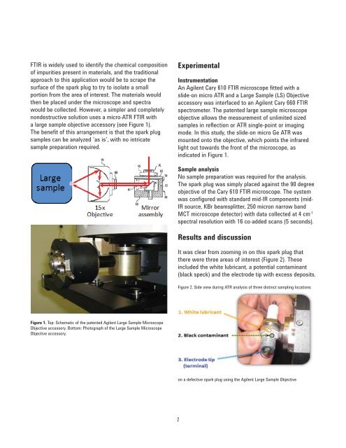
It was clear from zooming in on this spark plug that<br />
there were three areas of interest (Figure 2). These<br />
included the white lubricant, a potential contaminant<br />
(black speck) and the electrode tip with excess deposits.<br />
Figure 2. Side view during ATR analysis of three distinct sampling locations<br />
on a defective spark plug using the <strong>Agilent</strong> Large Sample Objective<br />
2


